R E S E A R C H
Open Access
Species delimitation based on mtDNA
genes suggests the occurrence of new
species of Mesocestoides in the
Mediterranean region
Antonio Varcasia
1*†, Daria Sanna
2†, Marco Casu
1, Samia Lahmar
3, Giorgia Dessì
1, Anna Paola Pipia
1,
Claudia Tamponi
1, Gabriella Gaglio
4, Gabriela Hr
čková
5, Domenico Otranto
6and Antonio Scala
1Abstract
Background: This study is the first contribution to the molecular taxonomy of Mesocestoides spp. from domestic and wild carnivores in the Mediterranean area. A total of 13 adult worms and 13 larval stages of Mesocestoides spp. were collected from domestic and wild carnivore hosts in Italy and Tunisia. Samples collected in the Slovak Republic were used as comparative samples from outside the Mediterranean. The genes cytochrome c oxidase subunit 1 (cox1) and NADH dehydrogenase subunit 1 (nad1) of the mitochondrial genome were used as molecular markers to investigate the presence of cryptic Mesocestoides species in the area analysed.
Results: Results were consistent in showing three well-supported clusters of Mesocestoides spp. in southern Italy and Tunisia, which were strongly divergent from Mesocestoides litteratus, M. corti and M. lineatus. High levels of genetic variation and no evidence of geographical structuring was found between the clusters.
Conclusions: Considering the low dispersal capability of the intermediate hosts of Mesocestoides spp., the lack of geographical structuring among the Mediterranean regions could be due to a high potential for dispersion of the definitive hosts. This study provides a foundation for future formal descriptions of new species of the genus Mesocestoides in the Mediterranean area.
Keywords: Mesocestoides, Dog, Cat, Fox, Genetic structuring, Species delimitation, Mediterranean region Background
The genus Mesocestoides (Cyclophyllidea, Mesocestoidi-dae) includes parasites with unique peculiarities in many aspects of their biology, which have yet to be revealed [1]. Two intermediate hosts are likely required for the completion of the Mesocestoides life-cycle [2], with the first larval stage developing in coprophagous arthropods, and the second (i.e. tetrathyridium) in a wide variety of hosts (e.g. rodents, amphibians, reptiles and birds) [1,3,4]. Adult Mesocestoides worms have been recorded in up to 13.8% of cats, 26.5% of dogs, 70% of jackals and 81.8% of foxes [2, 5–8]. The latter species seem to be the most
important definitive hosts for these parasites as confirmed by a recent paper that highlighted a prevalence of 84.1% in red foxes from Poland [9]. Wild and domestic carnivores could also serve as second intermediate hosts [10] since tet-rathyridium larvae can multiply asexually by longitudinal fission, penetrate the intestinal wall, invade the peritoneal cavity of the hosts and eventually cause life-threatening peritonitis [11–13]. In addition, Mesocestoides spp. are po-tentially zoonotic, being reported in human infections (at least 27 cases) following the consumption of raw or under-cooked snake, chicken and wild game viscera [14,15].
The distribution of Mesocestoides species is not well de-lineated due to their high degree of phenotypic plasticity, which hinders a clear morphological delineation of the species [5]. Furthermore, identification at the species level is not possible for the larval stages from intermediate
* Correspondence:[email protected]
†Antonio Varcasia and Daria Sanna contributed equally to this work. 1Dipartimento di Medicina Veterinaria, Università di Sassari, Sassari, Italy Full list of author information is available at the end of the article
© The Author(s). 2018 Open Access This article is distributed under the terms of the Creative Commons Attribution 4.0 International License (http://creativecommons.org/licenses/by/4.0/), which permits unrestricted use, distribution, and reproduction in any medium, provided you give appropriate credit to the original author(s) and the source, provide a link to the Creative Commons license, and indicate if changes were made. The Creative Commons Public Domain Dedication waiver (http://creativecommons.org/publicdomain/zero/1.0/) applies to the data made available in this article, unless otherwise stated.
hosts [16] or also when parasites are recovered incomplete, as gravid proglottids. This might have led to the failure of correct species identification in the past [5]. Seven species of Mesocestoides have been recorded in Europe (in the Czech Republic, Slovak Republic and Spain) [4, 5, 16, 17] with M. litteratus and M. lineatus being the most widely distributed species. Although adults of Mesocestoides linea-tusand Mesocestoides litteratus may be differentiated mor-phologically by subtle differences in the structure of the cirrus-sac, the number of testes and the position of the ovary and vitellarium [18,19], a biomolecular confirmation of the morphological diagnosis is not exhaustive [20–25]. To date, information regarding intermediate and paratenic hosts of M. litteratus and M. lineatus in natural conditions is lacking [4].
Recently, a clear genetic distinction of M. lineatus and M. litteratushas been investigated in specimens display-ing minor differences in male and female reproductive organs of worms collected from red foxes in Slovak Republic [5]. Although these parasites are common in the Mediterranean region [2, 6, 13, 26, 27], very few studies have investigated the taxonomy and the molecu-lar characterization of Mesocestoides in this area.
In the present study, several individuals of Mesoces-toides spp. from different hosts in southern Italy and Tunisia have been studied by sequencing of the cyto-chrome c oxidase subunit 1 (cox1) and NADH dehydro-genase subunit 1 (nad1) mitochondrial genes, in order to shed new light on the possible occurrence of new genetic variants and/or species. Molecular species de-limitation methods were therefore applied. Furthermore, cox1 and nad1 sequences from Mesocestoides litteratus and M. lineatus specimens collected in Slovak Republic and deposited in the Parasitic Worms Collection at the Natural History Museum, London, were obtained in this study to be used as comparative material.
Methods
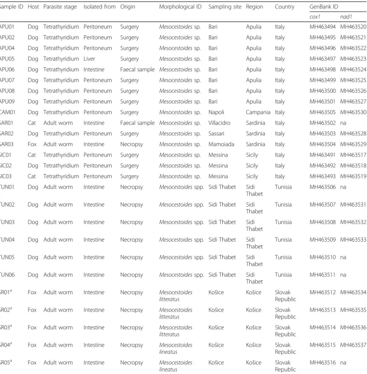
The study was carried out on a total of 13 adult worms and 13 larval stages (tetrathyridia) of Mesocestoides spp. These were collected between 2014 and 2017 from ani-mals (dogs and cats) referred for clinical visits and elect-ive surgeries or recovered during necropsy (dogs and foxes) at the Veterinary Teaching Hospitals of the Uni-versities of Sassari, Bari, Messina and Naples (Italy), and at the National School of Veterinary Medicine, Sidi Tha-bet (Tunisia). Details on hosts, parasites and sampling locations are reported in Table1and in Fig. 1. Morpho-logical identification of parasites to the genus level was performed, when possible, according to available keys [5]. Fragments from five adult individuals of M. littera-tus and one of M. lineatus found in foxes from Slovak Republic were also included in the study [5] in order to perform phylogenetic analysis. These specimens were
identified according to Skrjabin [18], mounted on slides and deposited in the Parasitic Worms Collection at the Natural History Museum, London under the accession numbers BMNH 2011.2.2.1-3 and BMNH 2011.2.2.19-20, and have been used as comparative/reference material, in this study focused on parasites of the Mediterranean region.
DNA was extracted using a commercial PureLink® Gen-omic DNA Mini Kit (Invitrogen, Carlsbad, California, USA) according to manufacturer’s instructions. Partial fragments of the mitochondrial cox1 and nad1 genes were amplified by polymerase chain reaction (PCR) following previously described protocols [5, 28–30]. PCR products were purified using a Nucleospin Gel and PCR Clean Up kit (Macherey-Nagel, Düren, Germany) and sent to an ex-ternal sequencing service (Eurofins Genomics, Ebersberg bei München, Germany). Sequence alignments were performed using BioEdit 7.2.5 [31] and deposited in the
GenBank database under the accession numbers
MH463491-MH463537 (see Table1and Additional file1: Table S1 and Additional file 2: Table S2 for details). The levels of genetic polymorphism within parasites from the Mediterranean region were assessed using DnaSP 5.10 [32]. A median-joining network [33] was constructed using Network 5.0.0.3 (www.fluxus-engineering.com) to infer the genetic relationships among the haplotypes. A 95% statistical parsimony network analysis was performed using TCS 1.21 [34], aimed at searching for possible dis-connections between groups of individuals. The
occur-rence of genetic structure among samples was
investigated by the Bayesian model-based clustering algo-rithm implemented in Baps 6 [35]. Each analysis was per-formed 10 times with a vector of values (1–10) for K each with 5 replicates.
For the phylogenetic analysis, two enlarged datasets for both cox1 and nad1 markers were built by aligning the sequences obtained in the present study with all comparable sequences available on GenBank for M. lit-teratus, M. lineatus and M. corti/M. vogae [5, 36, 37] (see Figs. 4and 5 for details). Two sequences from Italy (Tuscany, GenBank: JQ740884; and Sicily, GenBank: KU821650) attributed to Mesocestoides spp. were also included in the cox1 dataset. A sequence of Echinococcus multilocularis from GenBank was used as the outgroup (Figs. 4, 5 and Table1). The cox1 gene dataset included 26 sequences obtained in the present study (see Table 1 for details) and 40 from GenBank. For the nad1 gene, the fragment analysed for phylogeny was restricted to 202 bp in order to obtain an overlapping segment be-tween our sequences and those from GenBank. The new nad1 dataset included 21 sequences obtained in the present study (see Table 1 for details) and 25 sequences from GenBank.
In order to test the phylogenetic signal [38] and the adequacy of taxonomic coverage, the likelihood-mapping
analysis of 10,000 random quartets was performed using TreePuzzle 5.3 [39,40]. The datasets were used to plot a phylogenetic tree using the maximum likelihood (ML) algorithm implemented in MEGA7 [41] with 1000 boot-strap replications, and the Kimura 2-parameter (K2P) as a molecular evolutionary model. The nodes of the trees with bootstrap values lower than 50% were considered not well-supported and thus collapsed.
The combined use of two species delimitation methods, the Automatic Barcode Gap Discovery (ABGD) [42] and the Nucleotide Divergence Threshold (NDT) [42], allowed us to make inferences on the occurrence of taxonomic en-tities by means of two alternative distance models (simple p-distance for ABGD and Kimura (K80) distance for NDT). ABGD was calculated by means of the ABGD on-line tool (available at http://wwwabi.snv.jussieu.fr/public/
Table 1 Data collection and list of the specimens and the sequences included in the analyses
Sample ID Host Parasite stage Isolated from Origin Morphological ID Sampling site Region Country GenBank ID cox1 nad1 APU01 Dog Tetrathyridium Peritoneum Surgery Mesocestoides sp. Bari Apulia Italy MH463494 MH463520 APU02 Dog Tetrathyridium Peritoneum Surgery Mesocestoides sp. Bari Apulia Italy MH463495 MH463521 APU04 Dog Tetrathyridium Peritoneum Surgery Mesocestoides sp. Bari Apulia Italy MH463496 MH463522 APU05 Dog Tetrathyridium Liver Surgery Mesocestoides sp. Bari Apulia Italy MH463497 MH463523 APU06 Dog Tetrathyridium Intestine Faecal sample Mesocestoides sp. Bari Apulia Italy MH463498 MH463524 APU07 Dog Tetrathyridium Peritoneum Surgery Mesocestoides sp. Bari Apulia Italy MH463499 MH463525 APU08 Dog Tetrathyridium Peritoneum Surgery Mesocestoides sp. Bari Apulia Italy MH463500 MH463526 APU09 Dog Tetrathyridium Peritoneum Surgery Mesocestoides sp. Bari Apulia Italy MH463501 MH463527 CAM01 Dog Tetrathyridium Peritoneum Surgery Mesocestoides sp. Napoli Campania Italy MH463505 MH463530 SAR01 Cat Adult worm Intestine Faecal sample Mesocestoides sp. Villacidro Sardinia Italy MH463502 na SAR02 Dog Tetrathyridium Peritoneum Surgery Mesocestoides sp. Sassari Sardinia Italy MH463503 MH463528 SAR03 Fox Adult worm Intestine Necropsy Mesocestoides sp. Mamoiada Sardinia Italy MH463504 MH463529 SIC01 Cat Tetrathyridium Peritoneum Surgery Mesocestoides sp. Messina Sicily Italy MH463491 MH463517 SIC02 Dog Tetrathyridium Peritoneum Surgery Mesocestoides sp. Messina Sicily Italy MH463492 MH463518 SIC03 Cat Tetrathyridium Peritoneum Surgery Mesocestoides sp. Messina Sicily Italy MH463493 MH463519 TUN01 Dog Adult worm Intestine Necropsy Mesocestoides spp. Sidi Thabet Sidi
Thabet
Tunisia MH463506 na TUN02 Dog Adult worm Intestine Necropsy Mesocestoides spp. Sidi Thabet Sidi
Thabet
Tunisia MH463507 MH463531 TUN03 Dog Adult worm Intestine Necropsy Mesocestoides spp. Sidi Thabet Sidi
Thabet
Tunisia MH463508 MH463532 TUN04 Dog Adult worm Intestine Necropsy Mesocestoides spp. Sidi Thabet Sidi
Thabet
Tunisia MH463509 MH463533 TUN05 Dog Adult worm Intestine Necropsy Mesocestoides spp. Sidi Thabet Sidi
Thabet
Tunisia MH463510 na TUN06 Dog Adult worm Intestine Necropsy Mesocestoides spp. Sidi Thabet Sidi
Thabet
Tunisia MH463511 na SR01a Fox Adult worm Intestine Necropsy Mesocestoides
litteratus
Košice Košice Slovak Republic
MH463512 MH463534 SR02a Fox Adult worm Intestine Necropsy Mesocestoides
litteratus
Košice Košice Slovak Republic
MH463513 MH463535 SR03a Fox Adult worm Intestine Necropsy Mesocestoides
litteratus
Košice Košice Slovak Republic
MH463514 MH463536 SR04a Fox Adult worm Intestine Necropsy Mesocestoides
lineatus
Košice Košice Slovak Republic
MH463515 MH463537 SR05a Fox Adult worm Intestine Necropsy Mesocestoides
lineatus
Košice Košice Slovak Republic
MH463516 na
a
Deposited in the Parasitic Worms Collection at the Natural History Museum, London under the accession numbers BMNH 2011.2.2.1-3 and BMNH 2011.2.2.19-20. Abbreviation: na not available as not amplified
abgd/abgdweb.html) with a prior P ranging from 0.001 to 0.12, steps = 10 and relative gap width (X) = 1. The NDT method was applied by means of a script written in the R statistical environment (available at https://cran.r-projec-t.org/) and described in [43–45]. Estimates of evolutionary divergence over sequence pairs between groups were con-ducted in MEGA7 to evaluate the genetic distance be-tween taxa by using the Kimura 2-parameter model. Results
The correct taxonomic attribution of specimens from Slovak Republic used as comparative material in the present study, was verified via a BLASTsearch against the available data in the GenBank nucleotide database. They were attributed to M. litteratus and M. lineatus re-spectively (see Table 1 for details on species and Gen-Bank accession numbers). The analysis of the cox1 dataset evidenced two haplotypes for M. litteratus (n = 3, S = 2, h = 0.667,π = 0.00660) and one haplotype for M. lineatus(n = 1). The analysis of the nad1 dataset evi-denced two haplotypes for M. litteratus (n = 3, S = 3, h = 0.667,π = 0.00536) and one haplotype for M. lineatus (n = 2). The phylogenetic analysis below reported for the Mediterranean region corroborated the taxonomic attri-bution of specimens from Slovak Republic.
Overall, high levels of genetic variation were found for the cox1 dataset (373 bp long) among 21 Mesocestoides
Fig. 1 Map of the Mediterranean indicating the sampling sites
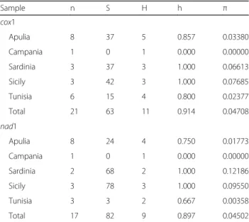
Table 2 Sample sizes and genetic diversity estimates obtained for the mitochondrial regions, cox1 (378 bp) and nad1 (558 bp). Sites with gaps were not considered. Sample codes are listed in Table1 Sample n S H h π cox1 Apulia 8 37 5 0.857 0.03380 Campania 1 0 1 0.000 0.00000 Sardinia 3 37 3 1.000 0.06613 Sicily 3 42 3 1.000 0.07685 Tunisia 6 15 4 0.800 0.02377 Total 21 63 11 0.914 0.04708 nad1 Apulia 8 24 4 0.750 0.01773 Campania 1 0 1 0.000 0.00000 Sardinia 2 68 2 1.000 0.12186 Sicily 3 78 3 1.000 0.09550 Tunisia 3 3 2 0.667 0.00358 Total 17 82 9 0.897 0.04502
Abbreviations: n sample size; S number of polymorphic sites, H number of haplotypes, h haplotype diversity, π nucleotide diversity
specimens from the Mediterranean region, with rather low indices of genetic divergence found for the samples from Tunisia (h = 0.800,π = 0.024) (see Table2for esti-mates of genetic divergence).
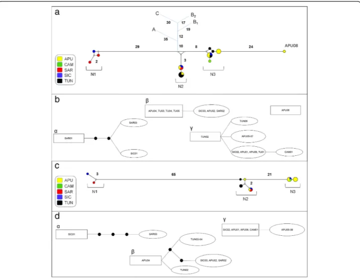
Four cox1 haplotypes were shared by 67% of the samples while the remaining haplotypes were unique to single indi-viduals (see Additional file1: Table S1). Median-joining net-work analysis revealed the occurrence of three main divergent groups of haplotypes (N1, N2 and N3) (see Fig.2a for details on the geographical distribution of haplotypes). Statistical parsimony network analysis revealed four discon-nected clusters within Mediterranean Mesocestoides speci-mens (Fig. 2b). Three of these clusters (α, β and γ) exactly matched the groups of haplotypes in the median-joining net-work (N1, N2 and N3, respectively). The highest root weight
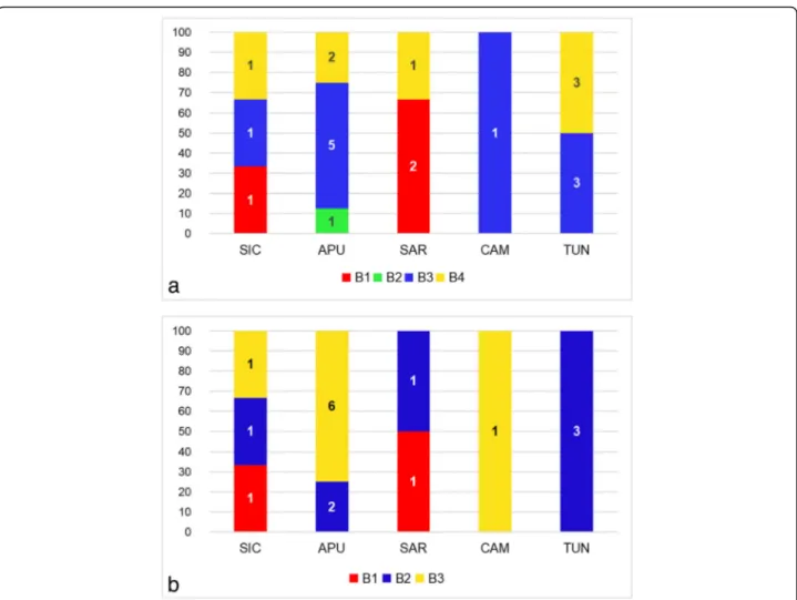
was shown by a haplotype found in Sardinia (SAR01) for cluster α, by haplotypes found in Tunisia and Apulia (TUN03-05, APU04) for clusterβ, and by a haplotype found in Tunisia (TUN02) for clusterγ. The Bayesian model-based clustering implemented in Baps 6 identified four distinct groups of haplotypes (B1, B2, B3 and B4) (see Fig.3a for de-tails on the geographical distribution of groups). B2 was the least frequent group, being only reported for the highly di-vergent haplotype from Apulia (APU08).
A 555 bp long alignment for the nad1 gene included se-quences belonging to 17 Mesocestoides specimens from Italy and Tunisia (Table1). As a possible consequence of a limited homology between universal primers used and the annealing region, scorable nad1 sequences were obtained for a reduced number of individuals. Overall, high levels
Fig. 2 Network analysis. a, b cox1 dataset; c, d nad1 dataset. a, c Median-joining networks with haplotypes coloured according to their geographical distribution. Small white dots on the nodes show median vectors representing hypothetical connecting sequences, calculated with a maximum parsimony method. The numbers of mutations between haplotypes greater than one are reported on the network branches. In the median-joining networks based on cox1 dataset (a) the short blue branches represent the connection with the other species. Abbreviations: A, Mesocestoides litteratus; B1and B2, M. lineatus from Mongolia and Slovak Republic, respectively; C, M. corti. b, d Clusters retrieved using 95% statistical parsimony networks. The
number of mutations greater than one are shown as black dots on the network branches. The haplotype in a square has the largest outgroup weight. Abbreviations: APU, Apulia; CAM, Campania; SAR, Sardinia; SIC, Sicily; TUN, Tunisia
of genetic variation were found in the Mediterranean re-gion, which resulted in a total genetic variability similar to that found for the cox1 dataset (see Table 2 for details). Three nad1 haplotypes were found in 65% of the samples, while the remaining were unique to single individuals (see Additional file 2: Table S2 for more details). The median-joining network analysis revealed the occurrence of three groups of haplotypes (N1, N2 and N3) (see Fig.2c for de-tails) almost corresponding to those found for the cox1 median-joining network analysis (see Fig.1a). Accordingly, the statistical parsimony network analysis highlighted the occurrence of three disconnected clusters (α, β and γ) within Mediterranean Mesocestoides spp. (see Fig.2d). The highest root weight was shown by a haplotype found in Si-cily (SIC01) for clusterα; by a haplotype found in Apulia (APU04) for clusterβ; and by haplotypes found in Apulia (APU01, APU09), Campania (CAM01) and Sicily (SIC02) for clusterγ.
The Bayesian model-based clustering implemented in Baps 6 (see Fig.3b for details) identified three groups of haplotypes (B1, B2 and B3) which were consistent with three (B1, B3 and B4) of the four groups reported for the cox1 Bayesian analysis.
The likelihood map based on cox1 gene dataset (see Add-itional file3: Figure S1a) indicated a strong phylogenetic sig-nal. The maximum likelihood (ML) tree analysis (Fig. 4) showed five supported and one unsupported (bootstrap value of 45%) clusters; three for M. litteratus, M. corti/M. vogaeand M. lineatus, and the remaining for the Mediterra-nean Mesocestoides spp. specimens analysed in the present study. Notably, the M. lineatus cluster included a GenBank sequence (KU821650) from Sicily. The individuals of Meso-cestoides spp. examined here grouped into three different clusters (M1, M2 and M3 in Fig.4). A consensus sequence of Mesocestoides sp., from GenBank (JQ740884) from Tuscany, Italy, was also included in the M3 group.
Fig. 3 Distribution of the groups identified by Bayesian model-based clustering implemented in Baps 6 within populations. a cox1 dataset. b nad1 dataset. X axis: populations; Y axis: relative frequency of distribution (%). Abbreviations: APU, Apulia; CAM, Campania; SAR, Sardinia; SIC, Sicily; TUN, Tunisia. The numbers in bars indicate the absolute frequency of distribution. B1, B2, B3 and B4 indicate the groups identified by Bayesian model-based clustering described in the text
The species delimitation methods, Automatic Barcode Gap Discovery (ABGD) and Nucleotide Divergence Threshold (NDT) converged on the same results; for this reason, only the ABGD results are reported. Four en-tities were identified within the Mediterranean Mesoces-toides spp. analysed. Overall, the composition of all the groups found by ABGD matched the clusters in the ML tree analysis. Evolutionary divergences were estimated between the ML clusters of Mesocestoides spp. (M. litter-atus, M.corti/M. vogae, M. linelitter-atus, M1, M2 and M3) (see Additional file 4: Table S3 for details). Consistent
with the previous analysis which converged in separating APU08 from the remaining samples, this specimen was considered as a further separate group to be tested.
The likelihood map based on the nad1 gene (see Additional file3: Figure S1b) indicated a low phylogenetic signal. The ML tree analysis was consistent in showing the same results for the cox1 dataset (see Fig.5for a compari-son). Additionally, the species delimitation methods (ABGD and NDT) converged on the same results, and the compos-ition of the groups obtained exactly matched the clusters obtained by ML tree analysis. The ABGD method identified Fig. 4 Maximum likelihood tree showing the interrelationships among Mesocestoides spp. based on the cox1 dataset. Outgroup: Echinococcus multilocularis. Only bootstrap support values > 45% are shown. The scale-bar indicates the number of substitutions per site. Sample codes are listed in Table1. M1, M2, and M3 indicate the entities found by species delimitation analysis described in the text
three entities within the Mediterranean Mesocestoides spp. analysed, with the position of the individual from Tunisia TUN02 representing the only discrepancy between the cox1 and nad1 ML tree and species delimitation analysis. Estimates of evolutionary divergences for nad1 gene are re-ported in the Additional file5: Table S4.
Discussion
This study on mitochondrial genetic variability of Meso-cestoides spp. from domestic and wild carnivores in the Mediterranean area allowed us to gather a deep defin-ition of the species delimitation among samples from southern Italy and Tunisia. Indeed, molecular analyses were consistent in pointing out three, well-defined,
taxonomic Mesocestoides entities from Italy and Tunisia with high levels of genetic variation among individuals and no evidence of geographical structuring among en-tities (namely M1, M2 and M3 as in ML analysis). The occurrence of a sequence (GenBank: JQ740884) from Tuscany belonging to the M3 entity suggests that the range of distribution of these parasites probably extends north and not only confined to the Mediterranean re-gion. Such a finding is consistent with a study [46], which highlighted the occurrence of a species genetically diver-gent from M. lineatus, M. litteratus and M. corti, in north-ern Italy. Two of the Mesocestoides entities in the present study (M2 and M3) displayed very low levels of genetic di-vergence among each other; the nad1 results evidenced Fig. 5 Maximum likelihood rooted tree showing the interrelationships among Mesocestoides spp. based on the nad1 dataset. Only bootstrap support values≥ 50% are shown. The scale-bar indicates the number of substitutions per site. Sample codes are listed in Table1. M1, M2 and M3 indicate the entities found by species delimitation analysis described in the text
the occurrence of a reciprocal monophyly between them, likely consistent with the presence of two Mesocestoides sister taxa in the Mediterranean area.
The statistical parsimony network analysis based on the cox1 dataset suggests that M2 and M3 probably originated in Tunisia, with an ancestor haplotype also present in Apulia. These findings could be consistent with the occur-rence of early polymorphisms, maybe common to the southern Italy and Tunisia, possibly due to the transloca-tion of domestic dogs since ancient times.
The levels of genetic divergence found between the Mediterranean Mesocestoides entities and M. lineatus, higher than those found between the M. lineatus geo-graphical internal subgroups from Slovak Republic, Mongolia and Italy, further support the occurrence, at least in Italy and Tunisia, of Mesocestoides entities strongly divergent from M. lineatus. Notably the latter, which is described in the Italian Peninsula [2], was not identified in this work. Conversely, M. lineatus has been previously identified in Italy in a cat from Sicily [27], as evidenced by sequence GenBank: KU821650 included as an outlier within the M. lineatus cluster. This finding suggests that possibly several Mesocestoides spp. may occur in sympatry in southern Italy.
From a systematic perspective, the co-occurrence of differ-ent molecular differ-entities, does not allow for unequivocal iddiffer-enti- identi-fication and description of the new taxa corresponding to the entities found. Furthermore, the inconsistency between morphological and molecular features supports the hypoth-esis that different environmental and ecological features interacted in the Mediterranean area to produce cryptic spe-cies within the genus Mesocestoides that are genetically di-vergent but morphologically indistinguishable from each other. In this context, it is important to underline the pivotal role of the molecular taxonomy, not only in identifying the cut-off to delimit species from each other, but also to the naming of the species itself, which result from the validation of the primary species hypotheses [47–53]. In fact, from a viewpoint that takes into account the appreciation of specific biodiversity, no species can be documented without a formal description, as well as no OTUs may substitute a species in any species checklist. [28,54]. For this reason, the combined analysis of molecular and morphological data, in the light of the“integrative taxonomy approach” [55] will be used in the near future to provide both a satisfactory insight on the evo-lutionary processes and taxonomic richness, with a formal description of new species of the genus Mesocestoides in the Mediterranean area.
Conclusions
The present study represents the first survey on mito-chondrial genetic variability of Mesocestoides spp. from domestic and wild carnivores in the Mediterranean area that allowed to point out three defined, taxonomic
Mesocestoides entities which are genetically divergent from M. lineatus, M. litteratus and M. corti.
Additional files
Additional file 1:Table S1. Distribution (absolute frequencies) of cox1 haplotypes in 21 specimens from five Mediterranean sites. Sample codes are listed in Table1. (DOCX 15 kb)
Additional file 2:Table S2. Distribution (absolute frequencies) of nad1 haplotypes in 17 specimens from five Mediterranean sites. Sample codes are listed in Table1. (DOCX 15 kb)
Additional file 3:Figure S1. Likelihood mapping. a cox1 dataset. b nad1 dataset. For both panels: (i) distribution map of dots P, where P represents the likelihoods of the three possible unrooted trees for a set of four sequences (quartets) [54]. Dots close to the corners and to the sides represent tree-like and network-like phylogenetic signal. Central area represents star-like signal (phylogenetic noise); (ii) percentage distribution of the three possible unrooted trees; (iii) partitions of the area of the triangle into seven regions. The three trapezoids at the corners represent the areas supporting strictly bifurcating trees, that is the presence of a tree-like phylogenetic signal. The three rectangles on the sides represent regions where the decision between two trees is not obvious. The centre of the triangle represents sets of points P where all three trees are equally supported. (TIF 5240 kb)
Additional file 4:Table S3. Estimates of evolutionary divergence over sequence pairs between groups based on the cox1 dataset. Genetic distances, represented by the number of base substitutions per site from averaging over all sequence pairs between groups, are shown below the diagonal and standard deviations above the diagonal. Analyses were conducted using the K2P model. Sample codes are listed in Table1. (DOCX 17 kb)
Additional file 5:Table S4. Estimates of evolutionary divergence over sequence pairs between groups based on the nad1 dataset. Genetic distances, represented by the number of base substitutions per site from averaging overall sequence pairs between groups, are shown below the diagonal and standard deviations are shown above the diagonal. Analyses were conducted using the K2P model. Sample codes are listed in Table1. (DOCX 18 kb)
Abbreviations
ML:Maximum likelihood; ABGD: Automatic Barcode Gap Discovery; NDT: Nucleotide Divergence Threshold
Acknowledgments
The authors would like to thank R. P. Lia, V. Tilocca, L. Rinaldi, P. Cabras and B. Boufana, for providing Mesocestoides spp. samples.
Funding Not applicable
Availability of data and materials
All relevant data are within the paper and its additional files. Nucleotide sequences obtained in this study are available in the GenBank database under the accession numbers MH463491-MH463537. Fragments from five adult individuals of M. litteratus and one of M. lineatus found in foxes from Slovak Republic were deposited in the Parasitic Worms Collection at the Natural History Museum, London under the accession numbers BMNH 2011.2.2.1-3 and BMNH 2011.2.2.19-20.
Authors’ contributions
Conceived and designed the experiments: AV. Performed the experiments: GD and APP. Analyzed the data: DS. Contributed reagents/materials/analysis tools: GD, APP and DS. Wrote the paper: AV, DS, MC, AS and DO. Collected biological samples: CT, GG, GH and SL. Revised the manuscript: AV, DS, MC, AS and DO. All authors read and approved the final manuscript.
Ethics approval and consent to participate
This study was performed following the recommendations of European Council Directive (86/609/EEC) on the protection of animals.
Consent for publication Not applicable. Competing interests
The authors declare that they have no competing interests.
Publisher’s Note
Springer Nature remains neutral with regard to jurisdictional claims in published maps and institutional affiliations.
Author details
1
Dipartimento di Medicina Veterinaria, Università di Sassari, Sassari, Italy. 2Dipartimento di Scienze Biomediche, Università di Sassari, Sassari, Italy. 3
National School of Veterinary Medicine, Laboratory of Parasitology, Sidi Thabet, Tunisia.4Dipartimento di Scienze Veterinarie, Università di Messina, Messina, Italy.5Institute of Parasitology, Slovak Academy of Sciences, Košice, Slovak Republic.6Dipartimento di Medicina Veterinaria, Università di Bari, Bari, Italy.
Received: 14 June 2018 Accepted: 6 November 2018
References
1. Loos-Frank B. One or two intermediate hosts in the life cycle of Mesocestoides (Cyclophyllidae, Mesocestoididae)? Parasitol Res. 1991;77:726–8.
2. Papini R, Matteini A, Bandinelli P, Pampurini F, Mancianti F. Effectiveness of praziquantel for treatment of peritoneal larval cestodiasis in dogs: a case report. Vet Parasitol. 2010;170:158–61.
3. McAllister CT, Bruce Conn D, Freed PS, Burdick DA. A new host and locality record for Mesocestoides sp. tetrathyridia (Cestoidea: Cyclophyllidea), with a summary of the genus from snakes of the world. J Parasitol. 1991;77:329–31. 4. Literák I, Olson PD, Georgiev BB,Špakulová M. First record of metacestodes of
Mesocestoides sp. in the common starling (Sturnus vulgaris) in Europe, with an 18S rDNA characterization of the isolate. Folia Parasitol. 2004;51:45–9. 5. Hrčkova G, Miterpáková M, O’Connor A, Šnábel V, Olson P. Molecular and
morphological circumscription of Mesocestoides tapeworms from red foxes (Vulpes vulpes) in central Europe. Parasitology. 2011;138:638–47.
6. Lahmar S, Boufana B, Ben Boubaker S, Landolsi F. Intestinal helminths of golden jackals and red foxes from Tunisia. Vet Parasitol. 2014;204:297–303. 7. Dalimi A, Sattari A, Motamedi G. A study on intestinal helminthes of dogs, foxes and jackals in the western part of Iran. Vet Parasitol. 2006;142:129–33. 8. Calvete C, Lucientes J, Castillo JA, Estrada R, Gracia MJ, Peribáñez MA, Ferrer
M. Gastrointestinal helminth parasites in stray cats from the mid-Ebro Valley, Spain. Vet Parasitol. 1998;75:235–40.
9. Karamon J, Dabrowska J, Kochanowski M, Samorek-Pieróg M, Sroka J, Różycki M, Bilska-Zajac E, Zdybel J, Cencek T. Prevalence of intestinal helminths of red foxes (Vulpes vulpes) in central Europe (Poland): a significant zoonotic threat. Parasit Vectors. 2018;11:436. 10. Crosbie PR, Boyce WM, Platzer EG, Nadler SA, Kerner C. Diagnostic
procedures and treatment of eleven dogs with peritoneal infections caused by Mesocestoides spp. J Am Vet Med Assoc. 1998;213:1578–83.
11. Siles-Lucas M, Hemphill A. Cestode parasites: application of in vivo and in vitro models for studies on the host-parasite relationship. Adv Parasitol. 2002;51:133–230.
12. Boyce W, Shender L, Schultz L, Vickers W, Johnson C, Ziccardi M, et al. Survival analysis of dogs diagnosed with canine peritoneal larval cestodiasis (Mesocestoides spp.). Vet Parasitol. 2011;180:256–61.
13. Montalbano Di Filippo M, Meoli R, Cavallero S, Eleni C, De Liberato C, Berrilli F. Molecular identification of Mesocestoides sp. metacestodes in a captive gold-handed tamarin (Saguinus midas). Infect Genet Evol. 2018;65:399–405. 14. Fuentes MV, Galán-Puchades T, Malone JB. A new case report of human
Mesocestoides infection in the United States. Am J Trop Med Hyg. 2003;68: 566–7.
15. Széll Z, Tolnai Z, Sréter T. Environmental determinants of the spatial distribution of Mesocestoides spp. and sensitivity of flotation method for the diagnosis of mesocestoidosis. Vet Parasitol. 2015;212:427–30.
16. Zaleśny G, Hildebrand J. Molecular identification of Mesocestoides spp. from intermediate hosts (rodents) in central Europe (Poland). Parasitol Res. 2012; 110:1055–61.
17. Tenora F. Mesocestoides litteratus (Batsch, 1786) (Cestoda), parasite of Vulpes vulpes (L., 1758) (Carnivora) in the Czech Republic. Acta Univ Agric Silvic Mendel Brun. 2005;53:185–8.
18. Skrjabin KI. Tetrabotrididae and Mesocestoididae - platyhelminths of birds and mammals. In: Skrjabin KI, editor. Osnovy Cestodologii, Volume 9. Moscow: Nauka; 1978. p. 156–215. (In Russian).
19. Jančev J. Morphology, taxonomy and distribution of the species of genus Mesocestoides Vaillant, 1863 in Bulgaria. Khelmintologiya. 1986;21:45–65. 20. De Jong Y, Verbeek M, Michelsen V, Bjørn PP, Los W, Steeman F, et al.
Fauna Europaea - all European animal species on the web. Biodivers Data J. 2014;2:e4034.
21. Andras T, Peter T. Data on worm infestation cats (Felis catus) in Hungarian hunting areas. Magy Allatorvosok Lapja. 2002;124:26–30.
22. Segovia JM, Guerrero R, Torres J, Miguel J, Feliu C. Ecological analyses of the intestinal helminth communities of the wolf, Canis lupus, in Spain. Folia Parasitol. 2003;50:231–6.
23. Martinez-Carrasco C, Ruiz De Ybanez MR, Sagarminaga JL, Garijo MM, Moreno F, Acosta I, et al. Parasites of the red fox (Vulpes vulpes Linneus, 1758) in Murcia, southeast Spain. Rev Med Vet. 2007;158:331–5. 24. Krone O, Guminsky O, Meinig H, Hermann M, Trinzen M, Wibbelt G.
Endoparasite spectrum of wild cats (Felis silvestris Schreber, 1777) and domestic cats (Felis catus L.) from the Eifel, Pfalz region and Saarland, Germany. Eur J Wildl Res. 2008;54:95–100.
25. Magi M, Macchioni F, Dell’Omodarme M, Prati MC, Calderini P, Gabrielli S, et al. Endoparasites of red fox (Vulpes vulpes) in central Italy. J Wildl Dis. 2009;45:881–5.
26. Bonfanti U, Bertazzolo W, Pagliaro L, Demarco B, Venco L, Casiraghi M, Bandi C. Clinical, cytological and molecular evidence of Mesocestoides sp. infection in a dog from Italy. J Vet Med A Physiol Pathol Clin Med. 2004;51: 435–8.
27. Lanteri G, Di Caro G, Capucchio MT, Gaglio G, Reina V, Lo Giudice C, Zanet S, Marino F. Mesocestoidosis and multivisceral tetrathyridiosis in a European cat. Vet Med Czech. 2017;62:356–62.
28. Otranto D, Varcasia A, Solinas C, Scala A, Brianti E, Dantas-Torres F, et al. Redescription of Cercopithifilaria bainae Almeida & Vicente, 1984 (Spirurida, Onchocercidae) from a dog in Sardinia, Italy. Parasit Vectors. 2013;6:132. 29. Littlewood DTJ, Waeschenbach A, Nikolov PN. In search of mitochondrial
markers for resolving the phylogeny of cyclophyllidea tapeworms (Platyhelminthes, Cestoda) - a test study with Davaineidae. Acta Parasitol. 2008;53:133–44.
30. Sanna D, Dedola GL, Lai T, Curini-Galletti M, Casu M. PCR-RFLP: a practical method for the identification of specimens of Patella ulyssiponensis s.l. (Gastropoda: Patellidae). Ital J Zool. 2012;79:50–9.
31. Hall TA. BioEdit: A user-friendly biological sequence alignment editor and analysis program for Windows 95/98/NT. Nucleic Acids Symp Ser. 1999;41: 95–8.
32. Librado P, Rozas J. DnaSP v5: a software for comprehensive analysis of DNA polymorphism data. Bioinformatics. 2009;25:1451–2.
33. Bandelt HJ, Forster P, Rohl A. Median-joining networks for inferring intraspecific phylogenies. Mol Biol Evol. 1999;16:37–48.
34. Clement M, Posada D, Crandall K. TCS: a computer program to estimate gene genealogies. Mol Ecol. 2000;9:1657–60.
35. Corander J, Cheng L, Marttinen P, Sirén J, Tang JBAPS. Bayesian analysis of population structure V. 6.0. Department of Mathematics and statistics. Helsinki: University of Helsinki; 2013.
36. Kashiide T, Matsumoto J, Yamaya Y, Uwasawa A, Miyoshi A, Yamada K, et al. Case report: first confirmed case of canine peritoneal larval cestodiasis caused by Mesocestoides vogae (syn. M. corti) in Japan. Vet Parasitol. 2014; 201:154–7.
37. Lesniak I, Heckmann I, Heitlinger E, Szentiks CA, Nowak C, Harms V, et al. Population expansion and individual age affect endoparasite richness and diversity in a recolonising large carnivore population. Sci Rep. 2017;7:41730. 38. Strimmer K, Von Haeseler A. Quartet puzzling: a quartet
maximum-likelihood method for reconstructing tree topologies. Mol Biol Evol. 1996;13: 964–9.
39. Schmidt HA, Strimmer K, Vingron M, von Haeseler A. TREE-PUZZLE: maximum likelihood phylogenetic analysis using quartets and parallel computing. Bioinformatics. 2002;18:502–4.
40. Schmidt HA, von Haeseler A. Phylogenetic inference using maximum likelihood methods. In: Lemey P, Salemi M, Vandamme AM, editors. The Phylogenetic Handbook. 5th ed. Cambridge: Cambridge University Press; 2012. p. 181–209.
41. Kumar S, Stecher G, Tamura K. MEGA7: Molecular Evolutionary Genetics Analysis Version 7.0 for bigger datasets. Mol Biol Evol. 2016;33:1870–4. 42. Hebert PDN, Cywinska A, Ball SL, deWaard JR. Biological identifications
through DNA barcodes. Proc Biol Sci. 2003;270:313–22.
43. Scarpa F, Cossu P, Lai T, Sanna D, Curini-Galletti M, Casu M. Meiofaunal cryptic species challenge species delimitation: the case of the Monocelis lineata (Platyhelminthes: Proseriata) species complex. Contrib Zool. 2016;85: 123–45.
44. Scarpa F, Cossu P, Sanna D, Lai T, Casu M, Curini-Galletti M. New insights on the genus Otoplana Du Plessis, 1889 (Platyhelminthes: Proseriata), with description of two new species from the Canary Islands. Mar Biodivers. 2017.https://doi.org/10.1007/s12526-017-0785-1.
45. Scarpa F, Sanna D, Cossu P, Lai T, Casu M, Curini-Galletti M. How to achieve internal fertilization without a vagina: the study case of the genus Archilina Ax, 1959 (Platyhelminthes, Proseriata) from Canary Islands. Mar Biodivers. (In Press)https://doi.org/10.1007/s12526-018-0890-9.
46. Jabbar A, Papini R, Ferrini N, Gasser RB. Use of a molecular approach for the definitive diagnosis of proliferative larval mesocestoidiasis in a cat. Infect Genet Evol. 2012;12:1377–80.
47. Varcasia A, Tamponi C, Tosciri G, Pipia AP, Dore F, Schuster RK, et al. Is the red fox (Vulpes vulpes) a competent definitive host for Taenia multiceps? Parasit Vectors. 2015;8:491.
48. Varcasia A, Canu S, Lightowlers MW, Scala A, Garippa G. Molecular characterization of Echinococcus granulosus strains in Sardinia. Parasitol Res. 2006;98:273–7.
49. Saarma U, Jõgisalu I, Moks E, Varcasia A, Lavikainen A, Oksanen A, et al. A novel phylogeny for the genus Echinococcus, based on nuclear data, challenges relationships based on mitochondrial evidence. Parasitology. 2009;136:317–28.
50. Varcasia A, Jia WZ, Yan HB, Manunta ML, Pipia AP, Garippa G, et al. Molecular characterization of subcutaneous and muscular coenurosis of goats in United Arab Emirates. Vet Parasitol. 2012;190:604–7.
51. Boufana B, Scala A, Lahmar S, Pointing S, Craig PS, Dessì G, et al. A preliminary investigation into the genetic variation and population structure of Taenia hydatigena from Sardinia, Italy. Vet Parasitol. 2015;214:67–74. 52. Scala A., Pipia AP, Dore F, Sanna G, Tamponi C, Marrosu R, et al.
Epidemiological updates and economic losses due to Taenia hydatigena in sheep from Sardinia, Italy. Parasitol Res. 2015;114:3137–43.
53. Kinkar L, Laurimaë T, Simsek S, Balkaya I, Casulli A, Manfredi MT, et al. High-resolution phylogeography of zoonotic tapeworm Echinococcus granulosus sensu stricto genotype G1 with an emphasis on its distribution in Turkey, Italy and Spain. Parasitology. 2016;143:1790–801.
54. Jörger KM, Schrödl M. How to describe a cryptic species? Practical challenges of molecular taxonomy. Front Zool. 2013;10:59.