1Department of Public Health, University of Naples “Federico II”, Naples, Italy
2Department of Gynecology and Obstetrics, ABC Foundation School of Medicine, Santo André, Brazil
3Unit of Gynecology and Obstetrics, Department of Human Pathology in Adulthood and Childhood ‘‘G. Barresi’’, University of Messina, Messina, Italy
4Department of Neuroscience and Reproductive and Odontostomatological Sciences, University of Naples “Federico II”, Naples, Italy Received: January 5, 2017
Accepted: March 8, 2017
Correspondence to: Antonio Simone Laganà E-mail: [email protected]
DOI 10.5152/eurasianjmed.2017.16215 ©Copyright 2017 by the Atatürk University School of Medicine - Available online at www.eurasianjmed.com
ABSTRACT
This case report of a 36-year-old woman with a diagnosis of cervical pregnancy describes a novel approach to this rare form of ectopic pregnancy, which was successfully treated with systemic and local methotrexate (MTX) therapy combined with hysteroscopic resection. After local and systemic administration of MTX, the patient underwent hysteroscopic resection of the cervical pregnancy using a 27 bipolar resectoscope with a 4-mm loop. The cervical pregnancy was completely treated, and satisfactory hemostasis was achieved with electrocoagulation. The reported case and literature review demonstrate that the combination of systemic and local (hysteroscopic) administration of MTX with hysteroscopic resection could offer the possibility of a safe, successful, minimally invasive, and fertility-sparing surgical treatment for cervical pregnancy.
Keywords: Hysteroscopy, methotrexate, cervical pregnancy
ÖZ
Servikal gebelik tanısı almış 36 yaşında bir kadın hasta hakkındaki bu vaka sunumunda ektopik gebeliğin, his-teroskopik rezeksiyon ile kombine sistemik ve lokal metotreksat (MTX) ile başarılı bir şekilde tedavi edilmiş olan bu nadir şekline karşı yeni bir yaklaşım tanımlanmaktadır. Lokal ve sistemik MTX uygulaması sonrasında hastaya servikal gebelik için, 4-mm loop ile 27 bipolar rezektoskop kullanılarak histeroskopik rezeksiyon ya-pıldı. Servikal gebelik tamamen tedavi edildi ve elektrokoagülasyon ile yeterli bir hemostaz sağlandı. Yapılan bu literatür taraması ve indeks olgu, sistemik ve lokal (histeroskopik) MTX uygulaması ile histeroskopik re-zeksiyon kombinasyonunun, servikal gebeliğin güvenli, başarılı, minimal düzeyde invaziv ve fertilite-koruyucu cerrahi tedavisinin mümkün olabileceğini göstermektedir.
Anahtar Kelimeler: Histeroskopi, metotreksat, servikal gebelik
Eurasian J Med 2017; 49: 66-8
Case Report
Introduction
Cervical pregnancy (CP) is a rare form of ectopic pregnancy associated with high morbidity and mortality rate [1]. It accounts for <1% of ectopic pregnancies, with an incidence of approxi-mately 1 in 9000 deliveries [2]. Risk factors for CP include utero-cervical anomalies, cervical stenosis, intrauterine device use, previous uterine surgery, pelvic inflammatory disease, and in
vitro fertilization [3].
Recent advances in high-resolution ultrasonography have led to earlier diagnosis, and there-fore to the development of several conservative treatment approaches (medical or surgical) that avoid hysterectomy and preserve fertility. The typical ultrasonographic image from color Doppler is an empty uterus and a gestational sac within the cervical area, invading the anterior or posterior wall of the cervix with a peri-trophoblastic blood flow [3]. Moreover, magnetic resonance imaging (MRI) is also used as a supplementary method [4]. MRI can be used in case of difficulties in distinguishing between a cervical and cervical-isthmic pregnancy. The combination of these two techniques allows better definition of disease evolution and early diagnosis [3, 4]. Multiple conservative approaches have been advocated, such as local or systematic methotrex-ate (MTX) injection, local potassium chloride injection, dilatation and curettage with intracervical tamponade, amputation of the cervix, cervical cerclage, Foley catheter placement in the cervical canal, stepwise devascularization of the uterus, internal iliac artery ligation, angiographic uterine artery embolization, intracervical carboprost injection and needle aspiration of the gestational debris,and hysteroscopic removal of the gestational sac [5]. The local or systemic
administra-Combined Systemic and Hysteroscopic Intra-Amniotic Injection of
Methotrexate Associated with Hysteroscopic Resection for Cervical
Pregnancy: A Cutting-Edge Approach for an Uncommon Condition
Servikal Gebelikte Histeroskopik Rezeksiyon ile İlişkili Kombine Sistemik ve Histeroskopik
İntra-Amniyotik Metotreksat Enjeksiyonu: Nadir Bir Durum İçin En Yeni Yaklaşım
Attilio Di Spiezio Sardo
1, Mariana da Cunha Vieira
2, Antonio Simone Laganà
3, Benito Chiofalo
3, Salvatore Giovanni
Eurasian J Med 2017; 49: 66-8 Di Spiezio Sardo et al.Hysteroscopy and Cervical Pregnancy •
67
tion of MTX, eventually followed by hystero-scopic resection, seems to minimize the risks for patients and preserve fertility [1].
Case Report
A 36-year-old woman, gravida II (one spontane-ous abortion 3 months previspontane-ously, treated with dilation and curettage), was referred to our clinic with a diagnosis of ectopic CP. She had a history of laparoscopic surgery for ovarian cyst 6 years previously.
Vital signs were stable. The patient was afebrile and did not present abdominal pain or vagi-nal bleeding. Routine laboratory findings were within the normal range. Transvaginal ultraso-nography (TVS) confirmed the presence of CP with a gestational sac measuring 1.16×0.6×1.0 cm, with a yolk sac and an embryo crown-rump length (CRL) of 2.4 mm. According to the last menstrual period and ultrasonography, gesta-tion was dated as 5 weeks and 5 days.
To evaluate peri-trophoblastic vascularization, we applied the same score system that the International Ovarian Tumor Analysis study used to describe the amount of blood flow within the solid components of an ovarian mass [6]. A score of 1 was given when no blood flow was found, a score of 2 was given when only minimal flow could be detected, a score of 3 was given when a rather strong flow was detected, and a score of 4 was given when peri-trophoblastic vascularization was profuse. At admission, peri-trophoblastic vascularization was scored as grade 4 in the patient. The patient was obese (BMI 34 kg/m2) and had a thrombophilic genetic mutation, and thus, was already under therapy with low molecular weight heparin (0.6 UI/die). The patient was counseled regarding the high risk of fetal loss, maternal hemorrhage, and hysterectomy associ-ated with this abnormal placental implantation. Thus, she elected to terminate the pregnancy and preserve fertility. The patient signed an informed consent for systemic and local MTX injection and it was administered the first dose of 100 mg MTX intramuscularly (i.m.; 1 mg/kg body weight) with folic acid (15 mg/die per os). β-human chorionic gonadotropin (βHCG) value was 19352 mUI/mL.
Seven days after admission, βHCG was 17352 mUI/mL and TVS showed an increase in the size of the gestational sac (1.5×0.42 cm diameter), with a CRL of 6.1 mm, and the presence of cardiac fetal activity. No vaginal bleeding was observed. The patient was advised to suspend heparin therapy. Due to the persistence of viable pregnancy, 8 days after admission, we
arranged for diagnostic hysteroscopy to inject MTX directly into the gestational sac to enhance the drug reaction.
A vaginoscopic hysteroscopy was performed using a 5-mm continuous-flow office operative hysteroscope, with a 2.9-mm rod lens (Bettocchi office hysteroscope size 5, Karl Storz, Tuttlingen, Germany). No analgesic or local anesthetic was administered. Distension of the uterine cavity was obtained using normal saline solution, and the intrauterine pressure was automatically controlled by an electronic irrigation and suc-tion device (Endomat, Karl Storz, Tuttlingen, Germany), set at 45 mmHg. Hysteroscopy detected a gestational sac implanted on the anterior wall of the right lateral cervical canal almost 1 cm above the external uterine ostium. A cautious coagulation of the superficial vessels was performed with a 5-Fr bipolar electrode. MTX (50 mg) was injected intra-amniotically using a 4-Fr needle introduced into the opera-tive channel of the hysteroscope.
The day after hysteroscopy, TVS showed a dys-morphic sac, approximately 2 cm in diameter, with a viable embryo. At day 10 after admis-sion, a second dose of 100 mg MTX i.m. was administered, and the same day, the patient complained of slight vaginal bleeding. At day 13 after admission, fetal heart beat was negative, and the patient complained of vaginal bleed-ing. Antibiotic therapy was started and anti-D prophylaxis was performed, since the patient was Rh-negative. TVS showed a collapsed ges-tational sac, but there was still profuse peri-trophoblastic vascularization with strong blood flow. At day 24 after admission, the last dose of MTX (100 mg) was administered, and the day after, βHCG decreased to 6435 mUI/mL. At day 26 after admission, TVS showed a significantly
reduced peri-trophoblastic vascularization and blood flow.
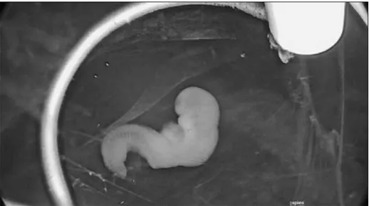
At day 28 after admission, the patient was scheduled for an operative hysteroscopy to remove completely the gestational sac. Hysteroscopic resection was performed under general anesthesia; the cervix was dilated up to 10 mm with Hegar’s dilators, and a bipolar resectoscope, 27-Fr Gynaecare with a 4-mm loop, was introduced. Saline solution was used for the distention and irrigation of the uterine cavity, and the intrauterine pressure was auto-matically controlled with Endomat. A dysmor-phic gestational sac measuring approximately 5 cm, mainly located on the anterior right lateral wall at approximately 0.5 cm from the internal uterine ostium, was visualized (Figure 1). With a 4-mm bipolar loop, a superficial coagulation of vessels was performed in the area of implanta-tion of the gestaimplanta-tional sac. A resecimplanta-tion of the gestational sac under continuous ultrasono-graphic control and continuous monitoring of distension medium was performed. The CP was almost completely resected, avoiding resection of the implanted area of pregnancy due to the presence of a particularly evident, highly vas-cularized peri-trophoblastic zone. Under ultra-sound guidance, an infusion of uterotonic agent was infiltrated and a Foley catheter inserted into the cervix. Rigorous control of hemostasis and meticulous fluid balance were maintained throughout the surgical procedure.
After 31 days from admission, vaginal bleeding was absent and the intrauterine Foley catheter was removed. TVS showed only a minimal flow in the peri-trophoblastic cavity by color Doppler and a βHCG value of 281 mUI/mL. The patient presented a transient leukopenia and stoma-titis that spontaneously resolved. Follow-up
included ultrasonography and a weekly βHCG exam. A month after discharge, no blood flow was detected on color Doppler ultrasonog-raphy and βHCG was negative. Before the hospital discharge, we obtained the patient’s informed consent for the publication of this case. This case report is in accordance with the Declaration of Helsinki and conforms to the Consensus-based Clinical Case Reporting Guideline Development and the Committee on Publication Ethics guidelines.
Discussion
Historically, CPs were difficult to diagnose and were identified at later gestational ages [1]. Because of profuse vascularization and strong blood flow of cervical tissue, CP was often associated with massive hemorrhage, which often led to life-threatening complications and necessitated emergency hysterectomy. Early diagnosis is associated with decrease in mor-bidity, once treatment options are capable of preserving the uterus and subsequent fertility [4]. According to the literature, no consensus exists for treatment.
Conservative management of early ectopic pregnancy using medical treatment alone or in association with minimally invasive procedures has been widely described as an effective strat-egy [7]. MTX is very successful at early gesta-tional ages. A 15% decrease in serum βHCG, between days 4 and 7, is a good indicator of the success of MTX treatment. Most common side effects are mild and include nausea, vomiting, stomatitis, diarrhea, and elevated liver function tests. Rare but severe side effects include neph-rotoxicity, interstitial pneumonitis, and alopecia dermatitis.
Several publications describe successful intra-amniotic MTX administration in monofetal or multifetal CPs as a single approach or combined with adjuvant conservative methods [8]. MTX combined with such methods has a success rate of almost 90%. Systemic administration of MTX in multiple doses has been described as one of the methods of treatment of CP, recommend-ing its use in cases of low gestational age fetuses and in the absence of fetal viability.
According to the most recent literature, conser-vative management of CP with MTX (systemic or local or both) has success rates of 92.7% for multi-dose treatment and 88.1% for single-dose treatment, respectively [1]. In some cases, curet-tage was used as a complementary treatment in combination with MTX [8]. The main problem of curettage is that being a blind method, it can cause injury to the cervical structure, and there
is a high risk of uncontrollable bleeding; there-fore, it is rarely performed.
In our case, we report a novel, conserva-tive, and successful approach for CP using the combination of systemic and local injection of MTX, associated with hysteroscopic resection. A local injection of MTX was performed after the failure of systemic therapy. We opted for hysteroscopic injection (as recently described), as this approach, in comparison with ultraso-nography, offers the main advantage of direct visualization of the site of implantation of the pregnancy, allowing identification of the avas-cular portion of the sac where the 5-Fr needle can be introduced. Indeed, no bleeding was present throughout and immediately after the procedure [1].
Operative hysteroscopy was performed in order to completely remove the gestational sac and prevent retaining of the CP tissue, which can cause serious complications such as per-sistent bleeding (also due to necrosis from the atonic cervix), and infection (i.e. residual tissue can be a culture medium for other infections). The preference for bipolar energy is to reduce the risk of hydro-electrolytic imbalance, when the distension medium used is saline solution, as well as to provide efficient and accurate hemostasis.
From a total of 18 articles found in the litera-ture, hysteroscopy was purely diagnostic in two studies, while in the remaining studies, it was used as a surgical technique with the purpose of hemostasis and/or for resection of the gesta-tional sac [9, 10].
Hysteroscopic resection was successfully per-formed in all of the cases, some of these fol-lowed by methods to reduce bleeding, such as the use of a Foley catheter, fibrillar net, or gauze packing. In none of the articles retrieved, hysterectomy was needed after conservative treatment. Some minor complications were reported, such as cervical diverticulum and intermittent vaginal bleeding.
From the available limited published evidence, the most efficient treatment for CP remains uncertain.
The hysteroscopic approach is a safer, faster, and more accurate technique in comparison with other methods such as curettage, since direct visualization provides a precise resection and coagulation of the ectopic tissue, achieving complete eradication with minimal bleeding.
In our experience, in case of CP with a viable embryo in a woman who desires to preserve her fertility, the combination of systemic and local (hysteroscopic) administration of MTX with operative hysteroscopy offers the possibility of a safe and successful manage-ment, preserving the woman’s reproductive capability.
Informed Consent: Written informed consent was obtained from patient who participated in this study. Peer-review: Externally peer-reviewed.
Author contributions: Concept - A.D.S.S., A.S.L., M.S., C.N., G.B.; Supervision - A.D.S.S., C.N., U.C., G.B.; Data Collection and/or Processing - M.D.C.V., B.C., S.G.V., M.D.F.; Analysis and /or Interpretation - M.D.C.V., A.S.L., S.G.V., M.D.F.; Literature Search - A.D.S.S., B.C., M.S., U.C.; Writing - M.D.C.V., A.S.L., B.C., U.C.; Critical Reviews - S.G.V., M.S., M.D.F., C.N., G.B.
Conflict of Interest: No conflict of interest was declared by the authors.
Financial Disclosure: The authors declared that this study has received no financial support.
References
1. Di Spiezio Sardo A, Alviggi C, Zizolfi B, et al. Cervico-isthmic pregnancy successfully treated with bipolar resection following methotrexate adminis-tration: case report and literature review. Reprod Biomed Online 2013; 26: 99-103.
2. Spitzer D, Steiner H, Graf A, Zajc M, Staudach A. Conservative treatment of cervical pregnancy by curettage and local prostaglandin injection. Hum Reprod 1997; 12: 860-6.
3. Jung SE, Byun JY, Lee JM, Choi BG, Hahn ST. Characteristic MR findings of cervical pregnancy. J Magn Reson Imaging 2001; 13: 918-22.
4. Oyelese Y, Elliott TB, Asomani N, Hamm R, Napoli L, Lewis KM. Sonography and magnetic resonance imaging in the diagnosis of cervico-isthmic preg-nancy. J Ultrasound Med 2003; 22: 981-3. 5. Taylor JE, Yalcinkaya TM, Akar ME. Successful
conservative management of cervical ectopic preg-nancy: a case series. Arch Gynecol Obstet 2011; 283: 1215-7.
6. Kaijser J, Bourne T, Valentin L, et al. Improving strategies for diagnosing ovarian cancer: a summary of the International Ovarian Tumor Analysis (IOTA) studies. Ultrasound Obstet Gynecol 2013; 41: 9-20. 7. De Greef I, Berteloot P, Timmerman D, Deprest J,
Amant F. Viable cervical pregnancy with levonorg-estrel containing intrauterine device, treated suc-cessfully with methotrexate and mifepristone. Eur J Obstet Gynecol Reprod Biol 2005; 120: 233-5. 8. Jeong EH, Kim YB, Ji IW, Kim HS. Triplet cervical
pregnancy treated with intraamniotic methothrex-ate. Obstet Gynecol 2002; 100: 1117-9. 9. Mayer RB, Yaman C, Ebner T, et al. Ectopic
pregnan-cies with unusual location and an angular pregnancy: report of eight cases. Wien Klin Wochenschr 2012; 124: 193-7.
10. Roussis P, Ball RH, Fleischer AC, Herbert CM 3rd. Cervical pregnancy. A case report. J Reprod Med 1992; 37: 479-81.