Role of Epstein-Barr virus in transformation of
follic-ular lymphoma to diffuse large B-cell lymphoma: a
case report and review of the literature
The Epstein-Barr virus (EBV) is the most common human virus, implicated in the pathogenesis of several human tumors, particularly B-cell lymphomas.1 EBV-induced lymphoproliferative disorders are the result of the outgrowth of EBV-infected B cells that would
normal-ly be controlled by an effective EBV-specific cytotoxic T-cell response.2,3The association between EBV and fol-licular lymphoma (FL) is not well recognized and only ten cases have been previously described.4-7 Nine of them showed EBV-encoded small RNA (EBER)-positivity in >75% of the tumor cells and one case in ~5–10%. EBV-positivity was associated with grade 3 FL (9/10 cases), CD30 expression and rapid progression of disease.6 However, it is still unclear whether EBV infection is an early event in the development of FL or a late event that
C
ASE
R
EPORTS
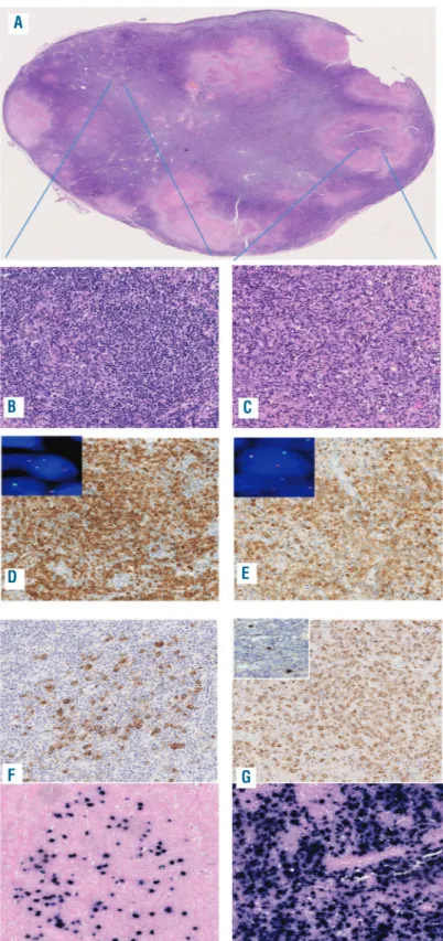
Figure 1. Histological examination of the 2017 specimen.
(A-C) Effacement of the nodal architecture and cellular necrosis (A) with areas of grade 2 follicular lymphoma (B) intermingled with a diffuse proliferation of blastic elements (C) were seen. (D,E) The neoplastic cells of the follicular component expressed BCL-2 (D) as did the diffuse compo-nent (E). Fluorescence in situ hybridization using break-apart probes was positive for the BCL2 translocation in both the follicular (inset 1D) and diffuse (inset 1E) areas. (F,G) Positivity for LMP-2A and EBV-encoded RNA in situ hybridization was present in scattered neoplastic cells with-in the follicles of the follicular areas (F) and with-in 90% of the neoplastic cells in the diffuse areas (G). Immunohistochemistry for Zebra was also positive in scat-tered larger cells in the diffuse areas (inset 1G). Original magnification: (A) 2.5x; (B-E) 10x; (F,G) 20x.
A
B
C
D
E
may contribute to the progression of a pre-existing lym-phoma. We describe a case of EBV-positive diffuse large B-cell lymphoma (DLBCL) that evolved from a previous FL. In April 2011, a 73-year old woman presented with systemic lymphadenopathy and a history of weakness. A right inguinal lymph node was removed for histological examination which showed complete effacement of the nodal architecture by closely packed follicles, with atten-uated mantle zones, loss of polarity and absence of tingi-ble body macrophages. The tumor cells expressed CD20, CD10, BCL-2, BCL-6 and, focally, CD30. The prolifera-tion index (Ki-67) was about 20%. Fluorescence in situ
hybridization using break-apart probes identified the typ-ical BCL2 translocation. Due to the involvement of mul-tiple extranodal sites, a diagnosis of FL, grade 2, stage IVA (Ann Arbor system) was made. The patient was treated with four cycles of rituximab plus fludarabine, mitox-antrone, and dexamethasone, followed by maintenance therapy (rituximab). The treatment was discontinued in 2013 because the disease was progressing. Biopsy of a right axillary lymph node was performed, confirming the previous diagnosis with an increased proliferative index (40%). The patient was placed in a "watch and wait" pro-gram. In March 2016, a follow-up computed tomography
C
ASE
R
EPORTS
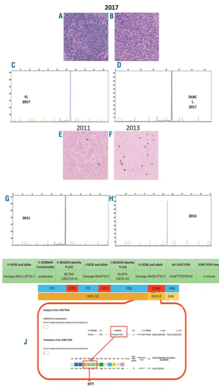
Figure 2. B-cell clonality, an identical N-glycosylation motif and Epstein-Barr virus positivity were demonstrated in all the specimens. (A,B) Laser capture microdissection of the follicular and dif-fuse areas of the 2017 lymph node biop-sy. (C,D) A peak of 316 base pairs (bp) in length was identified in both the follicu-lar and diffuse components. (E,F) EBV-encoded RNA in situ hybridization was positive in the 2011 and 2013 follicular lymphoma samples of the patient, increasing from the first (5%) to the sec-ond (10%) biopsy. (G,H) The clonal analy-sis of the 2011 and 2013 follicular lym-phoma samples identified a peak with the same length as that of the 2017 sample (316 bp). (I) The corresponding FR1-JH polymerase chain reaction prod-ucts of all the samples were directly sequenced and a productive IGH-VDJ rearrangement (V1-18*01, D3-9*01 and J4*02) with 86.76% homology with the germ-line sequence was detected, confirming the clonal relationship. (J) Furthermore, in all the biopsies, we detected an identical somatic hypermu-tation pattern and the same N-glycosyla-tion motif (NTT) in the HCDR3 region, characteristic of follicular lymphoma. Original magnification: (A,B,E,F) 10x. FL: follicular lymphoma; DLBCL: diffuse large B-cell lymphoma.
A
B
C
D
E
G
I
J
H
F
scan revealed enlargement of mediastinal and abdominal lymph nodes and laboratory tests showed increased serum lactate dehydrogenase levels. The patient started the rituximab plus cyclophosphamide, vincristine and prednisolone protocol which was interrupted in March 2017 because of pulmonary intolerance. A new regimen with rituximab and fludarabine per os was started. In November 2017, the patient was hospitalized for recur-rent non-infectious fever and a left inguinal-crural lymph
node biopsy was performed. Histological examination showed the presence of a diffuse proliferation of large lymphoid cells intermingled with areas of FL grade 2 and areas of necrosis (Figure 1A-C). The neoplastic cells of both components expressed PAX-5, CD10, 2, BCL-6, MUM-1 and CD30. The proliferative index was 70% in the areas of diffuse proliferation. Fluorescence in situ hybridization using break-apart probes identified the
BCL2 translocation in both areas (Figure 1D,E). Ninety
C
ASE
R
EPORTS
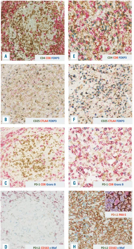
Figure 3. Time trend of evolution of the tumor microenvironment evolution.(A-D) In the follic-ular lymphoma sample, the neoplastic follicles showed a low number of regulatory T cells (A,B) and exhausted T cells (C). Numerous M2 macrophages were also found in the interfollic-ular areas accompanied by occasional M2 macrophages expressing PD-L1 (D). (E-H) The diffuse large B-cell lymphoma biopsy showed increased regulatory T cells (E,F) and exhaust-ed T cells (G). The number of M2 macrophages was increased and the great majority of them expressed PDL-1 (H). PD-L1 expression was also demonstrated in scattered neoplastic cells by double staining with PAX-5 and PD-L1 (H inset). Original magnification: (A-H) 20x.
A
E
F
G
H
B
C
D
percent of the neoplastic cells in the diffuse area and scat-tered cells in the follicular area were positive for Epstein-Barr latent membrane protein (LMP)-2A and EBER in situ hybridization (Figure 1F,G). A diagnosis of DLBCL trans-formed from FL was proposed. Due to the patient’s criti-cal clinicriti-cal condition and high viral loads of cytomegalovirus (32,461 copies) and EBV (24,212 copies) in the serum, the patient was treated with rituximab and lenalinomide plus valgancyclovir for the cytomegalovirus infection. To evaluate whether we were dealing with the transformation of the pre-existing FL, a comparative eval-uation of the original FL and DLBCL specimens was made in terms of clonality, EBV status and tumor microenvironment. To assess B-cell clonality, IGH-VDJ rearrangements were studied in all the specimens accord-ing to the BIOMED-2 protocol. Specifically, to evaluate whether the EBV-positive population observed in the his-tological sections of DLBCL corresponded to the follicu-lar population, we performed laser capture microdissec-tion of the diffuse and follicular areas of the 2017 biopsy (Figure 2A,B). A peak of 316 base pairs (bp) in length was identified in both (Figure 2C,D). We then checked the EBV status also in the 2011 and 2013 FL samples and found the virus in 5% (2011) and 10% (2013) of neoplas-tic cells within the follicles (Figure 2E,F). After laser cap-ture microdissection, the clonal analysis of the 2011 and 2013 FL samples identified a peak with the same length as the 2017 one (316 bp) (Figure 2G,H). The correspon-ding FR1-JH polymerase chain reaction (PCR) products of all the samples were directly sequenced and a productive
IGH-VDJ rearrangement (V1-18*01, D3-9*01 and J4*02)
with 86.76% homology of the germline sequence was detected, confirming the clonal relationship (Figure 2I). Furthermore, in all the biopsies, we detected an identical somatic hypermutation pattern and the same N-glycosy-lation motif (NTT) in the HCDR3 region (Figure 2J). Since the oncogenic role of EBV in leading the transformation of FL to a high-grade form is still controversial, a com-plete genetic assessment of the virus latency by quantita-tive reverse transcription PCR and immunohistochem-istry was performed. The expression of EBV nuclear anti-gen (EBNA)-1 and the negativity for LMP-1 and EBNA-2 in the FL specimen suggested a type I latency. In the DLBCL sample, we detected a non-canonical latency pat-tern of neoplastic cells characterized by the expression of LMP-2A along with some genes/proteins involved in the lytic cycle [BMRF1/Ea-D, BHRF1/Ea-R/p17 and BZLF1/Zebra (Figure 1G inset)]. Finally, we studied the tumor microenvironment of the FL and DLBCL speci-mens to identify a possible alteration of the EBV-host bal-ance responsible for the evolution. In the FL sample, the neoplastic follicles showed a low number of regulatory T cells (CD4+/FOXP3+ and CD25+/FOXP3+/CTLA-4+) (Figure 3A,B) and CD8+/PD1+/Granzyme B+ T cells (Figure 3C). The scenario was different in the trans-formed DLBCL biopsy, which showed increased regula-tory and CD8+/PD1+/Granzyme B+T cells (Figure 3E-G). The latter were concentrated at the periphery of the necrotic areas; the center of the necrotic area showed a significant increase of CD8+ T cells. In the FL sample
numerous CD163+/c-MAF+ macrophages (M2
macrophages) were found in the interfollicular areas accompanied by occasional CD163+/PD-L1+cells (Figure 3D). In DLBCL, the M2 macrophages increased and the great majority expressed PD-L1 (Figure 3H). PD-L1 expression was also demonstrated in scattered neoplastic cells (Figure 3H inset).
The patient’s clinical condition worsened and she died in June 2018.
The process of transformation has some confounding factors: the impact of chemotherapy on the immune microenvironment; the role of the immune system; the proliferative advantage provided by EBV; the impact of EBV latency program modification and expression of additional EBV products; and the natural evolution of FL independently of EBV. Our case favors the hypothesis that EBV was the trigger responsible for the transforma-tion of FL in DLBCL, under permissive immunological conditions. Indeed, the evidence of EBV lytic cycle reac-tivation in the DLBCL sample and the expression of a non-canonical latency program, as demonstrated by quantitative reverse transcription PCR and immunhisto-chemistry, may shed light on the effective role of the virus in the transformation of the disease and in the
maintenance of an immunosuppressive niche.
Conventionally, EBV-driven lymphomagenesis is
thought to involve primarily the latent cycle, but there is increasing evidence that lytic gene products contribute to the development and maintenance of malignancies through the induction of growth factors, oncogenic cytokine production such as interleukin-10, transforming growth factor-β9-11and the evasion of the inflammatory response by attenuation of interferon-g.12 Furthermore, we were able to demonstrate the clonal relationship between the diffuse areas and follicular components in all three biopsies and direct sequencing of the FR1-JH PCR products showed an identical IGH-VDJ rearrangement. These findings were also consolidated by the identifica-tion of an identical somatic hypermutaidentifica-tion pattern and the same NTT which are characteristic of FL and uncom-mon in other B-cell malignancies.8
The role of the virus in the transformation is further supported by the changes induced in the tumor microen-vironment. In fact, a valuable CD4/PD-1 T-cell popula-tion controls the response against the virus in the follicu-lar components.2 Following chemotherapy, the balance between EBV and the host immune system might have been disrupted leading to unchecked EBV activation resulting in non-canonical latency of the virus in the DLBCL sample and changes in the inflammatory microenvironment.9In fact, in the diffuse areas, we found an increased number of T-regulatory cells and CD8+
/PD1+
/Granzyme B+
T cells as well as the expres-sion of PD-L1 on M2 macrophages and lymphoid cells. The PD-1/PD-L1 pathway is a key check-point in
con-trolling the re-education of the tumor
microenvironment,13
diminishing activation of tumor infiltrating lymphocytes and inhibiting phagocytosis and tumor immunity, promoting lymphoma growth.14 This pattern seems to be different from EBV-negative DLBCL in which the number of M2 macrophages expressing PD-L1 is lower than in our case (preliminary, unpublished results from Marafioti et al.).
Notwithstanding this evidence, the precise role of EBV lytic activity and its ability to reshape the tumor microen-vironment remains elusive and further investigation is warranted. However, our findings expand the spectrum of recognized EBV-associated B-cell lymphomas and raise the question as to whether EBV association should be investigated in cases of FL to establish its relationship with lymphomagenesis or transformation.
Massimo Granai,1Maria Raffaella Ambrosio,1Ayse
Akarca,2Lucia Mundo,1Federica Vergoni,3Raffaella Santi,3
Virginia Mancini,1Gioia di Stefano,3Teresa Amato,1
Cristiana Bellan,1Benedetta Puccini,3Ester Sorrentino,1
Kikkeri N. Naresh,4Lorenzo Leoncini,1Teresa Marafioti2and
Stefano Lazzi1
1Section of Pathology, Department of Medical Biotechnology,
University of Siena, Italy; 2Department of Histopathology, University
College London Hospital, UK; 3Section of Pathology, University of
Florence, Italy and 4Hammersmith Hospital & Imperial College,
London UK
Correspondence: LORENZO LEONCINI [email protected]
doi:10.3324/haematol.2018.215053
Information on authorship, contributions, and financial & other disclo-sures was provided by the authors and is available with the online version of this article at www.haematologica.org.
References
1. Dojcinov SD, Fend F, Quintanilla-Martinez L. EBV-positive lympho-proliferations of B- T- and NK-cell derivation in non-immunocom-promised hosts. Pathogens. 2018;7(1):28.
2. Cocco M, Bellan C, Tussiwand R, et al. CD34+ cord blood cell-trans-planted Rag2-/- gamma(c)-/- mice as a model for Epstein-Barr virus infection. Am J Pathol. 2008;173(5):1369-78.
3. Linke-Serinsöz E, Fend F, Quintanilla-Martinez L. Human immuno-deficiency virus (HIV) and Epstein-Barr virus (EBV) related lym-phomas, pathology view point. Semin Diagn Pathol. 2017;34(4):352-363.
4. Orlandi E, Paulli M, Viglio A et al. Epstein–Barr virus-positive aggres-sive lymphoma as consequence of immunosuppression after multi-ple salvage treatments for follicular lymphoma. Br J Haematol. 2001;112(2):373-376.
5. Menon MP, Hutchinson L, Garver J, Jaffe ES, Woda BA.
Transformation of follicular lymphoma to Epstein-Barr virus-related Hodgkin-like lymphoma. J Clin Oncol. 2013;31(5):e53-e56. 6. Mackrides N, Campuzano-Zuluaga G, Maque-Acosta Y, et al.
Epstein-Barr virus-positive follicular lymphoma. Mod Pathol. 2017;30(4):519-529.
7. Mackrides N, Chapman J, Larson MC, et al. Prevalence, clinical char-acteristics and prognosis of EBV-positive follicular lymphoma. Am J Hematol. 2019;94(2):E62-E64.
8. Zhu D, McCarthy H, Ottensmeier CH, Johnson P, Hamblin TJ, Stevenson FK. Acquisition of potential N-glycosylation sites in the immunoglobulin variable region by somatic mutation is a distinctive feature of follicular lymphoma. Blood. 2002;99(7):2562-2568. 9. Cohen M, Vistarop AG, Huaman F, et al. Epstein-Barr virus lytic
cycle involvement in diffuse large B cell lymphoma. Hematol Oncol. 2018;36(1):98-103.
10. Ma SD, Hegde S, Young KH, et al. A new model of Epstein-Barr virus infection reveals an important role for early lytic viral protein expression in the development of lymphomas. J Virol. 2011;85(1):165-177.
11. Hong GK, Gulley ML, Feng WH, Delecluse HJ, Holley-Guthrie E, Kenney SC. Epstein-Barr virus lytic infection contributes to lympho-proliferative disease in a SCID mouse model. J Virol. 2005;79(22):13993-14003.
12. Long X, Li Y, Yang M, Huang L, Gong W, Kuang E. BZLF1 attenuates transmission of inflammatory paracrine senescence in Epstein-Barr virus-infected cells by downregulating tumor necrosis factor alpha. J Virol. 2016;90(17):7880-7893.
13. Kim JM, Chen DS. Immune escape to PD-L1/PD-1 blockade: seven steps to success (or failure) Ann Oncol. 2016;27(8):1492-1504. 14. Pham LV, Pogue E, Ford RJ. The role of macrophage/B-cell
interac-tions in the pathophysiology of B-cell lymphomas. Front Oncol. 2018;8:147.