ABSTRACT
Aim In this article the fabrication and use of new type 3D
printed splint of retainer after orthodontic treatments is reported.
Case report Two cases, one of an adoescent female patient
and the other of an adult female, are presented, describing step-by-step the clinical and laboratory procedures. The controls after 6 months are also reported.
Conclusion Further randomized clinical trials are required
in order to evaluate durability and efficacy and periodontal parameters in patients treated with this new type of retainer.
iNTRoDuCTioN
Retention is considered an important phase of orthodontic treatment, as there is a tendency for teeth to return to their initial position if retention is not prepared and luted (1).
In order to avoid relapse after orthodontic treatment, in 1973 Knierim introduced the direct bonded retainer (2, 3), which is still frequently used to prevent the relapse of crowding in the mandibular anterior region (2, 3, 4). In 1977, Zachrisson introduced the multistranded bonded lingual retainer, which, although varying in different shapes, has become the gold standard (5, 6). Orthodontic retainers are also known to have an effect on periodontal health. Various types of retainers have been described with wires of differing materials,
properties and diameters (7), or using different types of composites for the adhesion (8) or with fiber reinforcement (9).
Two recent articles reported the fabrication of a custom lingual retainer cut from a nickel-titanium block with CAD/CAM technology and a CAD/CAM Zirconium bar as a bonded mandibular fixed retainer (10, 11).
In recent years a new possibility is available for dental practitioners: CAD-CAM technology and 3D printing (12, 13), which find their application in all aspects of prosthodontics (14-18).
These new types of technologies can be useful in order to perform different dental treatments, from implantology (19, 20) to prosthodontics (21). More specifically, different indications to the use of CAD-CAM and 3D printers can be found also in orthodontics (22-24). Orthodontists are already familiar with several products that use 3D printers (i.e. invisible aligners), also known as additive manufacturing, 3D printing is a technology whereby sequential layers of material are layered on top of one another to form an object (25). The American Society for Testing and Materials defined additive manufacturing as “the process of joining materials to make objects from 3D model data, usually layer upon layer, as opposed to subtractive manufacturing methodologies” (12).
Since 1986, when Hull established the 3D Systems Company to market the first machine for rapid prototyping, which he called stereolithography (SLA), dozens of 3D printers employing variations of SLA, fused deposition modeling (FDM), and PolyJet photopolymer (PPP), technologies have become available (26).
Besides, different materials are now available for 3d printing: metal, ceramic and polymers. Within the category of polymers, photoinitiated resins can be found (27). Stereolithography (SLA) machines usually use this type of polymer as a build material, curing one layer at a time using UV light or laser. These polymers offer much more flexibility and options in color, rigidity, and modification of components (15).
The accuracy of the structures produced varies according to the geometries being replicated, the method of manufacture, and the materials being used. SLA can fabricate structures with a layer thickness of 25
Department of Periodontology, Universidad Complutense, Madrid, Spain
3 Department of Restorative Dentistry, Leeds University, UK
To CITe ThIS aRTICLe
Doldo T, Di Vece L, Ferrari Cagidiaco e, Nuti N, Parrini S, Carboncini F, Ferrari M. a new generation of orthodontic retainer using 3d printing technology: report of two cases. J osseointegr 2018;10(4):142-148.
DoI10.23805 /Jo.2018.10.04.06
μm (16). Precision and accuracy of fit are also important in terms of patient comfort and gingival health.
In daily dentistry, in order to make a customized lingual retained, a digital impression and its .stl file are needed in the first place. The .stl file can be generated by a digital impression made with an Intra Oral Scanner (iOS) or after scanning the stone model made by conventional procedure. In both cases the generated .stl file will be elaborated by the software of the 3D printer and the final piece designed and printed.
The main advantages to use an iOS for digital impressions are essentially the higher comfort for the patient, the less working time needed, the relatively lower cost (14). Also, it should be considered the possibility to avoid any physical model, following a complete digital workflow procedure (14) or, in case it is preferred by operators, to prototype the model printing it in resin using the same 3D printer (22). However, both procedures can be performed successfully.
For these reasons, in the present article, for the first time to our knowledge, the use of a 3D printing technique for the production of customized lingual retainers is reported
CASe RePoRTS
Case 1
A 19 year old patients (AB) had undergone orthodontic therapy for the resolution of a complex malocclusion. At the end of orthodontic treatment, the brackets of the upper arch were removed.
Due to an altered Bolton index, the patient underwent a conservative treatment of the upper lateral incisors (Fig. 1).
Digital impressions (Aadva iOS) for both arches were made.
The upper model, prepared with 3D printer, was used to make a clear thermoplastic retainer that was delivered to the patient within 5 days from the removal of the brackets. Computer-aided design and manufacturing (CAD/CAM) software was used to further process the file and prepare it for printing (Fig. 2).
The .stl file was sent to a 3D printer (ASIGA) (Fig. 3). The retainer was prepared using an experimental resin (Genial Printing Resin, GC).
To avoid problems during cementation of the retainer a copy was made.
Once the retainer was prepared, the patient, who had previously undergone professional oral hygiene, was
FIg. 1orthodontic patient at the end of her treatment in need to stabilize lower anterior teeth. The Upper laterals were restored with direct resin composite restorations for esthetic purpose.
FIg. 2after taking a intra-oral digital impression, the retainer was designed by the software.
FIg. 3The retainer project was transferred to the 3D printer software and finalized.
prepared for cementation of the retainer. The operative field was isolated by rubber dam.
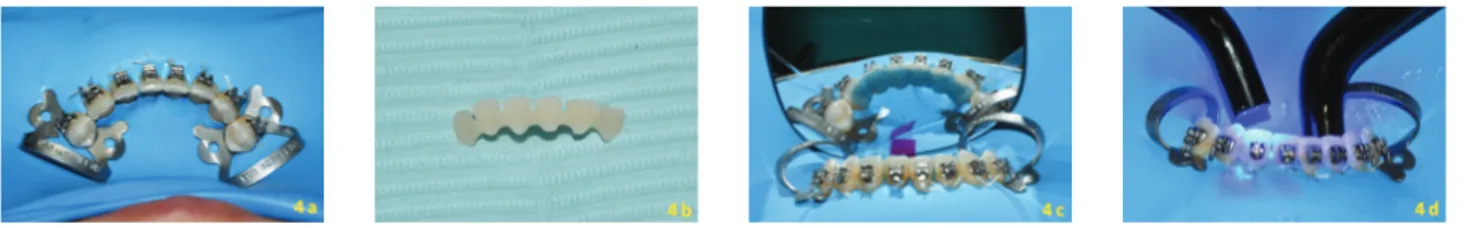
The lingual aspect of the lower anterior teeth (from canine to canine) were etched with 32% orthophosphoric acid for 45 seconds, then washed with water and air-dryed. Then the teeth were treated with adhesive (Premio Bond Universal Adhesive System, GC) and light-cured for 40 seconds. Meanwhile the retainer was silanized for 1 minute with MultiPrimer (GC). MutliLink Force (GC)
was used for cementation (Fig. 4). The internal surface of the resin splint was sandblasted with CoJet device (3M-ESPE).
Once the surfaces of teeth and retainer were prepared, a very thin layer of cement was applied to the retainer, which was gently placed in contact with the dental surfaces.
A constant pressure was applied on the splint to allow the excess cement to be removed before polymerization. Once the polymerization was completed, the rubber dam removed, the brackets of the lower arch were debonded and the patient was sent home with instructions to clean the retainer (Fig. 5).
The patient returned to controls after 7 days, one and six months and no detachment nor fractures of the retainer was noted (Fig. 6). Also, the patient showed no accumulation of plaque on the splinted teeth. At the 6-month recall there was not any bleeding on probing nor any other sign of gingivitis (Fig. 6).
Case 2
An adult female patient required orthodontic treatment to align the lower incisors. It was decided to make a fast movement of the incisors using an elastic device (Fig. 7). After two weeks the teeth were in the same line and an intraoral digital impression was made and a .stl file was generated (Fig. 8).
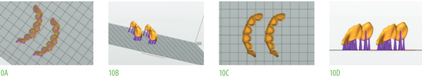
Using alab scanner (Aadva) the .stl file was processed, and the project of an adhesive lingual retainer (Fig. 9). Then the new file was transferred to the 3D Printer (Asiaga) and the adhesive retainer was programmed to be printed (Fig. 10).
Two twin retainers were obtained (Fig. 11).
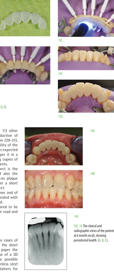
Next, the lower teeth were isolated with rubber dam and the luting procedure was performed, as described in case 1 (Fig. 12, 13). Thin microbrushes were used to protect interproximal spaces and for an easier and
FIg. 4The field was isolated by rubber dam, interproximal spaces protected by microbrushes, and the 3D printed retainer, after being sandblasted, was luted with dual-cure resin composite cement (LinkForce, gC).
FIg. 5The retainer immediately after removing the rubber dam. FIg. 6The retainer at a 6 month recall.
FIg. 7Clinical situation of a periodontal involved lower incisors of a elderly patient. The incisors were repositioned simply with an elastic placed buccaly.
better cleaning of these areas from resin cement excess. After 6 months the patient showed good esthetics, and comfort and good plaque control (Fig. 14).
DiSCuSSioN
For the first time cases a reported in which orthodontic retainers were made using a 3D printer.
Since there is no literature on this topic, comparisons cannot be made with other retainers made in the same technique.
The techniques presented above offers several
advantages compared to the direct techniques for chairside splinting (multistrand stainless steel retainers or fiber-reinforced composites).
First of all the possibility of drawing the retainer taking into account the different anatomical details (more or less accentuated tracks, height of the contact points, etc.) with an adaptability of the retainer that cannot be achieved with direct techniques. It can be possible to obtain a larger contact surface and therefore a greater adhesion of the retainer to the teeth and decidedly superior aesthetics than that of direct techniques. When this new splint is compared to the indirect digital techniques (subtractive manufacturing) already present
FIg. 8Different views of the screen of the ioS (a, B, C, D) (aadva, gC) and two views of the generated .stl file (e, F).
FIg. 9Some views of the designed lingual retainer (a, B, C, D, e, F).
8a 9a 8D 9D 8B 9B 8e 9e 8C 9C 8F 9F
FIg. 12Clinical steps of luting procedure: rubber dam always in place (C, D, e, F, g). 12a 11a 11D 12D 12B 11B 11e 12e 12C 12g 11C 12F FIg. 10Some views of the software ready to print the buccal retainer (a, B, C, D).
FIg. 11Two retainers were printed for the same patient (a, B, C, D, e).
in the literature (zirconia and similar) (10, 11) other advantages can be found, such as the reduction of wasted raw material, the speed of fabrication (29-31), the greater flexibility and therefore adaptability of the final retainer, the cost of the product (which is expected to decrease) and also the possibility to reaper it in a short time and with reasonable cost making copies of the retainer in case of breakages or detachments. However, the most important positive aspect is the facility to clean resin cement excess and also the easy maintenance at home. The fact that no plaque accumulation was not noted, although after a short time of clinical service, is a very positive aspect.
The possibility of repairing a fractured retainer and of course a long-term follow-up of the cases treated with this new technique remains to be investigated.
A randomized clinical trial on patients in need to be splinted after orthodontic treatment is on the road and will be reported in due time.
CoNCluSioNS
Based on specific need of the patients, two cases of resin printed splints were presented. From the short term clinical observations reported in this paper the following conclusions can be drawn: the use of a 3D printed retainer may be considered as a possible alternative to conventional multistrand stainless steel retainers or fiber-reinforced composites retainers for
FIg. 14The clinical and radiographic views of the patient at 6 month recall, showing periodontal health (a, B, C). 13a 14a 14B 14C 13D 13B 13e 13C 13g 13F
FIg. 13...the sandblasted internal surface of the retainer (B), protection with microbrushes of the interproximal spaces (C, D, e) and the incisors after the retainer has beinf luted (F, g, h).
ReFeReNCeS
1. Littlewood SJ, Millett DT, Doubleday B, Bearn DR, worthington hV. Retention procedures for stabilising tooth position after treatment with orthodontic braces. The Cochrane Library. 2016 Jan 1.
2. al yami ea, Kuijpers-Jagtman aM, van’t hof Ma. Stability of orthodontic treatment outcome: follow-up until 10 years postretention. american Journal of orthodontics and Dentofacial orthop 1999 Mar 1;115(3):300-4. 3. Knierim Rw. Invisible lower cuspid to cuspid retainer. The angle
orthodontist 1973 apr;43(2):218-220.
4. Dahl e. Long-term experience with direct-bonded lingual retainers. J Clin orthod 1991;25:619-30.
5. Zachrisson BU. Clinical experience with direct-bonded orthodontic retainers. am J orthod 1977 apr 1;71(4):440-8.
6. Bearn DR. Bonded orthodontic retainers: a review. am J orthod Dentofacial orthop 1995 aug 1;108(2):207-13.
7. Baysal a, Uysal T, gul N, alan MB, Ramoglu SI. Comparison of three different orthodontic wires for bonded lingual retainer fabrication. Korean J orthodontics 2012 Feb 1;42(1):39-46.
8. Pandis N, Fleming PS, Kloukos D, Polychronopoulou a, Katsaros C, eliades T. Survival of bonded lingual retainers with chemical or photo polymerization over a 2-year period: a single-center, randomized controlled clinical trial. am J orthod Dentofacial orthop 2013 aug 1;144(2):169-75.
9. geserick M, Ball J, wichelhaus a. Bonding fiber-reinforced lingual retainers with color-reactivating flowable composite. J Clin orthod 2004 oct;38:560-2. 10. Zreaqat M, hassan R, hanoun aF. a CaD/CaM Zirconium bar as a bonded
mandibular fixed retainer: a novel approach with two-year follow-up. Case Reports Dentistry 2017;2017.
11. Kravitz ND, grauer D, Schumacher P, Jo yM. Memotain: a CaD/CaM nickel-titanium lingual retainer. am J orthod Dentofacial orthop 2017 apr 1;151(4):812-5.
12. Nikzad, a., The evolution of rapid prototyping in dentistry: a review. Rapid Prototyping J 2009; 216-222.
13. Van Noort R. The future of dental devices is digital. Dental Materials 2012 Jan 1;28(1):3-12.
14. Joda T, M. Ferrari M. Chairside protocol for posterior single-unit implant
digital impression of sound teeth. accepted in J osseointegration 2018. 18. Pecciarini M, Biagioni a, Ferrari M. a systematic review of clinical trials on
digital impression of prepared teeth. accepted in J osseointegration 2018. 19. Sykes LM et al. applications of rapid prototyping technology in maxillofacial
prosthetics. Int J Prosthodont 2004; 17(4): 454-9.
20. Chan hL, Misch K, wang hL. Dental imaging in implant treatment planning. Implant Dent,2010. 19(4): 288-98.
21. Miyazaki T, hotta y. CaD/CaM systems available for the fabrication of crown and bridge restorations. aust Dent J 2011; 56 Suppl 1: p. 97-106.
22. Ciuffolo F et al. Rapid prototyping: a new method of preparing trays for indirect bonding. am J orthod Dentofacial orthop 2006; 129(1): 75-7. 23. goracci C, Franchi L, Vichi a, Ferrari M.accuracy, reliability, and efficiency
of intraoral scanners for full-arch impressions: a systematic review of the clinical evidence. eur J orthod 2015.
24. Tuncay oC. The Invisalign System. New Malden: Quintessence Publishing Co., Ltd. 2006.
24. Mandelli F, gherlone eF, Keeling a, gastaldi g, Ferrari M. Full-arch intraoral scanning: comparison of two different strategies and their accuracy outcomes. J osseointegr 2018;3: 65-74.
25. Shulman h, Spradling D, hoag C. Introduction to additive manufacturing. Ceramic Industry 2012;162(12):15-9.
26. hull C. StereoLithography: Plastic prototypes from CaD data without tooling. Modern Casting 1988 aug;78(8):38.
27. Barazanchi a, Li KC, al‐amleh B, Lyons K, waddell JN. additive technology: update on current materials and applications in dentistry. J Prosthod 2017 Feb;26(2):156-63.
28. Venkatesh KV, V.V. Nandini, Direct metal laser sintering: a digitised metal casting technology. J Indian Prosthodont Soc 2013; 13(4): 389-9.
29. groth Ch, Kravitz ND, Jones Pe, graham Jw, Redmond wR. Three-dimensional printing technology. J Clin orthod 2014 aug;48(8):475-85. 30. Berman B. 3-D printing: The new industrial revolution. Business horizons.
2012 Mar 1;55(2):155-62.
31. abduo J, Lyons K, Bennani V, waddell N, Swain M. Fit of screw-retained fixed implant frameworks fabricated by different methods: a systematic review. Internat J Prosthod 2011 May 1;24(3).
32. Madurantakam P, Kumar S. Fixed and removable orthodontic retainers and periodontal health. evid Based Dent 2017 Dec 22;18(4):103-104.