J. Mol. Biol. (1983) 165, 125-138
X-ray Absorption Near Edge Structure (XANES)
Determination of Calcium Sites of Troponin C and
Parvalbumin
A. BIANCONI, A. GIOVANNELLI, L. CASTELLANI t
Istituto di Fisica dell' Universit5 di Roma, 00185 Roma, Italy. S. ALEMA
Istituto di Biologia Cellulare del CNR, Via Romagnosi 18A, 00196 Roma, Italy P. FASELLA, B. OESCH
Istituto di Chimica Biologiea d'ell' Universitd di Roma, 00185 Roma, Italy AND
S. MOBILIO
P U L S , Laboratori Nazionali di Frascati, 00044 Frascati, Italy (Received 7 April 1982, and in revised form 30 October 1982) Using synchrotron radiation at the Frascati storage ring ADONE, the X-ray Absorption Near Edge Structure (XANES) has been applied to determine homologies and modifications of the local structure of the caicium binding sites of troponin C. In all four calcium binding sites, Ca 2+ appears to be co-ordinated to earboxyl and carbony! groups in a characteristic configuration. No structural difference has been found between high and low-affinity sites. A distortion of the Ca 2+ site geometry by binding of Mg 2+ has been observed.
The XANES of parvalbumin has been measured and found to be different from troponin C. A tentative identification of the characteristic XANES spectra of the two different Ca 2 ÷ sites in this protein is reported.
1. I n t r o d u c t i o n
Several biological functions are regulated b y changes in the concentrations of cytosolic calcium. Ca 2+ sensitivity is conferred b y specialized, sequence- homologous proteins such as troponin C, calmodulin and p a r v a l b u m i n (Kretsinger, 1980). I n carp p a r v a l b u m i n (Moews & Kretsinger, 1975), and in the v i t a m i n D- d e p e n d e n t calcium-binding protein (Szebenyi et al., 1981), the only two Ca 2+ m o d u l a t e d proteins whose crystal s t r u c t u r e has been d e t e r m i n e d , a unique configuration called " E F h a n d " has been identified in the region of the two Ca 2+- binding sites. In this configuration, Ca 2 + is b o u n d m a i n l y to c a r b o x y l and carbonyl t Present address: Rosenstiel Basic Medical Science Research Center, Brandeis University, Waltham, MA, 02254, U.S.A.
125
126 A. BIANCONI ET AL.
groups in a loop between two s-helices. A comparison of amino acid sequences of different Ca2+-binding proteins with that of parvalbumin indicates that homologous, helix-loop-helix domains are present in many proteins of this class (Kretsinger, 1980) like TnCt (Kretsinger & Barry, 1975).
Calcium ion, having the electronic configuration of a rare gas atom, is devoid of magnetic and optical response and is silent to spectroscopic analysis such as electron paramagnetic resonance or optical absorption. An additional difficulty in the study of the binding of calcium in inorganic and organic systems arises from the large variety of possible configurations and the asymmetry of the binding sites. For instance, a large variety of Ca 2 +-ligand distances has been generally observed in the first co-ordination shell (Williams, 1977).
X-ray absorption spectroscopy, using synchrotron radiation (Doniach et al., 1980) is a powerful emerging technique for high-resolution studies of metal binding sites in proteins because it provides a probe for a specific atom in a complex system, and it gives information on the local structure of the metal binding site, being sensitive to short-range order in atomic arrangements rather than to long-range order. This technique makes feasible the study of local structures when single crystals are not available and appears particularly suitable for the investigation of Ca2+-binding sites in biological systems (Bianconi et al., 1978,1980; Powers et al., 1978; Miller et al., 1982).
Three parts of an X-ray absorption spectrum can be distinguished on the basis of both structural information and physical photoionization processes:
(1} the edge region, extending over ~ 8 eV at the absorption threshold;
{2) the XANES (Bianconi, 1981) in energy range of ~ 4 0 eV above the edge; and (3) the E X A F S (Doniach et al., 1980) at higher energy.
The edge region is determined by the first allowed transitions from the core to empty molecular orbitals. It is therefore sensitive to the covalency of metal-ligand bonds and to the effective charge of the absorbers (Bianconi, 1981). E X A F S has been widely used as a structural technique for proteins both to determine completely unknown local structures, as in cytochrome oxidase (Powers et al., 1981), and to obtain more accurate interatomic distances in proteins already well- studied by X-ray diffraction, as in haemoglobin (Eisenberger et al., 1978). The advantageous simple characteristics of E X A F S are also its limitations. Generally, no information on co-ordination geometry (bonding angles) can be extracted and serious limitations appear with non-ordered structures (Eisenberger & Lengeler, 1980). Only in the presence of first and second neighbours in colinear fashion can bonding angles be determined by E X A F S through the "focusing" effect (Teo, 1981). In the EXAFS region, the wave function of the excited photoelectron can be described b y a simple theory: that the high kinetic energy photoelectron, extracted from the absorber (the central atom), is weakly backscattered by one of the neighbour atoms in a single-scattering process. This gives information about local structures only in terms of atomic radial distribution (distances) around the central atom within only ~ 4 A (short-range).
t Abbreviations used: TnC, troponin C; XANES, X-ray absorption near edge structure; EXAFS, extended edge X-ray absorption fine structure.
TROPONIN C AND PARVALBUMIN CALCIUM SITES 127 T h e X A N E S (Belli el al., 1980; Bianconi, 1981) contains information on t h e stereochemical details (co-ordination g e o m e t r y and bond angles) t h a t are particularly i m p o r t a n t for complex s y s t e m s such as proteins, characterized b y weak order a n d low s y m m e t r y . I n the p h o t o i o n i z a t i o n process, the low kinetic energy (10 to 40 eV) excited photoelectron is strongly backscattered b y neighbour atoms, generating a multiple-scattering process. I t is for such multiple-scattering involving several a t o m s t h a t X A N E S is i n f o r m a t i v e on the relative positions of the neighbour atoms. R e c e n t theoretical progress ( D u r h a m et al., 1981 ; K u t z l e r et al., 1980; Bianeoni et al., 1982) shows t h a t X A N E S is determined b y higher order pair- correlation functions of neighbour a t o m distribution, while E X A F S gives only the first order pair-correlation function. Because X A N E S is a s t r u c t u r a l probe o f a cluster of 15 to 30 a t o m s including the second shell o f neighbour atoms, the proposed e x p e r i m e n t a l approach (Bianconi et al., 1978) to the s t u d y o f local s t r u c t u r e s by X A N E S can be used.
In this paper, we h a v e used X A N E S spectroscopy to s t u d y the Ca2+-binding sites of TnC and p a r v a l b u m i n . TnC is the Ca 2 +-binding s u b u n i t of the troponin complex t h a t is p a r t of the r e g u l a t o r y system of muscle contraction. I t binds four Ca 2+ ( P o t t e r & Gergeley, 1975) : two a t high affinity, which also bind Mg 2+, and two a t low affinity, specific for Ca 2+. The presence of Mg 2+ appears to equalize the affinity of the four binding sites. We have f o u n d t h a t all four Ca2+-binding sites have the same co-ordination n u m b e r and similar s t r u c t u r e up to the second shell of neighbour atoms. Mg 2+ induces a modification of the Ca 2+ binding. T h e X A N E S spectrum of the two Ca 2+,Mg2+-binding sites o f carp p a r v a l b u m i n is different from t h a t of TnC, and is consistent with the presence of two different local co-ordinations (Moews & Kretsinger, 1975).
2. Materials and M e t h o d s (a) Materials
Salts in crystalline form were Suprapur products from Merck. Water, double-distilled and deionized, was further filtered through a Chelex-100 resin to reduce metal contamination. Plastic-ware was used during the preparation of proteins and samples to avoid calcium contamination from glass.
(b) Proteins
TnC was prepared from rabbit skeletal muscle as described by Perry & Cole (1974). Parvalbumins were isolated from carp white muscle according to the methods of Pech~re et al. (1971) and Kretsinger & Nockolds {1973). The pl. 4.25 component was used in this study. The purity of each preparation was checked by sodium dodecyl sulphate]polyacrylamide gel electrophoresis (Laemmli, 1970). The biological activity of TnC was tested by ATPase measurements in a reconstructed acto-myosin S1 system (Castellani et al., 1980).
(c) Sample preparation
Protein-metal complexes were prepared by dissolving freeze-dried proteins in Ca 2 +-free water (Ca 2 +concn 10- 6 M) and by adding CaC12 or TbC12 to obtain the desired protein-metal steichiometric ratio. Mg 2+ was added in molar excess (2 to 4 mM). Pr0tein-metal complexes in solution were then freeze-dried and the powder was placed into plastic holders mounted with Parafilm windows. The concentration of Ca 2 + in the samples was determined by atomic
128 A. BIANCONI ET AL.
absorption before and after the addition of calcium. Protein concentration was quantified by optical absorption measurements using molar extinction coefficient values of 2670 for TnC and 2000 for carp parvalbumin. Serial recordings of XANES spectra of the same samples were very similar, indicating that no major change occurred during the exposure to the X- ray beam. TnC samples were further checked for denaturation and capability to respond to binding of Ca 2 +. Intrinsic protein fluorescence measurements and circular diehroism spectra indicated no major difference in TnC before and after exposure to radiation.
(d) Methods
The storage ring ADONE at the synchrotron radiation facility PULS at Frascati was used as the X-ray source. I t was operated at 1"5 GeV and I = 60 mA. The X-ray beam was monochromatized by a Si(220) channel-cut single crystal at about 17 m from the source. The X-ray absorption data were obtained by transmission measurements. The high stability, high collimation (/18 -- 5 x 5 -5 rad) and the high intensity of the X-ray beam gave spectra of high resolution (AE/E ~ 10 -4) and good signal-to-noise ratio. XANES spectra were recorded in the energy range 4000 to 4150 eV with steps of 0-1 eV. The energy scale was calibrated at each electron beam injection in the storage ring, using calcium formate as a reference sample. The electron beam position in the ADONE storage ring at the emission point was directly controlled so that no change in the energy positions of absorption peaks larger than _+0-1 eV was observed during the lifetime of the electron beam in the storage ring. The electron beam size at the emission point was 0"7 mm, and an entrance slit of the same size was used in the X-ray monochromator. The angle of the X-ray monochromator is measured by an absolute encoder independent of the stepping motor drive. Data analysis of XANES spectra was carried out by plotting the relative absorption ~M(Ii¢o)/~A, where a M is the measured absorption coefficient and aa is the value of the high energy Ca 2 + atomic X-ray absorption, obtained by fitting the EXAFS oscillations. This procedure allows the normalization of the spectra of different Ca 2+ compounds to the same a A value. EXAFS data analysis was carried out using the EXAFS set of programs developed at the facility (S. Mobilio, F. Comin & L. Ineoecia, unpublished results).
3. Results
(a) E X A F S and X A N E S of T n C
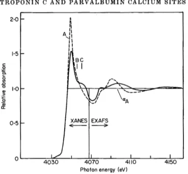
Figure 1 shows the E X A F S a n d X A N E S of TnC a n d of the model c o m p o u n d calcium f o r m a t e . T h e s e p a r a t i o n b e t w e e n t h e X A N E S a n d E X A F S region has been fixed a t the p h o t o e l e c t r o n kinetic e n e r g y ~¢o-Eo(eV ) = 151/d2(A), as discussed b y Bianconi (1981), where E o is t h e a b s o r p t i o n threshold a n d d is t h e C a 2 + - o x y g e n i n t e r a t o m i c distance. This criterion gives the threshold of E X A F S oscillations a b o v e the value of 2"6 A - 1 of the p h o t o e l e c t r o n w a v e v e c t o r .
T h e analysis of E X A F S gives an " a v e r a g e " C a 2 + - o x y g e n i n t e r a t o m i c distance o f 2"4 A for all the C a 2 + - m o d u l a t e d p r o t e i n s we h a v e studied. Ca2+-binding proteins belong to a special case where t h e E X A F S analysis is s t r o n g l y l i m i t e d b y m a n y g r e a t l y s c a t t e r e d distances in t h e first co-ordination shell. I t is c h a r a c t e r i s t i c of Ca 2 + bonding in Ca 2 + complexes a n d p r o t e i n s t h a t m a n y (more t h a n four) Ca 2 + - o x y g e n distances are s p r e a d o v e r a 0"3 to 0.6 A r a n g e (Moews & K r e t s i n g e r , 1975 ; E i n s p h a r & Bugg, 1974,1977 ; B u r g e r et al., 1977). I n this case, a single " a v e r a g e " distance is d e t e r m i n e d b y E X A F S analysis f r o m t h e p h a s e d e t e r m i n a t i o n o f the oscillations due to the first n e i g h b o u r a t o m s , o b t a i n e d b y F o u r i e r filtering of t h e m e a s u r e d s p e c t r u m . This single oscillation is given b y (Doniach et al., 1980):
c'.a. A(k)
X(k) = 2 --/:-~sin (2kR,+O(k)),
T R O P O N I N C A N D P A R V A L B U M I N C A L C I U M S I T E S 129 2,0 1.5 c .o I'0 0.5 405O I ii L / .
X~ NES l EXAFSS
._.L ~ L ~
i
,
I
4070 41 I0 4150Pho?on energy (eV)
FIG. 1. X-ray absorption spectra of TnC (TnC + 2CA 2 + and TnC + 4Ca 2 + ; broken line) and calcium formate (unbroken line). The EXAFS and XANES regions are indicated. The relative absorption ~M0~)~A is plotted, where aM(~5~) is the measured absorption, after subtraction of the pre-edge continuum background, and a A is the value of the high energy Ca 2 + atomic X-ray absorption continuum obtained by fitting the measured spectrum in the E X A F S region.
where C.N. is the co-ordination number (from 6 to 8 in calcium-binding proteins) and Ri are the distances of oxygen atoms from the calcium atom. Since only oxygen atoms are expected to be co-ordinated by Ca 2 +, A ( k ) and 0(~k), characteristic of the Ca 2 + - 0 2- pairs, can be determined easily from model compounds. In the case of six or eight different distances Ri distributed in a complex way, it is clear that E X A F S gives an average distance that is not directly related to the arithmetic mean distance. The analysis of the low-energy range of E X A F S (see Fig. I) can give information on the interatomic distances (and their modifications) of a larger cluster (including the second and possibly the third shell) of the same size as that determining the XANES (Belli et al., 1980) but its analysis is not simple in these complex structures.
In order to avoid the limitations of E X A F S applied to strongly asymmetric Ca2+-binding sites, we have analysed the XANES region of the absorption spectrum. The multiple-scattering resonances, which determine the features of XANES of Ca 2 + complexes, are determined by the spatial arrangements of the first and second neighbours around the central atom Ca 2+ (Durham et al., 1981; Bianconi et al., 1982). Since in Ca2+-modulated proteins Ca 2+ is co-ordinated by carboxyl groups of aspartic acid and glutamic acid residues, and by carbonyl groups of the main-chain, the X A N E S are determined both by the co-ordination geometry of oxygen first neighbours and b y the carbon second neighbours. I t has been pointed out (Bianconi et al., 1982) that if the central atom is co-ordinated by molecular groups with multiple bonds like COO- and CO, the XANES are strongly determined by "shape resonances" of the excited photoelectron within the
130 A. B [ A N C O N I E T AL. 2 . 0 .Q o 1.0; 0 o (.9 LU $ 0 - ( o ) B - (b)
/
/ (c) Et E2 /7
I 4 0 4 0 I I I i I I I 4 0 5 0 4 0 6 0 4 0 7 0 Photon energy (eV)FIO. 2. X A N E S spectra of TnO (Tn0-2Ca 2+ and TnO-4Ca 2+ : unbroken line) and Ca-EGTA (broken line). (a) Absorption spectra; (b) derivative of absorption spectra; and (c) difference between the absorption spectra in the "edge region" of TnO-2Ca and Ca-EGTA. Data acquisition time for each s p e c t r u m was l h with 5 s integration t i m e for each point. T h e error on t h e a b s o r p t i o n coefficient due to s t a t i s t i c s is as small as t h e t h i c k n e s s of t h e curves. T h e low f r e q u e n c y noise d u e to instability of t h e source a n d / o r a p p a r a t u s gives t h e s p u r i o u s weak s t r u c t u r e s in t h e d e r i v a t i v e spectra, which can be well seen overlapping t h e broad X A N E S features B a n d C in t h e s p e c t r a of t h e proteins.
neighbour molecular groups. The characteristic multiple-scattering resonances A, B and C of calcium-binding proteins and model compounds, shown in Figure 1, should be derived by the ~r and a-type shape resonances in scattering of low kinetic energy electrons (4 to 20 eV) observed in CO 2 and CO (Lynch et al., 1979). Therefore, the XANES of Ca 2+ co-ordinated by carboxyl and carbonyl groups should depend,
TROPONIN C. AND PAI:tVALBUMIN CALCIUM SITES 131 through the variation of intensity and energy shift, on the co-ordination number C.N., on the Ca2+-oxygen distances and on the Ca2+-O-C bonding angles.
In order to find out experimentally the effect of local structure on XANES, we have studied some calcium complexes where Ca 2 + is co-ordinated by a carboxyl group, such as calcium formate (Burger et al., 1977), calcium glutamate (Einsphar & Bugg, 1974), Ca-EDTA (Weakliem & Hoard, 1959; Smith & Hoard, 1959) and Ca-EGTA (ethyleneglycol-bis(fl-aminoethyl ether) N,N'-tetra-acetic acid). The energy position of the main resonance A shifts toward higher energy by about 1 eV by increasing C.N. from six to eight, in agreement with the finding of Powers et al. (1978). The splitting between peaks A and B goes from 6 eV in 6-fold co-ordinated sites of EDTA to 7"5 eV for the 7-fold co-ordinated site of calcium formate. In the 6- fold co-ordination, where C O 0 - is generally in the unidentate configuration with a Ca-O-C bonding angle of about 120 to 150 ° (Einsphar & Bugg, 1977), peak B is stronger than for the 7 or 8-fold co-ordination, with at least one carboxylate in the bidentate configuration with the bonding angle 0 - 90 °.
The comparison of XANES spectra of TnC with the model compound calcium formate (Fig. I) shows a general feature of XANES for calcium-binding proteins: peak A is stronger in proteins (about a factor of 2 larger than the high-energy atomic continuum A) than in simple compounds. In TnC the energy of peak A is at lower energy than in calcium formate and the two spectra are quite different. In Figure 2 the XANES spectrum of CaEGTA shows a close resemblance to t h a t of TnC. In the lower part of the Figure, the derivative functions of the spectra show t h a t the energies of the A, B and C structures are the same in TnC and EGTA. The energy position of peak A at 10"5_ 0"2 eV above the first structure E 1, is in the range of 6-fold co-ordinated calcium complexes like EDTA and Ca 2 + in aqueous solution (Licheri et al., 1976; Cummings et al., 1980), as well as the energy splitting between A and B (5"5 eV). We assign this similarity to the same number of oxygen atoms in the first co-ordination shell (C.N. -- 6) and to a similar geometry of the second co- ordination shell in the two systems, where Ca 2+ is co-ordinated by the same number of COO- groups (four).
The difference between these two Ca2+-binding structures can be further analysed in the "edge region" of the absorption spectrum below the structure S. We assign the maximum of derivative S, corresponding to the rising absorption threshold in calcium-binding proteins, to the threshold of allowed dipole transitions l s - * ~ p (Bianconi, 1981). The intensity of the peaks E I and E 2 is due both to quadrupole transitions and to the p-like components of the t2~ and eg molecular orbitals of the distorted octahedral cluster Ca06. The difference in absorption between TnC and CaEGTA plotted in Figure 2 shows two large minima separated by 2-5 eV, which is related to the crystal field splitting. The negative value of the difference spectrum indicates t h a t the p-like components of the empty 3d-derived orbitals in the protein are lower. However, the similar geometry is indicated by the same relative variation of both peaks.
(b) High and low-affinity sites of T n C
We have measured the XANES of n Ca 2 + moles per mole of TnC for the values of n = 2, 4 and 6. No difference, within the noise level, between the spectra of TnC-
132 A . B I A N C O N I E T AL. 2 - 0 -- :'-
h
!/A
B
]
"%.
c
"~ %... 1.0 "~" ... o r - - - . " 2 I I I 4 0 2 5 4 0 3 5 4 0 4 5 4 0 5 5 4 0 6 5 4 0 7 5Pho,on energy (eVl
FIc. 3. X A N E S spectra of TnC-2Ca 2+ (unbroken line), TnC-4Ca 2+ (broken line) and TnC-2Ca 2+-
2 T b 3 + ( d o t t e d line). T h e s p e c t r a c o i n c i d e w i t h i n t h e e x p e r i m e n t a l e r r o r s .
2Ca and TnC-4Ca have been found, as is shown in Figure 3. A large change has been found for n = 6. In the case of n -- 2, only the high-affinity Ca-Mg sites I I I and IV of TnC should be occupied by Ca 2+, while in the case of n = 4, both the high- affinity a n d low-affinity, Ca-specific I and I I sites are occupied (Kretsinger & Barry, 1975; P o t t e r & Gergeley, 1975). The i d e n t i t y between the two spectra suggests t h a t the s t r u c t u r e of the high and low-affinity sites with Ca 2 + b o u n d to the protein is the same. We have also measured the X A N E S of TnC-2Tb3+-2Ca 2+ Tb 3 + has greater affinity for the high-affinity sites t h a n Ca 2 +, and removes Ca 2 + from sites I I I and IV (Levis et al., 1980). I f this is the case, measuring the X - r a y absorption at the energy of the Ca-edge we do n o t see T b 3+, and therefore the measured X A N E S should be assigned to Ca 2 + bound to sites I I and I of TnC. T h e measured TnC-2Ca 2 +-2Tb 3 + and TnC-2Ca 2 + spectra are identical, within the noise level, as shown in Figure 3. This result confirms t h a t the s t r u c t u r e s o f high a n d low- affinity sites are the same within the sensitivity of X A N E S to local structure. Our results are in agreement with unpublished results obtained b y Powers et al. on E X A F S and " e d g e " of TnC-2Ca 2 + and TnC-4Ca 2 + in solution, which do n o t reveal differences. The X A N E S region, studied here, has been found in our s t u d y to be the p a r t o f the calcium K a b s o r p t i o n spectrum of calcium proteins and complexes most sensitive to small differences in local s t r u c t u r e ; however, one should r e m e m b e r t h a t X A N E S is d e t e r m i n e d b y a cluster a r o u n d the calcium ion formed mainly b y the first and the second shell, and therefore long-range differences between sites are not detected b y this method.
T R O P O N I N C A N D P A P ~ V A L B U M I N C A L C I U M S I T E S 133 o 2°0 : - 1.0 (o)
'\,
7¢:
I I I I I - 0 . 5 I I I I I I I 4 0 4 0 4 0 5 0 4 0 6 0 4 0 7 0 P h o t o n e n e r g y (eV)Fro. 4. Effect o f Mg ~ + on X A N E S o f troponin (' (TnC-2Ca 2+ a a d TnC-4Ca 2 +). (a) X A N E S o f TnC- 4Ca 2+ with {broken line} and without (unbroken line} added Mg z+. (b) Derivative spectra of the curves in (a).
shown). Tile measured spectrum is the sum of the a b s o r p t i o n spectra of Ca 2 + bound to sites I, ]I, I I I and IV, and of two Ca 2+ possibly b o u n d with very low affinity to some external sites of this largely acidic protein. W h a t e v e r the n a t u r e of the two l a t t e r sites, it is interesting to note t h a t t h e y seem to have quite different geometries.
(c) Effect of Mg ~+ on T n C
The effect of Mg 2+ on the s t r u c t u r e of Ca2+-binding sites is shown in Figure 4. T h e same effect has been observed both in TnC-4Ca 2+ and TnC-2Ca 2+. The intensity o f peak B decreases, and it is hard to distinguish peaks B and C, as can be
134 A. B I A N C O N I E T A L . 1.5 ,1"0 0.5 o o r r 1"5 1.0 0,5 0.5 o - 0 . 5
(o)
4040 4050 4060 4070Phofon energy (eV)
Fla. 5. XANES spectrum of carp parvalbumin (CPa). (a) XANES of CPa-2Ca 2+ (unbroken line) compared with TnC-2Ca 2+ (broken line); (b) XANES of the calcium site in the E F hand region (unbroken line) obtained by subtracting the XANES of TnC-2Ca 2+ fi'om the XANES of parvalbumin CPa-2Ca 2+ (CPa-TnC: unbroken line). This calculation implies that the CD hand calcium site of CPa (broken line) has the same feature of the Ca 2 +-binding sites of TnC. (c) Derivative spectra of the curves in (b). In the difference spectrum, the noise level is larger than in the raw spectra by about a factor of 2, as can be seen in the derivative spectrum in (c) fi'om the intensities of the spurious structures overlapping the broad XANES peaks B and C.
seen from the derivative spectrum in Figure 4. The energy shift (0"4 eV) is close to the shift between Ca-EGTA and calcium formate, where Ca 2 + is 7-fold co-ordinated (0"6 eV). This effect is specific for Mg 2 +. In order to test w h e t h e r a positive charge can modify the Ca 2+-binding site, we have added N a + to TnC as a probe. No effect on the X A N E S o f TnC has been observed when Na + is present.
(d)
X A N E S of carp parvalbumin
Figure 5 shows the X A N E S s p e c t r u m of carp p a r v a l b u m i n c o m p a r e d with t h a t o f TnC. T h e two spectra are clearly different in the X A N E S region, while only
TROPONIN C AND PARVALBUMIN CALCIUM SITES 135 subtle differences are o b s e r v e d in the edge region. I n p a r v a l b u m i n , p e a k A as well as p e a k s C a n d B, are shifted to higher e n e r g y b y 0"8 eV a n d are b r o a d e r c o m p a r e d with TnC. T h e e n e r g y splitting between p e a k s A a n d B is 7 _+ 0"2 eV. We k n o w f r o m c r y s t a l l o g r a p h i c d a t a t h a t p a r v a l b u m i n binds t w o Ca 2 + a t sites with different co- o r d i n a t i o n (Moews & K r e t s i n g e r , 1975). Therefore, the m e a s u r e d X A N E S s p e c t r u m o f p a r v a l b u m i n is t h e s u m of two different s p e c t r a c h a r a c t e r i s t i c of each site: t h e " C D h a n d " site, where Ca 2+ is co-ordinated b y six o x y g e n a t o m s a n d t h e " E F h a n d " site, where Ca 2 + is 8-fold co-ordinated.
TABLE 1
Amino acids bound by Ca ~+ in troponin (TnC) and parvalbumin (Parv) sites (from Kretsinger, 1980)
X Y Z - Y - X - Z
Parv CD Asp Asp Ser C = O Olu Glu
EF Asp Asp Asp C = 0 H20 Glu
TnC I Asp Asp H207 C = 0 Ser Glu
II Asp Asp Set C = 0 Asp Glu
III Asp Ash Asp C = 0 Asp Glu
IV Asp Ash Asp C = 0 Asp Glu
T o o b t a i n the X A N E S of the t w o p a r v a l b u m i n sites s e p a r a t e l y , we h a v e a s s u m e d the m e t a l binding sites in TnC as a model for the CD h a n d site of p a r v a l b u m i n , whose sequence is v e r y similar to t h a t of sites I I , I I I and I V of T n C ( K r e t s i n g e r & B a r r y , 1975: a n d see T a b l e 1). We h a v e t h e n s u b t r a c t e d the c o n t r i b u t i o n of this site to the m e a s u r e d s p e c t r u m of p a r v a l b u m i n in order t o o b t a i n the s p e c t r u m of t h e E F h a n d site. T h e result is p l o t t e d in F i g u r e 5(b). T h e o b t a i n e d s p e c t r a of the CD and E F h a n d sites in the edge region (from E l to S) show a different change of the E 1 a n d E 2 peaks. Clear differences a p p e a r on the m u l t i p l e - s c a t t e r i n g resonances of the X A N E S : p e a k A o f the o b t a i n e d E F h a n d site is as n a r r o w as p e a k A o f TnC, a n d it is shifted t o w a r d a higher e n e r g y of 1-2 eV. T h i s shift was e x p e c t e d f r o m analysis of model c o m p o u n d s when the C.N. value increases f r o m six to eight. T h e changes in i n t e n s i t y a n d t h e larger shift of p e a k B are also in a g r e e m e n t with this i n t e r p r e t a t i o n .
W e h a v e m e a s u r e d t h e effect of Mg 2+ on the X A N E S of p a r v a ] b u m i n to t e s t possible analogies w i t h TnC. No effect of Mg 2 + on t h e s t r u c t u r e of Ca 2 + sites in p a r v a l b u m i n has been found.
4. Discussion (a) Affinity and specificity
One of t h e p r i m a r y aims of this i n v e s t i g a t i o n w a s to e x p l o r e w h e t h e r specific configurations o f the Ca 2 +-binding sites could be correlated w i t h p r o p e r t i e s such as affinity (or s t a b i l i t y o f the c o m p l e x ) a n d s e l e c t i v i t y for Ca 2+ r e l a t i v e to t h a t of o t h e r cations. F r o m the s t u d y of Ca 2+ b o u n d to T n C a n d p a r v a l b u m i n , t h e s e correlations h a v e n o t been found.
136 A. BIANCONI ET AL.
In the proteins studied here, Ca 2 + is e x p e c t e d to be bound in the helix-loop-helix domain. The loop is characterized by a sequence of 12 residues. The amino acid sequences of the four' loops o f TnC show some differences concerning the order of the amino acids and the positions of C 0 0 - groups. Generally, Ca 2 + is co-ordinated in an ideal octahedron b y four C O 0 - groups from aspartic acid and glutamic acid residues, and b y a carbonyl peptide (Kretsinger, 1980), as is shown in Table 1. In this paper, we r e p o r t t h a t all the sites of TnC have similar s t r u c t u r e in spite of the sequence differences between sites. F r o m E X A F S and X A N E S spectra, we estimate t h a t both the average distance Ca2+-oxygen and the a r r a n g e m e n t of oxygens and Ca-O-C bonding angles are similar in all four C,a 2 + sites. Therefore, our d a t a confirm the prediction of K r e t s i n g e r & B a r r y (1975), t h a t TnC sites I I I , IV and I I are like the Ca 2+ sites of CD hand b u t no evidence for a different s t r u c t u r e predicted for site I (see Table 1 ) has been found.
The absence of a characteristic local s t r u c t u r e for Ca 2 + bonding in sites I and II, which are the calcium-specific sites responsible for muscle contraction, d e m o n s t r a t e s t h a t the answer to the question raised by Kretsinger (1980): " w h y are TnC-I and TnC-II the only loops (between calcium-modulated proteins) t h a t do not bind Mg 2+ with pKd(Mg 2+) > 37" c a n n o t be found in the local chemical bonding of calcium.
T h e different affinities for Ca 2+ exhibited by TnC sites seem not to be related to significant differences in near-neighbour atomic a r r a n g e m e n t ; therefore, the energy necessary to form the complex should depend on several factors, such as total amino acid sequence, fold energy and inter-domain interactions. All these factors m a y c o n t r i b u t e to the affinity and selectivity for Ca 2+ of the binding sites. Differences between binding sites are expected when Ca 2 + is not bound. According to circular dichroism measurements, in fact, the binding of Ca 2+ to the high- affinity sites occurs to domains of the protein t h a t do n o t contain a preformed ~- helical region in the Ca2+-free s t a t e (Reid et al., 1981). Similar m e a s u r e m e n t s in the region of the low-affinity sites indicate t h a t a-helices are present. The binding of Ca 2+ to the low-affinity sites p r o b a b l y requires only a m o v e m e n t of the two "helical fingers" of the helix-loop-helix CD hand domains relative to each other.
(b)
Effect of
M ~ +Binding of Mg 2+ to TnC appears to modify the s t r u c t u r e of the Ca 2 ÷-binding sites. The same changes are observed both in TnC-2Ca 2 + and TnC-4Ca 2 +. So far, the observed reduced affinity for Ca 2+ of the Ca2+-Mg2+ sites in the presence o f millimolar concentrations of Mg 2+ has been i n t e r p r e t e d simply on competition grounds. However, a conformational change of those sites induced b y Mg 2 + b o u n d to the so-called Mg 2+-specific sites c a n n o t be excluded. E v e n if the e x p e r i m e n t a l conditions for the preparation of the samples (Ca 2 +/Mg 2+ concentration ratio = 0"I to 0"2; Kca/KMg = 10 3) would predict a full occupancy of the high-affinity sites by Ca 2 + in both TnC-2Ca 2 + and TnC-4Ca 2 +, Mg 2 + binding confined to the Mg 2 + sites, we c a n n o t exclude a partial occupancy of the Ca 2 +-Mg 2 + sims b y some Mg 2 +. I f the f o r m e r prediction is correct, the effect o f Mg 2+ would be e x e r t e d on M1 four sites. At this stage of our s t u d y it is p r e m a t u r e to speculate on the biological
TROPONIN C AND PARVALBUMIN CALCIUM SITES 137 relevance of this finding and, clearly, f u r t h e r e x p e r i m e n t s are needed. A t a n y rate, the Mg2+-induced co-ordination g e o m e t r y of Ca 2+ in all T n C - b i n d i n g sites represents a third t y p e o f Ca 2+ co-ordination d e t e c t e d b y X A N E S spectra, different from t h a t o b s e r v e d in p a r v a l b u m i n a n d Mg 2 +-free TnC. T h e specificity of t h e effect of Mg 2+ on T n C Ca 2 +-binding sites is s u p p o r t e d b y the f a c t t h a t an excess of m o n o v a l e n t cations is unable to p r o d u c e similar changes a n d b y the non- specific effect of Mg 2+ on Ca2+-binding sites of p a r v a l b u m i n .
The a.uthom thank the director and st~ff of the Frascati Synchrotron radiation facility. This research was partially supported by Consiglio Nazionale delle l~icerche.
REFERENCES
Belli, M., Scafati, A., Bianconi, A., Mobilio, S., PMladino, L., Reale, A. & Burattini, E. (1980). Sol. St. Commun. 35, 355-361.
Bianconi, A. (1981). In E X A F S f o r Inorganic Systems (Hasnain, S. S. & Garner, C. D., eds), pp. 13-22, Daresbury Report DL/SCI/R17 E.
Bianconi, A., Doniach, S. & Lublin, D. (1978). Chem. Phys. Letters, 59, 121-124.
Bianconi, A., Oesh, B., AlemA, S., Castellani, L.. Davoli, I., Fasella, P. & Mobilio, S. (1980).
In Calcium-Binding Proteins: Structure and Function (Siegel, F. L., Carafoli, E.,
Kretsinger, R. N., MacLennan, D. H. & Wasserman, B. H., eds), pp. 297-301, Elsevier North Holla.nd, Amsterdam.
Bianconi, A., Dell'Ariccia, M., Durham, P. & Pendry, J. B. (1982). Phys. Rev. 15 Dec. 1982. Burger, N., Fuess, H. & Mason, S. A. (1977). Acta Crystallogr. sect. B, 33, 1968-1970. Caste|lani, L.. Morrison, E. P. & O'Brien, E. J. (1980). Biochem. Biophys. Res. Commun. 96,
558-565.
Cummings, S., Enderby, J. E. & Howe, R. A. (1980). J. Phys. sect. C, 13, 1-8.
Doniach, S., Eisenberger, P. & Hodgson, K. O. (1980). In Synchrotron Radiation Research
(Winick, H. & Doniach. S., eds), pp. 425-458. Plenum Press, New York. Durham, P. J., Pendry, J. B. & Hodges, C. H. (1981). Sol. St. Commun. 38, 159-162. Einsphar, H. & Bugg, C. E. (1974). Acta Crystallogr. sect. B, 30, 1037-1043.
Einsphar, H. & Bugg, C. E. (1977). In Calcium Binding Proteins and Calcium Function
(Wasserma.n, R. H., Corradino, R. A., C, arafoli, E., Kretsinger, R. H., MacLennan, D. H. & Siegel. F. L., eds), pp. 13-20, North Holland, Amsterdam.
Eisenberger, P. & Lengeler, B. (1980). Phys. Rev. sect. B, 22, 3551-3562.
Eisenberger, P., Shulman, R. G., Kincaid, B. M., Brown, G. S. & Ogawa, S. (1978). Nature
(London), 274, 30.
Kretsinger, R. H. (1980). Crit. Rev. Biochem. 8, 119-174.
Kretsinger, R. H. & Barry, C. D. (1975). Biochim. Biophys. Acta, 405, 40-52. Kretsinger, R. H. & Nockolds, C. E. (1973). J. BioL Che'm. 248, 3313-3326.
Kutzler, F. W., Natoli, C. R., Misemer, D. K., Doniaeh, S. & Hodgson, K. O. (1980). J.
Chem. Phys. 73, 3274-3288.
Laemmli, U. K. (1970). Nature (London), 227, 680-685.
Levis, P. C., Nagy, B., Lehrer, S. S., Biolokowska, H. & Gergeley, I. (1980). Arch. Biochem.
Biophys. 2D0, 17-21.
Licheri, G., Piccalunga, G. & Pinna, G. (1976). J. Chem. Phys. 64, 2437-2441.
Lynch. M. G., Dill, D., Siegel, J. & Dehmer. J. L. (1979). J. Chem. Phys. 71, 4249-4254. Miller, R. M., Hukins, D. W. L., Hasnain, S. S. & Lagarde, P. (1982). Daresbury, Lab.
Report DL/SCI/P250E, in the press.
Moews, P. C. & Kretsinger, R. H. (1975). J. Mol. Biol. 91,201-228.
Pech~re, J. F., Demaille, J. & Capony, J. P. (1971). Biochim. Biophys. Acts, 23{}, 391-408. Perry, S. V. & Cole, H, A. 0974). Biochem. J. 141, 733-743.
138 A. BIANCON1 ETAL.
Powem, L., Eisenberger, P. & Stamatoff, J. (1978). Ann. New York Acad. Sci. 307, 113-124. Powem, L., Chance, B., Ching, Y. & Angiolillo, P. (1981). Biophys. J. 34,465-498.
Reid, R. E., Gariepy, J., Saund, A. K. & Hodges, R. S. (1981). J. Biol. Chem. 256, 2742- 2751.
Smith, G. S. & Hoard, J. L. (1959). J. Amer. Che'm. Soc. 81,556-561.
Szebenyi, D. M. E., Obendorfl S. K. & Moffat. K. (1981). Nature (London), 294, 327-332. Teo, B. K. (1981). J. Amer. Chem. Soc. 103, 3990-4001.
Weakliem, H. A. & Hoard, J. L. (1959). J. Amer. Chem. Soc. 81,549-555.
Williams, R. J. P. (1977). In Calcium Binding Proteins (Wasserman, R. H., Corradino, R. A., Carafoli, E., Kretsinger, R. H., MacLennan, D. H. & Sigel, F. L., eds), pp. 3-12, North Holland, Amsterdam.