1 University of Siena – Department of Medical Biotechnologies
Doctorate in Genetics, Oncology and Clinical Medicine (GenOMeC) XXXIII cycle (2017-2020)
Coordinator: Prof. Francesca Ariani
COVID-19 pneumonia in a wide Italian cohort of sarcoidosis
patients
Scientific disciplinary sector: MED/10 – Respiratory Diseases
Tutor PhD Candidate
Prof.ssa E. Bargagli Annalisa Fui
Academic Year 2019/2020 Firmato digital mente da FUI ANNALI SA C: IT
3 TABLE OF CONTENTS 1.INTRODUCTION ... 4 1.1 SARCOIDOSIS... 4 1.1.2 Epidemiology ... 5 1.1.3 Risk factors ... 6
1.1.4 Comorbidity and mortality ... 7
1.1.5 Clinical features ... 7
1.1.6 Diagnosis ... 11
1.1.7 Therapy ... 13
1.2 SARS-CoV-2 ... 14
1.2.1 Epidemiology ... 15
1.2.2 Etiology and transmission ... 15
1.2.3 Pathogenesis ... 16 1.2.4 Clinical features ... 17 1.2.5 Diagnosis ... 18 1.2.6 Treatment ... 20 1.2.7 Prognosis ... 22 2.PERSONAL CONTRIBUTION ... 23
2.1 Materials and methods ... 24
2.2 Results ... 24
2.3 Discussion ... 26
3. BIBLIOGRAPHY ... 29
4 1.INTRODUCTION
1.1 SARCOIDOSIS
Sarcoidosis is a systemic inflammatory disease characterized by granulomas in different organs, especially the lungs [1].
It is extremely heterogeneous and has an unpredictable clinical course. The different clinical phenotypes arise from interactions between patient genotype and environmental factors [1,2]. Sarcoidosis affects quality of life, impairs working capacity and increases mortality[3].
Genetic factors play an important role in the etiology of sarcoidosis, as demonstrated by the fact that first-degree relatives of sarcoidosis patients are at higher risk of developing the disease. HLA alleles and variants of other genes, such as TNF, can be related to disease course and prognosis [4].
Sarcoidosis is characterized by a T-helper response in which CD4 lymphocytes and activated macrophages accumulate in affected organs. An undiscovered antigen processed by activated macrophages induces an immune response modulated by T cells and macrophages that releases a broad range of mediators, including cytokines, chemokines and oxygen radicals involved in etiopathogenesis [5]. This leads to the development of non-necrotizing epithelioid cell granulomas [1,6]. In a subset of patients, T cells accumulate in the lungs and express identical T cell receptors, suggesting that these patients were exposed to the same antigen (microorganisms or mycobacterial antigens) that could trigger the disease [7]
Molecular mimicry could cause an autoimmune reaction. This hypothesis is sustained by detection of T cell receptors for vimentin and anti-vimentin
5
antibodies in sarcoidosis patients. Vimentin is an important constituent of the cytoskeleton, mainly expressed by mesenchymal cells and produced by activated macrophages in sarcoid granulomas. Anti-vimentin antibodies have been detected in bronchoalveolar lavage (BAL) of subjects with sarcoidosis (HLA-DR3 +) [8]. Vimentin is involved in autoimmune diseases such as systemic lupus erythematosus, in which levels of anti-vimentin antibodies are correlated with the severity of the disease[9]
1.1.2 Epidemiology
The incidence and prevalence of sarcoidosis worldwide is difficult to determine because many patients are asymptomatic. A higher incidence of the disease is reported in northern (about 60 per 100,000) than in southern European countries, including Italy (<10 per 100,000). The disease is more common in females than males, non-smokers than smokers, and in certain races, such as African Americans[10].
The median age of onset is 40-55 years but children and elderly subjects may also be affected. The clinical manifestations of sarcoidosis are heterogeneous and related to patient characteristics. African Americans have more severe disease at diagnosis and are at higher risk of advanced disease or multiorgan involvement than other ethnicities [11,12]. White patients have the highest reported incidence of Löfgren's syndrome, a clinically distinct phenotype of sarcoidosis characterized by erythema nodosum, bilateral hilar lymphadenopathy, fever, and migratory polyarthritis. In Sweden, Löfgren's syndrome affects one-third of all cases of sarcoidosis and patients usually have the HLADRB1 * 03 (HLA-DR3) allele which is associated with good prognosis and remission in 70–80% of cases [13].
6
Mortality is estimated to affect about 2–4% of cases and is generally related to lung fibrosis and respiratory failure, occasionally to heart involvement (sudden cardiac death)[10]
1.1.3 Risk factors
Genetic factors
Familial forms of sarcoidosis account for less than 10% of cases. The disease is polygenic, associated with specific HLA antigens (class I: HLA-A,B and C; class II: HLA-DP, DQ, DR) [13,14]. Having a family member with the disease is associated with a two to four-fold increase in the risk of developing sarcoidosis and the risk increases with the number of affected relatives [15,16]. Environmental factors
The ACCESS study was a retrospective study of occupational exposure in sarcoidosis patients[15], identifying agents such as moulds, insecticides, agricultural environments and silica dust[17–19]. Several studies have shown that the inhalation of inorganic material can induce sarcoidosis. Smoking has been associated with a lower risk of sarcoidosis, possibly due to the immunomodulatory effects of nicotine[19].
Immunotherapy-induced sarcoidosis has been described in patients with malignant or chronic diseases treated with immunostimulating drugs (anti-IFNα, anti-IFNγ for psoriasis, anti-PD1, and BRAF inhibitors for melanomas [20–24]. Another study reported that recipients of bone marrow transplant from donors with sarcoidosis subsequently developed the disease[25].
7 1.1.4 Comorbidity and mortality
Mortality from sarcoidosis is estimated at 9-14 cases per 1,000 person-years and 5-year survival is 93–95% [26–29]. The mortality rate is higher in subjects with severe disease at diagnosis, as shown in a French study of patients with stage IV sarcoidosis[29].
Sarcoidosis is associated with increased risk of infections, especially in patients receiving immunosuppressant therapy [30], or with congestive heart failure [31,32], cerebrovascular events[31], venous thromboembolism[33,34] or autoimmune disorders (especially autoimmune thyroiditis, Sjogren's syndrome, ankylosing spondylitis and systemic lupus erythematosus) [35]. In a systematic review and meta-analysis of 16 studies, sarcoidosis was associated with an increased risk of blood, skin, upper digestive tract, kidney, liver, and colorectal cancer[36]
Epithelioid granulomas are composed of activated macrophages surrounded by T-helper-CD4+ lymphocytes and occasional B cells. Langerhans cells with Schaumann and asteroid bodies can be detected inside granulomas [1]. Granulomas are the result of exposure of genetically predisposed individuals with immune dysregulation and activated T-helper1 (Th1) lymphocytes to unknown antigens[37]. Clinical studies in sarcoidosis patients have demonstrated peripheral lymphocytopenia and intense lymphocyte activation in BAL with a prevalence of CD4+ T-helper lymphocytes [38–40].
1.1.5 Clinical features
Sarcoidosis has unpredictable clinical expression, natural history and prognosis and lacks reliable indicators of clinical outcome [40,41]. Because the disease has different phenotypes, it is not easy to identify suitable biomarkers.
8
Biomarkers proposed for sarcoidosis include various cytokines, chemokines and macrophage- or lymphocyte-derived mediators, such as neopterin, lysozyme angiotensin converting enzyme (ACE) and IL2R. [42–44] .
Sarcoidosis may have acute, subacute or chronic onset and it may be asymptomatic or remit spontaneously[1]. Spontaneous remission occurs in more than half of all patients with acute sarcoidosis, including Löfgren syndrome[45]. Symptoms in Löfgren patients include hyperpyrexia, lower extremity joint pain and in some cases acute anterior uveitis. Heerfordt-Waldestrom syndrome is another acute form of sarcoidosis and is associated with hyperpirexya, uveoparotitis and facial nerve palsy[46]
Onset of chronic sarcoidosis usually features insidious lung involvement and progresses slowly with variations. Symptoms include dry cough, dyspnea, chest pain and occasionally slight hyperpirexya. Progressive forms of sarcoidosis with fibrotic lung involvement are associated with pulmonary hypertension and hypoxiemia [1,2,6,47].
Recent phenotype classifications proposed for sarcoidosis [48] include the Scadding with its four radiological stages of lung involvement (stage I: bilateral hilar lymphadenopathy, stage II: bilateral hilar lymphadenopathy with parenchymal infiltrates, stage III: parenchymal infiltrates alone, stage IV: fibrosis) less used now because it does not consider extrapulmonary localizations and the Sarcoidosis Clinical Activity Classification (SCAC) based on onset and need for treatment [48–50].
Pulmonary sarcoidosis
Sarcoidosis most frequently affects the lungs and mediastinal lymph nodes (90% of cases) specifically with inflammation of the alveoli, small bronchi and
9
small blood vessels. At least 5% of patients develop pulmonary artery hypertension. Lung function test may be normal or indicate a restrictive pattern. Airflow obstruction caused by endobronchial disease, stenosis or airway distortion related to parenchymal disease is common especially in the African American population [51][52,53]. Reduced diffusion capacity suggests interstitial lung involvement or sarcoidosis-associated pulmonary hypertension [52]. Other findings may include pleural effusion, pericardial effusion, pneumothorax and lymph-node calcifications [10]
Lymph node involvement
The most common sarcoid localisations are hilar and mediastinal lymphadenopathies. More than 20% of patients have peripheral lymphadenopathies involving the cervical, axillary, inguinal and epitrochlear glands. Affected lymph nodes are moderately swollen, and usually nonpainful. Differential diagnosis should consider lymphoproliferative disorders such as Hodgkin lymphoma and infectious diseases such as Leishmaniasis, tuberculosis and toxoplasmosis[54]
Cardiac involvement
Cardiac involvement is clinically found in 2-7% of patients with sarcoidosis [55–57]. Cardiac sarcoidosis can be present in the absence of pulmonary or systemic involvement.
The clinical presentation consists of ventricular arrhythmias, high-grade heart blocks, progressive heart failure, due to myocardial infiltration by granulomas[55]. The specific diagnostic test to recognize cardiac sarcoidosis is cardiac-MRI with gadolinium 19[54].
10
Neurosarcoidosis
It occurs in 4-10% of sarcoidosis cases and mainly affects cranial nerves, meninges, brain, spinal cord, hypothalamus, neurohypophysis and peripheral nerves[46,58]. Hypothalamic and pituitary involvement can lead to hyperprolactinemia, reduction of circulating testosterone and follicle-stimulating hormone (FSH), diabetes insipidus[59].
Recently small fiber neuropathy (SFN) was recognized as a non-granulomatous sarcoidotic syndrome. SFN presents with peripheral pain and symptoms of autonomic dysfunction[60].
Endocrine and exocrine involvement
Dysregulated vitamin D production may occur in sarcoidosis. Activated macrophages in granulomas convert vitamin D to its active form, raising plasma concentrations of 1,25-dihydroxyvitamin D [61]. About 10% of patients manifest hypercalciuria and hypercalcemia. Osteoporosis and osteopenia leading to bone fractures and lithiasis are common [62]. About 5% of cases develop thyroid dysfunction. The eyes, endocrine glands or parotid glands may be involved in 20–50% of cases, causing xerostomia and xerophthalmia[10] Skin involvement
About 20–30% of patients manifest skin involvement, the most common acute manifestation being erythema nodosum [63,64]. Tender red nodules without ulceration are mostly located on the extensor surfaces of limbs [45]. Lupus pernio is a painful skin localisation characterized by facial, finger or toe lesions that may lead to permanent skin damage.[10]
11
Eye involvement
More than 60% of patients show eye involvement [65]. The form of anterior uveitis depends on onset and evolution: chronic iridocyclitis is associated with glaucoma, formation of cataracts and blindness, whereas acute iridocyclitis is characterized by pain, photophobia and hyperaemia, sometimes associated with the Löfgren or Heerfordt syndrome. It may be associated with posterior uveitis which can lead to papilledema, retinal bleeding and chorioretinitis [65,66] Liver and spleen involvement
Sarcoidosis patients often develop hepatomegaly and splenomegaly. Gastrointestinal involvement, frequently asymptomatic, appears as nodular lesions in ultrasound or CT scans, or as an impaired liver function and thrombocytopenia in routine blood tests [67,68]
1.1.6 Diagnosis
Sarcoidosis is diagnosed on the basis of typical clinical and radiological findings, confirmed by histological evidence of non-necrotic granulomas. Differential diagnosis with respect to other disorders is also necessary [54]. Laboratory tests including quantification of serum concentrations of calcium, vitamin D, angiotensin-converting enzyme (ACE) and soluble interleukin 2 receptor (an indicator of lymphocyte activation) are usually performed first[40,41,69].
The first signs suggestive of pulmonary sarcoidosis may be detected by conventional chest X-ray and followed up with high resolution computed tomography (HRTC) of the chest[49]. The features detectable by HRCT include: nodules (usually small, bilateral, with regular margins, in bronchial walls and clefts or subpleurally), hilar-lobar and mediastinal adenopathies,
12
opacities resulting from confluence of nodules, and signs of air trapping. Contrast medium improves the results of lymph-node CT scan in cases of suspected lymphoma[70,71]
HRCT is also useful for detecting complications of immunosuppressant therapy (aspergilloma) and non-infectious complications such as pulmonary hypertension[72].
Echocardiography may detect left and right ventricle alterations (changes in kinetics and increase in diameters due to pulmonary hypertension), indicative of poor prognosis Magnetic resonance imaging with gadolinium (MRI) may be used to asses heart and brain involvement.[56]
Nuclear radiology with positron emission tomography (PET-CT) with fluorodeoxyglucose (18-FDG) is useful to identify occult inflammatory lesions, indicative of disease activity [73,74].
Bronchoscopy is currently a fundamental procedure for performing BAL and obtaining a biopsy in cases where endobronchial granulomas are detected. BAL is useful to exclude alternative diagnoses, and is strongly diagnostic of sarcoidosis in the event of significant lymphocytosis with an increase in the CD4 +/CD8 + ratio> 3.5 [75,76].
Ultrasound-guided transbronchial lymph node aspiration (EBUS-TBNA) is useful to exclude other pathologies responsible for mediastinal lymphadenopathy (eg lymphoma and infections)[77].
Lung function tests have no diagnostic role in sarcoidosis but are used to assess the degree of lung involvement and response to therapy[54].
13 1.1.7 Therapy
The clinical course of sarcoidosis is highly variable. Many patients go into spontaneous remission, while others develop severe forms with heart and lung involvement which may require up to lung transplant[78].
Glucocorticoids are the first line of treatment. In patients with evident functional deterioration, steroid therapy leads to stabilization or improvement. Initial treatment consists of prednisolone 0.5-0.75 mg/kg /day for 4 weeks, which is then reduced or modified in relation to patient response. Therapy is usually continued for 12 months, and suspended on the basis of functional improvement and disappearance of symptoms [79–81].
A low maintenance dose for 6-12 months may often be associated with onset of weight gain, insomnia, glaucoma, cataract, diabetes, myopathy, osteoporosis[82]. Relapse of the disease may sometimes occur after the suspension of chronic cortisone therapy. In such cases, corticosteroid therapy should be reintroduced at 0.5 mg/kg/day for 8-12 weeks then continued at maintenance dose of 5-10 mg/day, with addition of an immunosuppressant. If this therapy is well tolerated it should be continued for at least two years[80,83,84].
Hypercalcemia occurs in 5-10% of patients with sarcoidosis and may be treated with bisphosphonates[61,85].
Immunosuppressants such as azathioprine, methotrexate and mycophenolate mofetil can be administered if corticosteroid therapy fails[81,83,84].
Cyclophosphamide is a further option. Such drugs should be administered together with a minimal dose of oral steroid therapy[86].
14
Several studies have indicated TNF-alpha as a possible therapeutic target[87].Monoclonal antibodies inhibiting this cytokine (infliximab, adalimumab) are used in extra-pulmonary forms of sarcoidosis refractory to therapy [88].
Lung transplant is a therapeutic option in the case of end-stage pulmonary sarcoidosis or pulmonary hypertension[89]
1.2 SARS-CoV-2
The new coronavirus belongs to a strain first identified in humans. It causes "coronavirus 2 Acute Respiratory Syndrome" and was named Severe Acute Respiratory Syndrome-Coronavirus-2 (SARS-CoV-2). The infection is characterized by a high rate of contagiousness and mortality. A common abnormality of patients infected with SARS-CoV-2 is lymphopenia which may be caused by apoptosis related depletion of T-lymphocytes. It belongs to a large family of viruses (Coronaviridae) that cause different diseases, ranging from the common cold to serious illnesses such as "Middle Eastern Respiratory Syndrome" (MERS) and "Severe Acute Respiratory Syndrome" (SARS). These positive-stranded RNA viruses have a corona-like appearance under the electron microscope. Coronaviruses were identified in the mid-1960s and are known to infect humans and some animals (including birds and mammals)[90,91] The epithelial cells of the respiratory and gastrointestinal tract are the primary target. To date, only seven coronaviruses are known to infect humans. The source of SARS-CoV-2 is not yet known. Available evidence suggests that the virus is of animal origin, the natural reservoir of SARS-CoV-2 most likely being in bats[90].
15 1.2.1 Epidemiology
SARS-CoV-2 was first isolated from a cluster of patients with acute pneumonia in the city of Wuhan (China) in December 2019.The disease, known as COVID-19 spread from China to most industrialized countries. It was declared pandemic by the World Health Organization (WHO) on 12 March 2020. About a month after the WHO declaration, confirmed cases in the world numbered one million. Great variability of Case Fatality Ratio (the ratio of the number dying from a specified disease to the number diagnosed with the disease in a given period of time ) varies widely between countries, shown by the forest plot produced by the Centre for Evidence-Based Medicine, University of Oxford (updated to 7 October 2020) [92]. This heterogeneity reflects differences in screening and reporting of data across countries. In countries under more aggressive screening campaigns a large number of asymptomatic/paucisymptomatic patients, whose prognosis is better, are diagnosed.
In Italy the statistics on deaths from COVID-19 indicate a mortality rate higher than that recorded in most countries[93].
1.2.2 Etiology and transmission
The subfamily Orthocoronavirinae (family Coronaviridae), contains four coronavirus genera: Alpha-, Beta-, Delta- and Gamma-coronavirus [94]. The Betacoronavirus genus, to which SARS-CoV-2 belongs,has five subgenera (including Sarbecovirus). Only seven coronaviruses are known to infect humans; they cause different manifestation ranging from common colds to severe lower respiratory tract infections. SARS-CoV-2 is believed to be of zoonotic origin, but its current predominant mode of transmission is between humans, generally through respiratory droplets.[95,p.19]It is possible to be infected by touching surfaces or objects where the virus is present and bringing
16
the hands to the mouth,nose or eyes[96]. Under ideal conditions, can persist on different surfaces for hours or days[96]. Fecal-oral transmission of the virus is being studied. In patients hospitalized with COVID-19, the virus was found in the feces of 53% of the sample and anal swabs were positive than oral swabs in advanced stages of the disease. The virus was identified in feces for periods ranging from 1 to 12 days. In 17% of patients, fecal tests remained positive even after the oral test became negative, indicating that gastrointestinal infection and fecal oral transmission may remain possible even after the virus has disappeared from the respiratory tract[97]. Several studies suggest that atmospheric pollution may play a role in the spread and persistence of the virus in the atmosphere [98,99].
1.2.3 Pathogenesis
There is still no consensus regarding pathogenesis.Although the pathogenesis behind COVID-19 is not well understood, the pathogenesis of MERS-CoV and SARS-CoV still can be the best source of information regarding COVID-19[100] When inhaled, SARS-CoV-2 probably binds to epithelial cells in the nasal cavity and starts replicating. Angiotensin-converting enzyme 2 (ACE 2) is the main receptor for SARS-CoV2 and SARS-CoV [101]. This transmembrane protein is ubiquitous, being found in type 1 and type 2 pneumocytes, in the endothelium of lung parenchyma, in enterocytes, myocytes and in the myocardium. The ACE2 enzyme is crucial for pulmonary homeostasis: its loss can increase susceptibility to acute lung damage. Injection of human recombinant ACE2 has been shown to mitigate the development of acute lung injury in wild-type mice[102,103]. ACE2 also regulates permeability of the alveolar-capillary interface and is a protective factor against lung damage induced by lipopolysaccharides. When the virus fuses with this transmembrane
17
protein, its nucleocapsid component is released in the cytoplasm, and replication begins in presence of protease TMPRSS2[104,105] .SARS-CoV-2 is processed by antigen-presenting cells, activating macrophages and triggering an immune reaction that leads to a massive release of pro-inflammatory cytokines (IFN-α, IFN-γ, IL-1β, IL-6, IL-12, IL-18, IL-33, TNF-α, TGFβ, etc.) and chemokines (CCL2, CCL3, CCL5, CXCL8, CXCL9, CXCL10, etc.) known as ’cytokine storm’[90,106]. This inflammatory cascade damage other organs[107].
In particular Interleukin 6 (IL-6) is a protein molecule produced by various types of cells, such as T lymphocytes and macrophages, in response to trauma or tissue damage. IL-6 is a crucial factor of COVID 19[108]. Interleukin-6 binds its IL-6R receptor based on the surface of cells, activating numerous pro-inflammatory actions[109] .One of the most important consequence is the production of the VEGF protein by fibroblasts, promoting increased permeability of endothelial cells of the blood vessels. Moreover, IL6 stimulates the production of pro-inflammatory and pro-coagulating proteins in the liver; stimulates B lymphocytes to produce antibodies against the virus; it promotes activation of Th17 lymphocytes, able to eliminate cells infected by the virus. For this reason, IL-6 has been identified for a possible target therapy for COVID-19 infection.
1.2.4 Clinical features
The virus incubation period ranges from 2 to 14 days, with an estimated median incubation period of 5 to 6 days. The clinical presentation of the disease may vary and the great variability is based on patients characteristics. Males are more susceptible to SARS-CoV-2 compared to females. People with the age of more than 60 are more sensitive to SARS CoV-2 [100]Patients may be asymptomatic
18
or develop symptoms with different manifestations. The most common clinical features of SARS-CoV-2 infection are fever (more than 80% cases), cough (more than 60% cases), fatigue (more than 35% cases), sputum production (more than 30% cases), and shortness of breath (more than 15% cases). Less common features include headache, muscle weakness, breathlessness, sore throat, and pleuritic pain (10–15%) [90] Due to the affinity between the virus and the cells of the gastrointestinal epithelium, symptoms such as dysgeusia, vomiting and diarrhea may also be evident[97]
The primary cause of severity and mortality of COVID-19 are respiratory failure (69.5%), sepsis or multi-organ failure (28%), cardiac failure (14.6%), and renal failure (3.7%)[110]
Studies have shown that these patients, starting from the sixth day after the onset of the first symptoms, begin to develop dyspnea, and this occurs independently of the prognosis From the tenth day, in the most vulnerable patients, sepsis or ARDS was observed[111].
1.2.5 Diagnosis
Diagnostic tests for COVID-19 are increasingly available in hospital and public health laboratories. In addition to serological tests, instrumental and histological methods can be used through imaging-guided biopsies that detect the the virus by PCR[90]
Nucleic acid amplification test (NAAT) is being used to confirm COVID-19 disease through a nasal swab or blood sample using a real-time fluorescence polymerase chain reaction (RT-PCR) [112]As reported in the recent literature, if a viral load is very less, a limited sensitivity is witnessed due to the low detection of virus nucleic acid. This may lead to false-negative results [113]
19
Laboratory analysis
Patients affected by COVID-19 showed an increase in non-specific inflammatory indices, such as ESR, PCR, LDH, fibrinogen, and D-dimer, especially in the acute phases. In these patients, leukopenia and thrombocytopenia were also noted. Laboratory tests are important to detect if an antibody immunization against the virus has occurred in the patient, evaluating IgM and IgG[114].
Instrumental diagnosis
High-Resolution computed tomography (HRTC) of the chest can play a role in the diagnosis, detection of complications, and prognostic evaluation of SARS-CoV-2 infection. HRCT highlights two main radiological patterns: in most patients the typical inflammatory picture of diffuse alveolar damage (DAD) with diffuse ground glass opacities and crazy paving (overlapping of ground glass opacities and septal thickening) is characterized by epithelial hyperplasia with alveolar exudation, macrophages and thickening of alveolar walls and inter and intralobular septa. The second pattern highlighted by HRTC is perilobular with the features of organizing pneumonia (OP)[115]. Studies have shown that SARS-CoV-2 infection evolves in stages. In the early stage 90% of patients show bilateral lung involvement and ground glass opacities, with posterior localization of damage [116]. Unilateral involvement is not infrequent in asymptomatic or paucisymptomatic patients at this stage. The "vascular dilatation sign" or vascular ectasias in the ground-glass areas are an expression of inflammation and possibly micro thromboses due to locally increased blood flow [117]. After 7-10 days, a second stage or progressive stage consists in progressive evolution of the ground-glass opacities which tend to consolidate in a perilobular pattern. The vascular ectasias that characterized the first stage
20
are still evident: the vessels dilate until pleural and pericardial effusion can be detected. Pneumothorax or pneumomediastinum may develop with enlarged mediastinal lymph nodes. As the disease progresses, diffuse alveolar damage occurs and ARDS may develop[116].
According to the Fleischner Society consensus statement, chest imaging is not indicated as a screening test for COVID-19 in asymptomatic patients or in patients with mild respiratory symptoms of COVID-19 (i.e. absence of significant lung dysfunction or damage)[118]
Pathology
Histological examination of lung biopsy samples has shown hyperplasia of second order pneumocytes linked to the radiological ground glass opacities [119]. Other aspects of lymphocyte infiltration or pneumocyte hyperplasia explain the crazy-paving pattern and typical organizing pneumonia patterns. Since the virus targets different organs, lymphocytic infiltrate may be found in the portal spaces with steatosis; the same infiltrate may be found among myocardiocytes expressing of heart damage. Micro-thromboses testify the affinity of this virus for the microvascular endothelium [119]
1.2.6 Treatment
Treatment of COVID-19 is supportive as no specific treatment is available. The various options are exploited according to the needs of individual patients. In cases of ARDS, the goal is to limit alveolar damage and above all the ensuing fibrosis.
Studies focused on corticosteroid therapy and recent WHO guidelines give a strong recommendation on the use of systemic corticosteroids in patients with severe COVID-19 even though is not recommended in mild and moderate
21
COVID 19 disease[120] .The positive role of steroids is based on the findings of a recent study demonstrating how dexamethasone significantly reduced mortality and the duration of hospital stay among 6425 patients with severe COVID-19[121]
Also the beneficial role of methylprednisolone has been reported [122,123]. Regarding experimental drugs, Lopinavir/Ritonavir, antiviral agents that inhibit HIV proteases, have been proposed [124] but remain controversial. After an initial recommandation, AIFA (the Italian Pharmaceutical Agency) decided to suspend the authorization for the off label use outside clinical trial.
Remdesivir showed prophylactic and therapeutic activity for MERS-CoV and SARS-CoV and is therefore also used to treat SARS-CoV-2 infection [125]. AIFA recommends the use of this drug in patients needing a low flow oxygen and with symptoms reported for less than 10 days.
Tocilizumab, a monoclonal antibody that binds to IL-6 receptors, has been studied in relation to the inflammatory pattern, the "cytokine storm", and the role that IL-6 plays in COVID-19. A recent retrospective, observational study showed that tocilizumab significantly reduced mortality among 630 COVID-19 patients requiring ICU[126]. A reduction in inflammation has been also demonstrated in patients affected by severe COVID-19[127]. On the other hand it has been shown that tocilizumab decreased mortality and duration of hospital stay in critically ill patients but leaded to a high risk of serious infections[128]. Moreover, studies demonstrated that tocilizumab did not reduce mortality or the number of ICU admission among 112 patients with severe COVID-19.In another study involving 66 patients, a limited clinical improvement in patients with severe COVID-19 was shown[129,130] .For this reason, more research is needed to elucidate tocilizumab role in COVID-19 therapy [131]
22
The pharmacological therapies for SARS-CoV-2 infection used so far have off-label indication and include chloroquine and hydroxychloroquine[132,133] by virtue of their potential antiviral and immunomodulating role against SARS-CoV-2 . However AIFA suspended the use of both drugs in hospital and home settings, due to their cytotoxic effects.
The efficacy of plasma from patients who have recovered from COVID-19 (convalescent plasma or hyperimmune plasma) has been studied[134].
1.2.7 Prognosis
The prognosis of patients with COVID-19 may be influenced by comorbidities and pre-existing chronic disease. The main sequela is interstitial lung disease[135]. Likewise progression to severe outcomes may be related to chronic respiratory diseases (chronic obstructive pulmonary disease, respiratory failure or bronchial asthma) and other pre-existing chronic conditions.
COVID-19 affects the endothelium of the pulmonary vessels and triggers thrombotic manifestations: this microvascular inflammatory thrombotic syndrome may become systemic involving vital organs and leading to multi-organ failure[136]. Cardiovascular and cerebrovascular disease, diabetes, metabolic dysfunction, obesity, hypertension, increase the likelihood of death in COVID-19 patients [136–138]. Since the beginning of the epidemic, the elderly population has been the most affected and the mean age of deceased remains high (80 years) [139]. Many of these patients had chronic diseases; the most frequent of which were heart disease (46%), diabetes (30%) and renal failure (20%).
23 2.PERSONAL CONTRIBUTION
At the beginning of coronavirus disease 2019 (COVID-19) outbreak, there was concern that infected sarcoidosis patients, likewise other patients with chronic lung disease, could be at higher risk of a poor outcome and death, due to sarcoid lung involvement, immunological dysfunction and steroid/immunosuppressant treatment[140,141].
Sarcoidosis is a systemic inflammatory disease characterized by the presence of granulomas occurring in different organs. The lung is the most common site.[1] Therefore, a survey was conducted both by Italian Amici contro la Sarcoidosi (ACSI) association and two sarcoidosis referral centers of Siena and Bari. Few months ago, an expert opinion paper, suggesting a proper management of sarcoidosis during COVID-19 pandemic, was published. The paper recommended to interrupt or to reduce treatments to the lowest effective dose, especially in stable patients. However, the authors acknowledged that their recommendations were made with minimal information regarding the impact of COVID-19 infection in sarcoidosis patients and felt more studies were needed. Literature data also hypothesized higher risk of poor outcome of sarcoidosis-COVID-19 patients as a consequence of comorbidities secondary to chronic glucocorticoid treatment [142,143]. In this period, the clinical management of sarcoidosis patients could be complicated by different pharmacological approaches to COVID-19 pneumonia, particularly the initial controversial role of corticosteroids [144] and the early optimism regarding hydroxychloroquine in the prevention and treatment of COVID-19 [145].
Aim of the study was to develop and perform a survey evaluating the percentage of sarcoidosis patients positive for COVID 19 and to describe their outcome.
24 2.1 Materials and methods
Questionnaire
To determine whether sarcoidosis patients really showed a high risk of poor outcome and death during COVID-19 pandemic, various surveys were performed worldwide. We collaborate with the University of Cincinnati in developing an electronic survey of the impact of COVID-19 on sarcoidosis patients [146] and this was proposed to many international sarcoidosis patients’ associations, including the Italian Amici Contro la Sarcoidosi (ACSI).
The questionnaire included questions regarding the patient's personal details (age, sex, ethnicity, profession), the characteristics of sarcoidosis (comorbidity, therapy), and any relationships with SARS-CoV-2 (infection or contacts with infected family members).
For patients who could not be reached electronically, the questionnaire was administered by telephone interview. In these cases, the telephone questions were the same as the online questionnaire.
Reference population
The clinical features of 1542 sarcoidosis patients, from all Italian regions, were anonymously collected by a telephone survey and online questionnaire from April to June 2020, with the contribution of ACSI, Siena and Bari sarcoidosis referral centers.
2.2 Results
Generalities and clinical characteristics of populations
Of the 1,542 sarcoidosis patients interviewed, 709 (46%) came from Northern Italy, 429 (28%) from the Center and 404 (26%) from the South.
25
Regarding the personal characteristics of the patients, 116 of them (7.5%) were healthcare workers, while 620 (40%) have comorbidities such as hypertension, obesity, diabetes, asthma and / or chronic obstructive pulmonary disease (COPD)
Furthermore, the pulmonary involvement of the disease was investigated: 973 patients (63%) declared having a pulmonary sarcoidosis, 459 (30%) declared having both pulmonary and extrathoracic involvement, while 110 (7%) reported only extrathoracic involvement.
Regarding the therapy, 69% of patients were treated with corticosteroids, immunosuppressants (including methotrexate, azathioprine and anti-TNF-alpha treatments) and 498 (31%) didn't undergo therapy.
Relationship between sarcoidosis and COVID-19
Of the 1,542 patients interviewed, 353 (23%) had contact with SARS-CoV-2 positive family members.
32 patients (2.1%) declared that they had contracted SARS-CoV-19 infection. 6 patients of them (19%) were hospitalized (among these, 2 patients were admitted to intensive care unit).
Of the 32 patients with sarcoidosis and virus positive, 23 had completed the online questionnaire and 9 had been reached by telephone.
Regarding the geographical distribution of this subgroup,14 (44%) come from Northern Italy, 13 (41%) from the Center and 5 (15%) from the South. 4 out of 32 patients (12%) were health care workers, while 15 (47%) reported having comorbidities.
26
Evaluating the pulmonary involvement in this subgroup, it emerges that: 14 out of 32 (44%) have a form of pulmonary sarcoidosis only, 12 (37%) have both pulmonary and extrathoracic involvement, 6 (19%) had an exclusively extrathoracic disease.
Regarding pharmacological therapy of the 32 these patients, it was seen that 21 of them (66%) underwent corticosteroid treatment, 11 (34%) didn’t take therapy.
Finally, of the 32 positive patients, 15 of them (47%) also stated that they had family contact with SARS-CoV-2 positive people
2.3 Discussion
This study evaluated a wide group of italian sarcoidosis patients during COVID-19 pandemic from April to June 2020.
The percentage of sarcoidosis patients positive for SARS-CoV-2 was evaluated through a telephone questionnaire and an online survey. Thanks to the contribution of the ACSI patient association and the collaboration with the University of Bari, analyzing the collected data emerged that 2.1% of the total patient population was positive for SARS-CoV-2.
We did not register any deaths for COVID-19 pneumonia in the population of patients with sarcoidosis, verifying the data from hospitals of Bari and Siena but also according to the data of the Register of Rare Diseases for Tuscany region.
As expected, our population was prevalent constituted by white Caucasian females.
27
Contrary to initial concerns about prognosis, our results suggest that patients with sarcoidosis did not have an increased risk of adverse outcome or death from COVID-19 infection compared to the general population. However, we cannot rule out that sarcoidosis patients adhered more closely to government-imposed protective measures against the spread of the pandemic than the general population. Similarly, patients with chronic arthritis treated with disease-modifying antirheumatic drugs (biologics, bDMARDs or synthetic targeting drugs, tsDMARDs) also do not appear to have an increased risk of respiratory or life-threatening complications from SARS-CoV compared to the general population[141]
Our data confirm what has already been described in the literature on the increased risk of contracting the infection among health workers compared to the general population and concerning family members of virus-positive patients.
About comorbidities, almost half of our patients with sarcoidosis who had contracted SARS-CoV-2 had at least one of the following diseases: hypertension, asthma, COPD, diabetes and obesity.
The geographical distribution of patients who voluntarily answered the questionnaires partly reflects a homogeneous distribution in the national territory, with a minimal prevalence in Northern Italy.
Interestingly, the subgroup of sarcoidosis patients developing COVID infection in our sarcoidosis cohort was characterized by lung involvement, high prevalence of comorbidities and treatment suggesting that these clinical parameters may be potential risk factors of developing COVID-19.
28
In conclusion, we wonder if, in light of our results in sarcoidosis patients in Italy and those obtained in other chronic inflammatory diseases during COVID-19 pandemic, it is still necessary to modify sarcoidosis therapy (as recently suggested) considering also the risk of relapse of the disease with the withdrawal or reduction of treatment2[141,147]. Moreover the concern about steroid treatment in COVID 19 is disappeared according to recent guidelines and since steroid has been introduced in guidelines. Of course, these findings need to be confirmed in great cohorts of patients with sarcoidosis collected in various countries, together with clinical data.
29 3. BIBLIOGRAPHY
[1] Costabel U, Hunninghake GW. ATS/ERS/WASOG statement on sarcoidosis. Sarcoidosis Statement Committee. American Thoracic Society. European Respiratory Society. World Association for Sarcoidosis and Other Granulomatous Disorders. European Respiratory Journal. 1999;14:735– 737.
[2] Kouranos V, Jacob J, Wells AU. Severe Sarcoidosis. Clinics in Chest Medicine. 2015;36:715–726.
[3] Kouranos V, Wells A, Walsh S. Why do people die from pulmonary sarcoidosis? Curr Opin Pulm Med. 2018;24:527–535.
[4] Grunewald J, Grutters JC, Arkema EV, et al. Sarcoidosis. Nature Reviews Disease Primers. 2019;5:1–22.
[5] Agostini C, Semenzato G. Cytokines in sarcoidosis. Semin Respir Infect. 1998;13:184–196.
[6] Prior C, Knight RA, Herold M, et al. Pulmonary sarcoidosis: patterns of cytokine release in vitro. Eur Respir J. 1996;9:47–53.
[7] Dubaniewicz A. Mycobacterium tuberculosis heat shock proteins and autoimmunity in sarcoidosis. Autoimmun Rev. 2010;9:419–424.
[8] Kinloch AJ, Kaiser Y, Wolfgeher D, et al. In Situ Humoral Immunity to Vimentin in HLA-DRB1*03+ Patients With Pulmonary Sarcoidosis. Front Immunol [Internet]. 2018 [cited 2021 Jan 15];9. Available from: https://www.ncbi.nlm.nih.gov/pmc/articles/PMC6046378/.
[9] Graham RR, Ortmann W, Rodine P, et al. Specific combinations of HLA-DR2 and DR3 class II haplotypes contribute graded risk for disease susceptibility and autoantibodies in human SLE. Eur J Hum Genet. 2007;15:823–830.
[10] Bargagli E, Prasse A. Sarcoidosis: a review for the internist. Intern Emerg Med. 2018;13:325–331.
[11] Baughman RP, Teirstein AS, Judson MA, et al. Clinical characteristics of patients in a case control study of sarcoidosis. Am J Respir Crit Care Med. 2001;164:1885–1889.
[12] Rabin DL, Richardson MS, Stein SR, et al. Sarcoidosis severity and socioeconomic status. Eur Respir J. 2001;18:499–506.
30
[13] Schürmann M, Reichel P, Müller-Myhsok B, et al. Results from a genome-wide search for predisposing genes in sarcoidosis. Am J Respir Crit Care Med. 2001;164:840–846.
[14] Baughman RP, Lower EE, du Bois RM. Sarcoidosis. Lancet. 2003;361:1111–1118.
[15] Rybicki BA, Iannuzzi MC, Frederick MM, et al. Familial aggregation of sarcoidosis. A case-control etiologic study of sarcoidosis (ACCESS). Am J Respir Crit Care Med. 2001;164:2085–2091.
[16] Rossides M, Grunewald J, Eklund A, et al. Familial aggregation and heritability of sarcoidosis: a Swedish nested case-control study. Eur Respir J. 2018;52.
[17] Vihlborg P, Bryngelsson I-L, Andersson L, et al. Risk of sarcoidosis and seropositive rheumatoid arthritis from occupational silica exposure in Swedish iron foundries: a retrospective cohort study. BMJ Open. 2017;7:e016839.
[18] Kern DG, Neill MA, Wrenn DS, et al. Investigation of a unique time-space cluster of sarcoidosis in firefighters. Am Rev Respir Dis. 1993;148:974–980.
[19] Newman LS, Rose CS, Bresnitz EA, et al. A Case Control Etiologic Study of Sarcoidosis. Am J Respir Crit Care Med. 2004;170:1324–1330. [20] Chiang C-H, Lai F-J. Sarcoidosis on the injection sites following
treatment of interferon-alpha and ribavirin for hepatitis C. J Formos Med Assoc. 2014;113:981–982.
[21] Jeon EK, Hong J, Hong SH, et al. First reported case of interferon-alpha-induced sarcoidosis in an Asian patient with malignant melanoma. Asia Pac J Clin Oncol. 2016;12:e347–e349.
[22] Viana de Andrade ACD, Brito ÉA, Harris OMO, et al. Development of systemic sarcoidosis and xanthoma planum during multiple sclerosis treatment with interferon-beta 1a: Case Report. Int J Dermatol. 2015;54:e140-145.
[23] Berthod G, Lazor R, Letovanec I, et al. Pulmonary sarcoid-like granulomatosis induced by ipilimumab. J Clin Oncol. 2012;30:e156-159.
31
[24] Abdel-Wahab N, Shah M, Suarez-Almazor ME. Adverse Events Associated with Immune Checkpoint Blockade in Patients with Cancer: A Systematic Review of Case Reports. PLoS One. 2016;11:e0160221.
[25] Heyll A, Meckenstock G, Aul C, et al. Possible transmission of sarcoidosis via allogeneic bone marrow transplantation. Bone Marrow Transplant. 1994;14:161–164.
[26] Gribbin J, Hubbard RB, Le Jeune I, et al. Incidence and mortality of idiopathic pulmonary fibrosis and sarcoidosis in the UK. Thorax. 2006;61:980–985.
[27] Park JE, Kim YS, Kang MJ, et al. Prevalence, incidence, and mortality of sarcoidosis in Korea, 2003-2015: A nationwide population-based study. Respir Med. 2018;144S:S28–S34.
[28] Rossides M, Kullberg S, Askling J, et al. Sarcoidosis mortality in Sweden: a population-based cohort study. Eur Respir J. 2018;51.
[29] Nardi A, Brillet P-Y, Letoumelin P, et al. Stage IV sarcoidosis: comparison of survival with the general population and causes of death. Eur Respir J. 2011;38:1368–1373.
[30] Ungprasert P, Crowson CS, Matteson EL. Sarcoidosis Increases Risk of Hospitalized Infection. A Population-based Study, 1976-2013. Ann Am Thorac Soc. 2017;14:676–681.
[31] Ungprasert P, Crowson CS, Matteson EL. Risk of cardiovascular disease among patients with sarcoidosis: a population-based retrospective cohort study, 1976-2013. Eur Respir J. 2017;49.
[32] Crawshaw AP, Wotton CJ, Yeates DGR, et al. Evidence for association between sarcoidosis and pulmonary embolism from 35-year record linkage study. Thorax. 2011;66:447–448.
[33] Ungprasert P, Crowson CS, Matteson EL. Association of Sarcoidosis With Increased Risk of VTE: A Population-Based Study, 1976 to 2013. Chest. 2017;151:425–430.
[34] Yaqoob ZJ, Al-Kindi SG, Zein JG. Sarcoidosis and Risk of VTE: Validation With Big Data. Chest. 2017;151:1398–1399.
[35] Wu C-H, Chung P-I, Wu C-Y, et al. Comorbid autoimmune diseases in patients with sarcoidosis: A nationwide case-control study in Taiwan. J Dermatol. 2017;44:423–430.
32
[36] Bonifazi M, Bravi F, Gasparini S, et al. Sarcoidosis and cancer risk: systematic review and meta-analysis of observational studies. Chest. 2015;147:778–791.
[37] Facco M, Cabrelle A, Teramo A, et al. Sarcoidosis is a Th1/Th17 multisystem disorder. Thorax. 2011;66:144–150.
[38] Ramstein J, Broos CE, Simpson LJ, et al. IFN-γ-Producing T-Helper 17.1 Cells Are Increased in Sarcoidosis and Are More Prevalent than T-Helper Type 1 Cells. Am J Respir Crit Care Med. 2016;193:1281–1291.
[39] Drent M, Mansour K, Linssen C. Bronchoalveolar lavage in sarcoidosis. Semin Respir Crit Care Med. 2007;28:486–495.
[40] Bargagli E, Mazzi A, Rottoli P. Markers of inflammation in sarcoidosis: blood, urine, BAL, sputum, and exhaled gas. Clin Chest Med. 2008;29:445– 458, viii.
[41] Lieberman J, Schleissner LA, Nosal A, et al. Clinical correlations of serum angiotensin-converting enzyme (ACE) in sarcoidosis. A longitudinal study of serum ACE, 67gallium scans, chest roentgenograms, and pulmonary function. Chest. 1983;84:522–528.
[42] Ivanišević J, Kotur-Stevuljević J, Stefanović A, et al. Association of serum amyloid A and oxidative stress with paraoxonase 1 in sarcoidosis patients. Eur J Clin Invest. 2016;46:418–424.
[43] Bargagli E, Rottoli P. Serum chitotriosidase activity in sarcoidosis patients. Rheumatol Int. 2007;27:1187.
[44] Bargagli E, Magi B, Olivieri C, et al. Analysis of serum amyloid A in sarcoidosis patients. Respir Med. 2011;105:775–780.
[45] Grunewald J, Eklund A. Löfgren’s syndrome: human leukocyte antigen strongly influences the disease course. Am J Respir Crit Care Med. 2009;179:307–312.
[46] Ibitoye RT, Wilkins A, Scolding NJ. Neurosarcoidosis: a clinical approach to diagnosis and management. J Neurol. 2017;264:1023–1028. [47] Moller DR, Chen ES. Genetic basis of remitting sarcoidosis: triumph of
the trimolecular complex? Am J Respir Cell Mol Biol. 2002;27:391–395. [48] Prasse A, Katic C, Germann M, et al. Phenotyping sarcoidosis from a
33
[49] Scadding JG. Prognosis of intrathoracic sarcoidosis in England. A review of 136 cases after five years’ observation. Br Med J. 1961;2:1165–1172. [50] Culver DA, Baughman RP. It’s time to evolve from Scadding:
phenotyping sarcoidosis. Eur Respir J. 2018;51.
[51] Kingah P, Alam M, Chugh K, et al. Role of Pulmonary Evaluation in Diagnosis of Neurosarcoidosis. Sarcoidosis Vasc Diffuse Lung Dis. 2016;33:209–215.
[52] McDonnell MJ, Saleem MI, Wall D, et al. Predictive value of C-reactive protein and clinically relevant baseline variables in sarcoidosis. Sarcoidosis Vasc Diffuse Lung Dis. 2016;33:331–340.
[53] Bonifazi M, Gasparini S, Alfieri V, et al. Pulmonary Sarcoidosis. Semin Respir Crit Care Med. 2017;38:437–449.
[54] Judson MA. The diagnosis of sarcoidosis. Curr Opin Pulm Med. 2019;25:484–496.
[55] Chau EMC, Fan KYY, Chow WH. Cardiac sarcoidosis: a potentially fatal but treatable form of infiltrative heart disease. Hong Kong Med J. 2006;12:65–67.
[56] Tan JL, Fong HK, Birati EY, et al. Cardiac Sarcoidosis. Am J Cardiol. 2019;123:513–522.
[57] West SG. Current management of sarcoidosis I: pulmonary, cardiac, and neurologic manifestations. Curr Opin Rheumatol. 2018;30:243–248.
[58] Culver DA, Ribeiro Neto ML, Moss BP, et al. Neurosarcoidosis. Semin Respir Crit Care Med. 2017;38:499–513.
[59] Tabuena RP, Nagai S, Handa T, et al. Diabetes insipidus from neurosarcoidosis: long-term follow-up for more than eight years. Intern Med. 2004;43:960–966.
[60] Hoitsma E, Marziniak M, Faber CG, et al. Small fibre neuropathy in sarcoidosis. Lancet. 2002;359:2085–2086.
[61] Baughman RP, Papanikolaou I. Current concepts regarding calcium metabolism and bone health in sarcoidosis. Curr Opin Pulm Med. 2017;23:476–481.
34
[62] Minisola S, Cipriani C, Occhiuto M, et al. New anabolic therapies for osteoporosis. Intern Emerg Med. 2017;12:915–921.
[63] Blake T, Manahan M, Rodins K. Erythema nodosum - a review of an uncommon panniculitis. Dermatol Online J. 2014;20:22376.
[64] Velter C, Lipsker D. [Cutaneous panniculitis]. Rev Med Interne. 2016;37:743–750.
[65] Tugal-Tutkun I. Systemic vasculitis and the eye. Curr Opin Rheumatol. 2017;29:24–32.
[66] Birnbaum AD, French DD, Mirsaeidi M, et al. Sarcoidosis in the national veteran population: association of ocular inflammation and mortality. Ophthalmology. 2015;122:934–938.
[67] Levitt DG, Levitt MD. Protein losing enteropathy: comprehensive review of the mechanistic association with clinical and subclinical disease states. Clin Exp Gastroenterol. 2017;10:147–168.
[68] Cunha BA, Sivarajah T, Jimada I. Sarcoidosis with fever and a splenic infarct due to CMV or lymphoma? Heart Lung. 2017;46:394–396.
[69] Kiani A, Abedini A, Adcock IM, et al. Association Between Vitamin D Deficiencies in Sarcoidosis with Disease Activity, Course of Disease and Stages of Lung Involvements. J Med Biochem. 2018;37:103–109.
[70] Silva M, Nunes H, Valeyre D, et al. Imaging of Sarcoidosis. Clin Rev Allergy Immunol. 2015;49:45–53.
[71] Al-Kofahi K, Korsten P, Ascoli C, et al. Management of extrapulmonary sarcoidosis: challenges and solutions. Ther Clin Risk Manag. 2016;12:1623–1634.
[72] Huitema MP, Spee M, Vorselaars VMM, et al. Pulmonary artery diameter to predict pulmonary hypertension in pulmonary sarcoidosis. Eur Respir J. 2016;47:673–676.
[73] Baratto L, Park SY, Hatami N, et al. 18F-FDG silicon photomultiplier PET/CT: A pilot study comparing semi-quantitative measurements with standard PET/CT. PLoS One. 2017;12:e0178936.
[74] Soussan M, Brillet P-Y, Nunes H, et al. Clinical value of a high-fat and low-carbohydrate diet before FDG-PET/CT for evaluation of patients with suspected cardiac sarcoidosis. J Nucl Cardiol. 2013;20:120–127.
35
[75] Heron M, Slieker WAT, Zanen P, et al. Evaluation of CD103 as a cellular marker for the diagnosis of pulmonary sarcoidosis. Clin Immunol. 2008;126:338–344.
[76] Mortaz E, Gudarzi H, Tabarsi P, et al. Flow cytometry applications in the study of immunological lung disorders. Iran J Allergy Asthma Immunol. 2015;14:12–18.
[77] Bonifazi M, Tramacere I, Zuccatosta L, et al. Conventional versus Ultrasound-Guided Transbronchial Needle Aspiration for the Diagnosis of Hilar/Mediastinal Lymph Adenopathies: A Randomized Controlled Trial. Respiration. 2017;94:216–223.
[78] Baughman RP, Nagai S, Balter M, et al. Defining the clinical outcome status (COS) in sarcoidosis: results of WASOG Task Force. Sarcoidosis Vasc Diffuse Lung Dis. 2011;28:56–64.
[79] Gibson GJ, Prescott RJ, Muers MF, et al. British Thoracic Society Sarcoidosis study: effects of long term corticosteroid treatment. Thorax. 1996;51:238–247.
[80] Hunninghake GW, Gilbert S, Pueringer R, et al. Outcome of the treatment for sarcoidosis. Am J Respir Crit Care Med. 1994;149:893–898.
[81] James WE, Baughman R. Treatment of sarcoidosis: grading the evidence. Expert Rev Clin Pharmacol. 2018;11:677–687.
[82] Broos CE, Poell LHC, Looman CWN, et al. No evidence found for an association between prednisone dose and FVC change in newly-treated pulmonary sarcoidosis. Respir Med. 2018;138S:S31–S37.
[83] Baughman RP, Winget DB, Lower EE. Methotrexate is steroid sparing in acute sarcoidosis: results of a double blind, randomized trial. Sarcoidosis Vasc Diffuse Lung Dis. 2000;17:60–66.
[84] Müller-Quernheim J, Kienast K, Held M, et al. Treatment of chronic sarcoidosis with an azathioprine/prednisolone regimen. Eur Respir J. 1999;14:1117–1122.
[85] Burke RR, Rybicki BA, Rao DS. Calcium and vitamin D in sarcoidosis: how to assess and manage. Semin Respir Crit Care Med. 2010;31:474–484. [86] Baughman RP, Nunes H, Sweiss NJ, et al. Established and experimental medical therapy of pulmonary sarcoidosis. Eur Respir J. 2013;41:1424– 1438.
36
[87] Ziegenhagen MW, Rothe ME, Zissel G, et al. Exaggerated TNFalpha release of alveolar macrophages in corticosteroid resistant sarcoidosis. Sarcoidosis Vasc Diffuse Lung Dis. 2002;19:185–190.
[88] Hostettler KE, Studler U, Tamm M, et al. Long-term treatment with infliximab in patients with sarcoidosis. Respiration. 2012;83:218–224. [89] Shlobin OA, Nathan SD. Management of end-stage sarcoidosis:
pulmonary hypertension and lung transplantation. Eur Respir J. 2012;39:1520–1533.
[90] Samudrala PK, Kumar P, Choudhary K, et al. Virology, pathogenesis, diagnosis and in-line treatment of COVID-19. European Journal of Pharmacology. 2020;883:173375.
[91] Chen Y, Liu Q, Guo D. Emerging coronaviruses: Genome structure, replication, and pathogenesis. J Med Virol. 2020;92:418–423.
[92] Global Covid-19 Case Fatality Rates [Internet]. The Centre for Evidence-Based Medicine. [cited 2021 Jan 15]. Available from: https://www.cebm.net/covid-19/global-covid-19-case-fatality-rates/.
[93] Scortichini M, Schneider dos Santos R, De’ Donato F, et al. Excess mortality during the COVID-19 outbreak in Italy: a two-stage interrupted time-series analysis. International Journal of Epidemiology [Internet]. 2020 [cited 2021 Jan 15]; Available from: https://doi.org/10.1093/ije/dyaa169. [94] Coronavirus Infections—More Than Just the Common Cold | Global
Health | JAMA | JAMA Network [Internet]. [cited 2021 Jan 15]. Available from: https://jamanetwork.com/journals/jama/fullarticle/2759815.
[95] Lotfi M, Hamblin MR, Rezaei N. COVID-19: Transmission, prevention, and potential therapeutic opportunities. Clin Chim Acta. 2020;508:254–266. [96] van Doremalen N, Bushmaker T, Morris DH, et al. Aerosol and Surface Stability of SARS-CoV-2 as Compared with SARS-CoV-1. New England Journal of Medicine. 2020;382:1564–1567.
[97] Gu J, Han B, Wang J. COVID-19: Gastrointestinal Manifestations and Potential Fecal–Oral Transmission. Gastroenterology. 2020;158:1518– 1519.
[98] Comunian S, Dongo D, Milani C, et al. Air Pollution and Covid-19: The Role of Particulate Matter in the Spread and Increase of Covid-19’s Morbidity and Mortality. Int J Environ Res Public Health. 2020;17.
37
[99] Fattorini D, Regoli F. Role of the chronic air pollution levels in the Covid-19 outbreak risk in Italy. Environ Pollut. 2020;264:114732.
[100] Li X, Geng M, Peng Y, et al. Molecular immune pathogenesis and diagnosis of COVID-19. J Pharm Anal. 2020;10:102–108.
[101] Xu Z, Shi L, Wang Y, et al. Pathological findings of COVID-19 associated with acute respiratory distress syndrome. Lancet Respir Med. 2020;8:420–422.
[102] Zhang H, Penninger JM, Li Y, et al. Angiotensin-converting enzyme 2 (ACE2) as a SARS-CoV-2 receptor: molecular mechanisms and potential therapeutic target. Intensive Care Med. 2020;46:586–590.
[103] Coates D. The angiotensin converting enzyme (ACE). Int J Biochem Cell Biol. 2003;35:769–773.
[104] Bilinska K, Jakubowska P, Von Bartheld CS, et al. Expression of the SARS-CoV-2 Entry Proteins, ACE2 and TMPRSS2, in Cells of the Olfactory Epithelium: Identification of Cell Types and Trends with Age. ACS Chem Neurosci. 2020;11:1555–1562.
[105] Lukassen S, Chua RL, Trefzer T, et al. SARS-CoV-2 receptor ACE2 and TMPRSS2 are primarily expressed in bronchial transient secretory cells. EMBO J. 2020;39:e105114.
[106] Cameron MJ, Bermejo-Martin JF, Danesh A, et al. Human immunopathogenesis of severe acute respiratory syndrome (SARS). Virus Res. 2008;133:13–19.
[107] Rothan HA, Byrareddy SN. The epidemiology and pathogenesis of coronavirus disease (COVID-19) outbreak. J Autoimmun. 2020;109:102433.
[108] McGonagle D, Sharif K, O’Regan A, et al. The Role of Cytokines including Interleukin-6 in COVID-19 induced Pneumonia and Macrophage Activation Syndrome-Like Disease. Autoimmun Rev. 2020;19:102537. [109] Tanaka T, Narazaki M, Kishimoto T. IL-6 in inflammation, immunity,
and disease. Cold Spring Harb Perspect Biol. 2014;6:a016295.
[110] Chen N, Zhou M, Dong X, et al. Epidemiological and clinical characteristics of 99 cases of 2019 novel coronavirus pneumonia in Wuhan, China: a descriptive study. Lancet. 2020;395:507–513.
38
[111] Huang C, Wang Y, Li X, et al. Clinical features of patients infected with 2019 novel coronavirus in Wuhan, China. Lancet. 2020;395:497–506. [112] Wu Y-C, Chen C-S, Chan Y-J. The outbreak of COVID-19: An
overview. J Chin Med Assoc. 2020;83:217–220.
[113] Verma N, Patel D, Pandya A. Emerging diagnostic tools for detection of COVID-19 and perspective. Biomed Microdevices. 2020;22:83.
[114] Zhang W, Du R-H, Li B, et al. Molecular and serological investigation of 2019-nCoV infected patients: implication of multiple shedding routes. Emerg Microbes Infect. 2020;9:386–389.
[115] Zhang S, Li H, Huang S, et al. High-resolution computed tomography features of 17 cases of coronavirus disease 2019 in Sichuan province, China. Eur Respir J. 2020;55.
[116] Jin Y-H, Cai L, Cheng Z-S, et al. A rapid advice guideline for the diagnosis and treatment of 2019 novel coronavirus (2019-nCoV) infected pneumonia (standard version). Mil Med Res. 2020;7:4.
[117] Wang J, Xu Z, Wang J, et al. CT characteristics of patients infected with 2019 novel coronavirus: association with clinical type. Clin Radiol. 2020;75:408–414.
[118] Rubin GD, Ryerson CJ, Haramati LB, et al. The Role of Chest Imaging in Patient Management During the COVID-19 Pandemic: A Multinational Consensus Statement From the Fleischner Society. Chest. 2020;158:106– 116.
[119] Carsana L, Sonzogni A, Nasr A, et al. Pulmonary post-mortem findings in a series of COVID-19 cases from northern Italy: a two-centre descriptive study. Lancet Infect Dis. 2020;20:1135–1140.
[120] A living WHO guideline on drugs for covid-19 | The BMJ [Internet].
[cited 2021 Jan 15]. Available from:
https://www.bmj.com/content/370/bmj.m3379.
[121] Dexamethasone in Hospitalized Patients with Covid-19 — Preliminary Report | NEJM [Internet]. [cited 2021 Jan 15]. Available from: https://www.nejm.org/doi/10.1056/NEJMoa2021436.
[122] Qin Y-Y, Zhou Y-H, Lu Y-Q, et al. Effectiveness of glucocorticoid therapy in patients with severe coronavirus disease 2019: protocol of a randomized controlled trial. Chin Med J (Engl) [Internet]. 2020 [cited 2021
39
Jan 15]; Available from:
https://www.ncbi.nlm.nih.gov/pmc/articles/PMC7147272/.
[123] GLUCOCOVID: A controlled trial of methylprednisolone in adults hospitalized with COVID-19 pneumonia | medRxiv [Internet]. [cited 2021
Jan 15]. Available from:
https://www.medrxiv.org/content/10.1101/2020.06.17.20133579v1.
[124] Lopinavir‐Ritonavir: A New Protease Inhibitor - Mangum - 2001 - Pharmacotherapy: The Journal of Human Pharmacology and Drug Therapy - Wiley Online Library [Internet]. [cited 2021 Jan 15]. Available from: https://accpjournals.onlinelibrary.wiley.com/doi/abs/10.1592/phco.21.17.1 352.34419.
[125] Wang M, Cao R, Zhang L, et al. Remdesivir and chloroquine effectively inhibit the recently emerged novel coronavirus (2019-nCoV) in vitro. Cell Res. 2020;30:269–271.
[126] Biran N, Ip A, Ahn J, et al. Tocilizumab among patients with COVID-19 in the intensive care unit: a multicentre observational study. Lancet Rheumatol. 2020;2:e603–e612.
[127] Guaraldi G, Meschiari M, Cozzi-Lepri A, et al. Tocilizumab in patients with severe COVID-19: a retrospective cohort study. Lancet Rheumatol. 2020;2:e474–e484.
[128] Moreno-Pérez O, Andres M, Leon-Ramirez J-M, et al. Experience with tocilizumab in severe COVID-19 pneumonia after 80 days of follow-up: A retrospective cohort study. J Autoimmun. 2020;114:102523.
[129] Colaneri M, Bogliolo L, Valsecchi P, et al. Tocilizumab for Treatment of Severe COVID-19 Patients: Preliminary Results from SMAtteo COvid19 REgistry (SMACORE). Microorganisms. 2020;8.
[130] Salvarani C, Dolci G, Massari M, et al. Effect of Tocilizumab vs Standard Care on Clinical Worsening in Patients Hospitalized With COVID-19 Pneumonia: A Randomized Clinical Trial. JAMA Intern Med. 2021;181:24– 31.
[131] Abubakar AR, Sani IH, Godman B, et al. Systematic Review on the Therapeutic Options for COVID-19: Clinical Evidence of Drug Efficacy and Implications. Infect Drug Resist. 2020;13:4673–4695.
40
[132] Hashem AM, Alghamdi BS, Algaissi AA, et al. Therapeutic use of chloroquine and hydroxychloroquine in COVID-19 and other viral infections: A narrative review. Travel Medicine and Infectious Disease. 2020;35:101735.
[133] Keyaerts E, Vijgen L, Maes P, et al. In vitro inhibition of severe acute respiratory syndrome coronavirus by chloroquine. Biochem Biophys Res Commun. 2004;323:264–268.
[134] Samad N, Sodunke TE, Banna HA, et al. Convalescent Plasma Therapy for Management of COVID-19: Perspectives and Deployment in the Current Global Pandemic. Risk Manag Healthc Policy. 2020;13:2707–2728.
[135] Xiong Y, Sun D, Liu Y, et al. Clinical and High-Resolution CT Features of the COVID-19 Infection: Comparison of the Initial and Follow-up Changes. Invest Radiol [Internet]. 2020 [cited 2021 Jan 15]; Available from: https://www.ncbi.nlm.nih.gov/pmc/articles/PMC7147282/.
[136] Perico L, Benigni A, Casiraghi F, et al. Immunity, endothelial injury and complement-induced coagulopathy in COVID-19. Nat Rev Nephrol. 2020;1–19.
[137] Li B, Yang J, Zhao F, et al. Prevalence and impact of cardiovascular metabolic diseases on COVID-19 in China. Clin Res Cardiol. 2020;109:531–538.
[138] COVID-19 and cardiovascular disease: from basic mechanisms to clinical perspectives | Nature Reviews Cardiology [Internet]. [cited 2021 Jan 15]. Available from: https://www.nature.com/articles/s41569-020-0413-9. [139] Wang C, Horby PW, Hayden FG, et al. A novel coronavirus outbreak of
global health concern. The Lancet. 2020;395:470–473.
[140] Sweiss NJ, Korsten P, Syed HJ, et al. When the Game Changes: Guidance to Adjust Sarcoidosis Management During the Coronavirus Disease 2019 Pandemic. Chest. 2020;158:892–895.
[141] Conticini E, Bargagli E, Bardelli M, et al. COVID-19 pneumonia in a large cohort of patients treated with biological and targeted synthetic antirheumatic drugs. Annals of the Rheumatic Diseases. 2021;80:e14–e14. [142] Garg S. Hospitalization Rates and Characteristics of Patients
Hospitalized with Laboratory-Confirmed Coronavirus Disease 2019 — COVID-NET, 14 States, March 1–30, 2020. MMWR Morb Mortal Wkly
41
Rep [Internet]. 2020 [cited 2021 Jan 15];69. Available from: https://www.cdc.gov/mmwr/volumes/69/wr/mm6915e3.htm.
[143] Guan W, Liang W, Zhao Y, et al. Comorbidity and its impact on 1590 patients with COVID-19 in China: a nationwide analysis. Eur Respir J [Internet]. 2020 [cited 2021 Jan 15];55. Available from: https://www.ncbi.nlm.nih.gov/pmc/articles/PMC7098485/.
[144] Russell CD, Millar JE, Baillie JK. Clinical evidence does not support corticosteroid treatment for 2019-nCoV lung injury. The Lancet. 2020;395:473–475.
[145] Hydroxychloroquine in patients with mainly mild to moderate coronavirus disease 2019: open label, randomised controlled trial | The BMJ [Internet]. [cited 2021 Jan 15]. Available from: https://www.bmj.com/content/369/bmj.m1849.
[146] Foundation for Sarcoidosis Research and University of Cincinnati Share Initial Results of COVID-19 Impact Survey [Internet]. BioSpace. [cited
2021 Jan 15]. Available from:
https://www.biospace.com/article/foundation-for-sarcoidosis-research-and-university-of-cincinnati-share-initial-results-of-covid-19-impact-survey/. [147] Monti S, Montecucco C. Prevalence of COVID-19 among patients with
rheumatic diseases: the need to await results from large collaborative studies. Response to: “COVID-19 pneumonia in a large cohort of patients treated with biological and targeted synthetic antirheumatic drugs” by Conticini et al. Ann RheumDis. 2021;80:e15.
42 4. TABLES AND FIGURES
Table 1 :Demographic and clinical features of our Italian sarcoidosis population. 1 hypertension, diabetes, obesity, COPD and asthma; 2 steroid, hydroxiclorochine
Demographic Data
Total n. of patients 1542
Age (median years, IQR) 52 (46-58)
Gender (Male/Female) 634/908
Ethnicity (Caucasian/others) 1542/3
Clinical and pharmacological Data of COVID19 sarcoidosis patients
Total n. of patients 32/1542
Age (median years, IQR) 52 (46-54)
Gender (Male/Female) 14/18
Ethnicity (Caucasian/others) 32/0
Comorbidities1 15/32
Sarcoidosis organ involvement
Lung and thoracic lymphonodes 13/32
Lung and other localizations 19/32
Ongoing pharmacological therapies2 (yes/no) 14/18
Hospitalization for COVID19 (yes/no) 6/26
43
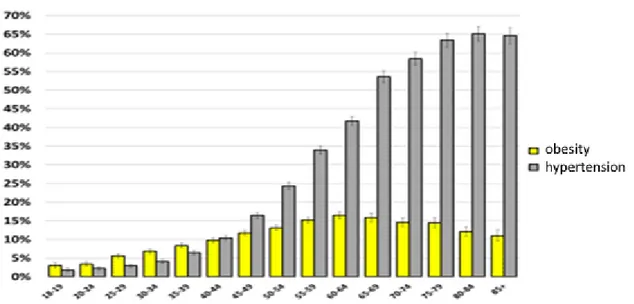
Figure 1: Prevalence of comorbidity in sarcoidosis patients with covid 19
46