Received: 26 September 2015 Accepted for publication: 8 March 2016 UDC 636.32/.39:616.441-006.5:632.15
ASSESSMENT OF CIRCULATING TOTAL AND FREE
IODOTHYRONINES’ PATTERNS IN ADULT OVINE AND
CAPRINE SPECIMENS: INFLUENCES OF ENDEMIC GOITRE
AREA
Esterina Fazio*, Pietro Medica, Cristina Cravana, Adriana Ferlazzo
Department of Veterinary Sciences, University of Messina, Polo Universitario Annunziata, 98168 Messina, Italy *Corresponding author, E-mail: [email protected]
Abstract:North-eastern Sicily is an area with iodine deficiency disorders occurring in both humans and animals. The aim of this study was to test the influences of an endemic goitre area on iodothyronine ranges and their pattern in adult ovine and caprine specimens stabled in different locations of Sicily, taking into account the different sexes. A total of 48 Comisana sheep and 51 Maltese goats was studied. The sheep included 10 females and 6 males stabled in a non-endemic goitre area (farm A: control group), and 16 females and 16 males stabled in an endemic goitre area (farm B: observational group). The goats included 6 females and 13 males stabled in a non-endemic goitre area (farm A: control group), and 16 females and 16 males stabled in an endemic goitre area (Farm B: observational group). The results showed lower T3 and higher fT4 (P<0.0001) levels in female and male sheep, and higher T4 levels in males (P<0.0001) stabled in farm B than in farm A. In comparison to farm A, goats stabled in farm B showed higher fT3 (P<0.0001) levels; males stabled in farm B showed lower T4 levels (P<0.0001), and females showed higher fT4 levels (P<0.0001). Significant effects of sex and of endemic goitre area on the total and free iodothyronines of sheep and goats were observed. The obtained data showed that an endemic goitre area for humans is not necessarily the same for ovine and caprine species, which seem to be able to adopt an adaptive strategy without presenting any clinical signs of thyroid disorders. Key words: thyroid hormones; sheep; goat; endemic goitre area
Introduction
Thyroid hormones play a pivotal physiological role in metabolic turnover and homeostasis (1) and in animal thermogenesis (2). These hormones exert pleiotropic effects in many different organs, including central nervous system development, and changes in cognitive function in animals and humans (3). Thyroid diseases are well described in companion animals, but less knowledge is
available for livestock, in which nutritional iodine deficiencies represent a serious problem, especially in endemic goitre areas. In ewes, severe iodine deficiency results in neonatal lamb death and goitre, alopecia, and poor skeletal development; at the radiographs and necropsies the epiphyses of most of the long bones were not mineralised and the remainder of the bone was poorly mineralised (4). Placental restriction of foetal growth and small size of lambs at their birth may have increased the activation of T4 to T3 and the sensitivity of soft tissues to thyroid hormone, which may have contributed to catch-up postnatal growth (5).
Maternal goats’ hypothyroidism, displayed from mid-gestation, resulted in decreased brain and cerebellum weights of affected goitrous foetuses; T4 and fT4 levels in affected goat foetuses were
dependent on the maternal phenotype, as was the degree of enlargement of the goitre (6). Nevertheless, in lactating goats, long-term dietary iodine supplementation significantly increased the fT3 and
fT3/fT4 ratios (7). The quantity of T4 and T3 available
to new-born lambs in milk suggested that thyroid hormones ingested with the colostrum may have a physiological role during the early postnatal life of suckling goats (8); in addition to that, T3 seemed to act as metabolic modulators for the establishment of puberty in goats (9). There is an assumption that the hairless gene is often responsible for congenital hypertrichosis in mammalian species, and the protein codified by this gene is a transcriptional corepressor for thyroid hormone receptors (10). Therefore, thyroid hormones affect the expression of the neuronal gene RC3 mRNA preferentially in the striatum in prenatal and adult caprine brains, suggesting a region-specific sensitivity for thyroid deficiency (6), and the mRNA expression levels of skin monodeiodinase II and III in Cashmere goats (11). The effects of iodine deficiency, especially in endemic goitre areas, have been reported both in humans and in different animal species (12-14). North-eastern Sicily is an area with iodine deficiency disorders occurring both in humans and in different domestic animals. Partial beneficial effects of the so called “silent iodine prophylaxis”, an additional percentage of iodine salt given in water and food, on iodine deficiency disorders have been described in humans (12). In contrast, even in endemic areas, the iodine deficiency disorder in both adult and young farm animals is sporadically treated, and routine iodine supplementation of pregnant animals is not recommended, because it is economically unsustainable. The aim of this study was to compare the total and free iodothyronine levels of sheep and goat specimens stabled in non-endemic and non-endemic goitre areas, by taking into account the different sexes.
Material and methods
Animals, diets and, experimental design
The study was carried out on a total of 48 Comisana sheep and 51 Maltese goats, ranging
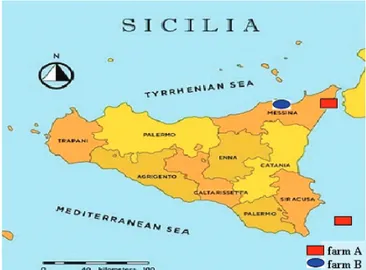
in age from 2 to 3 years and weighing 38.3±0.57 kg, and 34.9±0.56 kg, respectively, which were stabled in two different areas of Sicily (Figure 1). The sheep included 10 females and 6 males stabled in a non-endemic goitre area (farm A), and 16 females and 16 males stabled in an endemic goitre area (farm B). The goats included 6 females and 13 males stabled in non-endemic goitre area (farm A), and 16 females and 16 males stabled in an endemic goitre area (farm B). The post-partum ewes and goats had delivered from 60-90 days previously. The rams and billy goats were destined for meat production. Farm A was situated near the city of Messina (at 100 meters above sea level; 38°13’19”92 N; 15°14’20”76 E); farm B was situated on the slopes of Nebrodi mountains (at 660 meters above sea level; 38°5’9” 96 N; 14°48’28”08 E). The last farm is in an area of severe endemic goitre with the presence of abnormalities in human thyroid function in the population. The stable management of farms A and B was superimposable: both sheep and goats were kept together with their respective flocks of more than 100 ovine and caprine herds on local pastures for most of the time, and fed twice a day on a similar diet of commercial concentrate (barn, corn and soya) (15.5% protein, 2.5% fat, 6.8% cellulose and 6.7% ash) and cereal straw; water was available ad libitum. No iodine supplementation was introduced into the diet. No plant known to be goitrogenic or a member of the Brassica family was present on pastureland.
Non endemic goitre area = farm A Endemic goitre area = farm B
Measurements
The study was carried out in late spring and early summer; the mean environmental temperature was 22 °C (18 °C to 26 °C), and the mean relative humidity was 44.60% (38.20% to 51.5%). These were monitored using a Hygrothermograph ST-50 (Sekonic Corporation, Tokyo, Japan). Blood samples from the jugular vein (5 ml) were collected, twice a month for three months (April–June), using evacuated tubes (Venoject, Terumo®, Leuven, Belgium) at 09:00–10:00 h in order to minimise the effect of circadian rhythm on hormone measurements. All samples were taken in quiet conditions by the same veterinary team.
Laboratory analysis
Blood samples were kept at 4 °C until centrifugation at 1500 × g for 15 min, and the serum was harvested and stored in polystyrene tubes at -20 °C until total and free iodothyronines were determined. Hormone assays were analysed in duplicate using a commercially available immunoenzymatic kit and carried out per the manufacturer’s instructions (SEAC-RADIM, Pomezia, Rome).
Limits of detection were 0.24 nmol/L for T3,
5.79 nmol/L for T4, 0.15 pmol/L for fT3 and 1.3
pmol/L for fT4. Intra- and inter-assay coefficients of variation (CV) were 7.3% and 11.4% for T3, 2.3%
and 5.7% for T4, 4.2% and 11.9 % for fT3, 6.6 % and
9.6% for fT4,respectively, based on measurements, in three different samples. The commercial kits were validated for total and free iodothyronines by establishing that dilutions of ovine serum resulted in curves identical to those obtained with the human standards supplied with the assay kits.
Statistical analysis
All the results are expressed as mean ± SD (Figures 2 and 3). Two-way analysis of variance for repeated measures (2-way RM ANOVA) was applied to test for the effects of different sexes and locations of flocks (farms A and B) and sampling times, as well as the interaction between them, on hormonal concentrations; when the F statistic was significant, the differences between individual mean over time were then assessed using a post hoc
multiple comparison test (Bonferroni). The level of significance was set at P<0.05. All calculations were performed using the PRISM package (GraphPad Software Inc., San Diego, CA, USA). The ratios for T3/T4 and fT3/fT4, and the percentages of fT4/T4
and fT3/T3 were also calculated.
Results
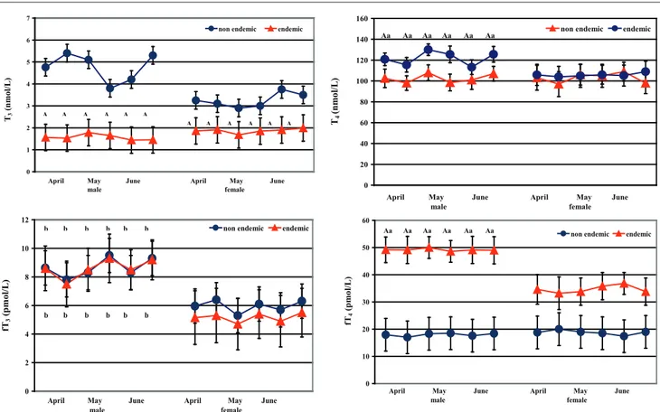
Ovine T3 levels ranged from 1.56 to 4.76 nmol/L, T4 levels ranged from 102.51 to 120.78 nmol/L, fT3
levels ranged from 5.15 to 8.63 pmol/L, fT4 levels
ranged from 17.95 to 49.18 pmol/L. Compared to the non-endemic area (farm A), female and male sheep stabled in the endemic area (farm B) showed lower T3 (P<0.0001) and higher fT4
(P<0.0001) levels, and only in males were higher T4 levels found (P<0.0001) (Figure 2). Compared
to the females of farm B, the males showed higher T4 (P<0.01) and fT4 (P<0.0001) levels. Males of
both farms showed higher fT3 (P<0.001) levels
than females did (Figure 2). Two-way RM ANOVA showed a significant effect of ovine sex on the fT3 (F=30.18; P<0.0001), and of the endemic goitre area on the T3 (F=30.18; P<0.0001) and fT4
(F=20.37; P<0.0001) changes.
Caprine T3 levels ranged from 2.20 to 3.60
nmol/L, T4 levels ranged from 101.50 to 110.87
nmol/L, fT3 levels ranged from 3.85 to 12.14
pmol/L, and fT4 levels ranged from 12.77 to 33.39
pmol/L. Compared to females, males showed higher T3 (farm A: P<0.0001; farm B: P<0.001)
and fT4 (farm B: P<0.0001) levels (Figures 2 and
3). Compared to farm A, female and male goats stabled in farm B showed higher fT3 (P<0.0001)
levels. Males stabled in farm B showed lower T4
levels (P<0.0001) and females higher fT4 levels (P<0.0001) (Figure 3). Compared to females of farm B, males showed higher fT3 (P<0.0001) and fT4
(P<0.0001) levels. Two-way RM ANOVA showed a significant effect of caprine sex on the T3 (F=29.40;
P<0.0001) and fT4 (F=86.91; P<0.0001) levels,
and of endemic goitre area on the fT3 (F=172.20; P<0.0001) and fT4 (F=18.64; P<0.0001) levels.
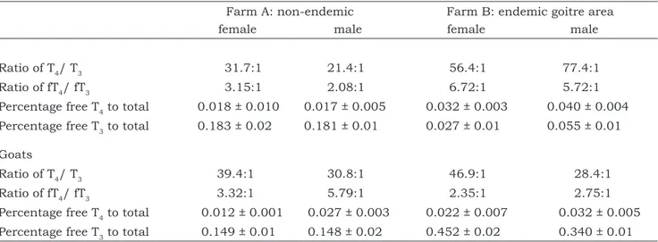
The T4/T3 and the fT4/fT3 ratios were lower
in both female and male sheep stabled in farm A (non-endemic goitre area) than those stabled in farm B (endemic goitre area) (Table 1). The percentages of fT4 to T4 and fT3 to T3 were lower in
sheep and goats of both sexes stabled in farm A than in farm B (Table 1).
0 1 2 3 4 5 6 7
non endemic endemic
T3
(nmo
l/L
)
April May June April May June male female A A A A A A A A A A A A 0 2 4 6 8 10 12
non endemic endemic
fT3
(pmo
l/L
)
April May June April May June male female b b b b b b b b b b b b 0 20 40 60 80 100 120 140 160
non endemic endemic
T4
(nmo
l/L
)
April May June April May June male female Aa Aa Aa Aa Aa Aa 0 10 20 30 40 50 60
non endemic endemic
fT4
(pmo
l/L
)
April May June April May June male female Aa Aa Aa Aa Aa Aa 0 0,5 1 1,5 2 2,5 3 3,5 4 4,5
non endemic endemic
T3
(nmo
l/L
)
April May June April May June male female a a a a a a b b b b b b 0 20 40 60 80 100 120 140
non endemic endemic
T4
(nmo
l/L
)
April May June April May June male female
A A A A A A
Figure 3:Total and free iodothyronine concentrations (M ± SD) in female and male goats stabled in non-endemic (farm A) and endemic (farm B) goitre area
Different superscripts show significant differences versus control group (non-endemic): A=P<0.001 and versus female: a=P<0.01; b=P<0.001; c=P<0.0001
Figure 2:Total and free iodothyronine concentrations (M ± SD) in female and male sheep stabled in non-endemic (farm A) and endemic (farm B) goitre area
Different superscripts show significant differences versus control group (non-endemic): A=P<0.001; and versus female: a=P<0.01; b=P<0.001 0 5 10 15 20 25 30 35 40 45
non endemic endemic
B B B B B B
c c c c c c b b b b b b
April May June April May June male female 0 2 4 6 8 10 12 14 16 18
non endemic endemic
fT3
(pmo
l/L
)
April May June April May June male female
A A A A A A Ab Ab Ab Ab Ab Ab
Table 1:Ratios and relative percentages in female and male sheep and goats stabled in non-endemic (farm A) and endemic (farm B) goitre area
Discussion
Thyroid hormone levels were within normal ranges in adult sheep and goats. The comparison of our results with previously published data reporting the circulating thyroid hormones of adult ovine (T3: 2.04-5.85 nmol/L; T4: 49.68-146.46 nmol/L; fT3: 3.63-4.35 pmol/L; fT4:
23.93-25.49 pmol/L) and caprine (T3: 2.21-3.56 nmol/L; T4: 65.64-142.85 nmol/L; fT3: 9.39-11.31 pmol/L;
fT4: 35.24-47.38 pmol/L) species (15, 16) did not
reveal any significant discrepancies. Some slight differences may also occur because of the different techniques, age, nutritional factors and/or geographic environmental variations (17–19). The higher fT4 levels recorded in female and male sheep
and in male goats stabled in the endemic goitre area (where no clinical symptoms of thyroid disorders were present), in comparison to the physiological ranges reported in literature (15, 16), suggest that hypothyroidism found in humans from the same endemic goitre area does not necessarily correlate with lower T4 levels and with evident clinical signs
in lambs, kids and/or their mothers. Indeed, like sheep and goats stabled in the endemic goitre area, with the highest T4 levels only in males,
sheep and goats stabled in a non-endemic area also showed high T4 levels. It is well known
that the fT4 fraction represents the biologically active hormone for tissues. Thus, the ratio of fT4 represents a primary factor to determine the
fractional turnover of thyroid hormones. Moreover, the binding fraction represents the hormonal
Farm A: non-endemic Farm B: endemic goitre area female male female male
Ratio of T4/ T3 31.7:1 21.4:1 56.4:1 77.4:1
Ratio of fT4/ fT3 3.15:1 2.08:1 6.72:1 5.72:1
Percentage free T4 to total 0.018 ± 0.010 0.017 ± 0.005 0.032 ± 0.003 0.040 ± 0.004
Percentage free T3 to total 0.183 ± 0.02 0.181 ± 0.01 0.027 ± 0.01 0.055 ± 0.01
Goats
Ratio of T4/ T3 39.4:1 30.8:1 46.9:1 28.4:1
Ratio of fT4/ fT3 3.32:1 5.79:1 2.35:1 2.75:1
Percentage free T4 to total 0.012 ± 0.001 0.027 ± 0.003 0.022 ± 0.007 0.032 ± 0.005
Percentage free T3 to total 0.149 ± 0.01 0.148 ± 0.02 0.452 ± 0.02 0.340 ± 0.01
reserve that balances the sudden increase and decrease of hormonal release to tissues. Although T3 is mainly an intracellular hormone and its
serum measurement is a less representative value of the total hormonal complex than serum T4 measurements, inherent physiological effects
are attributed almost exclusively to T3 (20).
Furthermore, peripheral T3 concentrations are
influenced mainly by extrathyroidal 5’-deiodinase activity, which represents an important control point for the regulation of metabolic status (21). In contrast, T4 has been known as the predominant
product of the thyroid gland, ant it has an intrinsic thyromimetic activity, as protection against hypothyroidism, which could be more pronounced in animals stabled in an endemic goitre area. Therefore, the lack of effects of an endemic area, and/or iodine deficiency, on thyroid hormones’ metabolism could show that enzyme activity is homeostatically regulated, and iodine is probably incorporated, so as to ensure the maintenance of total and free hormone homeostasis, in accordance with the species.
Furthermore, sheep stabled in the endemic goitre area had higher T4/T3 and fT4/fT3 ratios,
and higher percentages of fT4 and fT3,compared to
T4 and T3. Another point is that goats stabled in
the endemic goitre area also had high percentages of fT4 and fT3,compared to T4 and T3. These results confirm that changes in free iodothyronines generally follow those for total iodothyronines (22) and suggest the presence of a synergism between total and free amounts. For this reason, it is
possible to presume that, under circumstances of endemic iodine deficiency, a shift in T4/T3 balance
will occur in favour of T4, considered to be a
reserve hormone.
Unexpectedly, the influences of an endemic area on the thyroid function of sheep and goats were different, with the significant involvement of T3 and fT4 in both ovine females and males, and
of fT3 changes in both caprine sexes. The obtained
data suggest that sheep stabled in an endemic goitre area are probably capable of synthesizing more adequate T4 and/or reducing its conversion
to T3, or to increase its metabolic clearance in peripheral tissues, as shown by the lowest T3
levels observed in animals stabled in the endemic area; this hypothesis is partially superimposable on the goats stabled in an endemic goitre area, with T3 levels unchanged.
These findings could probably be supported by suitable amounts of iodine in the diet for thyroid function for the species, or by an efficient iodine recycling system via gastrointestinal tract, which conserves iodine and can protect the animals against low dietary iodine, as has been reported in cows (23); however, animals stabled in endemic goitre area were kept on pasture most of the time, with plants being the primary source of iodine.
The obtained data confirm that the physiological ranges of thyroid hormones in ovine and caprine species are wide, because of the many intrinsic and extrinsic variables that can influence physiological thyroid hormone concentrations. In addition, in these small ruminants, a relative lack of information has been found regarding the use of thyroid hormone measurements to evaluate thyroid disorders and dysfunctions (24); therefore, the diagnosis of thyroid diseases in adult ovine and caprine populations, as well as in other species, has usually been difficult to perform, and the synthesis and mechanism of action of the thyroid hormones in ruminant physiology have been extrapolated from the extensive canine and feline knowledge (18).
Our data confirm the existence of significant differences between ovine and caprine sexes in thyroid hormone concentrations, although 2-way RM ANOVA showed a significant effect of sex on the fT3 and fT4 changes in sheep, and on
T3 and fT4 changes in goats. The role of thyroid
hormones in controlling seasonal reproduction in several mammalian species, including small ruminants, is well known (20). In ewes’ rendered
hypothyroids, the end of the reproductive season occurred later than in the controls (25). The permissive role of thyroid hormones seemed to be represented by the increase of the responsiveness to the oestradiol negative feed-back, but they are also involved in steroid-independent seasonal cycles in luteinising hormone pulse frequency (26). In male sheep, thyroidectomy abolished seasonal cycles of gonadotropin secretion and testicular size (27), and in male goats’ testes T3 level stimulates androgen release (28). Moreover, these observations do not provide additional evidence for the different involvement of thyroid hormones in accordance with female and male total and free iodothyronine concentrations. Moreover, our data are not in accordance with the results obtained in sheep, in which no statistically significant differences in thyroid hormone levels were found, due to sex (29), neither with lower T3 levels reported in juvenile male goats than female (30). Higher T4 levels observed in male than female
sheep and goats are not in accordance with data reported by Celi et al. (30), which showed that T4 levels of goats were not affected by sex.
Thus, differences in T4 levels could be explained
on the basis of oestrogen-reduced catabolism of thyroxine-binding globulin, and androgen-inhibited TSH secretion by the pituitary (31).
Moreover, in sheep and goats stabled in the same areas of endemic human goitre, with severe hypothyroid cases of goitre, cretinism and deafness, no signs of any abnormal clinical symptoms associated with hypothyroidism were observed. Indeed, sheep and goats stabled in the endemic goitre area showed paradoxically higher T4 concentrations, than the physiological and control values. It would, therefore, appear that ovine and caprine species, as also observed in equines (32), cope with an endemic environment through a significant thyroidal response in order to synthesize a representative reserve of T4.
However, it is not possible to exclude the existence of an inhibition of the enzyme 5’- deiodinase, which is responsible for the conversion of T4 to
T3, which could represent an adaptive mechanism
to decrease the metabolic rate during iodine deficiency, as reported by Duckett (33) during illness and stress. The results would suggest a physiological adaptive response of the ovine and caprine species to a low iodine environment. In addition to that, animals of small farms A and B, which did not receive iodine supplementation,
are fully dependent on the natural local iodine source. Therefore, the results obtained in sheep and goats, fed roughage of local harvests, can be regarded as another valuable information on the iodine status in the Sicilian area.
Using the values found in humans (34) as the basis for comparison, the obtained data showed that the sheep exhibit higher total circulating T4
at 102.79 nmol/L (non-endemic goitre area) and 113.31 nmol/L (endemic goitre area), and the goats exhibit lower total circulating T4 at 106.64 nmol/L (non-endemic goitre area) and 102.33 nmol/L (endemic goitre area), than the values found in euthyroid humans (95.23 nmol/L) and goitre humans (82.36 nmol/L).
The possibility that hypothyroidism cases are present in adult sheep and goats is thus excluded; this is supported not only by the lack of clinical signs, but also by the rarity of the cases reported, being limited exclusively to lambs and lambings and as a possible consequence of foetal or precocious death.
In conclusion, endemic areas for human goitre are not necessarily so for small adult farm animals that show an adaptive strategy without presenting any clinical signs of thyroid disorders. Evidence to prove this hypothesis should be obtained by assessing iodine status not only in urine, but also in the milk of sheep and goats, especially in animals living and stabled in endemic goitre areas, as suggested in humans (35) and reported by the World Health Organization (36). However, the goitre areas of Sicily remain a serious environmental and health problem for humans and represent a potential economic threat for animal production.
Acknowledgements
The authors are grateful to the ovine and caprine farmers and veterinarians for their availability and invaluable contribution to the present study.
References
1. Budge H, Edwards LJ, McMillen IC, et al. Nu-tritional manipulation of foetal adipose tissue depo-sition and uncoupling protein 1 messenger RNA abundance in the sheep: differential effects of tim-ing and duration. Biol Reprod 2004; 71: 359–65.
2. Darwish RA, Ashmawy TA. The impact of lambing stress on post-parturient behaviour of sheep with consequences on neonatal homeother-my and survival. Theriogenology 2011; 76: 999– 1005.
3. Long DT, Voice TC. Role of exposure analysis solving the mystery of Balkan endemic nephropa-thy. Croat Med J 2007; 48: 300–11.
4. Campbell AJ, Croser EL, Milne ME, Hodge PJ, Webb Ware JK. An outbreak of severe io-dine-deficiency goitre in a sheep flock in north-east Victoria. Aust Vet J 2012; 90: 235–9.
5. De Blasio MJ, Gatford KL, Robinson JS, Owens JA. Placental restriction alters circulating thyroid hormone in the young lamb postnatally. Am J Physiol Regul Integr Comp Physiol 2006; 201: R1016–24.
6. Piosik PA, van Groenigen M, van Doorn J, Baas F, de Vijlder JJ. Effects of maternal thyroid status on thyroid hormones and growth in congen-itally hypothyroid goat fetuses during the second half of gestation. Endocrinology 1997; 138: 5–11.
7. Nudda A, Battacone G, Bomboi G, Floris B, Decandia M, Pulina G. Effect of dietary iodine on thyroid hormones and energy blood metabolites in lactating goats. Animal 2013; 7: 60–5.
8. Slebodziński AB, Twardon J. Thyroid hor-mone (TH) and 5’-monodeiodinase (5’-MD) activity in goat’s milk from the early, mid- and late lacta-tion period. Acta Vet Hung 2004; 52: 349–59.
9. Meza-Herrera CA, Torres-Moreno M, Ló-pez-Medrano JI, et al. Glutamate supply positive-ly affects serum release of triiodothyronine and insulin across time without increases of glucoses during the onset of puberty in female goats. Anim Reprod Sci 2011; 125: 74–80.
10. Potter GB, Beaudoin GM 3rd, DeRenzo CL, Zarach JM, Chen SH, Thompson CC. The hairless gene mutated in congenital hair loss disorders en-codes a novel nuclear receptor corepressor Genes Dev 2001; 15: 2687–701.
11. Qin F, Li J, Zhu X, Zhou J, Yang J, Jia Z. Dietary iodine and selenium affected the mRNA expression levels of skin monodeiodinase (II, III) in Liaoning Cashmere goats. Biol Trace Elem Res 2013; 151: 360–4.
12. Vermiglio F, Finocchiaro MD, Lo Presti VP, La Torre N, Nucifora M, Trimarchi F. Partial ben-eficial effects of the so called “silent iodine pro-phylaxis” on iodine deficiency disorders (IDD) in northeastern Sicily endemia. J Endocrinol Invest 1989; 12: 123–6.
13. Maberly GF. Iodine deficiency disorders: contemporary scientific issues. J Nutr 1994; 124: 1473S–8S.
14. Pugliese M, Medica P, Scardillo A, Parisi F, Fazio E. Thyroid function evaluation in Valle del Belice sheep affected by congenital hypotricosis. In: The 15th International Congress of Mediterra-nean Federation for Healthy Production of Rumi-nants. Kuşadasi, Tűrkiye, 2007: 244–9.
15. Nazifi S, Saeb M, Abangah E, Karimi T. Studies on the relationship between thyroid hor-mones and some trace elements in the blood se-rum of Iranian fat-tailed sheep. Vet Arh 2008; 78: 159–65.
16. Paulíková I, Seidel H, Nagy O, Tóthová C, Kováč G. Concentrations of thyroid hormones in various age categories of ruminants and swine. Acta Vet Beograde 2011; 61: 489–503.
17. Nazifi S, Gheisari HR, Shaker F. Serum lip-ids and lipoproteins and their correlations with thyroid hormones in clinically healthy goats. Vet Arh 2002; 72: 249–57.
18. Matamoros R, Contreras PA, Wittwer F, Mayorga MI. Hipotiroidismo en rumiantes. Arch Med Vet 2003; 35: 1–11.
19. Todini L. Thyroid hormones in small rumi-nants: effects of endogenous, environmental and nutritional factors. Animal 2007; 1: 997–1008.
20. Huszenicza Gy, Kulcsar M, Rudas P. Clini-cal endocrinology of thyroid gland function in ru-minants. Vet Med Czech 2002; 47: 199–210.
21. Kaplan MM. Regulatory influences on iodo-thyronine deiodination in animal tissues. In: Hen-nemann G, ed. Thyroid hormone metabolism. New York : Dekker, 1986: 231–53.
22. Fazio E, Medica P, Cravana C, Messineo C, Ferlazzo A.Total and free iodothyronines levels of growing thoroughbred foals: effects of weaning and gender. Livest Sci 2007; 110: 207–13.
23. Miller JK, Swanson EW, Spalding GE. Io-dine absorption, excretion, recycling, and tissue distribution in the dairy cow. J Dairy Sci 1975; 58: 1578–93.
24. Trávniček J, Kursa J. Iodine concentration in milk of sheep and goats from farms in South Bohemia. Acta Vet Brno 2001; 70: 35–42.
25. Hernandez JA, Hallford DM, Wells NH.
Ovarian cyclicity in thyroid-suppressed ewes treated with propylthiouracil immediately before onset of seasonal anestrus. J Anim Sci 2003; 81: 29–34.
26. Anderson GM, Connors JM, Hardy SL, Va-lent M, Goodmann RL. Thyroid hormones mediate steroid-indipendent seasonal changes in luteiniz-ing hormone pulsatility in the ewe. Biol Reprod 2002; 66: 701–6.
27. Parkinson TJ, Douthwaite JA, Follett BK. Responses of prepubertal and mature rams to thyroidectomy. J Reprod Fertil 1995; 104: 51–6.
28. Jana NR, Halder S, Bhattacharya S. Thyroid hormone induces a 52 kDa soluble protein in goat testis Leydig cell which stimulates androgen re-lease. Biochim Biophys Acta 1986; 1292: 209–14.
29. Aliefy MM, Zaki K, Abul-Fadle W, Ayoub L. Effects of season and sex on thyroid hormone level in the blood of Osimi sheep. Zentralbl Veter-inärmed A 1970; 17: 476–80.
30. Celi P, Seren E, Celi R, Parmeggiani A, Di Trana A. Relationships between blood hormonal concentrations and secondary fibre shedding in young cashmere-bearing goats at their first moult. Anim Sci 2003; 77: 371–81.
31. Christianson D, Roti E, Vagenakis AG, Bra-verman LE. The sex-related difference in serum thyrotropin concentration is androgen mediated. Endocrinology 1981; 108: 529–35.
32. Medica P, Fazio E, Cravana C, Ferlazzo A. Influence of endemic goitre areas on thyroid hor-mones in horses. Animal 2011; 5: 82–7.
33. Duckett WM. Thyroid gland. In: Reed SM, Bayly WM, eds. Equine internal medicine. Phila-delphia : Saunders , 1998: 917–23.
34. Vigneri R. Studies on the goiter endemia in Sicily. J Endocrinol Invest 1988; 11: 831–43.
35. Verheesen RH, Schweitzer CM. Iodine defi-ciency, more than cretinism and goiter. Med Hy-potheses 2008; 71: 645–8.
36. de Benoist B, Andersson M, Egli I, Takk-ouche B, Allen H, eds. Iodine status worldwide: WHO Global database on iodine deficiency. Ge-neva, Switzerland: Department of Nutrition for Health and Development World Health Organiza-tion, 2004: 1–47.
DOLOČANJE VEZANIH IN PROSTIH ŠČITNIČNIH HORMONOV V KRVI PRI ODRASLIH
OVCAH IN KOZAH: VPLIV ENDEMIČNIH OBMOČIJ Z GOLŠAVOSTJO
E. Fazio, P. Medica, C. Cravana, A. Ferlazzo
Povzetek: Severovzhodna Sicilija je območje, kjer se redno pojavljajo motnje, povezane s pomanjkanjem joda pri ljudeh in živalih. Cilj raziskave je bil raziskati pojavnost endemične golšavosti pri odraslih ovcah in kozah z merjenjem ravni jodotironinov na različnih lokacijah na Siciliji. Upoštevane so bile tudi razlike med spoloma. Skupno je bilo v raziskavo vključenih 48 ovc pasme comisana in 51 malteških koz. Pri ovcah je bilo vključenih 10 samic in 6 samcev z območij brez golšavosti (kmetija A: kontrolna skupina) ter po 16 samic in samcev iz endemičnih območij z golšavostjo (kmetija B: opazovana skupina). Pri kozah je bilo vključenih 6 samic in 13 samcev iz območij brez golšavosti (kmetija A: kontrolna skupina) in po 16 samic in samcev iz endemičnih območij z golšavostjo (kmetija B: opazovana skupina). Rezultati so pokazali nižje T3 in višje T4 vrednosti (p <0,0001) pri samcih in
samicah ovac in višje ravni T4 pri samcih (p < 0,0001), ki so bili nastanjeni na kmetiji B v primerjavi s kmetijo A. V primerjavi s kmetijo
A so pri kozah, ki so bile nastanjene na kmetiji B, ugotovili višje ravni prostega T3 (p < 0,0001), pri samcih na kmetiji B nižje ravni T4
(p < 0,0001), pri samicah pa višjo raven prostega T4 (p < 0,0001). Ugotovljene so bile statistično značilne razlike med skupinami
tako glede na spol kot glede na endemičnost področja na ravni vezanih in prostih jodotironinov pri ovcah in kozah. Pridobljeni podatki so pokazali, da območja z endemično golšavostjo pri ljudeh nimajo nujno enakega učinka pri ovcah in kozah, za katere se zdi, da so sposobne ustvariti prilagoditveno strategijo, zaradi katere se ne pokažejo klinični znaki bolezni ščitnice.