Università degli Studi di Catania
Facoltà di Medicina e Chirurgia
Dottorato di ricerca in Neurobiologia
XXVI Ciclo
Sede Amministrativa: Università di Catania Sede Consociata: Università di Roma “La Sapienza”
RELAZIONE FINALE
Anno accademico 2013-2014
Cholinergic enhancing strategies and brain
neuroplasticity. From preclinical evidence to
clinical applications.
Dott. Enea Traini
Tutor: Prof. Roberto Avola
Cotutor: Prof. Francesco Amenta
Coordinator: Prof. Roberto Avola
INTRODUCTION Pg.2
CHOLINE ALPHOSCERATE Pg. 6
Pharmacokinetics Pg. 8
Toxicology and safety Pg. 10
PRECLINICAL STUDIES Pg. 12 CLINICAL EFFICACY Pg. 25 ASCOMALVA TRIAL Pg. 31 Behavioural symptoms Pg. 41 Apathy evaluation Pg. 48 CONCLUSIONS Pg. 56 REFERENCES Pg. 58
2
INTRODUCTION
The observation of a reduction of choline acetyltransferase (acetylcholine biosynthetic enzyme) in the cerebral cortex of patients with Alzheimer's disease and subsequent preclinical and clinical evidence have led, between 1970 and 1980, to the identification of the cholinergic system as the most neurotransmitter systems involved in processes of memory and learning (Amenta et al., 2001). These studies have allowed to develop the, so-called, cholinergic hypothesis of cognitive dysfunction observed in adult. Starting from this hypothesis and from the correlation between brain levels of acetylcholine and the cognitive performance degree, several pharmacological approaches have been attempted to correct the cholinergic deficit that is observed in the central nervous system of patients suffering from the Alzheimer's disease (Giacobini, 1998; Amenta et al., 2001; Gauthier 2002). Similarly to what was observed in Alzheimer's disease, involvement of the cerebral cholinergic system was also suggested in the pathophysiology of vascular dementia VaD, the most common form of adult-onset cognitive dysfunction after Alzheimer's disease (Brashear, 2003; Roman, 2003).
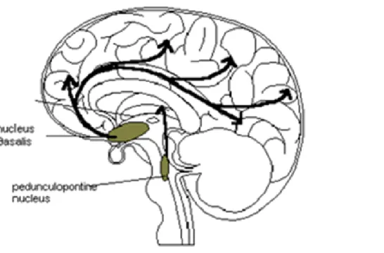
Figure 1 summarizes the cholinergic projections that innervate the cerebral cortex and that originate from the nuclei of the basal forebrain.
Figure 1. Schematic representation of the ascending cholinergic projections that, from the base of the forebrain
cholinergic nuclei (in particular the magnocellular basal nucleus), innervate the cerebral cortex. The figure also represents the projection from the nucleus pontine peduncle reaches the diencephalon and is considered to be responsible for attentional processes.
3
The possible attempts to restore cholinergic neurotransmission proposed or tried consist essentially in using:
Cholinergic precursors (drugs that increase the levels of acetylcholine, increasing the availability of the substrate for the neurotransmitter synthesis);
Inhibitors of acetylcholinesterase (AChE) or cholinesterase (ChE) (drugs that increase the levels of acetylcholine, delaying the degradation at the synaptic level);
Nicotinic or muscarinic agonists (drugs that mimic the action of acetylcholine at pre- or post-synaptic level);
Enablers of the release of acetylcholine (drugs to facilitate the release of acetylcholine from pre-synaptic terminals).
Among the 4 categories of compounds potentially capable of increasing or facilitate cholinergic neurotransmission, only the first two have led to the development of drugs used in therapy, although with different results (Giacobini, 1998; Amenta et al., 2001; Grutzendler and Morris, 2001; Gauthier 2002; Grantham and Geerts, 2002). Cholinesterase inhibitors are the first drugs that have received regulatory indication of the symptomatic treatment of Alzheimer's disease.
The activities of the three cholinesterase inhibitors available in the Italian pharmaceutical market was subject of a careful evaluation called "Project CRONOS". It is a study of post-marketing surveillance nationwide aimed at evaluating the transferability, in clinical practice, of the results obtained in clinical trials conducted before the marketing authorization of the medicines in question. In particular, the CRONOS, whose adherence was subject, for a time, the dispensation of cholinesterase inhibitors, was aimed at:
improve knowledge of the characteristics of the population suffering from Alzheimer's disease and treated with cholinesterase inhibitors:
assessing the appropriateness of the treatments;
broaden the definition of tolerability profiles of the drugs;
identify possible predictive variables for the optimization of the profile of drug efficacy. CRONOS Project was conducted on a representative sample of patients with Alzheimer's disease being treated at the over 500 Alzheimer Evaluation Unit (UVA) established in all Italian regions. The sample considered sufficient for objectives of the observational study was considered corresponding to about 1/5 of the total UVA (118 UVA). The UVA identified provided all the patients records from the period September 2000-December 2001 to February 2003. The analyzed
4
sample included 7,395 patients. From the total number of persons, 1,933 patients were excluded because they are already being treated with cholinesterase inhibitors before the CRONOS. So the analysis of the treatment was carried out on 5,462 patients. In terms of evaluating the results of the CRONOS, about 2 in 10 patients showed a response at 3 months. Patients responders to the treatment (considered those with a difference of 2 points in MMSE from baseline) only one has maintained the response at 9 months. One patient treated every 7 went to meet adverse events, that in almost 36% of cases have resulted in the discontinuation of therapy. The conclusion of CRONOS was a modest size response to drug therapy. (Bollettino Informazione Farmaci, 2004).
There is not an analogue evaluation to project CRONOS for Vascular Dementia, although an analysis of prescriptions from Italian UVA for different types of patients with adult-onset dementia has shown that cholinesterase inhibitors are prescribed to 90% of patients with Alzheimer's disease, 80% of patients with Lewy bodies dementia (LBD) and at 35-45% of patients with VaD and frontotemporal dementia (Frisoni et al., 2007). Careful evaluation of the effectiveness of treatment with cholinesterase inhibitors on cognitive dysfunction associated with VaD or otherwise found in mixed forms of neurodegenerative and vascular adult-onset dementia, was conducted by a recent meta-analysis that took examined 2,633 patients in active treatment and 1,650 patients treated with placebo in three trials with donepezil, 2 with galantamine and 2 with rivastigmine (Kavirajan and Schneider, 2007). The meta-analysis concluded that cholinesterase inhibitors produce a modest improvement in cognitive function of uncertain clinical significance in patients with mild to moderate VaD. This effect is considered insufficient to support a widespread use of cholinesterase inhibitors in the treatment of VaD, suggesting that a more accurate analysis of individual patient data is necessary to identify groups of patients who may benefit from the drugs in question (Kavirajan and Schneider, 2007).
The cholinergic system of the forebrain base plays an important role both in the elaboration of attention, memory and behavior, that in the regulation of cerebral blood flow (Everitt and Robins, 1999). The cholinergic structures of the forebrain base, as well as numerous brain areas involved in cognitive activities, are particularly sensitive to ischemia. This consideration would explain the strong cholinergic deficit that is observed in the associated neurodegenerative and vascular forms of dementia (Everitt and Robins, 1999; Roman, 2003). From the point of view of symptoms, these forms, which are reported in the literature with the term vascular dementia are characterized by progressive cognitive decline, disesecutive syndromes and behavioral changes. Symptoms dependent, in all likelihood, from ischemic damage or oligaemia against brain areas involved in the development of cognitive functions and complex behaviors (Roman, 2003).
5
Preclinical studies reported a neuroprotective effect of inhibitors of AChE / ChE, which could slow the progressive evolution of the disease in question (Francis et al., 2005). However these benefits in front a low cost / benefit ratio (especially in terms of adverse reactions) are modest and of doubtful clinical significance (Kavirajan and Schneider, 2007). One of the more negative aspects of therapy with inhibitors of AChE / ChE lies in the loss of efficacy of treatment with time. Another problem is the treatment of specific patient groups in which inhibitors of AChE / ChE are contraindicated; particularly the very elderly (over 85 years) or those suffering from bradycardia or obstructive pulmonary diseases, such as bronchial asthma and CPOD (Parnetti et al., 2007).
Cholinergic precursors represent one of the first approaches to treat adult-onset dementias, but their effectiveness has been demonstrated only for some molecules (Amenta et al., 2001). Studies conducted in experimental animals have shown that an association between choline alphoscerate (alpha-glyceryl phosphorylcholine, GFC) and an inhibitor of cholinesterase induces an higher increase acetylcholine levels compared to treatment with the individual substances (Tayebati et al., 2006). This effect on the levels of acetylcholine was particularly pronounced especially in brain areas involved in higher cognitive processes. A positive effect of an association precursor (CDP-choline) and the Ache inhibitor (galantamine) was reported by a pilot study in patients with schizophrenic psychosis; in this study was also highlighted the perfect tolerability of the association (Deutsch et al., 2008).
Choline alphoscerate is a cholinergic precursor probably among the most active and certainly more effective respect CDP-choline in increasing brain levels of acetylcholine (Amenta et al., 2001).
6
CHOLINE ALPHOSCERATE
Changes in cholinergic function are implicated in the pathogenesis of learning and memory alterations occurring in adult-onset cholinergic dysfunction including dementia disorders (Davies and Maloney, 1976; Bowen, 1977; Perry et al., 1977; Bartus et al., 1985; Gottfries et al., 1994). The cholinergic system is not the only neurotransmitter system affected in cognitive dysfunction common of Alzheimer’s disease or vascular dementia, but analysis of its involvement in cognitive functions has shown that central cholinergic receptors might be involved in learning and memory through complex mechanisms (Davies and Maloney, 1976; Bowen, 1977; Perry et al., 1977; Bartus et al., 1985; Gottfries et al., 1994).
Studies of the brain of aged subjects or of patients suffering from Alzheimer’s disease have shown marked loss of the acetylcholine synthesizing enzyme choline acetyltransferase and of nicotinic cholinergic receptors (Davies and Maloney, 1976; Bowen, 1977; Perry et al., 1977; Bartus et al., 1985; Gottfries et al., 1994). Administration of the muscarinic antagonist scopolamine to healthy young subjects induces a cognitive impairment resembling that found in adult-onset dementia (Amenta and Tayebati, 2008). A correlation between the loss of cortical cholinergic synapses and cognitive decline and a relationships between this loss and the decrease of high affinity cholinergic receptors were reported (Davies and Maloney, 1976; Bowen, 1977; Perry et al., 1977; Bartus et al., 1985; Gottfries et al., 1994). These findings have contributed to the development of the cholinergic hypothesis of geriatric memory dysfunction (Perry et al., 1977).
Cholinergic strategies were therefore developed for restoring deficient cholinergic neurotransmission which occurs primarily in basal forebrain (Terry and Boccafusco, 2003). Cholinergic precursors have represented an old approach to treat cholinergic dysfunction and cognitive decline in adult-onset dementia disorders (Amenta et al., 2001; Parnetti et al., 2007). Many of these precursors were early leaved because their efficacy was not clearly demonstrated. This is not true for choline alphoscerate, a cholinergic precursor available in the pharmaceutical market of several countries, which has been studied both in preclinical paradigms and in clinical trials (Tayebati and Amenta, 2013).
Choline and the choline-containing phospholipid phosphatidylcholine, are essential for maintaining cell membrane integrity and structure. Choline is also required to transport fats in and out of cells, and is a precursor of acetylcholine, the neurotransmitter required for several brain functions including learning and memory. Choline is probably one of the most basic nutrients necessary for optimal cognitive function being the precursor for acetylcholine and is also used by cellular
7
machinery for synthesizing phosphatidylcholine (Amenta and Tayebati, 2008; Gui et al., 2010; Hoff et al., 2010).
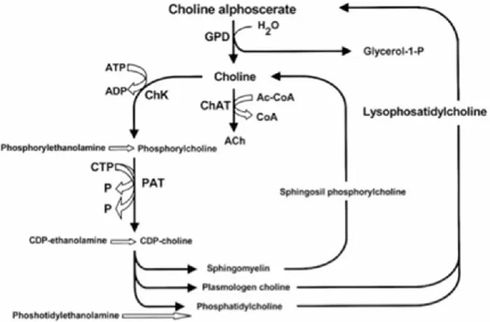
Figure 2 summarizes acetylcholine anabolic pathways (Amenta and Tayebati, 2008). As shown, the enzyme glyceryl- phosphorylcholine diesterase transforms alpha glyceryl-phosphorylcholine into a molecule of choline and another of glycerol-1-phosphate.
Figure 2: Acetylcholine synthetic pathways . Interference of choline-containing compounds. Figure modified from
(Amenta et al., 2001)
This figure shows the steps in which choline alphoscerate can influence neurotransmitter biosynthesis. Cythidin diphosphate (CDP); Cythidin triphosphate (CTP); Glyceryl-phosphorylcholine diesterase (GPD); Choline acetyltransferase (ChAT); Choline kinase (ChK); Phosphocholine cytidyl transferase (PCT).
Choline can be used to synthesize acetylcholine, whereas glycerol-1-phosphate after being phosphorylated can enter in the pool of phospholipids (Dross and Kewitz, 1972; Amenta and Tayebati, 2008) These pathways provide both free choline and phospholipids for preparing acetylcholine and to reorganize nerve cell membrane components. Free choline administration increases brain choline availability but it does not increase acetylcholine synthesis/or release
8
(Amenta and Tayebati, 2008). Acetylcholine is a neurotransmitter derived from choline and acetyl coenzyme A, the biosynthesis of which is catalyzed by choline acetyltransferase. Nervous tissue is unable to synthesize choline, that derives from the diet and is delivered to neurons through the blood stream. Acetylcholine released from cholinergic synapses is hydrolyzed by acetylcholinesterase into choline and acetyl coenzyme A. Approximately 50% of choline derived from acetylcholine hydrolysis is received through a high affinity transporter. Therefore neurons require a further supply of choline for synthesis of acetylcholine (Amenta and Tayebati, 2008).
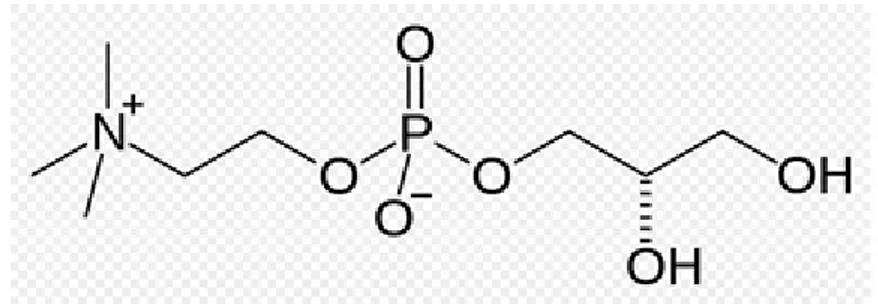
Choline alphoscerate or alpha-glycerylphosphorylcholine (ATC code N07AX02) (GPC) (Figure 3) is a semi-synthetic derivative of lecithin. Following oral administration, it is converted to phosphorylcholine, a metabolically active form of choline able to reach cholinergic nerve terminals where it increases acetylcholine synthesis, levels and release (Amenta et al., 2001; Amenta and Tayebati, 2008)
Figure 3: Chemical structure of choline alphoscerate
Although choline alphoscerate is in the pharmaceutical market since 1987, the interest on it was remarkably reduced after the introduction in therapy of cholinesterase inhibitors. In the last 10 years a renewed attention on the compound was also seen in literature with preclinical studies, clinical investigations and review articles published on it (Suchy et al., 2009; Tomassoni et al., 2012; Scapicchio, 2013; Tayebati and Amenta, 2013; Tayebati et al., 2013).
Pharmacokinetics
The kinetics and metabolism of choline alphoscerate (GPC) were investigated in male and female rats after i.v. (10 mg/kg) and oral doses (100-300 mg/kg), using the compound labelled with [14 C]-glycerol ([14G]-GPC) or [14C]-choline ([14C]-GPC). Different kinetic and metabolic profiles were observed after i.v. and oral administration. Choline alphoscerate is hydrolyzed by
9
phosphodiesterases in the gut mucosa. The labelled metabolites have different kinetic properties of absorption, distribution and clearance, leading to different blood concentration-time curves of total radioactivity. Both labelled compounds gave a wide distribution of radioactivity, particularly concentrated in the liver, kidney, lung and spleen compared to blood. Brain concentrations of [14 C]-GPC were comparable to ([14C]-GPC) or lower than ([14C]-GPC) total blood radioactivity. The metabolite profile in the perfused brain showed a small amount of choline and two unknown metabolites, probably the same as in blood. In addition, choline was incorporated into brain phospholipids in increasing amounts within 24 h of dosing. In all cases renal and fecal excretion of radioactivity was low and comparable for [14C]-GPC and [14C]-GPC. Mostly the administered radioactivity was exhaled as 14CO2, this degradation being faster and more pronounced for the glycerol-labelled metabolites than for the choline-labelled metabolites for both routes of administration (Abbiati et al., 1993).
Plasma choline levels were higher in eight healthy volunteers after intramuscular administration of choline alphoscerate compared to placebo (Fossati et al., 1994). Mean plasma choline levels in the placebo group ranged from 10.6 to 12.0 µM. After choline alphoscerate administration, the plasma choline level reached 35.1 µM in 30 min, then decreased gradually. Plasma choline levels became comparable in the treated and placebo groups 6-8 h after administration (Fossati et al., 1994). These pharmacokinetic data further support the view that choline alphoscerate increases the bioavailability of choline that can be then converted in acetylcholine.
An increased of brain cholinergic tone by administration of choline alphoscerate was demonstrated by the clinical pharmacology study mentioned below (Ceda et al., 1992). Growth hormone (GH) secretion is decreased during aging in humans and in rodents. This decrease may be due to increased hypothalamic somatostatin release, which is inhibited by cholinergic agonists, or to decreased secretion of GHRH. GH secretion is greater in younger subjects than in old individuals. Administration of choline alphoscerate elicited a greater GH response to the GHRH compared to placebo. The potentiating effect of choline alphoscerate on GH secretion was more pronounced in the elderly subjects. These findings support the view that increased cholinergic tone elicited by administration of choline alphoscerate enhances GH release (Ceda et al., 1992).
10
Toxicology and safety
Studies on the toxicological profile and safety of choline alphoscerate are summarized in a recent review article (Bronawell et al., 2011). The oral LD50 is equal to or greater than 10,000 mg/kg in rats and mice. In some cases deaths are preceded by convulsions. Dosing of dogs with up to 3000 mg/kg choline alphoscerate resulted only in reduced activity. Sub-chronic and chronic oral toxicity studies in rats (up to 1000 mg/kg/day) and beagle dogs (up to 300 mg/kg/day) produced symptomology primarily consisting of reduced activity, slight decreases in food consumption and body weight gain, slight reduction in liver weight, paralleled by significant decreases in plasma triglycerides, bilirubin, and alkaline phosphatase. These changes are no accompanied by histopathological correlates. In vivo and in vitro assays indicate that choline alphoscerate is devoid of mutagenic activity. This indicates that choline alphoscerate is not genotoxic in vitro or in vivo (Bronawell et al., 2011).
To sum up, toxicology studies indicate that choline alphoscerate has low acute oral toxicity and, has an oral NOAEL of 150 mg/kg bw/day following 26 weeks oral exposure (Bronawell et al., 2011).
Choline alphoscerate is a safe drug and in more than 20 years of clinical experience side effects were reported only rarely. Pharmacovigilance data analysis did allow to identify a single report of moderate adverse reactions in the elapse of time from 1 January 1998 to 16 May 2011.
Analysis of adverse reactions in controlled clinical trials revealed a slightly higher incidence of adverse reactions with choline alphoscerate compared to placebo, but these reactions were always mild and never required discontinuation of treatment (Parnetti et al., 2001; De Jesus Moreno, 2003; Levin et al., 2011; Amenta et al., 2012).
Compared with (acetyl)cholinesterase inhibitors that, although investigated in a larger number of clinical trials did not demonstrate in controlled studies a higher efficacy on cognitive domains compared to choline alphoscerate, the compound under analysis has undoubtedly an extremely more favorable safety profile.
The case of cardiovascular side effects of the (acetyl)cholinesterase inhibitors and of choline alphoscerate was reviewed (Amenta et al., 2010). Analysis included a literature search, of all randomized clinical trials and meta-analyses published from 1999 to July 2009. Efficacy outcomes and adverse reactions requiring treatment discontinuation were considered with attention. Alzheimer’s disease, vascular dementia, cholinesterase inhibitors, and choline alphoscerate were the terms considered. Second generation (acetyl)cholinesterase inhibitors induced modest improvement
11
in cognitive performances in mild and moderate Alzheimer’s disease patients. In demented aged individuals treated with (acetyl)cholinesterase inhibitors, a greater risk for syncope, bradycardia, pacemaker insertion, and hip fracture, was reported. The effect of choline alphoscerate on cognitive outcomes was not inferior to that of (acetyl)cholinesterase inhibitors. Treatment with choline alphoscerate was associated with a low incidence of adverse reactions and no changes in heart rate (Amenta et al., 2010). It was concluded that association of bradycardia in demented patients treated with (acetyl)-cholinesterase inhibitors suggests that closer surveillance for bradycardia may be warranted in patients with cardiovascular risk treated with these drugs. In view of the good effectiveness/safety profile of choline alphoscerate, this compound may be considered as an option in older adults with dementia at risk of cardiovascular events (Amenta et al., 2010).
12
PRECLINICAL STUDIES
Choline alphoscerate interferes with brain phospholipid metabolism and increases brain choline and acetylcholine levels and release (Amenta et al., 2001; Amenta and Tayebati, 2008). Pharmacodynamic studies on choline alphoscerate during phases of development of the compound were focused primarily on its role in potentiating brain cholinergic neurotransmission and in interfering with brain phospholipid metabolism. Pre-clinical studies have demonstrated that choline alphoscerate increases the release of acetylcholine in rat hippocampus (Parnetti et al., 2007), facilitates learning and memory in experimental animals (Sigala e al., 1992), improves brain transduction mechanisms (Schettini et al., 1990; Lopez et al., 1991) and decreases the age-dependent structural changes occurring in the rat frontal cortex and hippocampus (Amenta et al., 1993). Moreover, the compound contributes to anabolic processes responsible for membrane phospholipid and glycerolipid synthesis, positively influencing membrane fluidity (Aleppo et al., 1994). In several animal paradigms of impaired cognitive function, choline alphoscerate was demonstrated to improve cognitive deficit in experimental models of aging brain (Canonico et al., 1990; Drago et al., 1990) and to reverse mnemonic deficits induced by scopolamine administration (Sigala et al., 1992; Parnetti et al., 2007). Based on the above evidence, the central parasympathomimetic activity of choline alphoscerate was defined, suggesting its clinical use in patients affected by cognitive decline. Consistently with the activity profile, choline alphoscerate was classified as a centrally acting parasympathomimetic drugs both in international pharmacopoeias (Reynolds, 1996) and in the Chemical Therapeutical Anatomical Classification.
A restorative role of choline alphoscerate on central cholinergic system was also documented by studies performed in old rodents. In these investigations the compound was able to counter age-related changes in brain acetylcholine synthesizing (choline acetyltransferase) and degrading (acetylcholinesterase) enzymes (Amenta et al., 1994a) and some subtypes of muscarinic cholinergic receptors (Amenta et al. 1994b; Muccioli et al, 1996).
Neuroprotective effects of choline alphoscerate were also documented in a rodent model of altered cholinergic neurotransmission caused by lesioning of the Nucleus Basalis Magnocellularis which represents the main source of cholinergic innervation of cerebral neocortex (Bronzetti et al., 1993; Amenta et al., 1995). A neuroprotective effect of treatment with choline alphoscerate on hippocampus microanatomy and glial reaction which represents an early sign of brain damage was documented in spontaneously hypertensive rats, used as an animal model of brain vascular injury (Tomassoni et al., 2006). A model used to mimic to some extent neuropathological changes occurring in vascular dementia (Tayebati et al., 2011). Among cholinergic precursors tested, choline
13
alphoscerate elicited the most relevant stimulation on vesicular acetylcholine transporter, and choline transporter in the same model of brain vascular injury suggesting that it represents a strong enhancer of central cholinergic neurotransmission (Antal et al., 1999).
Effects of choline alphoscerate were limited not only in rodent models of aging of lesioning of brain cholinergic nuclei, but also in Rhesus monkeys. In this species the compound revealed general facilitatory properties on retinal neurotransmission as well as specific spatial frequency tuning effects on retinal information processing (Amenta et al., 2006).
More recent studies have demonstrated that association of choline alphoscerate with (acetyl)cholinesterase inhibitors potentiates effects on cholinergic neurotransmission. In fact, administration of choline alphoscerate plus the acetylcholinesterase inhibitor rivastigmine induced an increase of brain acetylcholine levels and of high affinity choline uptake binding sites more pronounced than single drugs (Amenta et al., 2006). This investigation has suggested that combination of a suitable precursor of brain acetylcholine such as choline alphoscerate and of an acetylcholinesterase inhibitor may represent an association worthwhile of being further investigated as a cholinergic replacement therapy in pathologies characterized by impaired cholinergic neurotransmission (Amenta et al., 2006). This working hypothesis was supported by the demonstration of a more sustained neuroprotective action by choline alphoscerate plus the acetylcholinesterase inhibitor galantamine than the two drugs administered alone (Tayebati et al., 2009).
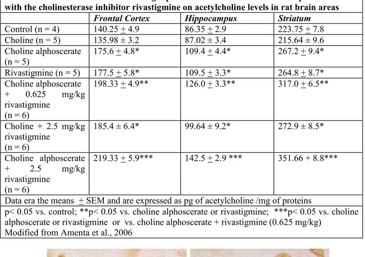
In a first phase of our study it was shown that brain levels of acetylcholine are increased by the association between choline alphoscerate and rivastigmine in a more relevant than single drugs (Table 1). The study has documented an increase of the immunoreactivity for acetylcholine in neurons of the nucleus basalis magnocellularis (Figure 4), as well as an increase of binding of the high affinity marker for the system of transport of choline [3H]hemicholinium-3 more evident in association than the single drugs (Amenta et al., 2006). Based on these data it seems that, similar to that observed for the dopaminergic therapy of Parkinson's disease several years ago, the association between a precursor of acetylcholine synthesis and inhibition of its catabolism, is associated with an enhancement of cholinergic neurotransmission.
14
Table 1
Effect of treatment with the cholinergic precursors choline and choline alphoscerate and with the cholinesterase inhibitor rivastigmine on acetylcholine levels in rat brain areas
Frontal Cortex Hippocampus Striatum
Control (n = 4) 140.25 + 4.9 86.35 + 2.9 223.75 + 7.8 Choline (n = 5) 135.98 ± 3.2 87.02 ± 3.4 215.64 ± 9.6 Choline alphoscerate (n = 5) 175.6 + 4.8* 109.4 + 4.4* 267.2 + 9.4* Rivastigmine (n = 5) 177.5 + 5.8* 109.5 + 3.3* 264.8 + 8.7* Choline alphoscerate + 0.625 mg/kg rivastigmine (n = 6) 198.33 + 4.9** 126.0 + 3.3** 317.0 + 6.5** Choline + 2.5 mg/kg rivastigmine (n = 6) 185.4 ± 6.4* 99.64 ± 9.2* 272.9 ± 8.5* Choline alphoscerate + 2.5 mg/kg rivastigmine (n = 6) 219.33 + 5.9*** 142.5 + 2.9 *** 351.66 + 8.8***
Data era the means + SEM and are expressed as pg of acetylcholine /mg of proteins
p< 0.05 vs. control; **p< 0.05 vs. choline alphoscerate or rivastigmine; ***p< 0.05 vs. choline alphoscerate or rivastigmine or vs. choline alphoscerate + rivastigmine (0.625 mg/kg)
Modified from Amenta et al., 2006
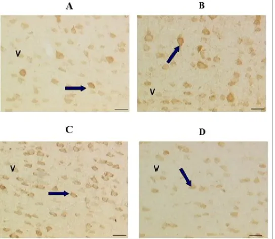
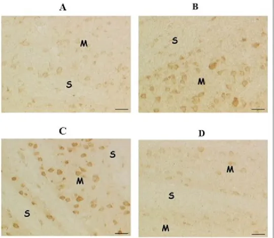
Figure 4: Immunohistochemical localization of acetylcholine in neurons of the magno cellular basal complex of control
Wistar rats (A) after treatment with choline alphoscerate 100 mg/Kg/die (B), galantamine 3 mg/Kg/die (C) and the association of the two drugs (D). Figure modified from Amenta et al., 2008
Calibration bar: 10 µm
A
B
C
15
Note how the drug treatment with choline alphoscerate or galantamine determines, compared with the control, an increase in immunoreactivity for acetylcholine (detectable by their color dark brown). The two drugs in combination induce a more pronounced immunoreactivity for the neurotransmitter.
To relate the probable neuroprotective activity of cholinesterase inhibitors (Akaike, 2006; Mori et al., 2006), however, also shared by memantine (Lipton, 2005) only to the increase in acetylcholine brain levels that this class of drugs induces it is a simplistic interpretation of a more complex pharmacological activity. It is known that the β-amyloid protein plays an important role in the mechanisms of neurotoxicity underlying neurodegenerative and vascular diseases of the nervous system by increasing the vulnerability of neurons induced by glutamic acid. Vulnerability determined by an over activation of the NMDA receptor of glutamic acid with a consequent increase of the entry of Ca2+ within neurons, formation of free radicals and neuronal damage. The neuroprotective action of memantine, a non-competitive antagonist of the NMDA receptor (Lipton, 2005) should be attributed to its interference with these mechanisms of neuronal damage. It is also speculated that the cholinesterase inhibitors implement its activity neuro protector through the same mechanism, mediated through the activation of the nicotinic cholinergic receptors α4-β2 and α7 (Akaike, 2006; Mori et al., 2006 ).
For an assessment of a possible neuroprotective effect of the association choline alphoscerate-cholinesterase inhibitor have been realized some experiments by treating spontaneously hypertensive rats (SHR), used as an animal model of vascular dementia, with choline alphoscerate, the cholinesterase inhibitor galantamine and the two drugs in combination.
The selection of the SHR rats as a model of vascular dementia is due to the relation between hypertension and adult-onset dementia. Long lasting arterial hypertension affects cerebrovascular tree and may be accompanied by brain damage leading to cognitive dysfunction. Cognitive impairment of vascular origin characterizes vascular dementia (VaD) (Birkenhager, 2006). Evidence developed in the last 10 years has suggested strong relationships between cognitive impairment and arterial hypertension (Frishman, 2002). Cerebrovascular pathology is acknowledged as an important cause of dementia (Jellinger, 2002). The involvement of cholinergic system in VaD cognitive impairment is less clearly established than Alzheimer’s disease (AD) one (Roman, 2003a; Roman, 2005).
Cholinergic neuronal processes are associated with cognitive functions, as well as with the control of cerebral blood flow. This may pertain to the pathogenesis of VaD, in which cognitive impairment results from decreased brain blood flow, hypoperfusion, and complete or incomplete ischemia
16
(Roman and Kalaria, 2006). Cholinergic structures and specific brain areas are rather vulnerable to ischemic damage. For instance, hippocampal CA1 neurons are particularly susceptible to experimental ischemia. Hippocampal atrophy is present in patient with VaD in the absence of AD (Vinters et al., 2000). Progressive cognition decline, functional ability impairment and behavior problems account for a central role of impaired cholinergic neurotransmission in the VaD and AD (Brashear, 2003).
The parameters considered in this micro anatomical study were, mainly, the number of nerve cells and the expression of glial reaction, early marker of damage to the nervous system.
The treatment of SHR rats of 32 weeks for 4 weeks with a daily oral dose of 100 mg / kg of choline alphoscerate, 3 mg / kg galantamine or the two drugs in combination has hindered the reduction of the number of nerve cells at the level of the frontal cortex , of the CA1 subfield of the hippocampus and the dentate gyrus (Figure 5), which occurs in the untreated SHR rats compared to normotensive control Wistar Kyoto rats.
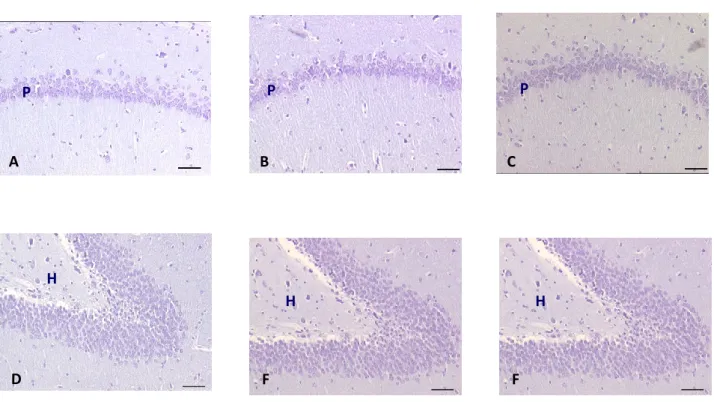
Figura 5: Microphotography of CA1 subfield (A-C) and Dentate gyrus (D-F) of hyppocampus of normotensive Wistar
Kyoto rats (A,D) spontaneously hypertensive SHR (B,E) and SHR rats treated with association between choline alphoscerate 100 mg/Kg/die and galantamine 3 mg/Kg/die (C,F). Slides coloured by Nissl stain (Cresyl violet 1.5%). Figure modified from Amenta et al., 2008
Calibration bar: 50 m P P P H H H A B C D F F
17
Note how, in this animal model of vascular dementia, the number of nerve cells in the CA1 subfield and to a lesser extent in the dentate gyrus is lower in SHR compared to the corresponding normotensive, indicating that the model in question spontaneous hypertension is accompanied by neurodegeneration. Treatment with choline alphoscerate galantamine and contrasts this neuronal depletion suggesting that drug treatment has a neuroprotective action.
Regarding glial reaction, which accompanies early neuronal injury, in our study was assessed using the protein gliofibrillare acidic (GFAP) as a marker. It is possible to note, in the SHR control, an increase in the number and, in some brain areas, in the volume of astrocytes immunoreactive for GFAP (Figure 6). Treatment with choline alphoscerate, but not with galantamine counteracts astrogliosis observed in brain of SHR rats.
A
B
C
D
18
The association between choline alphoscerate-galantamine, compared to the two drugs given alone, shows a more pronounced neuroprotective action (Figures 5 and 6), suggesting that this association merits clinical insights as a possible way to enhance a deficit in cholinergic neurotransmission disorders such as disease AD and VaD.
Other preclinical studies published in the last few years were focused on the activity of choline alphoscerate on neurotransmitter transporter systems. Plasma membrane and vesicular transporters for acetylcholine, and biogenic amines (dopamine, norepinephrine, and serotonin) are specific proteins with a crucial role in the regulation of neurotransmission and may represent a relevant target for the action of therapeutic agents (Canonico et al., 1990; Tomassoni et al., 2012). Cholinergic transporters including high-affinity choline uptake and vesicular acetylcholine transporters respectively control cellular mechanisms of acetylcholine synthesis and release at presynaptic terminals and loading of it into synaptic vesicles.
The process of synthesis, storage and release of acetylcholine (ACh) requires the expression of specialized systems, including the ACh vesicular transporter (VAChT) and high affinity choline transporter (CHT). Nervous tissue cannot synthesize choline, which is ultimately derived from diet and is delivered to neurons through the bloodstream. ACh released from cholinergic synapses is hydrolyzed by acetylcholinesterase (AChE) into choline and acetyl coenzyme A. Almost 50% of choline derived ACh hydrolysis is recovered by a high-affinity CHT.
The status of cholinergic system is investigated by assaying of specific cholinergic markers. VAChT is the cholinergic marker more recently identified and represents a protein selective for ACh transport into vesicles for evoked release at the cell surface (Weihe, 2006).
AChE/cholinesterase(ChE) inhibitors have been suggested as a possible symptomatic therapy of VaD patients. A relatively recent large-scale trial using galantamine (GAL) in subjects with VaD and AD with cerebrovascular disease has represented the first placebo-controlled trail showing clinically relevant benefits of GAL in this patient population (Brashear, 2003). Among AChE/ChE
Figura 6: Immunohistochemical demonstration of glial reaction, identified through the localization of the protein
gliofibrillare acidic (GFAP) in hippocampus of a rat normotensive Wistar Kyoto WKY (A) of spontaneously hypertensive control SHR (B) and SHR treated with choline alphoscerate 100 mg/Kg/die (C), galantamine 3mg/Kg/die (D) or the association of the two drugs (E). Figure modified from Amenta et al., 2008
Calibration bar: 50 m
Note the increase in glial reaction in rats SHR compared to WKY control reference and the activity on glial reaction carried out by the choline alphoscerate alone or in combination with galantamine, but not by galantamine alone.
19
inhibitors, GAL has an interesting pharmacological profile, being both a reversible AChE inhibitor and an allosteric modulator of nicotinic cholinergic receptors (Bullock, 2004).
The influence of treatment for 4 weeks of spontaneously hypertensive rats with choline alphoscerate was investigated (Tomassoni et al., 2012).
The immunochemical analysis of Elisa and Western blot are showed in figure 7 e 8.
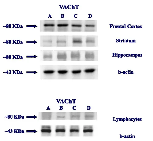
Exposure of membranes of cerebral cortex, hippocampus, striatum to anti-VAChT antibody caused the development of a band identified approximately at 80 KDa that represents the mature, post-translationally modified form of VAChT protein. Comparative densitometric analysis of different bands formed in WKY rats, SHR and SHR treated with GPC and GAL, normalized to the respective actin intensity for each sample, revealed slightly higher expression of VAChT in control SHR compared to the WKY rats. An increase of the intensity of the VAChT bands was evident in the different brain areas of the GPC treated with SHR, while the expression of VAChT in the SHR treated with GAL is similar to the WKY control group. The pattern of the changes expression of the VAChT is similar in the lymphocytes (Fig.7).
Figure 7: Western blot of brain area and Blood lymphocytes of rats: WKY Control (A); SHR Control (B); SHR treated
20
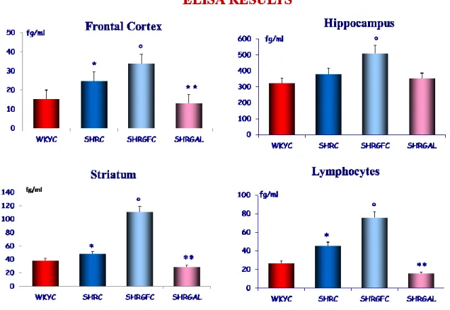
Quantitative ELISA determination, shown high expression of VAChT in hippocampus than other cerebral areas investigated. In the different treatment groups, the results were similarly to the WB determination, with an increase of the concentration of VAChT in the SHR group, respect WKY rats. An increase of VAChT was found in the SHR treated with GPC compared to WKY rats and/or SHR control rats. VAChT expression in the SHR treated with GAL was similar to the WKY control rat (Fig.8).
Figure 8: Western blot of brain area and Blood lymphocytes of rats: WKY Control (WKYC); SHR Control (SHRC );
SHR treated with GPC (SHRGPC); SHR treated with GAL (SHRGAL). Data are the means ± S.E.M. Significance are p<0,05
Figures 9-11 show the results of VAChT immunohistochemistry.
In all cerebrocortical layers of WKY rats sparse polygonal I shaped nerve cell bodies expressing in their external portions VAChT-IR were seen. These cell bodies were concentrated primarily in layers V e VI In SHR, the network of VAChT-positive nerve fibers and cell bodies was more evident than in normotensive WKY rats. Treatment of SHR with GPC results in an increase of the intensity of the VAChT-positive nerve fibers, while the treatment with GAL resulted in a
21
cerebrocortical picture of VAChT immunoreactivity more similar to that observed in WKY rats than in control SHR (Fig. 9).
Figure 9: VAChT immunohistochemistry in Frontal Cortex of rat brain: WKY Control (A); SHR Control (B); SHR
treated with GPC (C); SHR treated with GAL (D). Calibration bar: 25 µm
In the striatum of WKY rats, immune reaction was localized both in striosomes and matrix. A strong VAChT immune reaction was found around some nerve cell bodies as well as a dense plexus of fibers carrying small varicosities. No VAChT immunoreactive nerve cells were observed in striosomes. In SHR the pattern of VAChT immunoreactivity was similar to that found in normotensive WKY rats, with an apparently higher intensity of immune reaction. Treatment with GAL did not affect VAChT immunoreactivity in the striatum of SHR (Fig.10).
22
Figure 10: VAChT immunohistochemistry in Striatum of rat brain: WKY Control (A); SHR Control (B); SHR treated
with GPC (C); SHR treated with GAL (D) Calibration bar: 25 µm
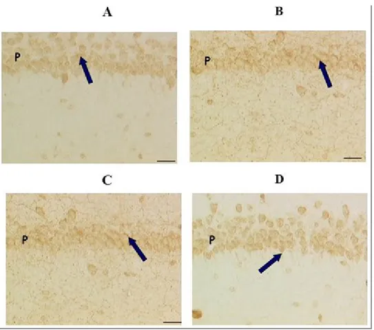
In the hippocampus of WKY rats, VAChT-immunoreactive fibers and varicosities were found throughout CA1 and CA3 subfields and the dentate gyrus. Particularly dense punctuates of immune reaction surrounded pyramidal cell layer in the CA1 and CA3 subfields. An intermediate level of varicosities and fiber staining was found in the stratum oriens, radiatum, molecular and hilum, whereas stratum lacunosum molecolare expressed a lower level of immunoreactivity. The pattern of VAChT immunoreactivity was similar in untreated SHR, but it displayed a higher intensity of immune reaction. Treatment of SHR with GPC increased the VAChT immunoreactivity, while treatment with GAL VAChT immunoreactivity of the hippocampus to a picture similar to that observed in WKY rats (Fig. 11).
23
Figure 11: VAChT immunohistochemistry in the Hyppocampus of rat brain: WKY Control (A); SHR Control (B);
SHR treated with GPC (C); SHR treated with GAL (D) Calibration bar: 25 µm
Choline alphoscerate induced an increase of high-affinity choline uptake and vesicular acetylcholine transporters in the frontal cortex, striatum and hippocampus as well as in peripheral blood lymphocytres considered as a marker of brain cholinergic transporters. This effect of choline alphoscerate was considered consistent with the increased synthesis of acetylcholine it induces (Tomassoni et al., 2012).
The role of choline-containing phospholipids on brain phospholipid biosynthesis may have influence on brain metabolism and neurotransmitter systems. Based on the observation that another choline-containing phospholipid, CDP-choline may have a monoaminergic profile, the activity of choline alphoscerate on brain dopamine, and serotonin levels and on dopamine plasma membrane transporter, vesicular monoamine transporters 1 and 2, serotonin transporter and norepinephrine transporter were also investigated in different rat brain areas. Administration of the compound increased dopamine levels in frontal cortex and cerebellum and serotonin levels in frontal cortex and striatum. It also stimulated dopamine plasma membrane transporter in frontal cortex and cerebellum. This investigation concluded that choline alphoscerate possesses also a monoaminergic
24
profile and interfere to some extent with brain monoamine transporters. Authors suggested that this profile may represent a first step for further analysis of therapeutic activity of choline alphoscerate in clinical settings (Tayebati et al., 2013).
25
CLINICAL EFFICACY
Cholinergic precursor loading therapy was the first approach tried to relief cognitive impairment in dementia disorders. Controlled clinical trials failed to show significant improvements with choline or phosphatidylcholine (lecithin), a choline-containing phospholipid (Blusztajn et al., 1987), alone or in association with cholinesterase inhibitors (tacrine plus choline, or physostigmine plus choline) (Higgins and Flicker, 2003). The reasons for the lack of effect of this precursor strategy are unclear (Amenta et al., 2001). The negative effects with choline or phosphatidylcholine (Amenta et al., 2001; Higgins and Flicker, 2003) cannot be generalized for all cholinergic precursors. It is thought that phosphatidylcholine increases brain choline and acetylcholine concentrations (Chung et al., 1995; Higgins and Flicker, 2003), although an effect of this precursor on neurotransmitter synthesis was not confirmed by all studies (Domino et al., 1983). Probably phosphatidylcholine can provide choline for acetylcholine synthesis only in conditions of stimulated neurotransmitter release (Jope, 1982).
Other cholinergic precursors such as cytidine 5′-diphosphocholine (CDP-choline) and choline alphoscerate increase acetylcholine content and release (Gatti et al., 1992; Sigala et al., 1992; Pinardi et al 1994; Dixon et al., 1997), being choline alphoscerate more effective than CDP-choline in rising plasma choline levels (Gatti et al., 1992). The reason of the different effects of the above compounds on acetylcholine synthesis and release is unclear. It cannot be excluded that the activity observed in clinical trials with different cholinergic precursors depends from the availability of the neurotransmitter they induce (Amenta et al., 2001).
The main clinical experiences with choline alphoscerate in adult-onset cognitive disorders are summarized below. The majority of clinical studies available on the effect of choline alphoscerate on cognitive function in neurodegenerative and cerebrovascular disorders were detailed in two reviews of our group (Parnetti et al., 2001; Parnetti et al., 2007) and are summarized in Table 2. Studies published before 2001 have investigated 1570 patients, 854 of which were included in controlled trials. The patients examined were affected by cognitive dysfunction dementia of degenerative, vascular or combined origin, such as senile dementia of the Alzheimer's type, vascular dementia and acute cerebrovascular diseases, such as transitory ischemic attack (TIA) and stroke (Parnetti et al., 2001). Test batteries for assessing the effect of choline alphoscerate on cognitive domains were primarily the Mini Mental State Evaluation (MMSE) in disorders of neurodegenerative origin and the Sandoz Clinical Assessment Geriatric (SCAG) scale in disorders of vascular origin (e.g. vascular dementia) (Parnetti et al., 2001).
26 CI of NDG origin CI of VAS origin CI of combined NDG and VAS origin TIA or stroke Total Total no. of trials 4 4 3 3 14 Controlled 4 4 1 0 9 Uncontrolled 0 0 2 3 5 Total no. of patients 826 789 216 2,484 4,315 Controlled 486 421 208 0 1,115 Uncontrolled 340 368 8 2,484 3,200
Table 2. Clinical trials on choline alphoscerate in cognitive dysfunction of neurodegenerative or vascular origin and in cerebrovascular disease.
Table included in the review article of Parnetti and coworkers (Parnetti et al., 2001) and in the trial of De Jesus Moreno (De Jesus Moreno, 2003) on Alzheimer's disease. Data summarized in this table were discussed critically in a paper (Parnetti et al., 2007). CI: Cognitive impairment NDG: Neurodegenerative; VAS: Vascular; TIA: Transient ischemic attack.
Overall, 565 patients with cognitive impairment of degenerative origin of mild to moderate grade were enrolled. Three homogeneous-case trials evaluated 186 patients, whereas the three combined-case trials included 379 patients with degenerative dementia. In four trials, choline alphoscerate was given orally at the dose of 1200 mg/day (466 patients treated for 6 months and 39 for 3 months). In the remaining studies it was administered intramuscularly at the dose of 1000 mg/day. The duration of treatment was 3 or 6 months for oral administration and 3 months for parenteral administration. These trials documented that choline alphoscerate improved the patients’ clinical condition with particular reference to memory and attention (Parnetti et al., 2001). Comparison between choline
27
alphoscerate and acetyl-l-carnitine gave scores more favorable to choline alphoscerate (Parnetti et al., 2001).
The activity of choline alphoscerate was also investigated in 789 patients with cognitive impairment of vascular origin. Three homogeneous-case trials evaluated 408 patients and three combined-case trials included 381 patients with vascular dementia. In four trials, choline alphoscerate was administered orally at the dose of 1200 mg/day for 3 or 6 months, while in the other three studies it was administered by intramuscular injection at the dose of 1000 mg/day for 3 months. Of the 431 orally-treated patients, 418 received the drug over 6 months and 13 over 3 months. A total of 358 were treated intramuscularly over 3 months. In these studies, investigators thoroughly assessed, cognitive impairment, behavioural disturbances, changes of interpersonal relations, affective disorders and somatic problems. Similarly as observed in degenerative dementia disorders in all trials on cognitive impairment of vascular origin, treatment with choline alphoscerate improved memory and attention impairment, as well as affective and somatic symptoms (fatigue, vertigo). Effects of choline alphoscerate were superior than those of placebo and of the same extent or superior of those of reference compounds (Parnetti et al., 2001). Comparison between choline alphoscerate and CDP-choline gave SCAG scores more favorable to choline alphoscerate (Parnetti et al., 2001).Oxiracetam was reported to have an activity comparable with choline alphoscerate in two uncontrolled trials (Parnetti et al., 2001).
Choline alphoscerate was also investigated in three uncontrolled studies examining its activity in acute cerebrovascular disease (Parnetti et al., 2001). Treatment consisted in the intramuscular administration of a daily dose of 1000 mg/day of choline alphoscerate in the 4 weeks following the acute event. Parenteral administration was followed by a 5-month oral administration of the drug at the dose of 1200 mg/day. In these trials, parenteral treatment with choline alphoscerate favored cognitive, functional and motor recovery in the acute phase. The subsequent oral treatment consolidated clinical results obtained in the acute phase and positively influenced the whole clinical course. Unfortunately, the typology of these studies makes them of marginal relevance (Parnetti et al., 2001).
A more recent trial has evaluated 261 patients aged 72.2 ± 7.5 years (132 treated for 180 days with 400 mg tablets of choline alphoscerate 3 times a day and 129 allocated to the placebo group) affected by mild to moderate dementia of the Alzheimer's type (De Jesus Moreno, 2003). In patients under active treatment, the mean decrease in ADAS-Cog score was 2.42 points after 90 days of treatment and 3.20 points at the end of the study (day 180), whereas in the placebo group a mean increase in ADAS-Cog score of 0.36 point <1 after 90 days and of 2.90 points after 180 days of
28
observation (p < 0.001 vs. baseline) was noticeable. Other parameters assessed (MMSE, GDS, ADAS-Behav, ADAS-Total, and CGI) improved after 90 and 180 days versus baseline, whereas in the placebo group they remained unchanged or worsened. Statistically significant differences were observed between treatments after 90 and 180 days in ADAS-Cog, MMSE, GDS, ADAS-Total, and CGI scores and after 180 days of treatment in ADAS-Behav and GIS scores (De Jesus Moreno, 2003). This last investigation presents the advantage of having been conducted using a modern clinical approach if compared with former investigations. On the other hand this trial, different from previous studies with choline alphoscerate (Parnetti et al., 2001), has used batteries of tests and time of observation comparable with studies assessing the activity of (acetyl)cholinesterase inhibitors (Moretti et al., 2004; Craig and Birks, 2005; Kuduszkiewicz et al., 2005; Roman, 2005; Roman et al., 2005; Birks, 2006; Birks and Harvey, 2006; Craig and Birks, 2006). A comparison of ADAS-Cog analysis from this investigation (De Jesus Moreno, 2003) with the results obtained on the same item in 4 trials with the cholinesterase inhibitor donepezil (Rogers et al., 1997; Rogers et al., 1998a; Rogers et al., 1998b; Burns et al., 1999) revealed a more positive trend with the cholinergic precursor choline alphoscerate compared with donepezil (Figure 12).
Figure 12: Comparison of the results of ADAS-Cog test in Alzheimer's disease patients treated with choline
alphoscerate or control (placebo choline alphoscerate) and 5 mg/day or 10 mg/day donepezil or control (placebo donepezil). Figure modified from Parnetti et al., 2007
Data of choline alphoscerate are taken from (De Jesus Moreno, 2003). Scores for donepezil and respective control groups were obtained by pooling average data obtained in 4 trials (Rogers et al., 1997; Rogers et al., 1998a; Rogers et al., 1998b; Burns et al., 1999). Data are expressed as means of the difference in the scores from baseline obtained in the ADAS-Cog test and were analyzed statistically by ANOVA. Standard error for each point was less than 5%.
29
A positive effect of treatment with choline alphoscerate on cognitive domains was also reported in an open 10-day study in which the therapeutic actions of the compound (in 40 patients at the dose of 1000 mg/day) were compared with those of piracetam (in 20 patients at a dose of 2000 mg/day) (Rogers et al., 1997). The two agents were given intravenously in patients suffering from Parkinson’s disease with cognitive impairments reaching the level of moderate cognitive disorder or dementia. Choline alphoscerate produced marked and moderate improvements on cognitive functions more frequently than piracetam (40% and 25%, respectively), while the incidence of deterioration was lower (5% and 15%, p < 0.05). Choline alphoscerate displayed a good profile of tolerability, with mild and short-term side effects only in the 15% of patients investigated Levin et al. 2011). Although limited both in terms of size of the sample investigated and of the length of treatment the results of this pilot study suggest that choline alphoscerate has cognitive enhancing capabilities worthwhile of being further investigated also in Parkinson’s disease accompanied by cognitive dysfunction. Other investigation of Russian groups have evaluated the effects of choline alphoscerate on ischemic and haemorragic stroke; they observed: regress of neurological deficit, better rehabilitation of cognitive functions and functional status in patients actively treated with choline alphoscerate (Builova et al., 2009; Ismagilov et al., 2009; Kamchatnov et al., 2012; Vaizova et al., 2012) and also stabilization of blood-brain barrier (Vaizova et al., 2012). A few studies investigates also chronic cerebrovascular disease (Stulin et al., 2009; Kostenko et al., 2012) with improvement of coordination neurological symptoms, cognitive and emotional functions, activity and mood. Language barriers (Russian with an English abstract), the small sample of patients recruited and/or the limited time of observation do not allow a detailed evaluation of these studies.
It should be pointed out that cognitive dysfunction is common in untreated patients in early Parkinson’s disease and affects attention, psychomotor function, episodic memory, executive function and category fluency (Elgl et al., 2009). Therapeutic intervention for cognitive symptoms in Parkinson’s disease are currently based on the use of (acetyl)cholinesterase inhibitors. These agents can produce a modest improvement in cognitive function, as well as in psychotic symptoms, generally without an adverse effect on motor function (Cavinesset al., 2011). The availability of another therapeutic option such as choline alphoscerate for these disorders could offer some alternatives primarily in patients in which (acetyl)cholinesterase inhibitors are not indicated.
Based on the preclinical results showing that association of choline alphoscerate with (acetyl)cholinesterase inhibitors potentiates effects of both drugs on cholinergic neurotransmission (Amenta et al., 2006; Tayebati et al., 2009) prompted the development in Italy of the independent (not supported by pharmaceutical companies) trial “Effect of association between a cholinesterase
30
inhibitor and choline alphoscerate on cognitive deficits in AD associated with cerebrovascular impairment” (ASCOMALVA).
31
ASCOMALVA TRIAL
ASCOMALVA is a clinical study of spontaneous generation; it is multicenter, randomized, placebo-controlled and double-blind study. It has included all the planned subjects (n = 210, 132 females and 78 males), aged between 59 and 93 years (average 77 years ) (Table 3).
Table 3: Demographic data of patients recruited for the ASCOMALVA trial
Sex (M/F) ♂ 78 (37%) / ♀ 123 (63%)
Age 77 ± 7
Min 59 Max 93 Median 79
Scolarity 7 ± 4
Min 2 Max 17 Median 5
MMSE at baseline 20 ± 3
Min 14 Max 24 Median 20
Donepezil Donepezil + choline alphoscerate Sex (M/F) ♂ 22 (40%) /♀ 34 (60%) ♂ 24 (42%) /♀ 33 (58%)
Age 78 ± 5 76 ± 8
Scolarity 7 ± 3 8 ± 5
MMSE at baseline 20 ± 3 20 ± 3
Table modified from Amenta et al., 2014
Centers involved were Alzheimer’s Unit of Cardarelli Hospital in Naples, Italy (Unità Valutativa Alzheimer e Malattie Involutive Cerebrali, Azienda Ospedaliera di Rilievo Nazionale A. Cardarelli, Napoli) and Division of Neurology of Poma General Hospital in Mantua, Italy (Unità Complessa di Neurologia, Azienda Ospedaliera C. Poma, Mantova ). Supervision, organization and information technology support and statistics were provided by Centro Ricerche Cliniche, Telemedicina e Telefarmacia of Camerino University (Camerino, Italy). Patients’ clinical data were downloaded into a computerized file on the website developed by Camerino University for ASCOMALVA (Amenta et al., 2012). Each researcher can access the clinical files using a personal password. For increasing the personal data protection, each centre prepared a numerical list of patients. The identity of subjects was kept not in the WEB, but was taken protected by the investigating centre only. The numerical list was sent to the Clinical Research Centre of Camerino University for randomization purposes and for allotting patients to one of the two treatments planned (donepezil + placebo or donepezil + choline alphoscerate). Randomization lists were prepared in groups of 20
32
subjects per centre. Hence, each centre could work independently with the advantage of an easy possible combination of the results obtained. This WEB-based procedure was followed also for economical reasons, being ASCOMALVA a clinical study of spontaneous generation.
Initially, the protocol considered to treat patients with donepezil + choline alphoscerate (treatment group) or donepezil plus placebo (control group) for 24 months starting from enrolment. Interim evaluations were planned at 3, 6, 9, 12, 18 months. A recent amendment of the protocol prolonged the study for additional two years. Hence, at the conclusion, ASCOMALVA will include observation of patients for 4 years of treatment.
Maintaining the double-blind study, the protocol provides that the supervising Center may assess via WEB the advancement of the parameters analyzed at the completion of the 3, 6, 9, 12, 18 and 24 months of treatment. The mid-term evaluation, without the opening of the blindness, was possible because all data of the study, excluding the identity of patients, were available in a web platform operated by the coordinating Center. Only the coordinator was aware of the type of treatment (active or reference) assigned to individual patients.
The trial recruited patients suffering from AD with concomitant cerebrovascular damage. These patients represent a population with a relevant cholinergic hypofunction (Auld et al., 2002; Schlieb and Arendt, 2006) that can obtain benefit from a sustained cholinergic load. Diagnosis of AD disease was established according to NICDS ADRDA criteria, and vascular damage was evaluated using the New Rating Scale for Age-Related White Matter Changes (ARWMC), based on cerebral ischemic injury evaluation with computed tomography and/or brain MRI. Patients recruited should have a score ≥ 2 at the ARWMC scale and present at least two of the following vascular risk factors: hypertension, diabetes, obesity, ischemic heart disease, dyslipidemia, hyperhomocysteinemia, smoking, previous cerebrovascular events and family history of cardio-cerebrovascular diseases.
The criteria for inclusion in the study were:
• Age>50 years;
• Mini-Mental State Evaluation (MMSE) (Folstein et al., 1975) between 24 and 12;
• Score ≥ 2 at the New Rating Scale for Age-Related White Matter Changes (ARWMC), the rating scale of cerebral ischemic injury evaluated with computed tomography and / or brain MRI (Wahlund et al., 2001);
33
• Presence of at least two of the following vascular risk factors: hypertension, diabetes, obesity, ischemic heart disease, dyslipidemia, hyperhomocysteinemia, smoking, previous cerebrovascular events and familiar history of cardio-cerebrovascular diseases.
Exclusion criteria were:
• Decompensated heart disease;
• Chronic renal failure;
• Severe liver failure;
• Incorrect dysthyroidism;
• Developmental disorders (eg cancer);
• Conditions that could interfere with assessments of safety / efficacy;
• Diagnosis of major depression (according to DSM IV criteria).
Eligible patients, after signing the informed consent, were randomly assigned to one of the treatment groups listed below.
• Active treatment: ChE-I (donepezil 10 mg/day) + precursor cholinergic (choline alphoscerate 1,200 mg/day)
• Reference treatment of ChE-I (donepezil 10 mg/day) + placebo.
Among the three ChE-Is available on the pharmaceutical market in Italy (donepezil, rivastigmine and galantamine), donepezil was chosen as it is the most largely prescribed and presents the advantage of once a day administration. Two years of treatment were reached by 113 patients (67 female and 46 male) and they underwent follow-up controls at 3, 6, 9, 12, and 18 months. During each follow-up visit, patients were examined using the tests listed below.
• Mini-Mental State Examination (MMSE) and Alzheimer’s Disease Assessment Scale Cognitive subscale (ADAS-cog) to assess global cognitive status.
• Basic Activities of Daily Living (BADL) and Instrumental Activities of Daily Living (IADL) for the evaluation of basic and instrumental activities of daily living.
• Neuropsychiatric Inventory frequency x severity (NPI-F) and distress of the caregiver (NPI-D) for assessing the severity of neuropsychiatric symptoms and caregiver distress.
34
Statistical analysis of possible differences between the scores of different tests in the two study groups (donepezil + choline alphoscerate vs. donepezil + placebo) was made by the analysis of variance (ANOVA). Significance of differences between the two groups was assessed by the two-tailed Student "t" test.
Twelve patients allotted to the reference treatment (11.4%) and 17 patients (16.2%) allotted to the association treatment withdrew from the study. Tolerability to treatment was similar in the two patient’s groups. Withdrawals reasons are summarised in Table 4.
Table 4: Patient withdrawals after two years of treatment Therapy
Causes of withdrawal
No. Donepezil + placebo No. Donepezil + choline alphoscerate
Total 12 17
Death 0 1
Lack of efficency
0 2 Worsening of the Disease
Non
compliance
3 Transferred to geriatric
homecare support
3 Transferred to geriatric homecare support Lack tollerability 3 1 Hallucinations, asthenia 3 1 Hallucinations, insomnia 1 Diarrhea, vomiting 2 Diarrhea, vomiting 1 Cutaneous rash Other reasons 6 5 Problems in reaching the hospital
8 3 Problems in reaching the
hospital
2 Home/city change
1 Unknown 3 Unknown
35
Data of cognitive assessment (MMSE and ADAS-cog) in AD patients throughout the study are summarized in Figs. 13A and 13B. In the control group, treated with donepezil + placebo a progressive time-dependent worsening of MMSE and ADAS-cog scores was observed. Treatment with donepezil + choline alphoscerate (active treatment) countered this decline in cognitive tests. The effect of association on psychometric tests was statistically significant after 12 and 24 months of treatment (Figs. 13A and 13B).
Functional evaluation showed a decrease in BADL and IADL values in both treatment groups. BADL scores were significantly different between control group and the donepezil + choline alphoscerate groups (Fig. 13C) after two years of treatment. IADL scores were improved in active treatment patients compared to the reference group at 12 and 24 months of observation (Fig. 13D).
Data of Neuropsychiatric Inventory (NPI), including analysis severity (NPI-F, Fig. 13E) and caregiver distress measures (NPI-D, Fig. 13F), at 12 months of observation and after two years of treatment, revealed a significant decrease in NPI severity and distress of caregiver scores in patients treated with donepezil + choline alphoscerate compared with those receiving treatment with donepezil alone.
36
Figure 13: Evaluation of the cognitive (MMSE, A; ADAS-cog, B) and functional (BADL, C; IADL, D ) and
behavioural (NPI-F , E; NPI-D, F) tests along the course of the ASCOMALVA trial. Data are the means ± S.E.M. * : p<0.05 vs. baseline; # : p<0.05 donepezil vs. association therapy. Figure modified from Amenta et al., 2014
Data from the above tests (psychometric, functional and behavioural), were also analyzed according to the MMSE score at the baseline. MMSE scores were divided into the three classes listed below: a) 24-21(mild dementia); b) 20-18 (mild-moderate dementia); c) 17-15 (moderate dementia).
Analysis of data of psychometric test revealed the most remarkable improvement in cognitive functions in the mild dementia patients group either for reference and active treatment. The most marked worsening in cognitive tests scores was observed in the group with intermediate MMSE. In
37
this group the association between donepezil and choline alphoscerate was more effective versus donepezil alone in countering cognitive impairment (fig 14A and 14B).
Similar results were observed in functional tests, in which treatment with donepezil + choline alphoscerate was more effective in the groups with mild to moderate and moderate dementia at baseline (fig 14C and 14D).
Behavioural analysis and the stress of the caregiver evaluation revealed a significant improvement in patients treated with donepezil + choline alphoscerate primarily in the group with higher MMSE at baseline after two years of treatment (fig 14E and 14F).
38
Figure 14: Evaluation of the progression in the cognitive (MMSE, A; ADAS-cog, B) and functional (BADL, C; IADL,
D) and behavioural (NPI-F , E; NPI-D, F) tests after stratification by MMSE at baseline: 25>MMSE>20; 21>MMSE>17; 18>MMSE>14. Panel summarizes the variation of the parameter in the different groups. Data are the means ± S.E.M.; # p<0.05 donepezil vs. association therapy. Figure modified from Amenta et al., 2014
Data of MMSE progression were interpolated by linear regression for estimating the time needed to reach a severe dementia score (MMSE<10). Regression line was calculated at least on 5-7 points (starting from the point of maximum MMSE value), and showed a mean coefficient of correlation of 90.9% (minimum 71.9%, maximum 98.7%) (Fig. 15). The results summarized in Table 5 show that association therapy increases the mean time needed to reach the severe dementia score. The most relevant differences were noticeable in the lower MMSE groups in which the estimated time values approximately doubled.