SCUOLA DOTTORALE IN BIOLOGIA
Sezione di Biologia Applicata alla Salute dell’Uomo (BASU) – XXIII Ciclo
ANALYSIS OF THE MOLECULAR AND CELLULAR
PATHWAYS INVOLVED IN THE GENESIS OF RARE
HEPATIC TUMOURS
PhD Student: Dr. Mara Viganotti
Supervisor: Prof. Caterina Tanzarella
Abbreviations...4
Summary...6
1.Italian...6 2.English...12Introduction...17
1.Childhood tumours...171.1 Liver tumours in children...17
1.1.1 Hepatocellular carcinoma...18
1.1.2 Hepatoblastoma: the most common pediatric liver tumour...18
1.2 Dysregulation of growth factor signalling and microRNA expression in childhood hepatic tumours...20
1.2.1 WNT pathway and its role in cancer...25
1.2.2 IGF system and its role in cancer...27
1.2.3 Altered microRNAs in hepatocarcinogenesis 29
1.3 Risk factors in HB: di(2-ethylhexyl)phthalate exposure and its biological effects...31
1.3.1 Molecular mechanisms involved in DEHP-mediated toxicity in humans...32
1.3.2 HB, altered fetal growth and exposure to phthalates...33
1.4 HB evidences in mouse...35
Rationale and aim of the project...38
Results...43
1.Characterization of HuH6, Hep3B, HepG2 and HLE liver cancer cell lines by WNT/β−catenin pathway and protein expression profile...43
1.1 Profililing of the expression of genes related to WNT/β−catenin pathway...43
1.2 Validation of the oligo GEArray® Human WNT Signalling Pathway Microarray data...47
1.3 Profiling of the expression of 224 proteins involved in the cell signalling pathways...50
1.4 Validation of the PanoramaTM Cell Signalling Antibody microarray by Western blot...55
2.Characterization of HB patients by the study of selected genes, mRNAs, proteins, and
analysis of the total content of microRNAs...56
2.1 CTNNB1, APC and IGF-II genes characterization in HB tissues...56
2.2 Analysis of NLK, RHOU, TCF7L2 and WNT10A trascripts and Grb-2 protein in HB tissues...57
2.3 MicroRNA expression profile in human HB...58
2.4 Five microRNAs differentially expressed in HB showed a different pattern compared to HCC...61
2.5 MicroRNA-214 and PTEN...64
3. The role of phthalates in hepatocarcinogenesis: in utero exposure to DEHP...66
3.1 General toxicity and reproduction data...66
3.2 Histological changes in liver...67
3.3 β-catenin localization in hepatocytes...69
3.4 Definition of a treatment-induced phenotype...69
3.5 AFP gene expression...70
4. The involvement of microRNAs in the DEHP-mediated alterations...71
4.1 IGF-II and miR-483 expression...71
4.2 Validation of a miR-483 target...72
Discussion...77
1.Characterization of HuH6, Hep3B, HepG2 and HLE liver cancer cell lines by WNT/β−catenin pathway and protein expression profile...77
2.Characterization of HB patients by the study of selected genes, mRNAs, proteins, and analysis of the total content of microRNAs...80
3. The role of phthalates in hepatocarcinogenesis: in utero exposure to DEHP...82
4. The involvement of microRNAs in the DEHP-mediated alterations...84
Abbreviations
AFP α-fetoprotein Akt Protein kinase B
AML Acute myeloid leukaemia APC Adenomatous polypolis coli
BF Benzofuran
BWS Beckwith-Wiedemann Syndrome CDK Cyclin-dependent kinase
CLL Chronic lymphoid leukemia DEHP Di-(2-ethylhexyl) phthalate
DKK Dickkopf
DVL Dishevelled
EGF Epidermal Growth Factor ELBW Extremely low birth weight FAP Familial adenomatous polyposis FFPE Formalin-fixed paraffin-embedded
FZD Frizzled
GD Gestational day
Grb-2 Growth factor receptor-bound protein 2 GSK-3β Glycogen synthase kinase-3β
HB Hepatoblastoma
HCC Hepatocellular carcinoma HGF Hepatocyte Growth Factor IGF Insulin-like Growth Factor
ITF-2 Immunoglobulin transcription factor-2 LBW Low birth weight
LOAEL Low-observed-adverse-effect level LPR Lipoprotein receptor-related proteins MAPK Mitogen-activated protein kinase
miR microRNA
MMP-7 Matrix metalloproteinase-7 NFAT Nuclear factor of activated T cells NLK Nemo-like kinase
NRs Nuclear receptor PAS Periodic Acid Schiff PI3K Phosphoinositide 3-kinase/
PM Plasmamembrane
PPARs Peroxisome proliferator activated receptors PPRE Peroxisome Proliferator Response Element
Ps Phthalate(s)
PVC Polyvinyl chloride
qRT-PCR Quantitative real-time reverse transcription PCR.
RD Rare disease
RR Relative Risk
TCF/LEF T-cell factor/lymphoid enhancer factor TGFα Transforming Growth Factor α TGFβ Transforming Growth Factor β
uPAR Urokinase-type plasminogen activator receptor UTR Untranslated region
Summary
1 Italian
In accordo con il Parlamento Europeo e con il Consiglio dell’Unione Europea (Regolamento EC n.141/2000), le malattie rare (rare diseases, RD) sono definite esclusivamente sulla base della loro prevalenza che non deve essere superiore a 5 casi su un totale di 10.000 cittadini europei. Le malattie rare costituiscono un gruppo ampio ed eterogeneo che include più di 6.000 malattie e coinvolge tutti gli organi e tessuti, spesso mostrando all’interno della stessa patologia subtipi clinici differenti.
La maggior parte delle malattie rare può causare la morte e/o la disabilità del paziente. Una diagnosi accurata e precoce è spesso di fondamentale importanza ai fini della prevenzione e della cura; molto spesso, però, la diagnosi e/o la prognosi di queste malattie risultano incerte.
Nel presente studio è stato affrontantato un approccio integrato sull’epatoblastoma (HB), un raro e severo tumore maligno del fegato che insorge in età pediatrica. Sebbene l’epatoblastoma, nei paesi occidentali, abbia un’ incidenza di soli 1.5 casi per milione di individui di età inferiore ai 15 anni, rappresenta il tumore maligno del fegato più frequente in età pediatrica (Reynolds et al., 2004; McLaughlin et al., 2006; Roebuck and Perilongo, 2006). La sua prognosi è basata su diversi fattori clinici e istologici. Bambini che mostrano una prognosi infausta, sono spesso caratterizzati da livelli anomali di α-fetoproteina (AFP) (<100 oppure >1,000,000 ng/ml) e dalla presenza di metastasi distanti (ad esempio al polmone e ai linfonodi); una progressiva diminuzione dei livelli di AFP nel corso della chemioterapia, è invece caratteristica dei pazienti con prognosi fausta (Roebuck and Perilongo, 2006; De Ioris et al., 2007). Evidenze scientifiche dimostrano la natura multifattoriale dell’epatoblastoma, che risulta associato sia a malattie genetiche (la sindrome di Beckwith-Wiedemann (BWS) e la Poliposi Adenomatosa Familiare (FAP)) (Roebuck and Perilongo, 2006), sia a fattori non genetici e ambientali che possono essere costituiti dallo status endocrino e metabolico della madre (giovane età, sovrappeso, uso di anticoncezionali, fumo) e preclampsia (McLaughlin et al., 2006). È stata inoltre suggerita un’associazione con l’esposizione occupazionale paterna a metalli, a prodotti del petrolio e a vernici/pigmenti, e il possibile coinvolgimento di contaminanti ambientali, quali gli ftalati (Buckley et al., 1989; Reynolds et al., 2004; McLaughlin et al., 2006).
Inoltre dati epidemiologici indicano il basso peso alla nascita (LBW) e l’aumento dei tassi di sopravvivenza di bambini LBW, come fattori associati ad un incremento di incidenza di epatoblastoma (Reynolds et al., 2004).
Nella prima parte del lavoro, studi in vitro su 4 linee cellulari umane di tumore del fegato e 9 biopsie ottenute da pazienti affetti da HB, hanno permesso di individuare nuovi marcatori dei tumori infantili del fegato. La nostra attenzione si è focalizzata sul pathway canonico e non canonico di WNT (Wingless-type), e sul pathway promosso da IGF-II (Insulin-Like
Growth Factor II); questi risultano infatti deregolati in FAP e in BWS
rispettivamente, le malattie genetiche che predispongono allo sviluppo di HB. È noto che i pazienti FAP sono portatori della mutazione germinale nel gene APC (adenomatosis polyposis coli), che codifica una proteina facente parte del complesso, costituito anche da Axina e GSK-3β (Glycogen
synthase kinase-3β) capace di regolare i livelli citoplasmatici di β-catenina
(Orford et al., 1997). Quest’ultima rappresenta l’effettore centrale del pathway canonico di WNT e la presenza di β-catenina nel citoplasma, determina l’induzione della trascrizione di geni coinvolti nella proliferazione cellulare (Rubinfeld et al., 1996). I pazienti affetti da BWS sono caratterizzati dalla perdita di imprinting (loss of imprinting, LOI) nella regione 11p15 dove è localizzato il gene IGF-II. La LOI causa l’espressione biallelica del gene con conseguente overespressione della proteina; è stato suggerito che tale fenomeno possa determinare un aumento nel tasso di proliferazione cellulare, mancato differenziamento e sviluppo di tumore (Veronese et al., 2010).
Saggi di array di mRNA e proteine, effettutati nelle linee cellulari di tumore del fegato, hanno rivelato diversi profili di espressione genica e proteica rispetto alla linea di controllo di epatociti normali. L’array di mRNA ha analizzato l’espressione di diversi geni (96) coinvolti nei pathway canonico e non canonico di WNT ed ha mostrato, nelle linee cellulari di tumore del fegato rispetto alla linea di controllo, la contemporanea upregolazione di geni antagonisti del pathway canonico di WNT, come NLK e SOX17, e la downregolazione dei suoi agonisti, come TCF7L2, TLE1, SLC9A3R1 e
WNT10A. Queste evidenze suggeriscono l’inattivazione del pathway
canonico di WNT, mentre la overespressione di RHOU dimostra l’attivazione del pathway non canonico.
La tecnologia innovativa dell’array di proteine ci ha permesso di valutare i livelli di espressione di 224 proteine coinvolte in diversi pathway biologici, come l’apoptosi, il ciclo cellulare e la trasduzione del segnale, rivelando che la proteina Grb-2 (growth factor receptor-bound protein 2) è overespressa
nelle linee cellulari di tumore del fegato rispetto alla linea di controllo. Grb-2 è un adattatore cellulare ubiquitario attivato, in condizioni normali dai pathway delle IGF (I e II). Mediante l’interazione con il pathway Raf/MAPK (mitogen-activated protein kinase), Grb-2 è capace di promuovere la proliferazione cellulare (Foulstone et al., 2005). In un secondo tempo abbiamo approcciato lo studio di 9 biopsie, 3 delle quali pervenute in forma congelata e 6 in paraffina, provenienti da pazienti affetti da HB. In questi rari campioni abbiamo potuto saggiare, i marcatori precedentemente individuati e validati nelle linee cellulari (trascitti di NLK, RHOU e WNT10A; la proteina Grb-2). Lo studio ha mostrato risultati simili tra le 3 biopsie congelate di HB e le linee cellulari di tumore del fegato, suggerendo la validità di questi marcatori anche nei campioni tissutali. I microRNA (miR) sono piccole molecole di RNA, a singolo filamento di 20-22 nucleotidi, capaci di legare per complementarità il 3’ UTR (untranslated region) di specifici mRNA target, e di reprimerne la traduzione. Poiché i profili di espressione dei microRNA sono alterati in diversi tumori rispetto alle loro controparti normali, queste molecole sono ampiamente utilizzate anche nel campo della classificazione dei tumori (Calin and Croce, 2006). Un array eseguito nelle 3 biopsie di HB congelate, ha permesso di evidenziare 51 microRNA specificatamente deregolati nella parte tumorale rispetto a quella normale. Dal confronto dei nostri risultati con dati precedentemente pubblicati (Varnholt et al., 2008), sono stati selezionati 9 microRNA da analizzare nei nostri 9 campioni. Dal confronto dei livelli dei microRNA tra la parte tumorale e quella non-tumorale, sono stati evidenziati 4 microRNA upregolati (miR-125a, -150, -199a e -214) e uno downregolato (148a). Dati in letteratura dimostrano che il miR-214 è capace di legare l’mRNA di PTEN e di downregolare il suo prodotto proteico che rappresenta un importante repressore del pathway mitogenico PI3K (Phosphoinositide 3-kinase)/Akt (PKB, protein kinase B), attivato in condizioni normali dai pathway di IGF (Foulstone et al., 2005; Yang et al., 2008). Anche nei nostri campioni, è stata evidenziata una correlazione tra alti livelli di miR-214 e bassi livelli proteici di PTEN nel tessuto tumorale rispetto a quello sano.
Lo studio di linee cellulari umane di tumore del fegato e di biopsie ottenute da pazienti affetti da HB mostrano l’inattivazione del pathway canonico di WNT e la contemporanea induzione di quello non canonico. Inoltre l’attivazione dei pathway di IGF è ipotizzata sulla base della deregolazione dei livelli proteici di Grb-2 e PTEN.
Nella seconda parte del progetto è stata studiata l’associazione tra l’insorgenza di HB e l’esposizione al di-etil-esil-ftalato (DEHP).
Quest’ultimo è lo ftalato più utilizzato, ed ha tra i suoi principali target il fegato. Studi effettuati sia nel topo che nell’uomo, dimostrano che il DEHP è capace di alterare del metabolismo del glucosio nel fegato (Latini et al., 2008). Il DEHP interagisce con la famiglia proteica dei PPARs (Peroxisome
proliferator activated receptors) (Feige et al., 2007; Latini et al., 2008), che
mediano i pathway di IGF-I e II responsabili della sintesi di glicogeno (Lopez et al., 1999). L’esposizione in utero di ftalati è significativamente associata alla prematurità (Latini et al., 2008), che a sua volta è stata fortemente associata all’HB (McLaughlin et al., 2006). L’epatoblastoma può insorgere sia in seguito a disturbi che possono interessare le fasi critiche dell’organogenesi, sia in seguito all’interazione, durante le vita prenatale o nelle prime fasi di quella postatale, con fattori di rischio, quali il DEHP (Salvatore et al., 2008).
Al fine di studiare gli effetti dell’esposizione pre-natale al DEHP sullo sviluppo del fegato, femmine di topi CD-1 sono state esposte al DEHP (25 e 100 mg/kg al giorno) durante il periodo critico dell’organogenesi ed istogenesi del fegato, compreso tra l’11esimo e il 19esimo giorno di gestazione (Duncan, 2003). Maschi e femmine della generazione F1, sono stati sacrificati nel giorno post natale (PND) 21 e 35, rappresentando rispettivamente lo svezzamento e l’inizio dell’età puberale. Studi istopatologici, istochimici e di espressione genica, eseguiti sui campioni di fegato di topi PND35, non hanno rilevato differenze significative tra i topi trattati e i loro rispettivi controlli. Per quanto riguarda i topi PND21 trattati rispetto ai controlli, è stata denotata la presenza di alterazioni nei pathway metabolici del glucosio e dei lipidi. L’inattivazione della GSK-3β è stata speculata sulla base dell’assenza di accumulo di glicogeno e della presenza di β-catenina nel citoplasma. L’accumulo di glicogeno in età fetale è promosso da IGF-II, ed è essenziale nell’assicurare livelli stabili di glicemia alla nascita, momento in cui il neonato si adatta alla vita extra-uterina. (Lopez et al., 1999; Hui et al., 2006). La presenza di β-catenina citoplasmatica indica l’attivazione del pathway canonico di WNT e l’induzione della trascrizione dei geni coinvolti nella proliferazione cellulare (Rubinfeld et al., 1996). Topi PND21 trattati mostrano inoltre una vacuolizzazione degli epatociti (epatosteatosi), fenomeno indotto da un eccesso di sintesi dei lipidi (Shimano et al., 2007), correlato all’inibizione della sintesi di glicogeno. I livelli di α-fetoproteina nel topo si abbassano progressivamente dopo la nascita, fino a spegnersi entro la terza settimana di vita, momento in cui il fegato è pronto ad accumulare energia sottoforma di glicogeno anziché AFP (Rusyn et al., 2006; Heudorf et al., 2007; Latini et al., 2009). Data l’associazione dell’espressione di AFP con lo stato
embrionale, tale gene viene anche utilizzato come marcatore della maturazione degli epatociti (Qin and Tang, 2004).
I nostri risultati hanno rivelato alti livelli di espressione genica di AFP nei topi PND21 trattati, e suggeriscono come il DEHP possa essere coinvolto sia nell’alterare lo switch da AFP a glicogeno, sia nel ritardare la maturazione degli epatociti.
In ultima analisi, è stato verificato il possibile coinvolgimento dei microRNA nelle alterazioni indotte da DEHP. IGF-II, essenziale nella vita fetale nel promuovere la sintesi del glicogeno, contiene all’interno del suo secondo introne, il miR-483 (Fu et al., 2005; www.ensembl.org). L’analisi dell’espressione di questo microRNA, ci ha permesso di stabile che anch’esso rappresenta un marcatore della maturazione degli epatociti, in quanto i suoi livelli si abbassano progressivamente dopo la nascita, fino a spegnersi. In topi PND21 trattati con la dose più alta di DEHP, è possibile osservare alti livelli di miR-483 che confermano un ruolo, per questa sostanza chimica, nel causare il ritardo della maturazione degli epatociti. Abbiamo inoltre dimostrato che questo microRNA è capace di downregolare i livelli proteici di β-catenina, costituendo quindi un limite allo stimolo proliferativo indotto dalla sua presenza nel citoplasma.
L’approccio multidisciplinare, costituito da modelli in vitro ed in vivo, ci ha permesso di individuare marcatori precoci e tardivi di epatoblastoma. I nostri studi hanno dimostrato che l’esposizione pre-natale a ftalati causa il ritardo della maturazione del fegato, alterando i pathway dei carboidrati e dei lipidi coinvolti nella nutrizione fetale. L’inibizione della sintesi di glicogeno, dovuta dall’inattivazione della di GSK-3β, determina l’induzione della sintesi dei lipidi, come dimostrato dall’epatosteatosi. L’aumento dei livelli d’espressione di AFP, in seguito all’esposizione pre-natale al DEHP, suggerisce sia un’alterazione nello switch da AFP a glicogeno, sia un ritardo nella maturazione degli epatociti, confermata anche dalla presenza di alti livelli di miR-483, un altro marcatore fetale. Inoltre è stato proposto un ruolo biologico per questo miR, che consiste nel limitare lo stimolo proliferativo indotto dalla presenza di β-catenina nel citoplasma.
La caratterizzazione dei modelli in vitro (linee cellulari umane di tumore del fegato e biopsie di pazienti HB) ha permesso di identificare dei trascritti (WNT10A, NLK e RHOU), delle proteine (Grb-2 e PTEN) e un microRNA (miR-214) capaci di rappresentare dei nuovi marcatori tardivi dell’epatoblastoma.
L’analisi dell’espressione genica ha mostrato l’inattivazione del pathway canonico di WNT e la contemporanea attivazione di quello non canonico. A livello proteico, l’upregolazione di Grb-2 nei campioni tumorali rispetto ai
normali, fa speculare l’attivazione del pathway mitogenico Raf/MAPK. L’upregolazione del miR-214 osservata nei campioni tumorali rispetto ai normali, correla con i livelli proteici di PTEN, che risultano più bassi nella controparte tumorale. Questa evidenza suggerisce che l’attivazione del pathway di PI3K/Akt possa determinare la deregolazione dei processi biologici da questo controllati, quali il metabolismo del glicogeno e la sopravvivenza cellulare (Foulstone et al., 2005).
Sebbene le alterazioni a carico dei pathway di WNT e delle IGF osservate negli studi in vivo che in vitro, interessino meccanismi differenti, l’importanza di questi e delle loro interazioni risulta di fondamentale importanza nell’insorgenza di epatolastoma e nella sua caratterizzazione.
2 English
According to the European Parliament and the Council of the European Union (Regulation EC n.141/2000), rare diseases are defined solely on the basis of low prevalence and affect not more than five individuals per 10.000 in the European population. RD are a large and diverse group of disorders; they include more than 6.000 conditions and involve all organs and tissues, often with several clinical subtypes within the same disease.
Almost all RD may cause early mortality and/or long-term disability and accurate and timely diagnosis is often of great importance for prevention and treatment. Very often information on many RD are insufficient, concerning either diagnosis and/or prognosis.
An integrated approach was set on a very rare and severe liver malignancy of childhood, hepatoblastoma. Although its annual incidence in western countries is 1.5 cases per million of individuals younger than 15 years, HB represents the most common liver cancer of childhood (Reynolds et al., 2004; McLaughlin et al., 2006; Roebuck and Perilongo, 2006). Its prognosis depends on numerous clinical and histological factors. Children with a poor prognosis are usually characterized by abnormal α-fetoprotein levels (<100 or >1,000,000 ng/ml) and distant metastases (i.e. lung and lymph nodes), whereas patients with good prognosis appear to have, among others, a decline in circulating AFP levels during chemotherapy (Roebuck and Perilongo, 2006; De Ioris et al., 2007). Scientific evidence points out HB as a multi-factorial condition associated with: genetic conditions (i.e. Beckwith-Wiedemann Syndrome and Familial Adenomatous Polyposis) (Roebuck and Perilongo, 2006), non genetic and environmental factors such as the endocrine-metabolic status of the mother (young age, higher body mass index, use of infertility treatments, smoking) and (pre)eclampsia (McLaughlin et al., 2006). An association with occupational paternal exposure to metals, petroleum products and paints/pigments as well the possible involvement of ubiquitous environmental contaminants, such as phthalates, have been suggested (Buckley et al., 1989; Reynolds et al., 2004; McLaughlin et al., 2006). Moreover, epidemiological data indicate low birth weight and increased survival of LBW newborns as consistently associated with increased risk of HB (Reynolds et al., 2004).
In the first part of the project, in vitro studies on 4 liver cancer cell lines and 9 matched biopsis collected from HB patients were performed in order to individuate new molecular markers of childhood liver cancers. Special
attention was dedicated to the canonical and non canonical WNT pathways and to the IGF-II signalling, since they are involved in the pathogenesis of FAP and BWS, the genetic disorders that predispose to HB onset. FAP patients carry a germline mutation in APC gene, which codifies for a protein that is part, together with Axin and GSK-3β, of the complex that controls the cytoplasmic levels of β-catenin (Orford et al., 1998), the central effector molecule of the canonical WNT pathway (Rubinfeld et al., 1996). BWS patients show a loss of imprinting LOI in the region 11p15, where IGF-II gene is localized. The LOI causes the biallelic expression and therefore, an increase of IGF-II levels, that could lead, as suggested by Veronese and coworkers, to enhanced cellular proliferation, differentiation failure and tumour development (Veronese et al., 2010).
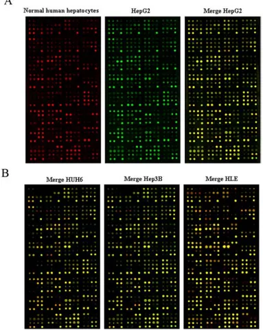
An mRNA- and protein-array, performed on the liver cancer cell lines; showed different signatures in the expression levels of genes and proteins, compared to normal human hepatocytes. The mRNA array analyzed the expression of several genes (96) involved in canonical and non canonical WNT signallings and revealed, in liver cancer cell lines compared to normal human hepatocytes, the contemporary upregulation of antagonists genes of the canonical WNT pathway, such as NLK and SOX17, and the downregulation of its agonists genes, such as TCF7L2, TLE1, SLC9A3R1 and WNT10A. These evidences suggest an inactivation of the canonical WNT signalling; at the same time the overexpression of RHOU transcript indicate the activation of the non canonical WNT signalling.
The very innovative protein array technology evaluated the epression levels of 224 proteins involved in biological pathways such as apoptosis, cell cycle, and signal transduction, and revealed that Grb-2 is over-expressed in the cell lines investigated, compared to normal human primary hepatocytes. Grb-2 is an ubiquitously expressed adapter induced by IGF (I and II) signallings, in normal conditions; through the interaction with Raf/MAPK, Grb-2 activation leads to the induction of cellular proliferation (Foulstone et al., 2005). In a second step, we approached the study of 3 snap-frozen and 6 paraffin-embedded matched tissues obtained from HB patients. In these rare samples we could evaluate the markers previously evidenced and validated in liver cancer cell lines (NLK, RHOU and WNT10A transcripts; Grb-2 protein). This study showed similar results in HB matched tissues when compared to liver cancer cell lines, suggesting that these markers are reliable also in HB tissue samples.
MicroRNAs are small RNA sequences, 20-22 nucleotides long, that can bind to the 3’UTR of specific mRNAs, and regulate their expression by leading to translational repression, mRNA cleavage, and mRNA decay.
Since microRNA expression profiles are altered in several tumours compared to their normal counterparts, these molecules are largely used also in the field of cancer classification (Calin and Croce, 2006). A microRNA-array, performed in the three snap-frozen HB tissues, revealed that 51 microRNAs could distinguish tumour from non-tumour samples. By comparing our results with previously published data (Varnholt et al., 2008), we selected 9 microRNAs to be analysed in our 9 matched samples. The comparison between these microRNAs expression in tumour and in non-tumour counterparts, highlighted four up-regulated microRNAs (miR-125a, -150, -199a and -214), and a down-regulated one (miR-148a). It has been demonstrated that miR-214 binds to the PTEN mRNA and downregulates its protein levels (Yang et al., 2008). PTEN is a negative regulator of the PI3K/Akt pathway which is activated by IGF signalling, in normal conditions (Foulstone et al., 2005). In our samples as well, the comparison of tumour and normal counterparts indicated a correlation between high miR-214 levels and low PTEN protein levels in the tumour samples.
The study of liver cancer cell lines and HB biopsis revealed the inactivation of the canonical WNT pathway and the contemporary induction of the non canonical one. Furthermore the activation of IGF-signalling is also speculated on the basis of the deregulation of Grb-2 and PTEN protein levels.
In the second part of the project the association between HB onset and the exposure to a particular phthalate, DEHP, was investigated. DEHP is the most abundant phthalate in the environment and liver represents one of its main targets. Studies performed on mice and humans demonstrated that DEHP alters the glucose metabolism (Latini et al., 2008). This process involve a family of proteins called PPARs that mediates the IGF-promoted signalling (Feige et al., 2007; Latini et al., 2008), responsible of glycogen storage (Lopez et al., 1999). In utero exposure to phthalates has been shown to be significantly associated with prematurity (Latini et al., 2008), that in turn has been strongly associated with HB (McLaughlin et al., 2006). HB can derive either from developmental disturbances during critical phases of organogenesis or from the interplay of risk factors (e.g. DEHP) during prenatal or early neonatal life (Salvatore et al., 2008).
In order to investigate the post-natal effects of DEHP prenatal exposure on liver development, pregnant CD-1 mice were exposed to DEHP (25 and 100 mg/kg BW pro die) during the critical period of liver organogenesis and histogenesis (starting from the 11th day of gestation and till the 19th) (Duncan, 2003). Male and female F1 mice were sacrificed at post natal day
21 and 35, respesenting weaning and puberty, respectively. Histopathological, histochemical and gene expression studies performed on livers of PND35 treated mice did not show significant differences compared to controls, while at PND21 treated mice showed alterations in glucose and lipid metabolism that were absent in controls. The inactivation of GSK-3β was speculated since these mice were characterized by lack of glycogen storage and presence of cytoplasmic β-catenin. Glycogen storage is triggered by IGF-II in fetal life and ensures stable levels of glycaemia at birth when the newborn makes the adjustment to extra-uterine life (Lopez et al., 1999; Hui et al., 2006). The presence of cytoplasmic β-catenin indicates that the canonical WNT pathway is active, and that the transcription of genes involved in cell proliferation is promoted (Rubinfeld et al., 1996). PND21 mice exposed to DEHP showed hepatocytes vacuolization (hepatosteatosis), a feature of an ecess of fatty acid synthesis, as a consequence of glycogen synthesis inhibition (Shimano et al., 2007). AFP levels in mice progressively decrease after birth and are almost completely switched off in the third week of life, when liver accumulates energy as glycogen instead of AFP (Rusyn et al., 2006; Heudorf et al., 2007; Latini et al., 2009) On the basis of the association between AFP expression and embryonal status, this gene is also used as a marker of hepatocytes maturation (Qin and Tang, 2004). Our results showed high levels of AFP gene expression in treated mice compared to controls, suggestable of a role for DEHP exposure in determining both the alteration of post-natal AFP to glycogen switch off, and a delay of hepatocytes maturation.
Furthermore the possible role of microRNAs in the DEHP-induced alterations was speculated. IGF-II gene, fundamental in the promotion of glycogen storage during fetal life (Lopez et al., 1999; Hui et al., 2006), harbours a microRNA, miR-483, within its second intron (Fu et al., 2005; www.ensembl.org). The analysis of miR-483 expression showed that this miR can be considered a fetal marker, since its levels progressively decrease after birth. High levels of miR-483 were detected in PND21 mice treated with DEHP 100 mg/kg bw pro die, in confirmation with a role of DEHP exposure in delaying the hepatocytes maturation. We also demonstrated that this miR is able to target and downregulate β-catenin, thus representing a limit in the proliferation stimulus induced by high levels of this protein within the cytoplasm (Rubinfeld et al., 1996).
The multidisciplinary approach, performed by using in vivo and in vitro studies, enabled us to identify early and tardive markers of hepatoblastoma. Our studies indicated that the prenatal exposure to DEHP delays liver maturation by affecting carbohydrate and lipid pathways involved in fetal
nutrition. The incapability to accumulate glycogen, due to the inactivation of GSK-3β function, is counterbalanced by an enhanced synthesis of lipid, as demonstrated by the presence of hepatocyte vacuolization. Furthermore the improper increased levels of AFP gene expression, suggested that DEHP alters the post-natal AFP-to-glycogen switch and may delay the post natal maturation of hepatocytes, as suggested also by high levels of miR-483, another fetal marker. A biological role for miR-483 in limiting the proliferation activation induced by β-catenin has been also proposed. The characterization of in vitro models (liver cancer cell lines and HB matched tissue samples) has identified new molecular tardive markers of chilhood liver cancers, at mRNA- (WNT10A, NLK and RHOU trascripts), protein- (Grb-2 and PTEN), and at microRNA- (miR-214) level. Gene expression analysis showed an inactivation of the canonical WNT signalling; and the contemporary activation of the non canonical one. At protein level, the upregulation of Grb-2 in tumour compared to non tumour samples, lets speculate the activation of the Raf/MAPK signalling pathway. The upregulation of miR-214 in tumour compared to non tumour samples seemed to affect PTEN protein levels, which resulted to be downregulated in the tumour counterpart. This evidence suggests that the activation of PI3K/Akt pathway could lead to the misregulation of the cellular functions that it controls, such as glucose metabolism, cell proliferation and survival (Foulstone et al., 2005).
Even though in in vivo and in vitro models the patterns of deregulation of WNT and IGF-signalling pathways involve different mechanisms, the importance of these pathways and their crosstalks can be considered pivotal in the onset and in the characterization of hepatoblastoma.
Introduction
1 Childhood tumours
Malignant tumours are relatively uncommon in children, when one compares them to adult cancers. However, cancer is the main cause of death due to disease in adolescents and children aged over 1 year old, and its incidence is comparable to the cumulative incidence of relatively common conditions, such as cerebral palsy, diabetes and bacteria meningitis, that affect quality of life and survival of childhood population (Ferrari et al., 2007). The annual incidence of cancer is estimated to be around 1 in 7000 children younger than 15 years of age; more than 12,000 new cases of children and adolescents less than 20 years with malignancies are diagnosed every year in the United States of America (Gurney and Bondy, 2006) (and about 1800 new cases a year in Italy) (Ferrari et al., 2007).
Though cancer is the main cause of disease-related mortality in children, all paediatric tumours could be virtually defined as ‘rare’. According to the European Parliament and the Council of the European Union (Regulation EC n.141/2000), rare diseases are defined solely on the basis of low prevalence and affect not more than five individuals per 10.000 in the European population. RD are a large and diverse group of disorders; they include more than 6.000 conditions and involve all organs and tissues, often with several clinical subtypes within the same disease. Almost all RD may cause early mortality and/or long-term disability and accurate and timely diagnosis is often of great importance for their prevention and treatment. Very often information on many RD are insufficient, concerning either diagnosis and/or prognosis. About 80% of RD have identified genetic origins; however in many cases the clinical phenotype and prognosis appear to be modulated by still unknown factors (e.g. different mutations on the same gene(s), epigenetic factors, etc.). Some important groups of RD, such as birth defects and childhood tumours, originate from gene-environment interactions still poorly clarified (www.eurordis.org).
1.1 Liver tumours in children
malignacies. Two cases of liver tumours out of three result malignants. While the predominant histology of liver tumours in adult are represented by hepatocellular carcinoma (HCC), in children hepatoblastoma (HB) accounts for the two thirds of liver tumours. Also sarcomas, germ cell tumours, rhabdoid tumours and the more familial hepatocellular carcinoma are liver pediatric tumours. The treatment and prognosis in children are guided by the histology and anatomy of the liver tumour (Litten and Tomlinson, 2008).
1.1.1 Hepatocellular carcinoma
Hepatocellular Carcinoma (HCC) represents the second most common malignant tumour in children. It is markedly distinct from HB and the age of diagnosis is typically observed after 10, representing the most common malignacy of liver in adolescents. HCC can be associated to known hepatic viral infections or cirrhosis and even though in some cases the tumour can take decades to develop, occasionally cases are seen in very young children (Litten and Tomlinson, 2008).
Previous reports from Southeast Asia cite an annual incidence of pediatric hepatic tumours that is roughly four times higher than western reports in children <15 years of age (Chen et al., 2005). This finding is largely based on the high hepatitis carrier rate, with a Taiwanese report stating that 80% of primary liver tumours in children were hepatocellular carcinoma.With the introduction of hepatitis B vaccine in Southeast Asia, however, there has been a marked reduction in the incidence of hepatocellular carcinoma, although the impact of the hepatitis B vaccine has mainly reduced the incidence of liver tumours in males (Chang et al., 1997). Occasionally, malignant tumours in children are seen with features of both hepatocellular carcinoma and hepatoblastoma. These tumours are more common in children with a diagnosis at later ages than that typical of hepatoblastoma (Litten and Tomlinson, 2008).
1.1.2 Hepatoblastoma: the most common pediatric liver tumour
HB is a very rare primary liver malignancy of infants and young children, with an annual incidence of 0.5–1.5 diagnoses per 1 million children below the age of 15 years in Western countries. Despite its low incidence,
hepatoblastoma represents the most common malignant tumour in children (Schnater et al., 2003; Roebuck and Perilongo, 2006). For poorly understood reasons, hepatoblastoma occurs in males significantly more frequently, compared to females (Weinberg and Finegold, 1983). HB is classified as an embryonal cancer, since these tumours are composed of cells resembling the developing fetal and embryonic liver. The cells comprising HB mark similarly to hepatic stem cells, defined as pluripotent hepatoblasts that can differentiate into hepatocytes or cholangiocytes (Ruck and Xiao, 1997; Ruck and Xiao, 2002). Hepatoblastoma tumour develops prenatally and is therefore diagnosed in very young children having an overall median age of 18 months; only 5% of new HB cases are diagnosed in children >4 years of age. Most commonly, these tumours are present in the right lobe of the liver (Exelby, 1975). These tumours can be histologically divided into epithelial or mixed epithelial/mesenchymal tissues. The majority of HB are epithelial and composed by embryonal and fetal cell types; HBs composed of small undifferentiated cells represent the 5% of HBs cases and are characterized by a worse prognosis (Haas et al., 2001). HB prognosis depends on numerous clinical and histological factors: children with a poor prognosis are usually characterized also by abnormal α-fetoprotein levels (<100 or >1,000,000 ng/ml) and distant metastases (e.g. lung and lymph nodes), whereas patients with good prognosis appear to have, among others, a decline in circulating AFP levels during chemotherapy (Roebuck and Perilongo, 2006; De Ioris et al., 2007).
Scientific evidences point out HB as a multifactorial condition associated with: genetic conditions (e.g. Beckwith-Wiedemann Syndrome and Familial Adenomatous Polyposis) (Roebuck and Perilongo, 2006), non genetic and environmental factors such as the endocrine-metabolic status of the mother (young age, higher body mass index, use of infertility treatments, smoking) and (pre)eclampsia (McLaughlin et al., 2006). An association with occupational paternal exposure to metals, petroleum products and paints/pigments as well the possible involvement of ubiquitous environmental contaminants such as phthalates have been suggested (Buckley et al., 1989; Reynolds et al., 2004; McLaughlin et al., 2006).
Conventional cytogenetic analyses of chromosomal aberrations in HB, performed using standard karyotyping,(Tonk et al., 1994; Sainati and Leszl, 1998; Nagata et al., 1999; Park et al., 1999; Yeh et al., 2000), fluorescence in situ hybridization (FISH),(Balogh et al., 1999; Parada et al., 2000; Sainati et al., 2002; Surace et al., 2002) and comparative genomic hybridization (CGH),(Hu et al., 2000; Weber et al., 2000), have been reported. Although
these analyses have identified several chromosomal aberrations in HB, predominantly the gains in chromosomes 1q, 2, 8q, 17q, and 20 and the loss in chromosome 4q, the tumour-associated genes of HB involved in these genomic copy number (CN) alterations are yet to be identified (Suzuky et al., 2008).
1.2 Dysregulation of growth factor signalling and microRNA expression in childhood hepatic tumours
HCC represents the most used model for the study of the pathogenesis of human childhood liver tumours, even though only limited knowledge has been gathered regarding genomic alterations occuring during its development and progression. There is vast evidence for the dysregulation in HCC of protumourigenic growth factor signalling affecting different signalling systems such as IGF-, WNT-, Hepatocyte Growth Factor (HGF)-, Transforming Growth Factor a (TGFα)/Epidermal Growth Factor (EGF)- and Transforming Growth Factor β (TGFβ) signalling. Dysregulation of these factors and their pathway components has been connected with essential tumour properties such as tumour cell proliferation, antiapoptosis neo-angiogenesis, invasive behavior and chemotherapy resistance (figure 1; Breuhahn et al., 2006).
Cancer is a complex genetic disease involving structural and expression abnormalities of both coding and non-coding genes. For almost three decades, the alteration of protein-coding oncogenes and/or tumour-suppressor genes (Bishop, 1991; Hunter, 1991; Weinberg, 1991) have been thought to be the causes of tumourigenesis.
With the discovery in the past few years of thousands of genes that produce non-coding RNA transcripts with no significant open reading frame, it has become evident that the genomic complexity of the cancer cell is far greater than expected.
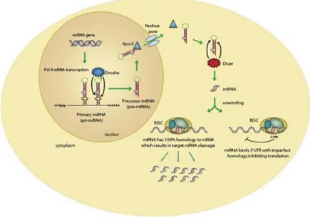
These microRNAs are small non-coding RNAs of 20–22 nucleotides, typically excised from 60–110 nucleotide foldback RNA precursor structures (Ambros, 2004; Bartel, 2004; Pasquinelli et al., 2005). The biogenesis of miRNAs involves a complex protein system, including members of the Argonaute family, Pol II-dependent transcription and the RNase IIIs
Drosha
andDicer
(Kim and Nam, 2006). MiRNAs are involved in crucial biological processes, including development,differentiation, apoptosis and proliferation (Bartel, 2004), through imperfect pairing with target messenger RNAs (mRNAs) of protein-coding genes and the transcriptional or post-transcriptional regulation of their expression (figure 2) (Bartel, 2004; Lim et al., 2005; Rajewsky, 2006).
Figure 1. Schematic and simplified display of different growth factor signalling pathways frequently involved in the development and progression of human hepatocellular carcinogenesis (IGF/IGF-IR, HGF/MET, TGFα/EGFR, WNT/FZD, and TGFβ/TβR) and their potential cross-talks. Predominantly dysregulated signalling components are highlighted in dark gray. Seldom regulated components (e.g. Smad2), molecules not expressed by tumour cells (e.g. HGF) and distinct protein familiy members dysregulated in HCCs (e.g. FZD-7) are highlighted in light gray. (Breuhahn et al., 2006).
Figure 2. In the nucleus, the RNase III–type enzyme Drosha processes the long primary transcripts (pri-miRNA), yielding a hairpin precursors (pre-miRNA) consisting of approximately 70 nt. The pre-miRNA hairpins are exported to the cytoplasm where they are further processed into unstable, 19-25 nt miRNA duplex structures by the RNase III protein Dicer (Sontheimer 2005). The less stable of the two strands in the duplex is incorporated into a multiple-protein nuclease complex, the RNA-induced silencing complex (RISC), which regulates protein expression (www.invitrogen.com).
The number of human miRNAs reported is continuously updated by miRBase, the microRNA database which accounts so far over 940 human hairpin sequences (the April 2010 release of
miRBase
, at the Sanger Institute15
,http://www.mirbase.org/).Initially identified in B-cell chronic lymphocytic leukaemia (
CLL
) (Calin et al., 2002), changes in the expression level of miRNAs have subsequently been detected by different groups in many types of human tumours (several reviews give various details of the link between miRNAs and cancer, such as McManus, 2003; Berezikov et al., 2005; Caldas and Brenton, 2005; Calin et al., 2005; Chen, 2005; Croce and Calin, 2005; Gregory and Shiekhattar, 2005; Calin and Croce, 2006; Esquela-Kescher and Slack, 2006; Hammond, 2006; Hwang and Mendell, 2006). MiRNAs have been proposed tocontribute to oncogenesis because they can function either as tumour suppressors (as is the case for miR-15a and miR-16-1) or oncogenes (as is the case for miR-155 or members of the miR-17–92 cluster). The genomic abnormalities found to influence the activity of miRNAs are the same as those for protein-coding genes, such as chromosomal rearrangements, genomic amplifications or deletions and mutations. In a specific tumour, abnormalities both in protein-coding genes and miRNAs can be identified (Calin and Croce, 2006).
To date, every type of tumour analysed by miRNA profiling has shown significantly different miRNA profiles (for mature and/or precursor miRNAs) compared with normal cells from the same tissue (
table
1,figure
3).Table 1. Facts about microRNAs-expression profiling in human cancers (Calin and Croce, 2006)
Figure 3. Examples of microRNA profiles in human solid and liquid cancers. The general consensus is that profiling enabled the signatures that are associated with diagnosis and prognois. (A) The profiling of 836 solid tissue and haematological primary cancers (and corresponding normal tissues samples) using the microarray technology (as described by Liu. et al., 2004) is shown. The exact number of analysed samples from each tumour type (corresponding normal controls included) is shown. (B) The common expression signature in six solid cancers indicates common altered regulatory pathways involving the same microRNA genes (named on the right side of the dendrogram) (Volinia et al., 2006). These miRNAs could be considered as ‘cancer markers’, because variations of their expression could identify the cancerous state. (C) The distinct clusters identified by miRNA profiles in chronic lymphocytic leukaemia (CLL) (Calin et al., 2004), were further confirmed to be associated with prognostic factors and disease progression (Calin et al., 2005). Note also that various normal haematopoietic samples cluster in a group that is distinct from all CLL samples. MNC, mononuclear cells; Ly, B lymphocytes; CD5, a subset of B lymphocytes largely accepted to represent the equivalent of malignant cells in CLL (Chiorazzi et al., 2005) (Calin and Croce, 2006).
In the case of HCC, the evaluation of the total content of microRNAs, showed that specific miRs are able to distinguish HCC from normal tissue with 97,8% accuracy. Also a correlation with differentiation of the tumours was found (Murakami et al., 2006).
Despite the abundance of studies focused on HCC alterations in pathways and microRNA expression, HB pathogenesis is poorly known. Several studies has focused their attention on the canonical WNT and IGF-II signallings, since their deregulation is involved in the pathogenesis of the genetic disorders, the Familial adenomatous polyposis and
Beckwith-Wiedermann Syndrome, that predispose to HB onset (Roebuck and Perilongo, 2006). FAP patients carry a germline mutation in APC gene, which codifies for a protein involved in the regulation of the cytoplasmic levels of β-catenin (Orford et al., 1997), the central effector molecule of the canonical WNT signalling pathway (Rubinfeld et al., 1996). BWS patients show a loss of imprinting in region 11p15 where IGF-II gene is localized. The LOI causes the biallelic expression and therefore, an increase of IGF-II levels, that could lead, as suggested by Veronese and coworkers, to enhanced cellular proliferation, differentiation failure and tumour development (Veronese et al., 2010).
1.2.1 WNT pathway and its role in cancer
The WNT/β-catenin signalling pathway, also named WNT/Frizzled (FZD) signalling cascade, is important for the determination of the cell fate during the embryonic development, as well as in maintaining tissue homeostasis in the adult (Katoh, 2002; Lee et al., 2006; Khan et al., 2007). WNT signals are transduced to the nucleus through two possible pathways (figure 4). The canonical WNT/β-catenin signalling pathway is initiated by the binding of WNT ligands to the transmembrane receptors. The resulting signals prevent β-catenin phosphorylation by a multiprotein complex composed by APC, GSK-3β, casein kinase 1, and Axins, and its subsequent proteosomal degradation (Blaker et al., 1999; Moon et al., 2004; Clevers, 2006). Nuclear β-catenin is then complexed with the T-cell factor/lymphoid enhancer factor (TCF/LEF) to activate the transcription of target genes (He et al., 1998; Pennica et al., 1998; Tetsu and McCormick, 1999; Kramps et al., 2002; Katoh and Katoh, 2003; Chamorro et al., 2005; Sareddy et al., 2009;). As observed in several types of tumours, the aberrant activation of the canonical WNT/β-catenin signalling pathway is an important contributor to tumourigenesis (Peifer and Polakis, 2000; Takigawa and Brown, 2008). On the contrary, the non canonical WNT signals are transduced through FZD family receptors and co-receptors (Oishi et al., 2003; Lu W et al., 2004; Lu X et al., 2004) to the Dishevelled (DVL)-dependent (i.e., RHOA, RHOU, RAC, CDC42, and JNK; Boutros et al., 1998; Tao et al., 2001), or the Nemo-like kinase (NLK) and nuclear factor of activated T cells (NFAT; Ishitani et al., 2003; Dejmek et al., 2006) signalling cascades (figure 4). Particularly, NLK is a serine/threonine kinase that suppresses the transcription activity of the β-catenin/TCF complex through
phosphorylation of TCF/LEF family transcription factors to inhibit the non canonical WNT signalling pathway (Ishitani et al., 2003). Approximately 80% HBs and 20% HCCs are characterized by somatic mutations in
CTNNB1, which codifies for β-catenin (Koch et al., 1999; Nhieu et al.,
1999; Wei et al., 2000; Yamamoto et al., 2003). Mutations in AXIN1 and
AXIN2 have been demonstrated to be important in an additional 10% of
HBs and HCCs, respectively (Taniguchi et al., 2002). Note that an altered expression of specific WNT/β-catenin target genes, playing key roles in proliferation and survival of cancer cells, has been already reported (Shtutman et al., 1999; Tetsu and McCormick, 1999). There is increasing evidence that regulatory mechanisms different from mutations either in
CTNNB1 or in proteins involved in the maintenance of β-catenin stability
may play a major role in hepatocarcinogenesis. Despite the identification of several target genes of the β-catenin/TCF transcription complex (e.g., c-MYC; He et al., 1998; Shtutman et al., 1999; Lee et al., 2006), cyclin D1 (He et al., 1998; Shtutman et al., 1999; Tetsu and McCormick, 1999), AXIN2 (Jho et al., 2002; Lustig and Behrens, 2003), matrix metalloproteinase-7 (MMP-7; Crawford et al., 1999), FRA-1, c-JUN, urokinase-type plasminogen activator receptor (uPAR; Mann and Smart, 2002), and immunoglobulin transcription factor-2 (ITF-2; Kolligs et al., 2002); their role in liver cancer development is still not clear.
Figure 4. WNT signalling cascades. WNT signals are transduced to the canonical WNT pathway for cell fate determination, and to the non canonical WNT pathway for control of cell movement and tissue polarity. Canonical WNT signals are transduced through FZD family receptors and LRP5/LRP6 co-receptor to the β-catenin signalling cascade. Non canonical WNT signals are transduced through FZD family receptors and the Knypek co-receptor to the DVL-dependent or the Ca2+-dependent (NLK and NFAT) signalling cascades. Both pathways promote the transcription of a specific set of genes involved in cell proliferation, cell polarity control and cell motility. For details, see text (di Masi et al., 2010).
1.2.2 IGF system and its role in cancer
The IGF signalling pathway is of central relevance in embryogenesis as well as lifespan regulation, and exhibits potent proproliferative and antiapoptotic properties. Key regulatory molecules of this axis are small
Canonical WNT signalling Frizzled WNT LRP5 LRP6 DVL β-catenin TCF/LEF SOX17 β-catenin GROUCHO NLK Non canonical WNT DVL K k Ca++ PKC RHOA DAAM Cytoskeletal reorganization CAMK MAP3K7 WNT Rac MAPKK JNK MAPKKK DKK APC GSK3 AXI Frizzled
WNT target genes (e.g., AXIN2,
DKK1, MYC, Cyclin D1, c-JUN)
Genes involved in cell polarity control and
cell movement
Nucleu Cytoplas
N
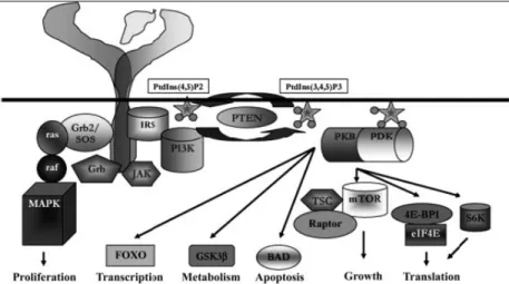
ligands (IGF-I and IGF-II, 7–8 kDa), IGF binding proteins (IGFBP1–6) and membrane-bound receptors (IGF-I receptor (IGF-IR), as well as the mannose-6- phosphate receptor (IGF-II/M6PR, IGF-IIR). Bioavailability of both IGFs is influenced by the presence of secreted IGFBPs (Murphy, 1998) as well as IGF-IIR, which directs IGFs to lysosomal degradation (Braulke, 1999). However, the system is far more complex, as both ligands not only bind to their conventional receptors, IR (Esposito et al., 1997) and IGF-IIR, but also to the insulin receptor (isoform A, INSR; Frasca et al., 1999), and INSR/IGF-16F-IR hybrid molecules (Sakai and Clemmons, 2003). After ligand binding, rapid phosphorylation of distal intracellular targets commences specific cellular downstream effectors such as INSR-substrates IR5 (IRS1,2,4), leading to the activation of, for example PI3K/Akt pathway. The PI3K/Akt pathway is negatively regulated by the lipid phosphatase PTEN whose activity is often reduced in several cancers (Simpson and Parsons, 2001), HCC included, underlying the key role of this pathway in cancer biology (Meng et al., 2007). Moreover, binding of Grb-2 leads to the activation of the Raf/ MAPK signalling pathway. Together, this regulatory network results in increased proliferation, increased cell survival, increased cell mass, and metabolic effects (figure 5; Foulstone et al., 2005).
An increased level of IGF-II gene expression compared with normal livers has been reported in a high proportion of human HB. In the fetal liver, promoters P2, P3, and P4 are active and expressed monoallelically; P3 is the most active promoter and P1 is inactive. However, in the adult liver, P1 becomes dominant and is biallelically expressed, and P2, P3 and P4 activities are decreased or lost (Li et al., 1998). In addition, P4 hypomethylation has been shown in several human cancers, including HCC, suggesting that it may be an early end common event in hepatocarcinogenesis (Tang et al., 2006). Although the exact mechanism leading to these alterations is not entirely certain, it seems that LOI and/or promoter demethylation are at least partly involved (Gray et al., 2000). It has been suggested that increased IGF-II could lead to an enhaned cellular proliferation, differentiation failure, and tumour development (Veronese et al., 2010).
Figure 5. Intra-cellular components of the insulin-like growth factor system. A simplified figure of the intra-cellular pathway is presented. Phosphorylation of the activation loop of the cytoplasmic domain of the IR and IGF-IR results in enhanced catalytic activity of the tyrosine kinase domain. Protein–protein interactions with insulin receptor substrates (IRS) establish priming of the PI3K enzymatic conversion of membrane phospho-inositols, activation of phospho-inositide-dependent protein kinase (PDK), protein kinase B (Akt/PKB), and downstream substrates that control transcription (forkhead transcription factors, FOXO), metabolism (GSK-3β), apoptosis (bcl-associated death promoter, BAD), cell growth and translation (mammalian target of rapamycin, mTOR; tuberous sclerosis gene product, TSC; Raptor; eukaryotic initiation factor 4E, eIF4E and its binding protein, 4E-BP1; and ribososmal protein S6 kinase, p70S6K). Via similar protein–receptor interactions, activation of proliferation is mediated via the Ras GTPase-mediated pathway leading to cell proliferation (Raf, and mitogen activated protein kinase family, MAPK) (Foulstone et al., 2005).
1.2.3 Altered microRNAs in hepatocarcinogenesis
Several studies on microRNA expression profile in HCC samples have suggested that many of the miRNA changes, that occur during hepatocarcinogenesis, do so early, so that many changes that predispose to HCC have already taken place in liver cirrhosis and other premalignant lesions (Varnholt, 2008). Subsequent changes in the miRNA expression in the transition from cirrhosis to HCC seem to be much less marked (Jiang et
al., 2008). A progressive downregulation of miR-145 and miR-198 from cirrhotic tissue to dysplastic nodules and further to HCCs of increasing histological grades has been observed. The fact that abnormal miRNA expression patterns are already present in premalignant lesions has also been shown for other organ sites such as miR-143 and miR-145, which are downregulated in colonic adenomas as well as adenocarcinomas (Michael et al., 2003) and miR-221, which is upregulated in papillary thyroid carcinomas and also in peritumoural thyroid parenchyma (He et al., 2005). Changes of miRNA patterns have been demonstrated to occur before tumour formation in a HCC-model of rats exposed to tamoxifen (Pogribny et al., 2007). Therefore, it remains a tantalizing possibility that miRNAs could serve as early warning markers for cancer initiation or progression (Caldas and Brenton, 2005; Calin and Croce, 2006; Cummins and Velculescu, 2006).
Interestingly, miR-122 expression is limited to the human liver, where it constitutes about 70% of the total microRNA content. In the absence of miR-122, liver functions and cholesterol levels are consistently compromised, probably because miR-122 regulates the expression of genes involved in cholesterol biosynthesis (Hutvagner, 2006; Lee et al., 2008). In HCV infection, miR-122 is required for efficient viral RNA expression: indeed, the virus can replicate in miR-122 expressing cells, such as HuH7 liver carcinoma cells, but not in HepG2, which does not express miR-122 (Mott et al., 2007).
MiR-21 was found to be upregulated in seven different studies performed on HCCs. Meng and coworkers, have demonstrated that miR-21 targets the tumour suppressor gene PTEN, which is a key contributor to HCC pathogenesis and growth, whose protein product is frequently absent in HCCs (Meng et al., 2007).
While miR-122 is the most abundant miRNA in adult livers, other miRNAs such as miR-92a and miR-483 are highly expressed in fetal livers (Girard et al., 2008). MiR-483 is localized in the second intron of IGF-II gene, pointing out again the importance of 11p15 locus (Fu et al., 2005). Veronese and coworkers tested the potential oncogenic activity of this miR, proving evidence for a role as an antiapoptotic oncogene involved in human tumourigenesis. In fact they demonstrated that an anti-miRNA oligonucleotide against miR-483 could suppress the tumourigenicity of HepG2, a cell line that overexpresses both miR-483 and IGF-II gene. Conversely, no antitumour affect was elicited by inhibition of IGF-II (Veronese et al., 2010).
Despite the identification of several altered microRNAs in HCC, no data are available concerning HB.
1.3 Risk factors in HB: di(2-ethylhexyl)phthalate exposure and its biological effects
The exposure of the general population to phthalates, or phthalate esters, is a global problem due to their possible health impact (Scientific Commitee on Emerging and Newly-Identified Health Risks, 2008). Phthalates are industrial chemicals commonly used to soften rigid polyvinyl chloride (PVC)-based plastics contained in food packaging, medical devices (e.g., intravenous bags and tubing), enteric coatings of some medications, consumer products (soaps, shampoos and other cosmetics), paints and pesticide formulations; as phthalates are not covalently bound to PVC, they may be released in the surrounding media, such as water, foods as well as blood (Reynolds et al., 2004).
Di(2-ethylhexyl)phthalate (DEHP) is the most abundant phthalate in the environment. In Europe, the exposure of general population to DEHP has been estimated at about 2 mg/day, based on ambient monitoring and scenario calculations (uptake of food, dust, water, etc.) (Scientific Commitee on Emerging and Newly-Identified Health Risks, 2008). The age-related mean (and maximum) estimated exposure were as follows: 16.16 (135.28) µg/kg body weight (bw)/day in infants (< 1 yr; 5.5 kg bw), 6.31 (62.10) µg/kg bw/day in toddlers (1-3 yrs; 13 kg bw), 2.54 (14.71) µg/kg bw/day in female adults (>18 yrs; 60 kg bw) and 2.85 (16.32) µg/kg bw/day in male adults (> 18 yrs; 70 kg bw) (Heudorf et al., 2007). The European Risk Assessment Report for DEHP established a Tolerable Daily Intake (TDI) of 20 µg/kg bw/day for newborns (<3 mo) and women in childbearing age and of 48 µg/kg bw/day for general population (Heudorf et al., 2007). Therefore, a significant proportion of infants and toddlers may have DEHP intake higher than TDI. Moreover, medical devices using PVC-based materials can provide higher exposures than the environmental ones: in the case of blood transfusion, an exposure to 250-300 mg of DEHP may occur, equivalent to 4.2-5.0 mg/kg for an adult weighting 60 Kg. Neonates in intensive care units are the population group undergoing the highest exposure to DEHP relative to body weight; the daily dose may increase up to 20 folds the TDI. Small children have also a higher production of oxidized metabolites, which have to be
considered in the biomonitoring of DEHP (Heudorf et al., 2007; Scientific Commitee on Emerging and Newly-Identified Health Risks, 2008).
1.3.1 Molecular mechanisms involved in DEHP-mediated toxicity in humans
In rodent toxicological studies, phthalates and their metabolites show several adverse effects in multiple organ systems, including the reproductive tracts, liver and thyroid; although some differences in potency and targets exist among the compounds, the developing organism appears consistently more vulnerable than the adult (Heudorf et al., 2007; Scientific Commitee on Emerging and Newly-Identified Health Risks, 2008).
In humans, cumulating data point out the possible risk of exposure to DEHP and other phthalates. Most reported adverse effects concern reproductive health such as increased risk of poor sperm quality and endometriosis.
However, human studies start to pay attention also to consequences of in
utero phthalate exposure: in particular, this can be associated with a
shorter duration of pregnancy and with altered homeostasis of placental essential fatty acids, potentially leading to abnormal fetal development. (Scientific Commitee on Emerging and Newly-Identified Health Risks, 2008).
DEHP can modulate, either directly or indirectly, different Nuclear Receptors (NRs), such as PPARs (Mulholland et al., 2005; Latini et al., 2008; Scientific Commitee on Emerging and Newly-Identified Health Risks, 2008). In rodents, PPAR-α acts as a proliferative inducer and hepatocarcinogen, affecting multiple signalling pathways through the transcriptional activation of PPAR-regulated genes, and regulating other NRs such as the androgen receptor (Mulholland et al., 2005).
In humans, DEHP does not induce peroxisome proliferation suggesting that effectors other than PPAR-α might be involved in liver tumour induction or promotion (Latini et al., 2008; Scientific Commitee on Emerging and Newly-Identified Health Risks, 2008). PPAR-γ is one major target of DEHP that mediates IGFs-promoted signalling (Feige et al., 2007; Latini et al., 2008). The molecule of PPAR-γ consists of an N-terminal domain (also called A/B domain), which is responsible for ligand-independent transcriptional regulation. The DNA-binding domain
(or C) contains two zinc-finger-like and an α-helical DNA binding motifs. Through the C domain, PPAR-γ recognizes PPRE (Peroxisome Proliferators Response Element) sequences in the regulated promoter regions. PPAR-γ interacts at different levels with IGF system and its downstream signalling pathways as MAPK, PI3K and mTOR. These crosstalks suggest that PPAR-γ acts as a an inhibitor of IGF-signalling(figure
6;
Belfiore et al., 2009).Figure 6. Crosstalk between PPAR-γ and IGF system downstream signalling pathways. The points of interaction between PPAR-γ and MAPK/PI3K pathways occur at different levels and are indicated as arrows (activation) or bars (inhibition). In some cell contexts PPAR-γ ligands reduce MEK1/2 protein expression (A) and inhibit ERK1/2 phosphorylation (B). However, in other cell systems, PPAR-γ agonists may activate ERK1/2 (C). Alternative mechanisms of interaction between PPAR-γ and MAPK/PI3K pathways include (D) ERKmediated PPAR-γ phosphorylation at Ser84/114; (E)MEK1/2-dependent PPAR-γ nuclear export followed by PPAR-γ degradation; (F) PTEN upregulation; (G) mTOR downregulation via activation of AMPK; (H) inhibition of p70S6K phosphorylation (Belfiore et al., 2009).
1.3.2 HB, altered fetal growth and exposure to phthalates
Significant exposure of human fetuses and newborns to DEHP has been demonstrated. Measurable concentrations of phthalates’ metabolites have been found in the amniotic fluid, thus linking fetal to maternal exposure, direct neonatal exposure can also occur. In particular, newborns in neonatal intensive care units are a group at particularly increased risk of phthalate toxicity in this subpopulation. Exposure may reach 3 mg/kg bw/day for a 4-kg newborn due to DEHP leakage from multiple PVC-based devices for extra corporal membrane oxygenation and parenteral nutrition, and such exposures may occur for a period of weeks or even months (Scientific Commitee on Emerging and Newly-Identified Health Risks, 2008). The increased risk of HB in premature (<38 weeks) and/or LBW(<2,500 g) babies has been indicated by different authors since ‘90s (Reynolds et al., 2004; McLaughlin et al., 2006). Extremely low birth weight (ELBW, less than 1,000 g) was associated with a strongly increased risk of HB with an estimated 56.9 relative risk (RR) of HB in comparison to normal birth weight (> 2,500 g) (McLaughlin et al., 2006). ELBW infants diagnosed with HB had received relatively longer duration of intensive care treatments, whose diffusion has greatly improved the survival of premature infants in industrialized countries during the last decades (Bunin, 2004; Reynolds et al., 2004; McLaughlin et al., 2006). Although the link between long-term perinatal phthalates exposure and HB pathogenesis might be biologically plausible, no direct evidence has been so far reached. Epidemiological data collected by Spector and collaborators revealed that among paediatric cancers, only hepatoblastoma risk is strongly associated to children with LBW, and unlikely to be attributable to chance and bias, having been observed in the United Kingdom, in Japan and in USA (Spector et al., 2008).
As showed in 1.3.1 paragraph, PPAR-γ is one major target of DEHP and mediates IGFs-promoted signalling. The activation of PPAR-γ, as a consequence of DEHP exposure, leads to the inactivation of the IGF-signalling (Feige et al., 2007; Latini et al., 2008). In particular of IGF-II is a key regulator of fetal growth and plays important roles in placental nutrient transfer; it is abundantly expressed in fetal liver, its concentration declining after birth (Rodriguez et al., 2007). Two transitional key events are known to occur post-natally in mammalian liver and both of them involve IGF-II: the adult switch of the hepatic glycogen storage and the
fetal-to-adult switch of IGF-II developmental-regulated transcripts. Hepatic glycogen storage is IGF-II-dependent during fetal life whereas post-natally is carried over by insulin; at the end of gestation, glycogen storage is triggered via IGF-II to ensure stable levels of glycaemia at birth when the newborn makes the adjustment to extra-uterine life (Lopez et al., 1999; Hui Tang et al., 2006).
In case of premature babies, both the hepatic transitional events may be affected: i) the IGF-II-dependent hepatic glycogen storage is shortened by the reduced gestation, and in turn ii) the switch of IGF-II developmental-regulated transcripts takes place earlier when the major fetal IGF-II transcript is not prone yet to silencing and, concurrently, when the hepatic mitotic signalling is further triggered to replenish the insufficient hepatic glycogen storage.
Moreover, in premature babies these two crucial events take place in presence of, and may be perturbed by, phthalate exposure that can induce effects in a more susceptible tissue similarly to in utero exposure (Salvatore et al., 2008).
1.4 HB evidences in mouse
HB is mostly studied in mice where, although rare, it may occur spontaneously. Mouse HB derives from pluripotent endodermal stem-like cells; thus the pathogenesis is similar, but not identical, to the human cancer (Ruck and Xiao, 2002; Turusov et al., 2005; Roebuck and Perilongo, 2006). Unlike in humans, mouse HB generally occurs in aged rather than juvenile animals: this can be due to the standard testing schedule for carcinogenicity in rodents, where dosing starts in juveniles and lasts through to two years of age, i.e., the major part of the rodent lifespan. A few chemicals can increase HB incidence; likewise human HB, chemically-induced murine HB shows a higher incidence in males. In particular, the lifetime exposure to benzofuran (BF), a recognised hepatocarcinogen in adult male mice, causes HB with higher specificity in comparison to other liver tumours, although no information are available on the specific mechanism of action. (Turusov et al., 2005). In mice, the critical window for liver development and differentiation starts at gestational day (GD) 12 and is completed at birth (GD21) (Duncan, 2003). However, at present, no data do exist on the induction of HB-like alterations in rodents at early life stages and/or upon intrauterine exposure.
in mouse HB: i) immunohistochemical studies have shown a loss of β-catenin membrane staining along with increased cytoplasmic and nuclear staining; ii) a high prevalence of deletion mutations affecting the GSK-3β-binding region of the β-catenin gene have been identified.
In mice, disruption of the IGF-I, IGF-II or IGF-IR genes retards fetal growth, whereas disruption of IGF-IIR or over-expression of IGF-II enhances fetal growth. On the other hand, high levels of IGF-II are often associated with perinatal lethality, whereas in humans the up-regulation of IGF-II has been described in a high percentage of HB (Gray et al., 2000; Kim et al.,.2005; Rodriguez et al., 2007). Finally. Moreover, in developing rodents, phthalates have been also shown to interfere with the IGF signalling (Bowman et al., 2005). So far, there are no data on DEHP-induced HB in mice; indeed, the range of chemicals inducing HB in mice has not been thoroughly investigated. Most important, no evidences do exist on whether prenatal chemical exposures might induce HB-related alterations in mice, especially in relation to intrauterine growth retardation and/or altered WNT/β-catenin signalling pathway.