Università Politecnica delle Marche
Facoltà di Ingegneria
Corso di Dottorato in Ingegneria
dell’Informazione
Curriculum Ingegneria Biomedica, Elettronica e delle
Telecomunicazioni
XXIX ciclo (XV ciclo n.s.)
Balance and Motor Control
in Dynamic Tasks
PhD Candidate:
Alessandro Mengarelli
Tutor:
When you have excluded the impossible whatever remains, however improbable must be the truth
Once you realize what a joke everything is being the Comedian is the only thing that makes sense
n1 grazie al Prof. Sandro Fioretti per avermi dato la possibilità di lavorare facendo ciò che più mi piace:
un privilegio concesso a pochi.
n! grazie a Francesco: senza di lui probabilmente niente sarebbe iniziato.
en grazie a tutte le persone che ho incontrato e con cui ho lavorato in questi anni: ognuno di loro nel
bene (molti) e nel male (pochi) ha lasciato qualcosa e spero di aver fatto lo stesso.
nn grazie a Stefano, Giacomo, Giulia e Michela: ognuno di loro sa il perché, con la speranza che non
finisca qui.
n grazie a Federica: non a tutti è concesso di conoscere persone veramente speciali. Sono stato fortunato.
Table of Contents
Part 1: Motor Control in Perturbed Balance
3
1. Introduction
4
1.1
Utility of Dynamic Posturography
6
2. Methods
23
2.1
Experimental protocol and setup
23
2.2
Dynamic data acquisition and processing
24
2.3
Kinematic data acquisition and processing
27
2.4
Electromyographic data acquisition and processing
34
3. Evaluation of alternative devices
for dynamic measurements
39
3.1
Wii Balance Board characteristics
40
3.2
Methods
41
3.3
Results and Discussion
47
4. Results
58
4.1
Fixed velocity
58
4.2
Increasing velocity
73
4.3
Sensory deprivation
84
5. Discussion
92
5.1
Fixed velocity
93
5.2
Increasing velocity
99
5.3
Sensory deprivation
102
6. Conclusions
108
7. Balance Response Modeling
111
7.1
Single Inverted Pendulum
112
Part 2: Motor Control in Walking Task
125
1. Introduction
126
2. Methods
132
2.1
Signal acquisition
132
2.2
Signal processing
133
2.3
Statistical gait analysis
136
2.4
Statistics
137
3. Results
140
3.1
Gender differences in lower limb muscles activity
140
3.2
Co-contraction patterns of ankle muscles
149
3.3
Gender-related differences in ankle muscles
co-contraction patterns
153
Part 1
Chapter 1
Introduction
Balance is defined as the ability to keep the body center of mass within the base of support limiting the sway. The study of human upright posture represents a well-known and rooted research field (Maki, 1986; Prieto et al., 1996) and in particular the instrumental assessment of balance is nowadays considered fundamental in order to characterize the principles governing the optimization and deterioration of postural control. However, in some cases a subject can maintain the upright stance without showing abnormal oscillation of both center of pressure and center of mass and at the same time exhibit abnormal responses when his balance encounters perturbations, of environmental as well as proprioceptive origins (Nardone et al., 2006). Starting with the seminal work of Nashner (1976, 1977), the analysis of human ability to maintain balance and recover upright stance after a perturbation has received growing interest throughout the years (Horak and Nashner, 1986; Horak et al., 1989), with the attempts to identify the existence of a single or a set of repeatable response patterns to various type of upright stance perturbations (Horak and Nashner, 1986; Alexander et al., 1992; Hughes et al., 1995; Runge et al., 1999). In recent years, the analysis of balance responses to various type of external stimuli, such as translation, tilt, rotation or backwa rd and forward shift of the base of support, has been applied with subjects suffering from diseases which lead to balance impairment, such as Parkinson’s disease, multiple sclerosis, peripheral vestibular deficits or stroke (Dimitrova et al., 2004; Cameron et al., 2008; Geurts et al., 2005,). In these cases, perturbed posturography resulted useful in order to evaluate the sensory-motor integration necessary for balance control, to estimate the risk of fall, to assess the effects of treatments and the impairment due to disease progress (Visser et al., 2008). Moreover, several manipulations can be introduced to make the balance task more challenging, such as reduction of the base of support or sensory deprivation. Indeed, postural control relies mainly on visual, vestibular and proprioceptive
afferent systems and the decreasing or deprivation of one or more of them may provide further insights in understanding physiological and neurological mechanisms which underlie the ability to maintain balance. The possibility to design such a high number of different experimental protocols is due to the high number of perturbation parameters that can be controlled by the examiner; these parameters include principally the type of perturbation, the velocity and the acceleration of the base of support and the direction of perturbation (Maki, 1986). Furthermore, also external perturbations have been used, aimed mainly at upper body segments, such as trunk and shoulders (Santos et al., 2010). An eventual advantage in the use of motor driven devices for the stimulus administration, is that those instruments, are able to provide the same perturbation to each examined subject, irrespective of weight and/or body mass distribution. This aspect represents an improvement on many clinical balance tests and provides an inter-subject independent assessment of balance; unfortunately, this aspect seems to remain, at the moment, the only standardizing element in the instrumental evaluation of balance through external perturbation of the base of support. Indeed, the earlier reported high number of parameters involved in this type of experiments, entails a high number of variables which can be modified separately, obtaining many different experimental conditions. This characteristic leads to a lack of standardization in perturbed balance tests (Visser et
al., 2008), hampering direct comparison of results obtained in different studies.
Despite the number of studies focused on understanding mechanisms joined to the upright stance maintenance after various type of perturbations in healthy adults (Horak and Nashner, 1986; Alexander et al., 1992; Hughes et al., 1995; Kavounoudias et al., 1999; Runge et al., 1999), a widespread interest for the assessment of fixed and repeatable balance recovery patterns as well as of the influence of perturbation characteristics, is still present in many research contexts (Creath et al., 2005; Campbell et al., 2009; Oude Nijhuis et al., 2009; Nonnekes et al., 2013; Schmid and Sozzi, 2016). The interest for the integration of postural and muscular adjustments in withstanding destabilizing stimuli is also reflected by the high number of studies in which a model for the human posture control is proposed (Alexandrov et al., 2005; Welch et al., 2008; Davidson et al., 2011; Li et al., 2012).
However, many aspects regarding balance recovery strategies remain still controversial, such as muscular features of the response (Welch et al., 2008; Horlings et al., 2009; Perucca et al., 2013), the role of further joints, respect to the ankle and hip, in controlling upright stance (Alexander et al., 1992; Runge et al., 1999; Creath et al., 2005; Corbeil et al., 2013; Cheng, 2016) and neural pathways involved in perturbation withstand (Kavounoudias et al., 1999; Nardone et al., 2012; Nonnekes et al., 2013). Thus, reference values of postural response to perturbations appeared valuable in order to obtain a more clear frame and some attempts have been yet made (Hughes et al., 1995; Perucca et al., 2013). Nevertheless, due to the high number of possible experimental configurations reported earlier, a characterization performed through a specific perturbative stimulus may be not reliable whether another experimental setup is considered (Visser et al., 2008). Thus, this study represents a first attempt to describe upright stance recovery in different perturbative conditions, related to both stimulus characteristics and sensory afferences, in order to give a frame of the different balance responses between different experimental conditions.
1.1
Utility of Dynamic Posturography
Upright posture analysis plays an important role in the instrumental evaluation of balance, which can help to understand the etiology of the considered balance disorder, evaluating at the same time its temporal evolution (Nardone et al., 2010). Symptoms of the disorder are often not evident through the analysis of the standing posture, while in perturbed conditions, e.g. base of support movement, the risk of falling heavily increases. Such kind of tests can support the clinical evaluatio n and clinical decision process, regarding whether a rehabilitation treatment is necessary, what kind of rehabilitation treatment is needed and the evaluation of the treatment results, thus covering the entire rehabilitative process (Hirsch et al., 2003).
The most used device to analyze upright stance is the force platform, which allows the evaluation of the center of pressure (CoP) displacement. CoP also in unperturbed configuration,
shows a certain degree of displacement, supported by the activity of postural muscles and the presence of intermittent mechanisms for the balance control (Burdet and Rougier, 2007; Mezzarane
et al., 2007). Conversely, the employment of moveable platforms with several degrees of freedom
underlines the need for an evaluation of the ability to withstand balance perturbations similar to those the subject could face in the everyday life (Nardone et al., 2006). In this type of test, not only the muscular system is considered, but also the visual, vestibular and proprioceptive systems are analyzed. One of the most used instrument for this kind of evaluations is the EquiTest (NeuroCom International Inc., Clackamas, OR, USA), which includes a platform able to tilt and translate within a movable visual environment (Susan et al., 2006; Geurts et al., 2005; Wrisley et al., 2007; Perucca et
al., 2013; Visser et al., 2008). These features allow to perform the Sensory Organization Test (Wrisley et al., 2007; Yeh et al., 2014), a widely used protocol where the visual reference and the base of
support can move according to the spontaneous body sway of the subject, in order to evaluate his ability to maintain upright posture through the remaining sensory information. The Sensory Organization Test (SOT) is composed by 6 different trials: 1) eyes open, fixed base of support, fixed visual reference 2) eyes closed, fixed base of support 3) eyes open, fixed base of support, movable visual reference 4) eyes open, movable base of support, fixed visual reference 5) eyes closed, movable base of support 6) eyes open, movable base of support, movable visual reference.
Others motor tests commonly used in perturbed posture analysis are the Motor Control Test (MCT), the Motor Adaptation Test (ADT) and the Postural Evoked Response Test (PERT), where the myoelectric evoked potentials are considered (Nardone et al., 2010; Perucca et al., 2013; Lee et al., 2012; Vervoort et al., 2013). MCT evaluates myoelectric potentials evoked by platform translations backward and forward, while in the ADT the platform tilts upward and downward and in PERT both type of potentials are evaluated. The most analyzed muscles in all the tests are gastrocnemius and tibialis anterior. The typical test configuration includes harness to secure the tested subject, who is instrumented through electromyography probes and reflective markers, placed in correspondence of joints of interest, in order to acquire body movements in response to the perturbation. Platform
movement is generally not higher than 10 cm and the frequency of the translation, comm only sinusoidal, not overtakes 1 Hz, where 0.2 and 0.6 Hz are used as references for a slow and fast movement. However, in some studies trials have been performed starting from 0.1 Hz up to 1 Hz, throughout progressive increases of 0.1 Hz. Generally, base of support inclination is around 10 degrees (Nardone et al., 2010; Nardone et al., 2006; Nardone et al., 2000; Schieppati et al., 1995; Nardone et al., 2005; Corna et al., 1999; Corna et al., 2003; Schieppati et al., 2002). Tilting velocity has high variability depending to the specific test performed and data are generally analyzed starting from at least 10 s after the beginning of the test, avoiding the initial period in which the subject adapts the postural response to the base of support movement. Translation perturbations are largely used in dynamic posturography (Diener et al., 1988; Dimitrova et al., 2004; Horak and Nashner, 1986; Horak et al., 1989; Hughes et al., 1995; Nonnekes et al., 2013; Chen et al., 2014), with a large variability in both velocity and displacement of the base of support (Maki 1986; Visser et al., 2008). CoP and CoM displacements are seldom considered (Oude Nijhuis et al., 2009), whereas the myoelectric activity has been commonly evaluated on the basis of the latency time, i.e. the temporal interval between the movement start and the arise of the first muscular burst, commonly classified as short latency response and medium latency response (Nardone et al., 2006; Schieppati et al., 1995; Nardone et al., 2005; Schieppati and Nardone, 1997; Nardone et al., 2008; Nardone et al., 2001; Nardone and Schieppati, 2004; Nardone et al., 2000; Chastan et al., 2008; Nardone and Schieppati, 1998).
Markers position allow to analyze relative joints movement and joint movements respect to the displacement of the base of support (Nardone et al., 2010; Nardone et al., 2006; Nardone et al., 2000; Schieppati et al., 1995; Corna et al., 1999; Schieppati et al., 2002; Chastan et al., 2008; Schieppati et al., 2003; Runge et al., 1999; Horak and Nashner, 1986; Nanhoe-Mahabier et al., 2012; Visser et al., 2010; Henry et al., 2001). A kinematic based parameter easy to represent is the standard deviation (SD) of the marker trace: SD of the marker placed in correspondence of the external auditory meatus is considered a simple but effective way to represent the head displacement and an index of the subject
stabilizing ability among different test configurations (Corna et al., 1999; Schieppati et al., 2002). Cross-correlation between pairs of marker displacement traces give an indication of the degree of coupling of the body segments (Corna et al., 1999; Schieppati et al., 2002).
Posturographic trials are often performed in eyes open (EO) and eyes closed (EC) conditions, highlighting different behaviors: in EO condition the subject acts as a classical pendulum keeping trunk and head stable and the movement is focused on the ankle joint. In EC condition, the subject acts as an inverted pendulum, controlling sway through the ankle joint, following the platform oscillation and withstanding the fall only when the platform movement inverts (Schieppati et al., 2002). In EO condition head movement resulted smaller than the hip, which presented a smaller movement respect to the lateral malleolus; on the contrary, in the EC condition the head presented the larger oscillation (Schieppati et al., 2002). In translational perturbations two strategies for maintaining balance have been suggested, the hip and ankle strategy, which arise with larger and smaller perturbation condition respectively (Horak and Nashner, 1986; Runge et al., 1999). More recently, evidences about a non-negligible role of the knee have been reported, suggesting the possibility to model upright posture in perturbed conditions with a multi-link pendulum rather than the classical double inverted pendulum (Hughes et al., 1995; Alexandrov et al., 2005; Cheng, 2016). Also, a characteristic muscular activation sequence is observed for ankle and hip strategy, with a distal-to-proximal activation pattern for the ankle strategy, where the first muscles to activate are the ankle plantar-flexors, and a proximal-to-distal sequence, where trunk and hip muscles presents the shorter latencies (Horak and Nashner, 1986; Diener et al., 1988; Schieppati et al., 1995; Perucca et
al., 2013).
Perturbed posturography is commonly used in clinical research. For example, a reduced ability to weigh different sensory inputs depending on changes in the environment has been identified in populations with different balance deficits: Parkinson’s disease (Colnat-Coulbois et al., 2005), peripheral vestibular deficits (Peterka, 2002), peripheral neuropathy (Reid et al., 2002) or stroke (Marigold et al., 2004). In PD patients posturographic tests in EO condition seem not able to
distinguish between fallers and non-fallers subjects, while in EC condition fallers PD subjects have shown a larger head oscillation with slower responses when the condition changes from EC to EO condition (Rossi-Izquierdo et al., 2014). Subjects with peripheral neuropathy showed better control indexes in dynamic conditions respect to the static ones; this aspect could refer to a feedforward control in dynamic conditions for this kind of patients (Nardone et al., 2000; Nardone and Schieppati., 2004; Nardone et al., 2005). Subjects with peripheral vestibular deficits maintain balance in static conditions, while the instability arises when sensory input are modified (Corna et al., 2003).
Risk of falling is one of the major problems connected to neurological diseases, thus an index capable to quantify or even predict this feature would have an overwhelming importance (Gu et al., 1996; Henry et al., 2006; Visser et al., 2008; Nardone et al., 2010; Ganesan et al., 2010; Kasser et al., 2011; Sosnoff et al., 2011; Johnson et al., 2013; Prosperini et al., 2011; Prosperini et al., 2013; Rossi-Izquierdo et al., 2014,). The use of moveable platform seems to be useful in evaluating those aspects related to the risk of falling, as the undesired stiffness effects and muscular activations, which resulted overrated in perturbation withstanding and thus increasing the risk of falling. Static upright stance sway seems to not represent a reliable indicator of the risk of falling, since no correlations have been recognized between sway and falling in PD patients. However, through moveable platform, the limits of stability and its critical points can be stressed and analyzed.
Dynamic posturography can be useful also in evaluating rehabilitation treatments: the SOT seems able to give a measure of the after treatment functional recovery in both PD and stroke patients (Geurts et al., 2005). However, in peripheral vestibular deficits, the measurement of the functional recovery appeared more difficult, with similar results in evaluation test between subjects with peripheral vestibular deficits and those with neuropathy (Nardone et al., 2005; Nardone et al., 2008). Static and dynamic posturography are seldom employed as diagnosis support (Visser et al., 2008; Nardone et al., 2010), because there are several pathologies which can lead to balance impairments and moreover the same postural deficit could be related to different pathologies in different subjects and on the contrary subjects suffering from the same pathologies can present
different balance impairments; the use of dynamic posturography is not always able to provide a clear and univocal indication about the nature of the pathology and thus has no large employment as a support for the diagnosis, considering also the requirement for a complex measurement setup. The use of dynamic posturography seems more oriented to the balance disorder analysis and quantification, with the opportunity to push the subject to his limits of stability and to manipulate the sensorial information used in balance control and perturbations withstanding.
In PD patients, as reported earlier, dynamic posturography appears particularly useful, due to their reduced ability in maintaining balance, also in unperturbed conditions and to their high risk of falling. Thus, a number of studies focused their analysis on the assessment of the risk of falling and on deriving index for a reliable distinction between fallers and non-fallers subjects. Perturbed posturography for this kind of patients appeared particularly useful due to the possibility to give destabilizing stimuli similar to those a subject can experiment during his everyday life, allowing an evaluation of the patient ability in withstanding this kind of threat. The most employed perturbatio n for this kind of subjects appeared to be the tilt of the base of support rather than its translation. This aspect reflects the frequent use of the SOT, where the platform tilt follows the spontaneous body sway and it is not imposed by the examiner (Ganesan et al., 2010; Lee et al., 2012). A moveable and sway referenced base of support can be used also without the SOT (Ebersbach and Gunkel, 2011), extracting in this case the path length of the cylinder which allows the platform movement rather than CoP displacement, as in the unperturbed posturography. When the platform tilt is imposed in a single direction (Nanhoe-Mahabier et al., 2012) or in multiple directions (Carpenter et al., 2004; Visser et al., 2010), the tilting angle is bounded between 8.25° and 4.25°, for anterior and posterior tilt respectively, in order to avoid falls (Lee et al., 2012). Base of support translation are often used in antero-posterior and medio-lateral directions in the MCT (Nardone et al., 2010) but often also others directions have been tested, up to 12 translation directions (Henry et al., 1998; Henry et al., 2001). Platform displacement in this case presents a narrow range of variation, between 6 and 9 cm, while acceleration and velocity are often tuned to the pathological condition of the subject, in order to
avoid falls during the test (Dimitrova et al., 2004). CoP and CoM are always measured in static tests but in dynamic tests they are often considered only with tilt perturbations (Ganesan et al., 2010).
Muscular activity is commonly acquired through the surface electromyography (sEMG) and the muscles of interest are usually ankle plantar-flexors, thigh and trunk muscles and also arms muscles (Henry et al., 1998; Henry et al., 2001; Carpenter et al., 2004; Dimitrova et al., 2004; Visser et al., 2010). Reflective markes are usually placed on ankle, knee and hip joints but often the interest is also in the movement of pelvis, arms, trunk and head, which play a significant role also in ankle and hip balance strategies (Henry et al., 2001; Chastan et al., 2008; Nanhoe-Mahabier et al., 2012), whereas trunk displacement resulted involved in balance maintenance (Henry et al., 2001; Visser et al., 2010; Nanhoe-Mahabier et al., 2012), together with arms and head (Henry et al., 2001; Chastan et al., 2008; Visser et al., 2010). The width between feet represents another perturbation parameter, where the distance between the heels defines a wide (≈30 cm) and a narrow (≈10 cm) stance; this distance can be changed asking the subject to modify feet position, in order to test two different conditions (Henry et al., 2001; Dimitrova et al., 2004; Dimitrova et al., 2004a). In translational trials, the measure of CoP and CoM displacement in static and perturbed period quantifies the spontaneous and induced body sway, which can be estimated also through the trunk (Carpenter et al., 2004; Nanhoe-Mahabier et al., 2012) and pelvis and head angular displacement (Chastan et al., 2008). The use of force platforms allows the evaluation of the GRF components, linked to the perturbation directions and provide a measure of the imbalance magnitude, also respect to the stance width (Henry et al., 2001; Dimitrova et al., 2004a). Ankle and hip angles represent the main measures of the body displacement, based on the classical modeling that involves ankle and hip strategies as the two most employed strategies to recover balance (Horak and Nashner, 1986). However, also the upper limbs have been recognized to play a significant role in withstanding perturbations of the base of support and recovering upright stance (Morris, 2000; Carpenter et al., 2004). In accordance with these observations, also muscles of the upper body have been considered in several studies (Henry et al., 1998; Henry et al., 2001; Dimitrova et al., 2004). Muscular activity is described not only by the
temporal delay between perturbation onset and muscular burst but also through the signal amplitude and its integration, which provides a characterization of the PD subjects muscular characteristics respect to healthy subjects (Henry et al., 1998; Henry et al., 2001; Carpenter et al., 2004; Dimitrova et al., 2004; Lee et al., 2012). Some authors (Henry et al., 1998; Henry et al., 2001; Dimitrova et al., 2004) use the muscle tuning curve, obtained from the integrated sEMG signal, in order to evaluate muscular activity in each direction of perturbed body sway. This feature gives an indication of which muscles activates in response to each perturbation direction, evaluating also their role in co-contraction activity; this kind of analysis provides information also about the direction where the risk of falling increases and also about the subject ability to tune his postural response when environmental conditions change. The analysis of muscular response allows also an evaluation of which neurological pathways are involved in parkinson’s disease and in which measure (Chastan et al., 2008; Dimitrova et al., 2004a).
Results obtained by the analyses performed through dynamic posturography are often compared with the outcomes provided by clinical test or balance scale, which assess the pathology development and the subject residual ability to maintain balance. The most used tests are the tandem stance and gait test, single leg standing test, timed up-and-go test and the sit-to-walk test (Ganesan et al., 2010; Ebersbach et al., 2011; Johnson et al., 2013). The most used balance scales are the Berg balance scale, Activities-specific balance confidence, balance evaluation system test, functional gait assessment and the unified Parkinson disease rating scale (Ganesan et al., 2010; Ebersbach et al., 2011; Nanhoe-Mahabier et al., 2012; Johnson et al., 2013). Parameters extracted from dynamic posturography test could support the evaluations made on the basis of the classical clinical tests and balance scales; in particular, it has been observed that a series of indexes obtained through the use of perturbed posturography correlate with the classical clinical evaluations, providing also a reliable instrument to distinguish fallers and non-fallers PD patients (Johnson et al., 2013). Another index of the balance ability is the evaluation of the limits of stability (LOS), which point out the maximum reachable body displacement without moving feet or make a step. LOS can be analyzed using visual
targets the subject has to follows up to the loss of balance (Ganesan et al., 2010; Vervoort et al., 2013), evaluating the sway in the bending direction, the velocity and the accuracy in maintaining CoM within a tolerance zone (Johnson et al., 2013).
Freezing of gait represents another feature of parkinson’s disease which has been analyzed through perturbed posturography (Vervoort et al., 2013; Jacobs et al., 2008). Surprisingly, freezers subjects have shown a better ability in integrating sensorial information and a better balance control respect to the non-freezers, but a lower accuracy in reaching the target (Vervoort et al., 2013). Difficulties in voluntarily moving CoM in the antero-posterior direction could indicate a relationship between freezing of gait and risk of falling, since the subject would not be able to counteract movement inertia caused by the sudden freezing of gait (Vervoort et al., 2013; Bloem et al., 2004). A central issue in Parkinson’s disease tests regards the pharmacological therapy: tests can be performed after 1 or 2 hours after the drug assumption (Visser et al., 2010; Ganeasn et al., 2010; Vervoort et al., 2013) or after 12 hours, when subject is in the OFF state of therapy (Nanhoe-Mahabier
et al., 2012; Dimitrova et al., 2004). Moreover, subjects are asked to perform two tests at a distance of
few hours, in order to evaluate the effects of pharmacological therapy on balance (Jacobs et al., 2008; Nallegowda et al., 2004) or conversely subjects which are following a pharmacological therapy that can affect balance or gait can be discarded before the beginning of the tests (Lee et al., 2012; Nardone
et al., 2012).
Regarding the number and the sequence of trials, for PD patients the first trial resulted the most analyzed, in part to avoid the habituation rate due to the ensuing trials and in part because the first trial reflects better the everyday conditions that could lead to the risk of falling (Visser et al., 2008; Visser et al., 2010). The first trial responses can also be compared to those of the ensuing trials, in order to evaluate the subject ability in elaborating and maintaining the postural balance strategy (Nanhoe-Mahabier et al., 2012).
Perturbed posturography shows a large employment also for balance evaluation in subjects affected by multiple sclerosis (MS), which leads, as in parkinson’s disease, to postural and balance
deficits (Sosnoff et al., 2011; Prosperini et al., 2011; Boes et al., 2012; Huisinga et al., 2012; Prosperini et
al., 2013; Prosperini et al., 2013a). Trials can be performed in EO and EC conditions and also in dual
task condition: This paradigm allows an evaluation of cognitive processing required to maintain standing balance, simply by applying a concurrent cognitive task (Boes et al., 2012; Prosperini et al., 2013a). The deterioration of the balance control appeared independent of pathology development or aging (Boes et al., 2012) and thus the hypothesis that balance and cognitive tasks may share neurological pathways, leading to the impossibility to perform both task at the same time.
In MS analysis, the effect of fatigue has been investigated also through the perturbed posturography (van Emmerik et al., 2010) and Hebert and Corboy (2012) assessed the relationship between fatigue and reduce ability to maintain balance through the use of the SOT. Their findings suggested that the fatigue could be influenced by the reduced ability to maintain balance, showing high correlations between fatigue and the most challenging configurations of the SOT Also translational perturbations have been used with MS patients (Cameron et al., 2008), measuring however only muscular responses, in terms of latencies and amplitude, to backward perturbations which lead to an anterior body sway. The low muscular strength seems not to affect balance, with an increased response in terms of signal amplitude respect to control subjects; this aspect could reflect the need to compensate a retarded muscular response observed in this kind of patients (Horak and Diener, 1994).
In conclusion, the use of dynamic posturography showed a wide employment with both healthy and pathological subjects. However, the several number of variables connected to this type of test (perturbation velocity, acceleration, displacement, direction, impulsive or continuous movement) leads to a lack of standardization which could bias outcomes comparison between different studies and hamper the diffusion of this technique in clinical practice. Moreover, the relative complexity of the instrumentation, the needed to synchronize several different acquisition systems and the weight of data processing can contribute to limit the diffusion of the dynamic posturography in clinical routine analysis. However, following what has been reported previously, dynamic posturography
appears to be valuable in both clinical and research contexts, to gain further insight in neurological and neuromuscular mechanisms which control upright stance and balance maintenance, to distinguish disease stages and in particular to understand neural and sensory pathways involved in each part of the global body response that follows a change in stance conditions.
Chapter References
Alexander N.B., Shepard N., Gu M.J., Schultz A., “Postural Control in Young and Elderly Adults When Stance Is Perturbed: Kinematics”, Journal of Gerontology, 1992.
Alexandrov A.V., Frolov A.A., Horak F.B., Carlson-Kuhta P., Park S., “Feedback equilibrium control during human standing”, Biological Cybernetics, 2005.
Bloem B.L., Hausdorff J.M., Visser J.E., Giladi N., “Falls and freezing of gait in Parkinson's disease: a review of two interconnected, episodic phenomena”, Movement Disorder, 2004.
Boes M.K., Sosnoff J.J., Socie M.J., Sandroff B.M., Pula J.H., Motl R.W., “Postural control in multiple sclerosis: Effects of disability status and dual task”, Journal of the Neurological Sciences, 2012. Burdet C., Rougier P.,” Analysis of center-of-pressure data during unipedal and bipedal standing using fractional Brownian motion modeling”, Journal of Applied Biomechanics, 2007.
Cameron M.H., Horak F.B., Herndon R.R., Bourdette D., “Imbalance in Multiple Sclerosis: A Result of Slowed Spinal Somatosensory Conduction”, Somatosensory and Motor Research, 2008.
Campbell A.D., Dakin C.J., Carpenter M.G., “Postural responses explored through classical conditioning”, Neuroscience, 2009.
Carpenter M.G., Allum J.H., Honegger F., Adkin A.L., Bloem B.R., “Postural abnormalities to
multidirectional stance perturbations in Parkinson’s disease”, Journal of Neurology, Neurosurgery and
Psychiatry, 2004.
Chastan N., Debono B., Maltête D., Weber J., “Discordance Between Measured Postural Instability and Absence of Clinical Symptoms in Parkinson’s Disease Patients in the Early Stages of the Disease”,
Movement Disorders, 2008.
Chen C.L., Lou S.Z., Wu H.W., Wu S.K., Yeung K.T., Su F.C., “Effects of the type and direction of support surface perturbation on postural responses”, Journal of Neuroengineering and Rehabilitation, 2014.
Cheng K.B., “Does knee motion contribute to feet-in-place balance recovery?”, Journal of
Biomechanics, 2016.
Colnat-Coulbois S., Gauchard G.C., Maillard L., Barroche G., Vespignani H., Auque J., Perrin P.P., “Bilateral subthalamic nucleus stimulation improves balance control in Parkinson’s disease Journal of
Neurology, Neurosurgery and Psychiatry, 2005.
Corbeil P., Bloem B.R., van Meel M., Maki B.E., “Arm reactions evoked by the initial exposure to a small balance perturbation: A pilot study”, Gait and Posture, 2013.
Corna S., Tarantola J., Nardone A., Giordano A., Schieppati M., “Standing on a continuously moving platform: is body inertia counteracted or exploited?”, Experimental Brain Research, 1999.
Corna S., Nardone A., Prestinari A., Galante M., Grasso M., Schieppati M., “Comparison of
Cawthorne-Cooksey Exercises and Sinusoidal Support Surface Translations to Improve Balance in Patients With Unilateral Vestibular Deficit”, Archives of Physical Medicine and Rehabilitation, 2003. Creath R., Kiemel T., Horak F., Peterka R., Jeka J., “A unified view of quiet and perturbed stance: simultaneous co-existing excitable modes”, Neuroscience Letters, 2005.
Davidson B.S., Madigan M.L., Southward S.C., Nussbaum M.A., “Neural Control of Posture During Small Magnitude Perturbations: Effects of Aging and Localized Muscle Fatigue”, IEEE Transactions
on Biomedical Engineering, 2011.
Diener H.C., Horak F.B., Nashner L.M., “Influence of Stimulus Parameters on Human Postural Responses”, Journal of Neurophysiology, 1988.
Dimitrova D., Horak F.B., Nutt JG., “Postural Muscle Responses to Multidirectional Translations in Patients with Parkinson's Disease”, Journal of Neurophysiology, 2004.
Dimitrova D., Nutt J., Horak F.B., “Abnormal force patterns for multidirectional postural responses in patients with Parkinson’s disease”, Experimental Brain Research, 2004a.
Ebersbach G., Gunkel M., “Posturography Reflects Clinical Imbalance in Parkinson’s Disease”,
Movement Disorders, 2011.
Ganesan M. Pal P.K., Gupta A., Sathyaprabha T.N., “Dynamic posturography in evaluation of balance in patients of Parkinson’s disease with normal pull test: Concept of a diagonal pull test”,
Parkinsonism and Related Disorders, 2010.
Geurts A.C., de Haart M., van Nes I.J., Duysens J., “A review of standing balance recovery from stroke”, Gait and Posture, 2005.
Gu M.J., Schultz A.B., Shepard N.T., Alexander N.B., “Postural control in young and elderly adults when stance is perturbed: dynamics”, Journal of Biomechanics, 1996.
Hebert J.R., Corboy J.R., “The association between multiple sclerosis-related fatigue and balance as a function of central sensory integration”, Gait and Posture, 2012.
Henry S.M., Fung J., Horak F.B., “EMG Responses to Maintain Stance During Multidirectional Surface Translations”, Journal of Neurophysiology, 1998.
Henry S.M., Fung J., Horak F.B., “Effect of Stance Width on Multidirectional Postural Responses”,
Journal of Neurophysiology, 2001.
Hirsch M.A., Toole T., Maitland C.G., Rider R.A., “The effects of balance training and high-intensity resistance training on persons with idiopathic Parkinson’s disease”, Archives of Physical Medicine and
Rehabilitation, 2003.
Horak F.B., Diener H.C., Nashner L.M., “Influence of Central Set on Human Postural Responses”,
Horak F.B., Nashner L.M., “Central programming of postural movements: adaptation to altered support-surface configurations”, Journal of Neurophysiology, 1986.
Horak F.B., Diener H.C., “Cerebellar control of postural scaling and central set in stance”, Journal of
Neurophysiology, 1994.
Horlings C.G., Küng U.M., van Engelen B.G., Voermans N.C., Hengstman G.J., van der Kooi A.J., Bloem B.R., Allum J.H., “Balance control in patients with distal versus proximal muscle weakness”,
Neuroscience, 2009.
Hughes M.A., Schenkman M.L., Chandler J.M., Studenski S.A., “Postural responses to platform perturbation: kinematics and electromyography”, Clinical Biomechanics, 1995.
Huisinga J.M., Yentes J.M., Filipi M.L., Stergiou N., “Postural control strategy during standing is altered in patients with multiple sclerosis”, Neuroscience Letters, 2012.
Jacobs J.V., Nutt J.G., Carlson-Kuhta P., Stephens M., Horak F.B., “Knee trembling during freezing of gait represents multiple anticipatory postural adjustments”, Experimental Neurology, 2008.
Johnson L., James I., Rodrigues J., Stell R., Thickbroom G., Mastaglia F., “Clinical and Posturographic Correlates of Falling in Parkinson’s Disease”, Movements Disorders, 2013.
Kasser L.S., Jacobs J.V., Foley J.T., Cardinal B.J., Maddalozzo G.F., “A Prospective Evaluation of Balance, Gait, and Strength to Predict Falling in Women With Multiple Sclerosis”, Archives of
Physical Medicine and Rehabilitation, 2011.
Kavounoudias A., Gilhodes J.C., Roll R., Roll J.P., “From balance regulation to body orientation: two goals for muscle proprioceptive information processing?”, Experimental Brain Research, 2009. Lee J.M., Koh S.B., Chae S.W., Seo W.K., Kwon D.Y., Kim J.H., Oh K., Baik J.S., Park K.W. , “Postural Instability and Cognitive Dysfunction in Early Parkinson's Disease”, Canadian Journal of Neurological
Sciences, 2012.
Li Y., Levine W.S., Loeb G.E., “A Two-Joint Human Posture Control Model With Realistic Neural Delays”, IEEE Transactions on Neural Systems and Rehabilitation Engineering, 2012.
Maki B.E., “Selection of perturbation parameters for identification of the posture-control system”,
Medical and Biological Engineering and Computing, 1986.
Marigold D.S., Eng J.J., Tokuno C.D., Donnelly C.A., “Contribution of muscle strength and
integration of afferent input to postural instability in persons with stroke”. Neurorehabilitation and
Neural Repair, 2004.
Mezzarane R.A., Kohn A.F., “Control of upright stance over inclined surfaces”, Experimental Brain
Research, 2007.
Morris M.E., “Movement Disorders in People With Parkinson Disease: A Model for Physical Therapy”, Physical Therapy, 2000.
Nallegowda M., Singh U., Handa G., Khanna M., Wadhwa S., Yadav S.L., Kumar G., Behari M., “Role of Sensory Input and Muscle Strength in Maintenance of Balance, Gait, and Posture in Parkinson’s Disease”, American Journal of Physical Medicine and Rehabilitation, 2004.
Nanhoe-Mahabier W., Allum J.H.J., Overeem S., Borm G.F., Oude Nijhuis L.B., Bloem B.R., “First Trial Reactions and Habituation Rates over Successive Balance Perturbations in Parkinson’s Disease”,
Neuroscience, 2012.
Nardone A. Grasso M., Tarantola J., Corna S., Schieppati M., “Postural Coordination in Elderly Subjects Standing on a Periodically Moving Platform”, Archives of Physical Medicine and
Rehabilitation, 2000.
Nardone A., Tarantola J., Miscio G., Pisano F., Schenone A., Schieppati M., “Loss of large-diameter spindle afferent fibres is not detrimental to the control of body sway during upright stance: evidence from neuropathy, Experimental Brain Research, 2000.
Nardone A., Galante M., Lucas B., Schieppati M., “Stance control is not affected by paresis and reflex hyperexcitability: the case of spastic patients”, Journal of Neurology, Neurosurgery and Psychiatry, 2001.
Nardone A., Schieppati M., “Medium-latency response to muscle stretch in human lower limb: estimation of conduction velocity of group II fibresand central delay”, Neuroscience Letters, 1998. Nardone A., Schieppati M., “Group II spindle fibres and afferent control of stance. Clues from diabetic neuropathy”, Clinical Neurophysiology, 2004.
Nardone A., Schieppati M.,” The role of instrumental assessment of balance in clinical decision making”, European Journal of Physical and Rehabilitation Medicine, 2010.
Nardone A., Galante M., Pareyson D., Schieppati M., “Balance control in Sensory Neuron Disease”,
Clinical Neurophysiology, 2006.
Nardone A, Grasso M., Schieppati M., “Balance control in peripheral neuropathy: Are patients equally unstable under static and dynamic conditions?”, Gait and Posture, 2005.
Nardone A., Galante M., Grasso M., Schieppati M., “Stance ataxia and delayed leg muscle responses to postural perturbations in cervical spondylotic myelopathy”, Journal of Rehabilitation Medicine, 2008.
Nardone A., Pasetti C., Schieppati M., “Spinal and supraspinal stretch responses of postural muscles in early Parkinsonian patients”, Experimental Neurology, 2012.
Nashner L.M., “Adapting reflexes controlling the human posture”, Experimental Brain Research, 1976. Nashner L.M., “Fixed patterns of rapid postural responses among leg muscles during stance”,
Experimental Brain Research, 1977.
Nonnekes J., Scotti A., Oude Nijhuis L.B., Smulders K., Queralt A., Geurts A.C.H., Bloem B.R., Weerdesteyn V., “Are postural responses to backward and forward perturbations processed by different neural circuits?”, Neuroscience, 2013.
Oude Nijhuis L.B, Allum J.H.J., Borm G.F., Honegger F., Overeem S., Bloem B.R., “ Directional Sensitivity of “First Trial” Reactions in Human Balance Control”, Journal of Neurophysiology, 2009. Peterka R.J., “Sensorimotor integration in human postural control”, Journal of Neurophysiology, 2002. Perucca L., Caronni A., Vidmar G., Tesio L., “Electromyographic latency of postural evoked responses from the leg muscles during EquiTest Computerised Dynamic Posturography: Reference data on healthy subjects”, Journal of Electromyography and Kinesiology, 2014.
Prieto T.E., Myklebust J.B., Hoffman R.G., Lovett E.G., Myklebust B.M., “Measures of Postural
Steadiness: Differences Between Healthy Young and Elderly Adults”, IEEE Transaction on Biomedical
Engineering, 1996.
Prosperini L., Kouleridou A., Petsas N., Leonardi L., Tona F., Pantano P., Pozzilli C., “The
relationship between infratentorial lesions, balance deficit and accidental falls in multiple sclerosis”,
Journal of the Neurological Sciences, 2011.
Prosperini L., Fortuna D., Giannì C., Leonardi L., Pozzilli C., “The Diagnostic Accuracy of Static Posturography in Predicting Accidental Falls in People With Multiple Sclerosis”, Neurorehabilitation
and Neural Repair, 2013.
Prosperini L., Pozzilli C., “The Clinical Relevance of Force Platform Measures in Multiple Sclerosis: A Review”, Multiple Sclerosis International, 2013a.
Reid V.A., Adbulhadi H., Black K.R., Kerrigan C., Cros D., “Using posturography to detect unsteadiness in 13 patients with peripheral neuropathy: a pilot study”. Neurology and Clinical
Neurophysiology, 2002.
Rossi-Izquierdo M. Basta D., Rubio-Rodríguez J.P., Santos-Pérez S., Ernst A., Sesar-Ignacio Á., Alberte-Woodward M., Guijarro-Del Amo M., Estany-Gestal A., San Román-Rodríguez E., Faraldo-García A., Zubizarreta-Gutiérrez A., Soto-Varela A., “Is posturography able to identify fallers in patients with Parkinson’s disease?”, Gait and Posture, 2014.
Runge C.F., Shupert C.L., Horak F.B., Zajac F.E., “Ankle hip postural strategies defined by joint torques”, Gait and Posture, 1999.
Santos M.J., Kanekar N., Aruin A.S., “The role of anticipatory postural adjustments in compensatory control of posture: 1. Electromyographic analysis”, Journal of Electromyography and Kinesiology, 2010. Schieppati M., Nardone A., “Medium-latency stretch reflexes of foot and leg muscles analysed by cooling the lower limb in standing humans”, Journal of Physiology, 1997.
Schieppati M, Nardone A., Siliotto R., Grasso M., “Early and late stretch responses of human foot muscles induced by perturbation of stance”, Experimental Brain Research, 1995.
Schieppati M., Giordano A., Nardone A., “Variability in a dynamic postural task attests ample flexibility in balance control mechanisms”, Experimental Brain Research, 2002.
Schieppati M. Nardone A., Schmid M., “Neck Muscle Fatigue Affects Postural Control in Man”,
Schmid M., Sozzi S., “Temporal features of postural adaptation strategy to prolonged and repeatable balance perturbation”, Neuroscience Letters, 2016.
Sosnoff J.J., Socie M.J., Boes M.K., Sandroff B.M., Pula J.H., Suh Y., Weikert M., Balantrapu S., Morrison S., Motl R.W., “Mobility, Balance and Falls in Persons with Multiple Sclerosis”, PLOSONE, 2011.
Susan L.W., Marchetti G.F., Schade A.I., “The Relationship Between Falls History and Computerized Dynamic Posturography in Persons With Balance and Vestibular Disorders”, Archives of Physical
Medicine and Rehabilitation, 2006.
Van Emmerik R.E., Remelius J.G., Johnson M.B., Chung L.H., Kent-Braun J.A., “Postural control in women with multiple sclerosis: effects of task, vision and symptomatic fatigue”, Gait and Posture, 2010.
Vervoort G., Nackaerts E., Mohammadi F., Heremans E., Verschueren S., Nieuwboer A., Vercruysse S., “Which Aspects of Postural Control Differentiate between Patients with Parkinson’s Disease with and without Freezing of Gait?”, Parkinson’s Disease, 2013.
Visser J.E., Carpenter M.G., van der Kooij H., Bloem B.R., “The clinical utility of posturography”,
Clinical Neurophysiology, 2008.
Visser J.E., Oude Nijhuis L.B., Janssen L., Bastiaanse C.M., Form G.F., Duysens J., Bloem B.R., “Dynamic Posturography in Parkinson’s Disease: Diagnostic Utility and the “First Trial Effect””,
Neuroscience, 2010.
Welch T.D.J., Ting L.H., “A Feedback Model Reproduces Muscle Activity During Human Postural Responses to Support-Surface Translations”, Journal of Neurophysiology, 2007.
Wrisley D.N., Stephens M.J., Mosley S., Wojnowski A., Duffy J., Burkard R., “Learning Effects of Repetitive Administrations of the Sensory Organization Test in Healthy Young Adults”, Archives of
Physical Medicine and Rehabilitation, 2007.
Yeh J.R., Hsu L.C., Lin C., Chang F.L., Lo M.T., “Nonlinear Analysis of Sensory Organization Test for Subjects with Unilateral Vestibular Dysfunction”, PLOSONE, 2014.
Chapter 2
Methods
Ten healthy and young adults volunteered for this study (nine females and one male). Mean values of age, height and weight were 23.6±1.2 years, 168±8 cm and 55.6±5.5 kg. None of them suffered from neurological or motor disorder that could affect balance in either static or dynamic condition. Before the experiment, purposes and procedures of the study have been explained and informed consent was obtained from each participant.
2.1
Experimental protocol and setup
A movable platform was used as perturbation device, actuated by electro-mechanical servos and able to translate horizontally in forward and backward direction, with variable velocity and displacement. Subjects stood on the platform, looking forward with arms hanging comfortably on their sides. Subjects self-selected their stance width and were asked to maintain balance in response to the perturbation, without any guidance about how to remain in the upright posture. Perturbation trials in which the subjects had to step in order to maintain balance were not included in the analysis and the trials were repeated. No practice trials were given to the subjects before the beginning of the test.
Each subject was exposed to three different perturbation blocks: the first type of perturbation block consisted of 5 consecutive backward perturbation trials with fixed velocity (15 cm/s) followed by five consecutive forward perturbation trial with fixed velocity (15 cm/s). This kind of experimental condition aimed to allow the evaluation of the first trial effect and the habituation rate. The second type of perturbation block consisted of 5 consecutive perturbation trials in backward direction, with increasing velocity sequence (15 cm/s, 20 cm/s, 25 cm/s, 30 cm/s, 35 cm/s), followed by 5 consecutive
perturbation trials in forward direction with the same platform velocity sequence. The purpose of this kind of trial block was the evaluation of the perturbation speed on the balance response. Platform displacement in the former case (fixed velocity trial block) was set at 5 cm, while in the latter trial block (increasing velocity) was fixed at 15 cm. The eventual type of perturbation consisted of two consecutive backward perturbation at 20 cm/s and with a displacement of 5 cm: during the first perturbation, the subject maintained eyes open and in the second perturbation subject was asked to maintain eyes closed. The same configuration was repeated with forward platform displacement. This kind of experimental test aimed at the evaluation of the sensorial deprivation weight on the balance maintenance. In all the trial blocks, platform acceleration was set at 3 m/s2
and a resting period of 2 minutes was included after each perturbation block, whereas consecutive trials were at most 10 seconds apart. The perturbation amplitudes and velocities were large enough to provoke body sway that is known to exceed natural sway (Winter et al., 2001) and were chosen according with the seminal study of Diener et al. (1988), where the influence of different amplitudes and velocities on postural responses were tested.
Dynamics, kinematics and electromyographic responses were recorded for each subject and all trials were analyzed 1 s before the platform movement onset and 3 s after the platform movement offset. All data were averaged across all trials of the same perturbation amplitude for each subject.
2.2
Dynamic data acquisition and processing
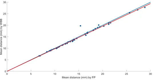
Ground reaction force (GRF) and center of pressure (CoP) displacements are seldom acquired and analyzed in dynamic posturography tests, however, they contributed in obtaining a broader characterization of balance response. Classical force plates (FP) are able to acquire 3 GRF components, i.e. the antero-posterior (AP), medio-lateral (ML) and vertical one, and the AP, ML and vertical momentum; then the CoP displacement in both AP and ML direction can be computed. The characteristics of the moveable platform, a pure mechanical actuator without any embedded
measurement system, the need to remain in a measurement volume defined by the stereophogrammetric system and a series of environmental limits, did not allow the use of a classical FP for dynamic data acquisition. Thus, after a reliability evaluation (see the next chapter), the employment of a commercial and low cost device (Nintendo Wii Balance Board, WBB) has been decided. This instrument allows the acquisition of the vertical GRF component and thus both the CoP displacement components can be extracted. The WBB was placed upon the moveable platform and connected to the latter in order to avoid undesired WBB shifting respect to the support base. Four reflective markers were placed in correspondence of the middle of each WBB side, in order to obtain the WBB position, the platform movement profile and to be able to refer kinematic-based data to the WBB own reference frame if needed. Due to the perturbation characteristics (backwar d and forward horizontal translation), only the AP CoP component has been considered and analyzed. The vertical component of the GRF was firstly low-pass filtered at 10 Hz (Quant et al., 2005, Nonnekes et al., 2013) through a zero-lag, second order Butterworth filter and then CoP displacement was computed according with the method reported by Leach et al. (2014) and explained in the next chapter.
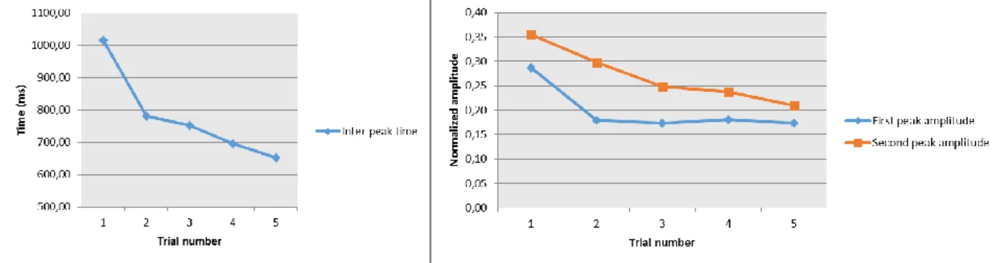
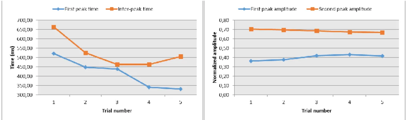
Due to its characteristic double peak shape (Fig. 1), a series of temporal and spatial parameters have been extracted from the CoP AP displacement for both backward and forward trial type:
Figure 1. Representative CoP displacement during a backward trial. Increasing values indicate anterior displacement while negative values stand for posterior displacement. First and second peaks are indicated by black dots. CoP displacement during platform movement is highlighted by red color line.
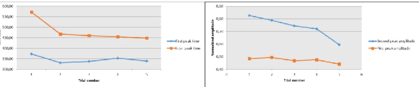
First peak time (FPT): defines the temporal delay between the perturbation onset and the first
peak achievement.
Inter peaks time (IPT): represents the temporal delay between the first peak and the second
peak.
First peak amplitude (FPA): amplitude value of the first peak respect to a mean value
computed on 1 s before the perturbation onset. The FPA was normalized to the distance between the heel and the first metatarsal head, considered as the foot length.
Second peak amplitude (SPA): amplitude value of the second peak respect to the value of the
FPA. The SPA was normalized to the foot length.
First peak space (FPS): CoP path length computed from the perturbation onset to the first
peak reaching. FPS was normalized to the foot length.
Second peak space (SPS): CoP path length computed from the perturbation onset to the
second peak reaching. SPS was normalized to the foot length.
Movement displacement (MD): CoP path length computed from the perturbation onset to the
perturbation offset. MD was normalized to the foot length.
Inter-peaks space (IPS): CoP path length computed between the first and the second peak. IPS
was normalized to the foot length.
Steady state time (SST): temporal interval between the perturbation offset and the reaching of
a steady state. Steady state value was computed as the time instant when CoP enters in an interval defined as its mean value computed in the last 1 s of data acquisition plus two standard deviations (Fig. 2).
Figure 2. Steady state time calculation of CoP AP displacement (blue line) in a representative case. Horizontal black line indicates mean CoP displacement value computed in the last 1 s, while horizontal red lines define confidence interval as mean value±2*standard deviation. Vertical black line indicates time instant of the steady state reaching.
2.3
Kinematic data acquisition and processing
Kinematic data were acquired through a six camera, 3D motion analysis system (BTS, Italy) with a sample rate of 100 Hz and a total of 26 reflective markers have been placed on subject anatomical landmarks, on bilateral sides (Fig. 3).
A total of four markers were placed on foot, in correspondence of the first (FM) and the fifth (VM) metatarsal heads, lateral malleolus (LM) and calcaneus (CA). Four markers have been placed on tibialis tuberosity (TT), head of the fibula (HF), lateral epicondyle (LE) and great trochanter (GT). Four markers were applied on anterior superior iliac crest (ASIS) and posterior superior iliac spinae (PSIS). This marker-set follows what has been reported by Leardini et al. (2007) and thus calibration through a pointer with two markers in known position respect to the tip was performed in order to track the position of second metatarsal head, medial malleolus and medial epicondyle (Leardini et
al., 2007) Eventually six markers were placed on 12th thoracic vertebra (T12), acromion (AC),
temporal bone (TR) and glabella (G).
Markers positions were acquired, low-pass filtered at 5 Hz and then used for angular joints displacement calculation. Anatomical reference frames for foot, shank, thigh and pelvis have been defined according with Cappozzo et al. (1995). For trunk and head, anatomical reference frames have been defined as follows: for the trunk, the origin is at the midpoint between right and left AC, z axis is oriented as the line passing through the ACs and with its positive direction from left to right, x axis is orthogonal to the quasi-frontal plane defined by the z axis and the T12, with its positive direction forward and the y axis is orthogonal to the zx plane with its positive direction distal (Fig. 4). For the head the origin is at the midpoint between left and right TR, z axis is oriented as the line passing through the TRs with its positive direction from left to right, y axis is orthogonal to the quasi-transverse plane defined by z axis and T12, with its positive direction distal and the x axis is orthogonal to the zy plane, with its positive direction forward (Fig. 4). Standard coordinate systems were adopted according with Grood and Suntay (1983), defining flexion/extension, internal/external rotation and abduction/adduction for all the considered joints. Trunk joint kinematics was considered as the relative movement between trunk (proximal) and pelvis (distal), whereas head joint kinematics was considered as the relative movement between head (proximal) and trunk (distal).
Figure 4. Graphical representation of trunk and head reference frames.
Trajectories of markers and joint kinematic were processed and analyzed from 1 s before the perturbation onset to 2.5 s after perturbation offset (Diener et al., 1988). For each joint, from the absolute angle variation, the offset value has been subtracted by the corresponding static posture angle, computed 500 ms prior the perturbation onset, when the subject maintains the upright posture on the moveable platform. Thus, the kinematic measurements were all referenced to the moving platform and to the standing position during the quiet period. As indicate in the previous section for dynamic data, translational backward or forward balance perturbations induce sagittal plane variations and thus flexion/extension angles for each joint and AP and vertical markers trajectories were considered mainly. In order to give a measure of which joints play the most important role in maintaining balance in different configurations, angular range of variation has been computed bilaterally for each joint as the difference between the maximum and the minimum value detected from perturbation onset until 2.5 s after perturbation offset. Moreover, to evaluate the inter-joint coordination and thus postural movement strategies adopted to maintain or restore balance, an ellipse embracing 95% of the values of the angle-angle plot for each couple of joints was fitted (de Lima Pardini et al., 2012, Fig. 5). Ratios of the short axis versus the long axis, ellipse area were then calculated and furthermore also the ellipse slope has been assessed through a regression analysis (Fig. 5). Albeit the ankle and hip strategies were identified as the two principal strategies for balance recovery (Horak and Nashner, 1986), evidences of an even more complex motor recovery
strategy have been reported (Alexandrov et al., 2005; Runge et al., 1999; Cheng, 2016), which involved the knee joint. Therefore, in order to assess which strategy has been employed under the different trail conditions and in what extent respect to the others, the slope of the ellipse embracing the 95% of the angle-angle plot values computed for ankle, knee and hip joints was considered.
Figure 5. Representative plot of ankle and knee joint angles. The two angle distributions were demeaned and thus the 0 value represents the center of the final distribution. Increasing variations indicate joint flexion whereas decreasing variations stand for joint extension. The ellipse fitting the 95% of the distribution values is represented by the red line whereas the ellipse slope is represented by the black line.
For each couple of joints, a slope equal to 1 indicated an equal extent of both the considered joints and thus an undefined prevalent strategy; instead, a slope value equal or lower than 0.8 indicated a prevalence of the balance strategy identified by the joint plotted on the abscissa axis. Eventually, a slope value equal or higher than 1.2 indicated a prevalence of the balance strategy identified by the joint plotted on the ordinate axis. The slope value was computed bilaterally for each three joints and after the identification of a dominant strategy in each trial and for each subject, the recurrence of the dominant strategy among all the subjects has been assessed in percentage of the total subjects.
Center of mass (CoM) displacement in AP and vertical directions has been estimated through kinematic measurements of body segments, following what has been reported by Winter et al. (1998), using foot, shank, thigh and HAT (head, arms, trunk) body segments. CoM ML displacement was assumed to be coincident with the center of pelvis ML movement, computed as the centroid defined by right and left ASISs and PSISs. As for the CoP, due to the characteristics of the perturbation, only the AP component of CoM displacement was considered (Fig. 6). CoM showed a smoother wave form (Fig. 7) respect to the CoP, as expected (Winter et al., 1998); however, AP CoM followed the double peaks path observed for the CoP (Fig. 1 and 7) and then all the aforementioned spatial and temporal parameters calculated for the CoP have been computed for the CoM as well. Also for the CoM the spatial parameters values were normalized to the foot length. Furthermo re, the 3D CoM spatial evolution was computed (Fig. 8) in order to assess the distance between the initial (perturbation onset) and final (steady state reaching time) position of the CoM. (de Lima et al., 2012). CoM coordinates have been referred to the WBB reference frame by subtracting the platform movement to the markers positions, allowing a direct comparison with the CoP displacement.
Figure 6. Representative case of CoP (blue line) and CoM (red line) displacement in ML direction. For both CoP and Co M the maximum variation do not exceed 20 mm.
Figure 7. Representative CoP (blue line) and CoM (red line) AP displacement. CoM shows a smoother but clear double peak wave form that follows that of the CoP.
Figure 8. Representative CoM 3D movement. Increasing displacement indicates forward lean of CoM. Red and black dots highlight initial and final position respectively.
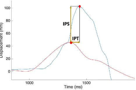
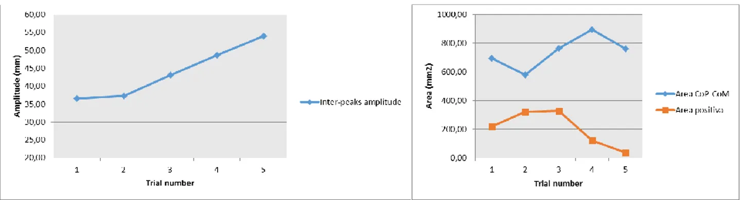
CoP and CoM AP displacements have been also related computing the IPA as the amplitude difference between the first peak of CoP and CoM and the IPT as the temporal gap between the first peaks of CoP and CoM (Fig. 9). Moreover, from the curve obtained subtracting CoM AP displacement to the CoP AP displacement (Winter et al., 1998, de Lima Pardini et al., 2012), the area under the curve has been computed from the perturbation onset until perturbation offset (Fig. 10),
with its positive and negative parts. Positive area values indicate that CoP moves ahead the CoM, while negative values stand for CoP behind CoM; this measure can be interpreted as a postural stability indicator, where the more the area value is high, the more the postural stability is substantial (Winter et al., 1998; de Lima Pardini et al., 2012).
Figure 9. Inter-peaks space and inter-peaks time calculation from CoP (blue line) and CoM (red line) displacement. Red dots indicate maximum peaks for both curves.
Figure 10. Area under the CoP-CoM curve computed during the platform movement. Positive values indicate CoP ahead of CoM, whereas negative values stand for a CoP behind the CoM. Area is highlighted by red color.