C. Publicacions
A
-Gumà, A., Testar, X., Palacín, M., Zorzano, A.; Insulin-stimulated
a-(methyl)aminoisobutyric acid uptake in skeletal muscle. (Evidence for a short-term
activation of uptake independent of Na* electrochemical gradient and protein
synthesis). BiochemJ. 253: 625-9, 1988.
Biochem. J. (1988) 253, 625-629 (Printed in Greal Britain) 625
Insulin-stimulated a-(methyl)aminoisobutyrie acid uptake in
skeletal muscle
Evidence for a short-term activation of uptake independent of Na+ electrochemical gradient and protein synthesis
Anna GUMÀ, Xavier TESTAR, Manuel PALACÍN and Antonio ZORZANO*
Unidad de Bioquímica y Biología Molecular B, Departamento de Bioquímica y Fisiología, Facultad de Biología, Universidad de Barcelona, Avda. Diagonal 645, Barcelona 08071, Spain
1. The present study was designed to explore the mechanisms by which insulin stimulates system A of amino acid transport in extensor digitorum longus (EDL) muscles, by using a system A analogue, a-(methyl)aminoisobutyric acid (MeAIB). 2, Insulin stimulation of MeAIB uptake was noted after only 30 min of incubation and was maximal at 60 min. Kinetics of the insulin effect on MeAIB uptake were characterized by an increased ^aje without modification of Km for MeAIB. 3. Incubation of EDL muscles
with cycloheximide for 90 min did not modify MeAIB uptake in either the presence or the absence of insulin, indicating the independence of insulin action from protein synthesis de novo. Incubations for 180 min with cycloheximide caused a decrease in basal MeAIB uptake; however, the percentage stimulation of amino acid transport by insulin was unaltered. Basal MeAIB uptake was increased by incubation for 180 min, but under these conditions no change in the percentage effect of insulin was found. 4. Ouabain, gramicidin D, or both, markedly decreased basal MeAIB uptake by EDL muscle, but the percentage effect of insulin was unaltered. 5. We conclude that insulin action on amino acid transport through system A in muscle is rapid, is characterized by an increased Vmn, and is independent of protein synthesis de novo and the Na+
electrochemical gradient. Our data are compatible with insulin acting directly on the system A transporter.
INTRODUCTION
Insulin, on interaction with its receptor, exerts a complex array of cellular actions that profoundly affect the physiology of the plasma membrane. Thus insulin activates enzymes such as Na++ K+-dependent ATPase
(Moore, 1973; Clausen & Kohn, 1977; Rosic et al., 1985) or phosphodiesterase (Loten & Sneyd, 1970; Kono et al., 1975) in several cell types, it modulates the number of cell-surface receptors for insulin-like growth factor-II or transferrin (Oka et ai, 1984; Wardzala et al., 1984; Davis et al., 1986), and it also enhances the transport of several important metabolites such as glucose or neutral amino acids that are taken up by system A (Kipnis & Noall, 1958 ; Kletzien et al., 1976 ;Gliemann & Rees, 1983). Much is known about the mechanisms by which insulin activates glucose transport in adipocytes or in muscle; a major mechanism by which insulin mediates this effect is to increase the number of glucose transporters residing in the plasma membrane of the cell (Cushman & Wardzala, 1980; Suzuki & Kono, 1980; Kono et al., 1981; Karnieli et al., 1981; James et al, 1987), as a consequence of a translocation of glucose transporters from an intracellular location to the plasma membrane. However, our knowledge of the mechanism involved in insulin action to activate amino acid transport through system A is scarce, as is structural information on the system A transporter.
In liver, the stimulatory effect of insulin on amino acid
transport is totally dependent on protein synthesis and microtubular function, and it is characterized by an increased Vmtx (Fehlmann et al., 1979; Prentki et al., 1981). In muscle, contradictory findings have been reported about the kinetics of insulin action on amino acid transport (Akedo & Christensen, 1962; Elsas et al., 1968, 1975; Manchester et al, 1971; Le Marchand-Brustel et al, 1982). However^ the stimulatory effect of insulin on a-aminoisobutyric aciíi (AIB) uptake in muscle is observed at an earlier time than in liver, and it is only partially prevented by cycloheximide (Elsas et al, 1968; Le Marchand-Brustel et al, 1982). In addition, insulin action on AIB uptake in perfused muscle seems to be independent of the Na+-K+- ATPase activity (Zorzano
et al, 1986è).
In the present study, we have attempted to delineate further insulin action on system A of transport in skeletal muscle by using the amino acid analogue a-(methyl)-aminoisobutyric acid (MeAIB). This analogue is taken up in skeletal muscle exclusively by system A, unlike AIB, which also enters the cell by the ASC system (Guidotti et al., 1978 ; Maroni et al., 1986). To that effect, the time course of insulin activation of MeAIB uptake, as well as its kinetics of stimulation by incubated rat skeletal muscle, were investigated. Special attention was paid to the dependence of insulin action on the basal transport activity, and whether insulin action on amino acid transport is mediated via changes in the Na+
electrochemical gradient.
Abbreviations used: AIB, aminoisobutyric acid; MeAIB, a-(methyl)aminoisobulyric acid; EDL, extensor digilorum longus. * To whom correspondence should be addressed.
626 A. Gumà and others
MATERIALS AND METHODS Animals and dissection procedures
Male Wistar rats (40-70 g) obtained from our own colony were used. The rats were fed on Purina Laboratory chow ad libitum. Animals were housed in animal quarters maintained at 22 °C with a 12 h-light/12 h-dark cycle. The dissection and isolation of the extensor digitorum longus (EDL) muscle were carried out under anaesthesia with pentobarbital (5-7mg/100g body wt., intra-peritoneally) as described previously (Maizels et al., 1977). The isolated EDL muscle was fixed to a stainless-steel clip in order to maintain the muscle under slight tension (approximating to resting length) during the incubation. Such muscles are able to maintain normal ATP and phosphocreatine concentrations during a 3 h incubation. EDL muscles from several animals were randomly assigned to different experimental groups. Incubations
EDL muscles were incubated in a shaking incubator at 37 °C for 1.5-3 h in 2 ml of Krebs-Henseleit buffer (as in Zorzano et al., 1985), pH 7.4, containing 5 mM-glucose, 0.1 % bovine serum albumin and 20 mM-Hepes. After addition of the muscles to the vials, they were stoppered and placed in a Dubnoff metabolic shaker set at 37 °C and a shaking rate of 70 cycles/min. Vials were gassed with O2/CO2 (19:1) during the whole incubation period. The incubation medium was kept for no longer than 90 min, and during prolonged incubations it was renewed thereafter. At different times, insulin (200 nM) was added to the incubation medium, as well as several inhibitors such as cycloheximide (0.1 HIM), ouabain (1 HIM) or gramicidin D (25 ¿wg/ml) (see details in Table legends). Measurement of ammo acid uptake into muscle
Amino acid uptake by system A was measured in EDL muscles by using the non-metabolizable amino acid analogue MeAIB. After the incubations with insulin and the above-mentioned inhibitors, muscles were blotted and transferred to a vial with 1.5 ml of Krebs-Henseleit buffer, pH 7.4, containing 5 mM-glucose, 0.1% bovine serum albumin, 20 mM-Hepes and 0.1 mM-a-[l-14 C]-MeAIB (800/iCi/mmol), 10 mM-pHJmannitol (33/tCi/ mmol) and insulin and modulators at the same con-centrations as during the preceding incubation period. The vials were stoppered and incubated at 37 °C in a shaking incubator for 30 min. The gas phase in the vials was Oj/CO2 (19:1). As these studies were conducted over a period of 1 year, and since seasonal variation in amino acid uptake by skeletal muscle has been previously reported (Arvill & Ahren, 1967; Zorzano et al., 1985, 1986fl), control and experimental groups were always performed during the same experimental day.
After incubation, muscles were quickly rinsed in cold saline (0.9 % NaCl), blotted briefly on filter paper and frozen in liquid N2. Samples were weighed and digested in 0.25 ml of NCS tissue solubilizer (The Radiochemical Centre, Amersham, Bucks., U.K.) at 50 °C in Teflon-sealed vials for 2 h. Muscle digests and samples of the incubation media were placed in scintillation vials containing 10 ml of scintillation cocktail and counted for radioactivity in a Packard scintillation counter with channels preset for simultaneous 3H and 14C counting. The amount of each radioisotope present in the samples was determined, and this information was used to
calculate the extracellular space. The extracellular space of EDL muscles, estimated by using [3H]mannitol, increased progressively with time, being 0.17 + 0.01, 0.19 ±0.01 and 0.25 ±0.01 ml/g after 10, 20 and 30 min respectively, and it was not modified by the presence of insulin. The intracellular concentration of 14C-labelled amino acid analogue was calculated by subtracting its amount in the extracellular space from the total label found in tissue, as previously reported (Zorzano et al., 1985). Student's / test was used for statistical analysis of the data.
RESULTS AND DISCUSSION
Characterization of insulin effect on MeAIB uptake by EDL muscle
Our initial objective was to delineate the stimulatory effect of insulin on MeAIB uptake by EDL muscles. MeAIB is a specific probe for system A of neutral amino acid transport by skeletal muscle (Maroni et al., 1986), unlike AIB, which enters the cell through the A and ASC systems. Preliminary studies showed that insulin-stimulated MeAIB uptake was linear during 30 min of incubation (results not shown). From this, uptake was determined after 30 min of incubation with the radio-active analogue in all subsequent experiments. To investigate the time course of insulin action, muscles were incubated for a total of 3 h, either in the absence of insulin (basal group) or with insulin present during the last 30, 60 or 120 min of incubation. At 30 min after insulin addition, MeAIB uptake was increased by 74% as compared with the basal group (23.2 + 5.5 versus 13.3+1.1 nmol/30min per g respectively). MeAIB up-take after 60 min of insulin addition was already maximal and indistinguishable from that at 120 min (44.4 ±4.1 and 37.9+1.8 nmol/30 min per g respec-tively). Kinetic analysis of the stimulatory effect of
Control
Insulin
-2 10
1/lMeAIB] imuT
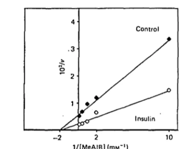
Fig. 1. Effect of insulin on the kinetic analysis of MeAIB uptake by EDL muscle
Muscles were incubated as described in the Materials and methods section for 180 min. Insulin when present was added during the last 60 min of the experiment. Uptake (v, nmol/h per g) was measured at different concentrations of MeAIB (mM) for 30 min. Each point represents the average of four to six muscles. Statistical analysis demonstrated that regression curves were significantly different in control (r = 0.997; y = 0.55 + 0.28 x) as_£ompared with insulin-treated group (r = 0.985; >• = 0.21 +0.13 x) at P < 0.05. 1988
Insulin-stimulated a-(methyl)aminoisobutyric acid uptake in muscle 627
insulin on MeAIB uptake (Fig. I) indicated that it was characterized by an increased Kmax (235 and 486 nmol/h per g in the absence and the presence of insulin respectively), without modifications of Km for MeAIB
(0.70 HIM and 0.62 mM in the absence and the presence of insulin respectively).
Thus we have substantiated a short-term effect of insulin stimulating MeAIB uptake by incubated muscle, which is already detected at 30 min after hormone addition, and attains a maximal effect by l h of incubation in the presence of hormone. That is in keeping with previous results, which showed acute modulation of AIB transport by insulin, exercise or electrical stimulation in the perfused or incubated muscle (Goldberg et al., 1974; Zorzano et al., 1985, 1986a,ft).
Kinetic analysis of the insulin effect on MeAIB uptake by incubated muscle demonstrated an increased Vmtx,
with no modification of Km. This coincides with other
reports of insulin augmenting the Vmix of AIB
(Manchester et al., 1971 ; Elsas et al., 1975 ; Le Marchand-Brustel et al., 1982); however, an effect of insulin on AIB uptake characterized by a decrease in the Km for AIB has
been described in diaphragm (Akedo & Christensen, 1962; Elsas et al., 1968, 1971). The reason for this discrepancy remains to be explained, and heterogeneity between diaphragm and skeletal muscle might be invoked. In any event, our results allow us to discard an effect of insulin increasing the affinity of MeAIB to the system-A transporter in skeletal muscle. Whether insulin modifies Km for Na+ in the incubated muscle was not determined in this study ; however, it does not invalidate our prior conclusion.
Effect of protein synthesis and adaptive regulation on insulin-stimulated MeAIB uptake by EDL muscle
To assess whether insulin activates amino acid transport system A by a mechanism that involves protein synthesis de novo, EDL muscles were incubated in the absence or the presence of 0.1 mM-cycloheximide, a concentration that completely inhibits protein synthesis (Forsayeth & Gould, 1983). When cycloheximide was present during the last 90 min of the experiment (added 30 min before insulin), basal as well as insulin-stimulated MeAIB uptake were unaltered (Table 1). Thus the maximal effect of insulin was not perturbed by cycloheximide added 30 min before the hormone. The data demonstrate that insulin stimulates MeAIB uptake by muscle independently of protein synthesis de novo. Thus insulin does not activate amino acid transport by increasing the transcription and/or translation of certain genes which could code for the amino acid transporter or another unknown modulator.
Incubation of EDL muscles with cycloheximide for 180 min caused a decrease in basal MeAIB uptake. Only under these conditions did the presence of cycloheximide result in a decrease in the absolute effect of insulin on MeAIB uptake (Table 1). However, under these circumstances the insulin effect was not modified when expressed as a percentage, in keeping with previous .observations (Elsas et al., 1968; Le Marchand-Brustel
et al., 1982). These data suggested that cycloheximide
acted not by altering the mechanism of insulin action, but through a modification of basal transport activity.
The next series of experiments was designed to test that hypothesis. To that effect, basal MeAIB uptake was increased by prolonged incubation of EDL muscles. It is
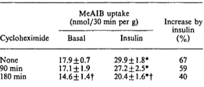
Table 1. Effect of cycloheximide on basal and insulin-stimulated MeAIB uptake by EDL muscle
Results are means+ S.E.M. for 14-25 observations per group. EDL muscles were incubated for 180min, with or without 200 nM-insulin (during the last 60 min of incuba-tion). Cycloheximide (0.1 mM) was added either at the beginning of the experiment (180 min group) or during the last 90 min of the experiment (90 min group). MeAIB uptake was determined during the last 30 min. * Value significantly different from that of the basal group (P < 0.05), f value significantly different from that of the no-cycloheximide group (P < 0.05).
MeAIB uptake
(nmol/30 min per g) Increase by insulin Cycloheximide Basal Insulin
None 90min 180min 17.9+0.7 17.1 + 1.9 I4.6+1.4f 29.9+1.8* 27.2 + 2.5* 20.4±1.6*t 67 59 40
Table 2. Effect of adaptive regulation on basal and insulin-stimulated MeAIB uptake by EDL muscle.
Results are means+ S.E.M. for 6-17 observations, except for the 150 min group, which represents the mean of two observations. Individual data of MeAIB uptake in the 150 min groups were 11.8 and 15.3 nmol/30 min per g in basal state and 22.4 and 22.8 nmol/30 min per g after insulin addition respectively. EDL muscles were incubated for 90, 150 or 180 min in the absence or in the presence 'of insulin (200 nM). When indicated insulin was present during the last 60 min of incubation. * Value significantly different from that of the basal group (P < 0.05), f value significantly different from that of the 90 min group (P < 0.05). Duration of experiment (min) 90 150 180 MeAIB uptake (nmol/30 min per g) Basal 11.4+1.2 13.5 19.1+0.9f Insulin 19.0 + 0.9* 22.6 29.9+1.9*t Increase by insulin (Of \ \/o) 67 66 59
well known that adaptive regulation is active in skeletal muscle (Guidotti et al., 1975; Le Marchand-Brustel
et al., 1982; Logan et al., 1982). A 70 % increase in basal
MeAIB uptake by EDL muscle was observed by increasing the total incubation time from 90 to 180 min (Table 2). Under those conditions, MeAIB uptake in the presence of insulin was also increased in the 180min group as compared with the 90 min. That is, the absolute effect of insulin was increased; nevertheless, the per-centage effect of insulin was similar in all groups (Table 2). Thus, again the insulin effect was dependent on basal transport activity.
In all, our interpretation of these results is that cycloheximide does not block insulin action on amino acid transport. In addition, when basal MeAIB uptake is either increased or lowered, the effect of insulin persists Vol. 253
628 A. Gumà and other unaltered. It may be proposed that cycloheximide at long
time periods or adaptive regulation alters basal transport activity or another related factor, such as the intracellular pool of transporters, which would be directly modulated by insulin.
Effect of Na+-electrochemical-gradient disrupters on insulin-stimulated MeAIB uptake by EDL muscle
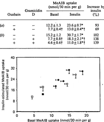
Insulin induces hyperpolarization (Zierler, 1959) and stimulates the Na+-K+ pump (Moore, 1973; Clausen & Kohn, 1977; Flatman & Clausen, 1979; Rosic et al., 1985) in skeletal muscle, and it is still a matter of controversy whether hyperpolarization is related or not to previous activation of Na+-K+-ATPase (Zierler & Rogus, 1981). Thus, in a further set of experiments we examined the effect of disruptors of the Na+ electro-chemical gradient, such as ouabain (1 HIM), an inhibitor of the Na+-K+-ATPase, or gramicidin D (25 /ig/ml), an ionophore known to abolish membrane potential (Kristensen & Folke, 1986). It has been previously reported that 1 mM-ouabain is sufficient to occupy the total number of membrane Na+-K+ pumps (Clausen & Flatman, 1987), and that 25 fig of gramicidin D/ml abolished membrane potential in liver 5 min after its addition (Kristensen & Folke, 1986). These experiments were carried out at a different time of the year, and, in keeping with previous observations (Arvill & Ahren, 1967), marked differences were detected in basal MeAIB uptake as compared with the results described above.
Initially we investigated the time course of the ouabain effect on basal MeAIB uptake by incubated EDL muscle. Ouabain rapidly caused a marked decrease in basal MeAIB uptake. After only 30 min of exposure to 1 mM-ouabain, MeAIB uptake decreased by 40% (from
11.1±1.5 to 6.2 ±1.4 nmol/30 min per g), and the maximal inhibitory effect of ouabain was attained l h after its addition (5.0 ±0.5 nmol/30 min per g). Ouabain action on MeAIB uptake was reversible, and 30 min of ouabain exposure followed by 30 min with no inhibitor caused a partial recovery of transport activity (7.5 + 1.7 nmol/30 min per g). On the basis of these findings, and in order to investigate whether insulin action on MeAIB uptake required an unaltered Na+-K+ pump activity, EDL muscles were incubated in the absence or the presence of insulin and ouabain. Results are presented in Table 3. Ouabain caused a 40 % decrease in basal MeAIB uptake. In the presence of insulin, MeAIB uptake was also decreased in the ouabain-treated group compared with the control group ; however, the percentage stimulation of MeAIB uptake induced by insulin was similar in ouabain and control groups (Table 3). Again, under those conditions, cycloheximide did not affect insulin-stimulated MeAIB uptake (results not shown).
Next, insulin action in the presence of gramicidin D, or of ouabain plus gramicidin D, in the incubation medium was investigated (Table 3). Incubation with gramicidin D during 30 min caused a 50 % decrease in basal MeAIB uptake, whereas ouabain plus gramicidin D caused a 70 % decrease in MeAIB uptake by EDL muscle. MeAIB uptake after insulin addition was also decreased in the presence of inhibitors compared with the control group; nevertheless, the percentage stimulation caused by insulin was similar under all these conditions (Table 3). These data demonstrate that insulin action on amino acid transport is not mediated by modification of the Na+
Table 3. Effect of ouabain and gramicidin D on basal and insulin stimulated MeAIB uptake by EDL muscle
Results are means + S.E.M. for 4 to 12 observations per group. EDL muscles were incubated for 180min. When indicated, ouabain (1 mM) and insulin (200 mM) were present during the last 60 min of incubation (a, b). In some studies (¿>)> gramicidin D (25 /¿g/ml) was added during the last 30 min of the experiment. Gramicidin D was dissolved in 60 % ethanol, so the final ethanol concentration in the incubation medium was 1 % ; in those experiments, the control group also contained 1 % ethanol in the medium. * Value significantly different from that of the basal (no insulin) group (P<0.05); f value significantly different from that of the control (no ouabain, no gramicidin D additions) group (P < 0.05). Gramicidiï Ouabain D (a) (b) MeAIB uptake (nmol/30 min per g) li
Basal 12.2±1.3 7.7±0.4f 15.2±1.2 7.7 + 0.8t 4.6±0.6f Insulin 23.6 ±0.7* 13.0±0.4*t 30.7+1.7* 18.3±2.1*t 11.0+1.8*t icrease by insulin 93 69 102 138 139 4U • v ço m "S < o. | c 24. •o É S o f -S 16 .i f vt C •È 8 c ~ n ""ï 1 •-•i-» ^ ^ ^ 5 O 5 10 15 20 Basal MeAIB uptake (nmol/30 min per g)
Fig. 2. Relationship between basal and insulin-stimulated MeAII uptake by EDL muscle
Points are means ± S.E.M. for 2-25 observations (see legends to Tables 1, 2 and 3). •, Control groups with different durations of incubations with no amino acids in the medium; O» cycloheximide groups; •, ouabain group; D, gramicidin D and gramicidin D plus ouabain groups. A significant linear regression was detected, with r = 0.909 and ;•= 5.32 +1.32 x.
electrochemical gradient, and is independent of the Na+-K+-ATPase and of membrane potential. However, as we discussed above regarding protein synthesis, foi insulin action to be maximal a preserved Na* electro-chemical gradient is required. These data agree with previous work performed in the perfused rat hindquarter, which showed that insulin and exercise stimulate AIB uptake in a fashion independent of -Na+-K+-ATPase activity (Zorzano et al., 1986è).
Finally, a significant correlation was found (r = 0.909,
P < 0.001) when means of basal MeAIB uptake were
plotted against insulin-stimulated MeAIB uptake for all 198Í
Insulin-stimulated a-(methyl)aminoisobutyric acid uptake in muscle 629
experimental groups (Fig. 2). That is, under conditions characterized by either increasing incubation time or the presence of cycloheximide, ouabain or gramicidin D, stimulation of MeAIB uptake induced by insulin was dependent on basal transport activity. That provides support to the contention that, whatever the mechanisms by which insulin stimulates amino acid transport in skeletal muscle, they are not mediated by protein synthesis de novo, adaptive regulation or modification of the Na+ electrochemical gradient. In addition, the
correlation between basal and insulin-stimulated MeAIB uptake by muscle implies that insulin action is somehow dependent on basal transport activity. These findings differ from what occurs in hepatocytes, where it has been described that insulin stimulates AIB uptake by a slower mechanism that involves protein synthesis (Fehlmann
et al, 1979, 1981). In fact, it has been proposed that
insulin probably stimulates transcription of a gene coding for the A transporter in liver. Therefore it can be concluded that the mechanisms by which insulin stimu-lates amino acid transport in muscle and in liver might be different.
In conclusion, the present study provides evidence that insulin stimulates system A of amino acid transport in skeletal muscle, by increasing the VmiK of transport. The mechanism that mediates this action is independent of protein synthesis and Na+ electrochemical gradient.
However, insulin-stimulated MeAIB uptake depends on basal transport activity in a variety of situations (cycloheximide, adaptive regulation, gramicidin D, ouabain). That fact allows us to postulate a possible direct effect of insulin at the level of the transporters, either by increasing their intrinsic activity (independent of the Na+ electrochemical gradient) or in consequence
of a translocation of them from a hypothetical intra-cellular pool to the plasma membrane, as described for glucose transporters.
This work was supported in part by a grant from the C.I.C.Y.T. (PB-573/86) and from C.I.R.I.T. (Generalität de Catalunya), Spain. We thank Miss Pepi Nieto for secretarial help. Pig insulin was kindly provided by Mr. M. L. Johnson (Eli Lilly, Indianapolis, IN, U.S.A.).
REFERENCES
Akedo, H. & Christensen, H. N. (1962) J. Biol. Chem. 237, 118-122
Arvill, A. & Ahrén, K. (1967) Acta Endocrino!. (Copenhagen) 56, 279-294
Clausen, T. & Flatman, J. A. (1987) Am. J. Physiol. 252, E492-E499
Clausen, T. & Kohn, P. G. (1977) J. Physiol. (London) 265, 19-42
Cushman, S. W. & Wardzala, L. J. (1980) J. Biol. Chem. 255, 4758^762
Davis, R. J., Corvera, S. & Czech, M. P. (1986) J. Biol. Chem. 261,8708-8711
Elsas, L.J., Albrecht, I. & Rosenberg, L. E. (1968) J. Biol. Chem. 243, 1846-1853
Elsas, L. J., II, MacDonell, R. C, Jr. & Rosenberg, L. E. (1971) J. Biol. Chem. 246, 6452-6459
Elsas, L. J., Wheeler, F. B., Danner, D. J. & De Haan, R. L. (1975) J. Biol. Chem. 250, 9381-9390
Fehlmann, M., Le Cam, A. & Freychet, P. (1979) J. Biol. Chem. 254, 10431-10437
Fehlmann, M., Samson, M., Koch, K. S., Leffert, H. L. & Freychet, P. (1981) Biochim. Biophys. Acta 642, 88-95. Flatman, J. A. & Clausen, T. (1979) Nature (London) 281,
580-581
Forsayeth, J. R. & Gould, M. K. (1983) Diabetologia 25, 429-432
Gliemann, J. & Rees, W. D. (1983) Curr. Top. Membr. Transp. 18, 339-379
Goldberg, A. L., Jablecki, C. & Li, J. B. (1974) Ann. N.Y. Acad. Sei. 228, 190-201
Guidotti, G. G., Gazzola, G. C-, Borghetti, A. F. & Franchi-Gazzola, R. (1975) Biochem. Biophys. Acta 406, 264-279 Guidotti, G. G., Borghetti, A. F. & Gazzola, G. C. (1978)
Biochim. Biophys. Acta 515, 329-366
James, D. E., Lederman, L. & Pilch, P. F. (1987) J. Biol. Chem. 262, 11817-11824
Karnieli, E., Zarnowski, M. J., Hissin, P. J., Simpson, I. A., Salans, L. B. & Cushman, S. W. (1981) J. Biol. Chem. 256, 4772-4777
Kipnis, D. M. & Noall, M. W. (1958) Biochim. Biophys. Acta 28, 226-227
Kletzien, R. F., Pariza, M. W., Becker, J. E., Potter, V. R. & Butcher, F. R. (1976) J. Biol. Chem. 251, 3014-3020 Kono, T., Robinson, F. W. & Sarver, J. A. (1975) J. Biol.
Chem. 250, 7827-7835
Kono, T., Suzuki, K., Dansey, L. E., Robinson, F. W. & Blevins, T. L. (1981) J. Biol. Chem. 256, 6400-6407 Kristensen, L. O. & Folke, M. (1986) Biochim. Biophys. Acta
855, 49-57
Le Marchand-Brustel, Y., Moutard, N. & Freychet, P. (1982) Am. J. Physiol. 243, E74-E79
Logan, W. J., Klip, A. & Gagalang, E. (1982) J. Celi. Physiol. 112, 229-236
Loten, E. G. & Sneyd, J. G. T. (1970) Biochem. J. 120,187-193 Maizels, E. Z., Ruderman, N. B., Goodman, M. N. & Lau, D.
(1977) Biochem. J. 162, 557-568
Manchester, K. L., Guidotti, G. G., Borghetti, A. F. & Luneburg, N. (1971) Biochim. Biophys. Acta 241, 226-241 Maroni, B. J., Karapanos, G. & Mitch, W. E. (1986) Am. J.
Physiol. 251, F74-F80
Moore, R. D. (1973) J. Physiol. (London) 232, 23-45 Oka, Y., Mottola, C., Oppenheimer, C. L. & Czech, M. P.
(1984) Proc. Nati. Acad. Sei. U.S.A. 81, 4028-4032 Prentki, M., Crettaz, M. & Jeanreniud, B. (1981) J. Biol.
Chem. 256, 4336-4340
Rosic, N. K., Standaert, M. L. & Pollet, R. J. (1985) J. Biol. Chem. 260, 6206-6212
Suzuki, K. & Kono, T. (1980) Proc. Nati. Acad. Sei. U.S.A. 77, 2542-2545
Wardzala, L., Simpson, I. A., Rechler, M. W. & Cushman, S. W. (1984) J. Biol. Chem. 259, 8378-8383
Zierler, K. L. (1959) Am. J. Physiol. 197, 515-523.
Zierler, K. L. & Rogus, E. (1981) Am. J. Physiol. 241, C145-C149
Zorzano, A., Balón, T. W., Garetto, L. P., Goodman, M. N. & Ruderman, N. B. (1985). Am. J. Physiol. 248, E546-E552 Zorzano, A., Balen, T. W., Goodman, M. N. & Ruderman,
N. B. (1986a) Am. J. Physiol. 251, E21-E26
Zorzano, A., Balón, T. W., Goodman, M. N. & Ruderman, N. B. (19866) Biochem. Biophys. Res. Commun. 134,
1342-1349
Received 16 November 1987/3 March 1988; accepted 3ist March 1988
B
-Gumà, A., Camps, M., Palacín, M., Testar, X., Zorzano, A.; Protein kinase C activators
selectively inhibit insulin-stimulated system A transport activity in skeletal muscle at a
post-receptor level. BiochemJ. 268: 633-9, 1990.
Biochem. J. (1990) 268. 633-639 (Printed in Great Britain)
Protein kinase C activators selectively inhibit insulin-stimulated
system A transport activity in skeletal muscle at a post-receptor
level
Anna GUMÀ. Marta CAMPS. Manuel PALACÍN. Xavier TESTAR and Antonio ZORZANO* Departament de Bioquímica i Fisiologia. Facultat de Biologia, Universitat de Barcelona, Avda. Diagonal 645, 08028 Barcelona. Spain
633
We have investigated the role of phorbol esters on different biological effects induced by insulin in muscle, such as activation of system A transport activity, glucose utilization and insulin receptor function. System A transport activity was measured by monitoring the uptake of the system A-specific analogue a-(methyl)aminoisobutyric acid (MeAIB), by intact rat extensor digitorum longus muscle. The addition of 12-0-tetradecanoylphorbol 13-acetate (TPA, 0.5 //M) for 60 or 180 min did not modify basal MeAIB uptake by muscle, suggesting that insulin signalling required to stimulate MeAIB transport does not involve protein kinase C activation. However, TPA added 30 min before insulin (100 nM) markedly inhibited insulin-stimulated MeAIB uptake. The addition of polymyxin B (0.1 mM) or H-7 (1 HIM), protein kinase C inhibitors, alone or in combination with TPA leads to impairment of insulin-stimulated MeAIB uptake. This paradoxical pattern is incompatible with a unique action of Polymyxin B or H-7 on protein kinase C activity. Therefore these agents are not suitable tools with which to investigate whether a certain insulin effect is mediated by protein kinase C. TPA did not cause a generalized inhibition of insulin action. Thus both TPA and insulin increased 3-O-methylglucose uptake by muscle, and their effects were not additive. Furthermore, TPA did not modify insulin-stimulated lactate production by muscle. In keeping with this selective modification of insulin action, treatment of muscles with TPA did not modify insulin receptor binding or kinase activities. In conclusion, phorbol esters do not mimic insulin action on system A transport activity; however, they markedly inhibit insulin-stimulated amino acid transport, with no modification of insulin receptor function in rat skeletal muscle. It is suggested that protein kinase C activation causes a selective post-receptor modification on the biochemical pathway by which insulin activates system A amino acid transport in muscle.
INTRODUCTION
The phospholipid, Ca"*- and diacylglycerol-dependent protein kinase (protein kinase C) plays a major role in controlling cell function, through phosphorylation of serine and threonine residues in cellular proteins [1,2]. Extracellular signals, which include neurotransmitters, hormones and growth factors [1], bind to specific cell-surface receptors and generate second messengers that activate protein kinase C. Protein kinase C is also the intracellular receptor of phorbol esters [3], which are potential tumour promoters. Phorbol esters, such as 12-O-tetradecanoylphorbol 13-acetate (TPA), bind to and activate the protein kinase C by interaction at the jn-l,2-diacylglycerol site [3,4]. In addition to tumour-promoting effects, phorbol diesters possess insulin-like activity in a variety of tissues. Thus, phorbol esters stimulate glucose transport and oxidation [5-7], lipogenesis [8] and pyruvate dehydrogenase activity [5].
Insulin is the major anabolic and anti-catabolic hormone acting in mammals. The actions of insulin at the cellular level are initiated by insulin binding to its plasma-membrane receptor [9,10]. The insulin receptor is a tyrosine-specific ligand-st.mulated protein kinase [11.12], and there is much evidence indicating that the tyrosine kinase activity of the insulin receptor is essential for insulin action [13,14].
Several types of interaction connecting insulin action and protein kinase C have been postulated. On the one hand, it has been reported that phorbol esters inhibit insulin action and insulin-receptor tyrosine kinase in Fao cells [2,15] and in isolated rat adipocytes [6,16]. This is consistent with the fact that protein
kinase C directly phosphorylates the insulin receptor in vitro and decreases its tyrosine kinase activity [17]. On the other hand, some investigators have described an insulin-induced activation of protein kinase C activity in several cell types. Insulin activates membrane protein kinase C activity in rat diaphragm [18] and membrane and cytosolic protein kinase C in BC3H-1 myocytes [19]. In adipocytes, insulin causes an increase in [3H]phorbol 12,13-dibutyrate binding to cytosol and a decrease in that to plasma-membrane fractions [20], and stimulates cytosolic protein kinase C activity [21]. Nevertheless, other studies have failed to detect an insulin-induced activation of protein kinase C in adipocytes, L6 or BC3H-1 muscle cell lines [22-24].
In the present work, we have investigated the effects of TPA on basal and insulin-stimulated system A amino acid transport activity, glucose transport and lactate production by intact rat skeletal muscle, as well as the effects of TPA on insulin-stimulated receptor tyrosine kinase activity after lectin-chromatography purification. TPA treatment of incubated muscle caused a blockade on the stimulatory effect of insulin on a-(methyl)-aminoisobutyric acid (MeAIB) uptake. This alteration of insulin action was not detected when 3-O-methylglucose uptake or lactate production was assessed. When insulin receptors were purified from TPA-treated muscles, the insulin-stimulated tyro-sine kinase activity of the receptors was found to be unaltered. These results suggest that in skeletal muscle protein kinase C does not mediate insulin action on system A transport activity; however, previous activation of protein kinase C modulates insulin action in a negative fashion by a mechanism that lies at a post-receptor level.
Abbreviations used: TPA. 12-O-tetradecanoylphorbol 13-acetate; MA, 4/î-phorbol 13a-monoacetate; EDL. extensor digitorum longus; MeAIB, *-(methyl)aminoisobutyric acid: H-7. l-(5-isoquinolinylsulphonyl)-2-methylpiperazine; WGA, wheat-germ agglutinin.
* To whom correspondence should be addressed. Vol. 268
634 A. Gumá and others
EXPERIMENTAL Materials
Pig monocomponent insulin was a gift from Mr. T. L. Jeatran, Eli Lilly and Co. [12M-TyrA"]Monoiodoinsulin was obtained from New England Nuclear and [l"I-Tyrl>ili]monoiodoinsulin was obtained from Amersham Corp. [y-3-P]ATP was prepared from [3iP]P, (New England Nuclear) by using a Gamma-prep kit from Promega Biotech. All electrophoresis reagents were obtained from Bio-Rad; wheat-germ agglutinin (WGA) bound to agarose was obtained from Vector, and disuccinimidyl suberate was from Pierce. BSA (fraction V, fatty-acid-free), TPA, 4//-phorbol 13a-monoacetate (MA), polymyxin B, l-(5-isoquinolinylsulphonyl)-2-methylpiperazine (H-7) and most commonly used chemicals were from Sigma.
Animals and dissection procedures
Male Wistar rats (50-60 g) obtained from our own colony were used. The rats were fed on Purina Laboratory chow ad libitum. Animals were housed in animal quarters maintained at 22 °C with a 12 h-light/12 h-dark cycle. The dissection and isolation of the extensor digitorum longus (EDL) muscle were carried out under anaesthesia with pentobarbital (5-7 mg/100 g body wt., intraperitoneally) as described previously [25]. The isolated EDL muscle was fixed to a stainless-steel clip in order to maintain the muscle under slight tension (approximating to resting length) during the incubation. Such muscles (20-30 mg wet wt.) are able to maintain normal ATP and phosphocreatine concentrations during a 3 h incubation.
Incubations
EDL muscles were incubated in a shaking incubator at 37 °C for 3 h in 3 ml of Krebs-Henseleit buffer, pH 7.4, containing 5 mM-glucose, 0.2 °0 BSA and 20 mM-Hepes. After addition of the muscles to the vials, they were stoppered and placed in a Dubnoff metabolic shaker set at 37 °C and a shaking rate of 60 cycles/min. Vials were gassed with O2/CO2 (19:1) during the whole incubation period. The incubation medium was kept for no longer than 90 min, and during prolonged incubations it was renewed thereafter. At different times, insulin ( l O O n M ) was added to the incubation medium as well as several drugs such as TPA (0.5 /¿M), MA (0.5 ,«M), polymyxin B (0.1 HIM) or H-7 (1 mM) (see details in Figure and Table legends). Experimental series were performed by comparing biological activity of one muscle with the contralateral one from the same rat (paired muscles).
Measurements of amino acid and glucose uptake and lactate production by muscle
Amino acid uptake by system A was measured in EDL muscles by using the non-metabolizable amino acid analogue MeAIB. After the incubations with insulin and the above-mentioned agents, muscles were transferred to vials with 1.5 ml of Krebs-Henseleit buffer, pH 7.4, containing 5 mM-glucose, 0.2 °0 BSA, 20 mM-Hepes and 0.1 mM-[l-'4C]MeAIB (800//Ci/mmol), 1 mM-[3H]mannitol (330 //Ci/mmol) and in-sulin and modulators at the same concentrations as for the preceding incubation period. The vials were stoppered and incubated at 37 °C in a shaking incubator for 30 min. The gas phase in the vials was O,/COj (19:1). In experiments designed to measure 3-O-methylglucose uptake, muscles were incubated in Krebs-Henseleit buffer containing 2 mM-pyruvate instead of glucose, and during the last 30 min of incubation the medium contained 0.1 mM-[14C]3-0-methylglucose (800 //Ci/mmol) and 1 mM-[aH]mannitol (330//Ci/mmol). After incubation, muscles were placed in 0.25 ml of NCS tissue solubilizer (Amersham
International), and radioactivity of muscle digests and samples of the incubation media was measured. The amount of each radioisotope present in the samples was determined, and this information was used to calculate the extracellular space. That of EDL muscles, estimated after 30 min of [3H]mannitol addition, was 0.222±0.003 ml/mg and 0.219±0.006 ml/mg in the absence and presence of insulin respectively. The intracellular concen-tration of [NC]MeAIB or [l4C]3-O-methylglucose was calculated as previously reported (26,27]. Lactate release to the incubation media was measured during the last 30 min of incubation as in [28]. Student's / test for paired data was used for statistical analysis.
Measurement of insulin binding by the incubated muscle
Insulin binding was assessed at 21 °C by the technique of Le Marchand-Brustel et al. [29]. EDL muscles were initially incubated for 30 min in the absence or in the presence of 0.5 /<M-TPA at 21 °C in 3 ml of Krebs-Henseleit buffer, pH 7.4. con-taining 5 mM-glucose, 0.2 °0 BSA and 20 mM-Hepes. The muscles were then incubated with 30 pM-['"I-TyrA14]monoiodoinsulin for 3 h. After that time, muscles were washed five times in cold 0.9°0 NaCl containing 0.25 °,0 BSA. The results presented have been corrected for non-specific binding, which was determined as the amount of 125I-insulin recovered in muscles incubated in the presence of a saturating concentration (1.5//M) of unlabelled insulin. Radioactivity was quantified in a Packard y-radiation counter. Non-specific binding was approx, 30 °0 of the total binding. The degradation of '"I-insulin, assessed by the increase in trichloroacetic acid-soluble radioactivity in the incubation media, was 5-10 °0 at 3 h in both control and TPA-treated groups.
Preparation of insulin receptors
EDL muscles were incubated for 3 h in 3 ml of Krebs-Henseleit buffer, pH 7.4, containing 5 mM-glucose, 0.20 °0 BSA and 20 mM-Hepes. For the last 90 min they were incubated in the absence or presence of 0.5 //M-TPA. At the end of the incubation period, muscles were frozen in liquid N2. Pools from 10-15 muscles (approx. 250-300 mg of tissue) were homogenized and solubilized in 1 °Q Triton X-100 as described [30]. The solubilized homo-genate was centrifuged at 150000 g for 90 min at 4°C. The 150000 g supernatant (1.8 ml) was recycled for 30 min (approx. 5-7 times) through a column containing 0.2 ml of WGA bound to agarose, at 4 °C. The resin was washed with buffer (20 ml) containing 25 mM-Hepes and 0.1 °0 Triton X-100, pH 7.4. Receptors were eluted from the WGA column with buffer containing 25 mM-Hepes, 0.1 °0 Triton X-100 and 0.3 M-,V-acetyl-D-glucosamine, pH 7.4.
Ligand binding and receptor cross-linking
Insulin binding was measured as in [30]. WGA eluate (20/<1) was incubated for l h at 22 °C in 30 mM-Hepes buffer containing 0.1 °0 BSA and 100 units of bacitracin/ml (pH 7.6; 200/il) and 20000 c.p.m. of ['"I-Tyr*u]monoiodoinsulin (=60pM). Non-specific binding was estimated as '"I-insulin bound in the presence of 1 //w-insulin (5-10 °0 of total binding). Binding data were expressed per fig of protein, with the latter measured by the method of Bradford [31]. Receptor cross-linking protocols were carried out essentially as described by Pilch & Czech [32], by using 0.5 nM-['"I-TyrB2']monoiodoinsulin.
Autophosphorylation and phosphorylatlon of an exogenous substrate
Autophosphorylation assays were performed as described previously [30]. Phosphorylation of an exogenous substrate was carried out with receptor preparations which were preincubated 1990
Effect of phorbol esters on insulin action in muscle 635
for l h in 30 Hepes buffer, pH 7.6. containing 50 mM-magnesium acetate, and various concentrations of insulin. The receptor kinase activity was initially activated by addition of 50 ,HM-[y-3îP]ATP (5-10 ^Ci) for 10 min. The reaction was initiated by the addition of the exogenous substrate (co-polymer of Glu/Tyr. 4:1; 0.25 mg/ml). The reaction was stopped after 30 min by applying samples to filter-paper squares (Whatman 3MM). which were immediately washed in 10°0 trichloroacetic acid containing lOmM-sodium pyrophosphate. Papers were washed, dried and counted as described [30].
RESULTS
Effect of phorbol esters on MeAIB uptake
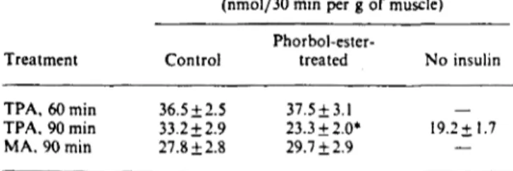
Incubation of EDL muscles with 0.5 /iM-TPA, a concentration that causes maximal effects in other cell types, only caused a minor increase in MeAIB uptake, which did not reach statistical significance (Table 1). This lack of effect on MeAIB uptake was observed after incubation for 60 or 180min in the presence of TPA. In fact, the same pattern was observed after incubation for 60 min with the inactive phorbol ester MA (11.5+1.4 and 14.1 ± 1.9 nmol/30 min per g in control and MA-treated groups respectively). Dimethyl sulphoxide at a concentration of 0.01 °0 had no effect on MeAIB uptake (results not shown). This lack of effect of TPA contrasts with the substantial effect of insulin at 60 min after its addition on MeAIB uptake (Table 1). These results indicate that TPA, a protein kinase C activator, does not mimic insulin action, and therefore insulin signalling required to stimulate MeAIB transport does not involve protein kinase C activation in EDL muscle.
Effect of phorbol esters on insulin-stimulated MeAIB uptake
We have previously shown that 100 nM-insulin maximally stimulates MeAIB uptake in EDL muscle after 60 min of incubation [27]. Therefore, in the present study we have selected these conditions to investigate the effect of TPA on insulin-stimulated MeAIB uptake. When 100 nM-insulin and 0.5 ftM-TPA were added simultaneously (both during the last 60 min of incubation), insulin-stimulated MeAIB uptake was ijot compromised (Table 2). However, when 0.5 //M-TPA was added 30 min before 100 nM-insulin (that is, TPA present during the last 90 min and insulin during the last 60 min of incubation),
Table 1. Effect of TPA and insulin on MeAIB uptake by EDL muscle Results are means±S.E.M. for 9-10 observations per group. EDL muscles were incubated for 180min by using three different protocols: (i) in the absence or presence of 0.5/iM-TPA during the last 60 min of incubation, (ii) in the absence or presence of 0.5 //M-TPA during the 180min of incubation, (Hi) in the absence or presence of 100 nM-insulin for the last 60 min of incubation. TPA was dissolved in dimethyl sulphoxide, in a final concentration of 0.01 V Control muscles were also incubated in the presence of 0.0! °o dimethyl sulphoxide. Uptake of MeAIB was determined during the last 30 min. * Value significantly different from that of the basal group (P < 0.05).
MeAIB uptake (nmol/30 min per g of muscle)
Table 2. Effect of phorbol esters on insulin-stimulated MeAIB uptake by EDL muscle
Results are means + s.E.M, for 8-11 observations per group. EDL muscles were incubated for 180 min in the absence or in the presence of 100 nM-insulin during the last 60 min of incubation. Muscles were incubated in the absence or in the presence of 0.5 //M-TPA. which was added either at the same time as the insulin (i.e. it was present during the last 60 min of incubation) or 30 min before insulin addition (TPA 90 min). In a different experimental series, muscles were incubated in the absence or presence of 0.5 //M-MA during the last 90 min of incubation. Phorbol esters were dissolved in dimethyl sulphoxide (final concn. 0.01 "„). Control muscles were also in-cubated in the presence of 0.01 % dimethyl sulphoxide. MeAIB uptake was determined during the last 30 min of incubation. * indicates a significant difference between control and TPA groups. at P < 0.05.
MeAIB uptake (nmol/30 min per g of muscle) Treatment TPA, 60 min TPA. 90 min MA. 90 min Control 36.5 + 2.5 33.2 ±2.9 27.8 + 2.8 Phorbol-ester-treated 37.5 + 3.1 23.3 + 2.0* 29.7 + 2.9 No insulin 19.2+1.7 Treatment TPA, 60 min TPA, 180min Insulin. 60 min Control group 15.6+1.6 17.2±1.2 17.3+1.7 Experimental group 17.9±1.8 18.3+1.9 3I.8±2.5*
insulin-stimulated MeAIB uptake was largely prevented (Table 2). This effect was specific to TPA, since incubation for 90 min (30 min before insulin addition) with the inactive phorbol MA did not modify insulin action (Table 2). Furthermore, dimethyl sulphoxide (0.01 °0) had no effect on insulin-stimulated MeAIB uptake, either (results not shown). These results provide evidence that protein kinase C rapidly compromises insulin action in the incubated skeletal muscle, promoting a situation of insulin resistance.
Additive inhibitory effects of TPA and protein kinase C inhibitors (polymyxin B and H-7) on insulin-stimulated MeAIB uptake
Polymyxin B and H-7 are well-known inhibitors of protein kinase C [33-35] in several cell types, and they block different biological effects induced by phorbol esters [36,37]. Thus we attempted to assess whether these agents could also prevent TPA effects on insulin-stimulated MeAIB uptake by muscle. To that end, we investigated, in a separate set of experiments, the effect of both inhibitors on the stimulation of MeAIB uptake induced by incubation for 1 h with 100 nM-insulin. Incubation for 2 h in the presence of O.I mM-polymyxin B (100 times its Kt for protein kinase C inhibition; [34]) caused a substantial inhibition of insulin-stimulated MeAIB uptake, with no effects on basal MeAIB uptake (Table 3). In fact, the effect of polymyxin B was very similar to the inhibitory effect caused by preincubation for 90 min with 0.5//M-TPA on insulin-stimulated MeAIB uptake (Table 3). Thus, whereas insulin induced an increase in MeAIB uptake by control muscle, this stimulation was decreased after incubation with TPA or polymyxin B (Table 3). When both polymyxin B (120 min) and TPA (90 min) were present in the incubation medium, insulin action was maximally inhibited (Table 3). The additive effects of TPA and polymyxin B inhibiting insulin-stimulated amino acid transport indicate that, in muscle, polymyxin B exerts actions other than the inhibitory effect on protein kinase C activity. That is, polymyxin B inhibits insulin action on amino acid transport by a mechanism not mediated by protein kinase C.
We also investigated the effect of H-7 on basal and insulin-Vol. 268
636 A. Gumà and others
Table 3. Effect of TPA and polymyxin B on insulin-stimulated MeAlB uptake by EDL muscle
Results are means+ S.E.M. for 6-8 observations per group. EDL muscles were incubated for 180 min in the absence or presence of 100 nM-insulin during the last 60 min of incubation. Muscles were incubated in the absence or presence of 0.5/iM-TPA (added during the last 90 min of incubation). 0.1 mM-poIymyxin B (added during the last 120 min of incubation ) or both TPA (90 min) and polymyxin B (120 min). MeAIB uptake was determined during the last 30 min of incubation, 'indicates a significant difference between basal and insulin groups, at P < 0.05.
MeAIB uptake (nmol/30 min per g of
muscle) Treatment No additions TPA Polymyxin B TPA + polymyxin B Basal 17.6+1.3 19.3+1.9 19.3 + 2.1 13.5±0.9 Insulin 32.3 ±3.6* 28.8 + 3.4« 25.8±3.3 I4.8±0.7 Effect of insulin (°u of basal) 83 49 34 10
Table 4. Effect of TPA and H-7 on insulin-stimulated MeAIB uptake by EDL muscle
Results are means±S.E.M. for 8-9 observations per group. EDL muscles were incubated during 180 min in the absence or presence of 100 nM-insulin during the last 60 min of incubation. Muscles were incubated in the absence or presence of 0.5 /iM-TPA (added during the last 90 min of incubation). 1 mM-H-7 (added during the last 120 min of incubation) or both TPA (90 min) and H-7 (120 min). H-7 was dissolved in ethanol. in a final concentration of 0.2 "„. Control muscles were also incubated in the presence of 0.2% ethanol. MeAIB uptake was determined during the last 30 min of incubation, «indicates a significant difference between basal and insulin groups, at P < 0.05.
MeAIB uptake (nmol/30 min per g of muscle) Treatment No additions TPA H-7 TPA + H-7 Basal 16.6+1.2 18.6 + 2.0 21.7 + 2.5 14.2+1.6 Insulin 34.4 ±3. 5* 26.1+3.1« 34.7 + 2.7» I8.0±l.6 (°0 of basal) 107 40 60 27
stimulated MeAIB uptake (Table 4). EDL muscles were incubated for 120 min in the presence of 1 mM-H-7, which is well above the AT, value (6 //M) for inhibition of protein kinase G [35]. Incubation with H-7 caused an increased MeAIB uptake under basal conditions, but not in the presence of insulin (Table 4). Similarly to the effects of polymyxin B, incubation of muscles with 1 mM-H-7 (for 120 min)andO,5/<M-TPA(for90 min) caused a further decrease in insulin-stimulated MeAIB uptake (Table 4). In summary, both H-7 and polymyxin B present additive effects with TPA with respect to their inhibitory effect on insulin action, and are not therefore suitable agents to investigate whether a certain insulin effect is mediated by protein kinase C activity. Effect of TPA on 3-O-methylglucose uptake and lactate production
In light of the marked inhibition of insulin-stimulated MeAIB uptake induced by TPA in EDL muscle, we analysed whether
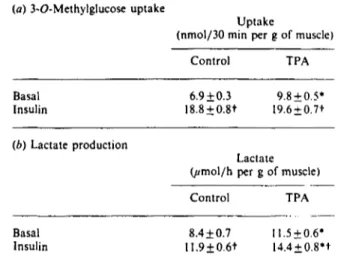
Table 5. Effect of TPA on 3-O-methylglucose uptake and lactate production by EDL muscle
Results are means+ S.E.M. for 5-6 observations per group for 3-O-methylglucose uptake, and for 14-21 observations per group for lactate production. EDL muscles were incubated for 180 min in the absence or in the presence of 100 nM-insulin during the last 60 min of incubation. Muscles were incubated in the absence or in the presence of 0.5 /(M-TPA. which was added 30 min before insulin addition (TPA 90 min). TPA was dissolved in dimethyl sulphoxide (final concn. 0.01 °„). Control muscles were also incubated in the presence of 0.01 °„ dimethyl sulphoxide. 3-O-Methylglucose uptake and lactate production were determined during the last 30 minutes of incubation. * indicates a significant difference between control and TPA groups, at P < 0.05. t indicates a significant difference between basal and insulin groups, at P < 0.05.
(a) 3-O-Methylglucose uptake
Uptake
(nmol/30 min per g of muscle)
Control TPA Basal Insulin 6.9 + 0.3 18.8±0.8t 9.8 + 0.5* 19.6±0.7t (b) Lactate production Lactate (jtmo\/h per g of muscle)
Control TPA Basal Insulin 8.4 + 0.7 11.9±0.6t ll.5±0.6* 14.4 + 0.8't
that was a consequence of a generalized TPA-dependent 'in-hibition of insulin action. To that end, we investigated the effect of TPA on insulin-stimulated glucose utilization by muscle. Incubation of muscles in the presence of TPA (0.5 //M, 90 min) caused a 40 °0 increase in the rate of 3-O-methylglucose uptake
by muscle (Table 5); insulin caused a larger increase (170°0) in
3-O-methylglucose uptake, and insulin and TPA effects were not additive (Table 5). TPA also caused an enhanced production of lactate, which was quantitatively similar to the activation caused by 100 nM-insulin (Table 5); under these conditions insulin and TPA showed additive effects (Table 5).
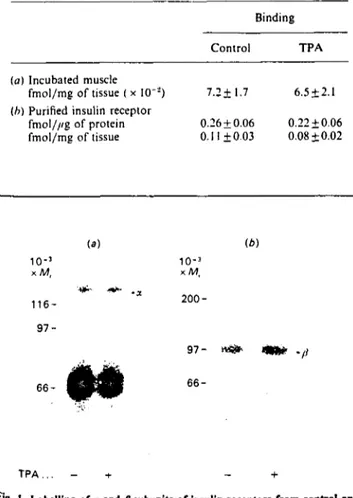
Effect of TPA on insulin binding and receptor tyrosine kinase In order to ascertain the mechanisms by which TPA compromises insulin-stimulated system A transport activity by skeletal muscle, specific insulin binding was determined by using the incubated EDL muscle (Table 6). TPA treatment for 30 min did not cause any significant change in the binding of tracer quantities of insulin by muscle.
Insulin binding was also assessed after team-affinity purification of insulin receptors. Insulin receptors from control and 90 min-TPA-treated EDL muscles were partially purified by WGA affinity chromatography. No differences in the yield of glycoproteins was detected in either group (0.44 + 0.03 and 0.37 ± 0.02 ¿ig/mg of muscle in control and TPA-treated muscles respectively). In addition, no significant differences in insulin binding were detected, either when expressed per //g of protein eluted from the column or when expressed as total insulin binding per mg of muscle (Table 6).
Studies of affinity cross-linking using 0.5 nM-[l"I-TyrB2
']-insulin (near high-affinity KA values) were also performed to
determine the Mr of the a subunit of the insulin receptor from
Effect of phorbol esters on insulin action in muscle 637
Table 6. Insulin binding by incubated rat EDL muscle and by partially purified insulin receptor: effect of TPA
Values are means+ S.E.M. of 5-6 observations per group, (a) EDL muscles were initially incubated for 30 min in the absence or presence of 0.5/'M-TPA. To assess insulin binding, the muscles were then incubated w i t h 30 pM-['-:'I-TyrA"]monoiodoinsulin at 21 °C for 3 h
in the absence or presence of l.5//M-insulin. The '"I-insulin specifically bound was determined by subtracting the binding observed in the presence of 1.5/iM-insulin from that observed in the absence of unlabelled insulin, (b) Insulin receptors were partially purified from control and TPA-lreated (90 min) EDL-muscle homogenates. after solubilization in Triton X-IOO and ultracentrifugation. by lectin affinity chromatography. Each prep-aration was obtained from 10-15 rats. The WGA eluate (20/d) was incubated for I h at 22 °C in buffer containing 30 mM-Hepes, 0.2°,, BSA, 100 units of bacitracin/ml and ['"I-Tyr^'^mono-iodoinsulin, in the absence or presence of 1 /;M-insulin. See the Experimental section for further details. Differences between control and TPA groups were statistically insignificant.
Binding
Control TPA
(a) Incubated muscle
fmol/mg of tissue ( x l O '2) 7.2±1.7 6.5 ±2.1
(h) Purified insulin receptor
fmol///g of protein 0.26 ± 0.06 0.22 ± 0.06 fmol/mg of tissue 0.11 ±0.03 0.08 + 0.02 — 9 o a. ñ O) 6 •
f t
o I 0.1 1.0 10 100 [Insulin] (nM)Fig. 2. Effect of TPA on exogenous kinase activity of insulin receptors from muscle
Insulin receptors from EDL muscles treated (O) or not (•) with TPA for 90 min were partially purified as described in the Ex-perimental section. Each preparation was obtained by pooling muscles from 10-12 rats. WGA eluates (10 //I) were incubated at 22 °C for 1 h in 30 mM-Hepes buffer, pH 7.6, containing 50 m.M-magnesium acetate and various concentrations of insulin. [-/• "PjATP (50//M) was added, and samples were incubated for an additional 10 min. The substrate (copolymer of Glu/Tyr, 4 : 1 : 0.25 mg/ml) was then added and allowed to react for 30 min. The reaction was stopped by applying samples to filter-paper squares and soaking in 10"0 trichloroacetic acid/10 miu-sodium
pyrophosphate. Papers were washed, dried and counted by Cerenkov radiation. All values have been corrected for non-specific association of 31P with the paper, which was estimated by incubating samples in
the absence of receptor addition. Each data point is the mean of 6 observations per group performed in triplicate.
(a) (b) 10-3 X / W , 116- 97-10-3 xM. 66-
INI
200-97- «Si* «fe* 66-T P A . . . - + - +F'g- I. Labelling of x and ß subunits of insulin receptors from control and
TPA-treated muscles
Partially purified insulin receptors were obtained from control and TPA-treated (90 min) EDL muscles as described in the Experimental section. Each preparation was obtained by pooling muscles from '0-12 rats, (a) Affinity cross-linking of '"I-insulin to the insulin receptor. Partially purified receptor (10//I) was incubated at 22 °C for 60 min in 30 mM-Hepes containing 0.5 nM-['"I-TyrB2<]insulin in
•he absence or presence of 1 //M unlabelled insulin. After incubation for 5 min at 0 °C. disuccinimidyl suberate was added (final concn. ' mM), and samples were incubated for a further 15 min at 0 °C. The faction was stopped by addition of Laemmli sample buffer with 0.1 M-dithiothreitol. (b) Labelling of ß subunit of insulin receptors. Partially purified receptors were phosphorylated in the presence of ly-^PjATP and insulin. The reaction was stopped by addition of Laemmli sample buffer with 0.1 M-dithiothreitol. Samples were subjected to electrophoresis in 7.5°0-polyacrylamide gels, followed
°y autoradiography.
Vol. 268
control and TPA-treated muscles (Fig. 1). When affinity-labelled insulin receptors from control and TPA-treated muscles were run on SDS/PAGE under reducing conditions, only one band (Mr
approx. 130000) was specifically labelled (Fig. 1). The migration characteristics of this specifically labelled band did not diner between control and TPA-treated muscles. The ß subunit, detected after autophosphorylation, showed similar apparent M, values when control and TPA-treated groups are compared (Fig. 1). This supports the observation that the integrity of the a and ß subunits of insulin receptors partially purified from control and from TPA-treated EDL muscles were similar.
The kinase activity of the insulin receptor in control and TPA-treated EDL muscle was next characterized by using an ex-ogenous substrate. The dose/response relationship between in-sulin and 32P incorporation into a copolymer of Glu/Tyr, in the
presence of purified insulin receptor, is presented in Fig. 2. Equal amounts of insulin binding (and protein) were used for control and TPA-treated groups. Insulin stimulated the exogenous kinase activity of the insulin receptor from control muscle as previously shown (Fig. 2). Thus, 1 nM-insulin caused more than a half-maximal stimulation of the rate of exogenous substrate phosphorylation, and at 10 nM-insulin stimulation was already maximal. Supra-maximal insulin caused a 3-fold increase in exogenous kinase activity from control insulin receptors. Insulin receptors partially purified from TPA-treated muscles exhibited a similar ability to phosphorylate the exogenous substrate in the absence as well as in the presence of insulin as compared with the control group (Fig. 2).
DISCUSSION
638 A. Gumá and others protein kinase C activation inhibits some effects of insulin, as
assessed by insulin-stimulated system A transport activity. How-ever, TPA treatment does not modify insulin-stimulated receptor kinase, glucose transport or lactate production by muscle. These are in keeping with previous reports in which no inhibitory effect of phorbol esters on insulin-stimulated receptor kinase or glucose transport by muscle was substantiated [38,39]. Therefore, we propose that activation of protein kinase C must block a step located at a post-receptor level in the biochemical pathway that leads to stimulation of amino acid transport. Whether this is the only effect caused by TPA on insulin action in muscle remains to be established. Our findings differ from other results reported for intact Fao hepatoma cells, in which TPA treatment led to inhibition of insulin action and insulin-stimulated tyrosine kinase activity [2,15], as well as with other reports in adipocytes [7,8]. In our study, we did not find a substantial modification of insulin binding after TPA treatment, which agrees with others [2,15,38.40]. However, alterations of insulin-binding properties have also been reported in isolated rat adipocytes, lymphocytes, macrophages, monocyte-like and promyelocytic leukaemia human cell lines after phorbol ester treatment [6,16,41,42].
The variable response to phorbol esters, as well as the variable interaction between insulin and protein kinase C discussed above, might be understood on the basis of tissue differences in the pattern of expression of protein kinase C isoenzymes. At least seven different subspecies of protein kinase C have been identified in mammalian tissues [43-45], and some kinetic differences among isoenzymes have been reported [44]. Further work is required to define the protein kinase C isoenzymes present in skeletal muscle, their kinetic properties and the similarities between the isoenzyme patte'rns found in muscles and other hormone-sensitive tissues.
Under our conditions, we have observed that in skeletal muscle the effect of insulin stimulating system A transport activity for neutral amino acids is not mimicked by the addition of TPA. a protein kinase C activator. From a mechanistic viewpoint, our data permit the conclusion that protein kinase C activation does not mediate insulin-stimulated system A trans-port in skeletal muscle. The lack of effect of TPA on basal system A transport activity contrasts with the marked increase that we found in glucose transport and lactate production. The stimulatory effect of TPA on glucose transport agrees with previous observations obtained in incubated muscle from mice [39] and contradicts another report, in which TPA failed to increase glucose transport in rat skeletal muscle [38]. The reason for these differences is unclear. In any event, the fact that TPA stimulates glucose transport and not MeAIB transport in skeletal muscle substantiates the contention that insulin causes activation of both transport systems by independent mechanisms. This is especially interesting in the light of the parallel regulation of glucose uptake and system A transport activity previously described in skeletal muscle under a variety of conditions [26,46,47].
It is worth noting that, although TPA causes a smaller stimulatory effect on glucose transport as compared with insulin, both agents enhance lactate production to a similar extent. Furthermore, whereas TPA and insulin did not show additive effects on glucose transport, their stimulatory action on lactate production by muscle was clearly additive. In order to explain this metabolic pattern, we should mention that (a) insulin-stimulated glucose uptake, in the incubated muscle, is mainly directed into glycogen and lactate production [25], (b) TPA inhibits glycogen synthesis from glucose in muscle [38], and (c) phorbol esters increase fructose 2,6-bisphosphate levels and activate glycolysis in several cell types [48,49], Based on that, it might be postulated that in the presence of TPA a smaller proportion of the glucose taken up by the cell is incorporated
into muscle glycogen. in favour of a greater activation through the glycolytic pathway.
Many inhibitors of protein kinase C have recently been reported. These include polymyxin B [33.34], calmodulin antagonists [33], H-7 [35], K252a [50], staurosporine [51]. sphin-gosine [52] and sangivamycin [53]. These inhibitors are sometimes used with the intention of gaining information on the role of protein kinase C in different cellular responses. Ideally, protein kinase inhibitors block, under appropriate conditions, different phorbol-ester-induced effects [36,37,50]. However, it has been reported that, for instance, potymyxin B also inhibits insulin-stimulated membrane transport processes in muscle and adipocytes [54,55], and in addition inhibits a Ca2*-activated K*
channel [56]. In the present study we have substantiated that both H-7 and polymyxin B block, to a different extent, insulin action and, in addition, that these inhibitors and phorbol esters present additive inhibitory effects on insulin-stimulated MeAIB uptake. Our data indicate that protein kinase C does not mediate the mechanism by which H-7 or polymyxin B blocks the insulin effect on system A transport activity. This suggests that H-7 or polymyxin B are not specific inhibitors of protein kinase C activity and are not therefore suitable agents with which to investigate whether a certain insulin effect is mediated by protein kinase C activity.
We have substantiated that phorbol esters do not induce an insulin-resistance-like situation as regards either glucose utilization or receptor tyrosine kinase activity in skeletal muscle. That allows us to hypothesize that situations of muscle insulin resistance characterized by altered peripheral glucose disposal and deficient receptor kinase activity, such as those reported in diabetes or obesity [57,58], cannot be attributable to altered catalytic properties of protein kinase C. However, protein kinase C must be envisaged as a negative modulator of the biochemical pathway by which insulin activates system A amino acid transport activity in skeletal muscle. This offers a molecular explanation for the generation of insulin-resistant states in which the im-pairment just involves certain biological effects of insulin.
This work was supported in part by a research grant from the Dirección General de Investigación Científica y Técnica (PB-573/86!. and from the Fondo de Investigaciones Sanitarias (87/1718 and 89/0174), Spain. A. G. and M. C. are recipients of predoctoral fellowships from the Ministerio de Educación y Ciencia. Spain.
REFERENCES
!. Nishizuka, Y. (1986) Science 233. 305-312
2. Takayama. S.. White. M. F.. Lauris. V. & Kahn. C. R. (1984) Proc. Nati. Acad. Sei. U.S.A. 81. 7797-7801
3. Casiagna, M.. Takai. Y.. Kaibuchi, K., Sano. K„ Kikkawa, U. & Nishizuka, Y. (1982) J. Biol. Chem. 257. 7847-7854
4. Ebeling, J. G-, Vandenbark, G. R., Kühn, L., Ganong. B., Bell, R. M. & Niedel, J, (1985) Proc. Nati, Acad. Sei. U.S.A. 82. 815-819
5. Farese, R. V., Standaert. M. L., Bames, D. E., Davis, J. S. & Pollet. R. J. (1985) Endocrinology (Baltimore) 116. 2650-2655
6. Kirsch. D., Obcrmaier. B. &Haring, H. U. (1985) Biochem. Biophys. Res. Commun. 128, 824-832
7. Martz. A.. Mookerjee, B. K. & Jung, C. Y. (1986) J. Biol. Chem. 261, 13606-13609
8. Van de Werve, G.. Proietto, J. & Jeanrenaud, B. (1985) Biochem. J. 225. 523-527
9. Freychet, P., Roth. J. & Neville. D. M. (1971) Proc. Nati. Acad. Sei. U.S.A. 68. 1833-1837
10. Cuatrecasas, P. (1972) Proc. Nati. Acad. Sei. U.S.A. 69. 1277-1281 11. Kasuga. M., Zick, Y.. Blithe, D. L.. Karlsson, F. A.. Häring, H. U.
& Kahn, C. R. (1982) J. Biol. Chem. 257. 9891-9894 12. Shia, M. A. & Pilch. P. F. (1983) Biochemistry 22. 717-721
Effect of phorbol esters on insulin action in muscle 639
13. Ebina. Y.. Araki. E.. Taira. M.. Shimada. F.. Mori. M.. Craik. C. S.. Siddle. K.. Pierce. S, B.. Roth. R. A. & Rutter. W, J. (1987) Proc. Nati. Acad. Sei. U.S.A. 84. 704-708
14. Chou. C. K.. Dull. T. J.. Russell. D. S.. Gherzi. R.. Lebwohl. D.. Ullrich. A. & Rosen. O. M. (1987) J. Biol. Chem. 262, 1842-1847 15. Takayama. S.. While. M. F. & Kahn. C. R. (1988) J. Biol. Chem.
263. 3440-3447
16. Häring. H.. Kirsch. D.. Obermaier. B.. Ermel. B. & Machicao. F. (1986) J. Biol. Chem. 261. 3869-3875
17. Bollag. G. E.. Roth. R. A.. Beaudoin, J.. Mochly-Rosen. D. & Koshland, D. E. (1986) Proc. Nati. Acad. Sei. U.S.A. 83. 5822-5824 18. Walaas. S. I.. Horn. R. S.. Adler. A.. Albert, K. A. & Walaas, O.
(1987) FEBS Lett. 220, 311-318
19. Cooper, D. R.. Konda, T. S.. Standaert, M. L, Davis, J. S., Pollet, R. J. & Farese. R. V. (1987) J. Biol. Chem. 262. 3633-3639 20. Pershadsingh, H.A., Shade. D. L. & McDonald, J. M. (1987)
Biochem. Biophys. Res. Commun. 145, 1384-1389
21. Draznin, B.. Leitner. J. W., Sussman. K. E. & Sherman, N. A. (1988) Biochem. Biophys. Res. Commun. 156, 570-575
22. Glynn. B. P., Colliton, J. W., McDermott, J. M. & Witters, L. A. (1986) Biochem. Biophys. Res. Commun. 135. 1119-1125 23. Spach, D. H., Nemenoff, R. A. & Blackshear, P. J. (1986) J. Biol.
Chem. 261, 12750-12753
24. Klip. A. & Ramlal. T. (1987) Biochem. J. 242, 131-136
25. Maizels, E. Z., Ruderman, N. B., Goodman, M. N. & Lau, D. (1977) Biochem. J. 162. 557-568
26. Zorzano, A., Balón, T. W., Garetto. L. P., Goodman, M. N. & Ruderman, N. B. (1985) Am. J. Physiol. 251, E546-E552
27. Gumà, A., Testar. X., Palacín. M. & Zorzano, A. (1988) Biochem. J. 253, 625-629
28. Goodman. M. N., Berger, M. & Ruderman, N. B. (1974) Diabetes 23. 881-888
29. Le Marchand-Brustel. Y., Jeanrenaud, B. & Freychet, P. (1978) Am. J. Physiol. 234, E348-E358
30. James, D. E., Zorzano, A., Boni-Schnetzler, M., NemenofT. R. A., Powers. A., Pilch, P. F. & Ruderman, N. B. (1986) J. Biol. Chem. 261. 14939-14944
31. Bradford, M. M. (1976) Anal. Biochem. 72. 248-254
32. Pilch. P. F. & Czech, M. P. (1980) J. Biol. Chem. 255, 1722-1731 33. Wise. B., Glass. D. B.. Jen Chou. C. K., Raynor. R. L., Katoh, N.,
Schatzman, R. C.. Turner, R. S.. Kibler, R. F. & Kuo, J. F. (1982) J. Biol. Chem. 257. 8489-8495
34. Mazzei. G. J.. Katoh. N. & Kuo. J. F. (1982) Biochem. Biophys. Res. Commun. 109. 1129-1133
35. Hidaka. H.. Inagaki, M., Kawamoto. S. & Sasaki, Y. (1984) Biochemistry 23. 5036-5041
36. Nel. A. E., Wooten. M. W.. Goldschmidt-Clermont. P. J.. Miller. P. J.. Stevenson. H. C. & Galbrahh. R. M. (1985) Biochem. Biophys. Res. Commun. 128. 1364-1372
37. Struhar. D. & Harbeck. R. J. (1987) FASEB J. I. 116-118 38. Sowell M. O.. Treutelaar, M. K.. Burant.C. F. & Buse. M. G. (1988)
Diabetes 37. 499-506
39. Tanti. J.-F.. Rochet, N., Grémeaux. T.. Van Obberghen. E. & Le Marchand-Brustel. Y. (1989) Biochem. J. 258. 141-146
40. Van de Werve, G.. Zaninetti. D., Lang. U.. Vallotton. M. B. & Jeanrenaud. B. (1987) Diabetes 36, 310-314
41. Grunberger, G. & Gorden, P. ( 1982) Am. J. Physiol. 243. E319-E324 42. Thomopoulos, P.. Testa. U., Gourdin. M. F.. Hervy, C., Titeux. M.
& Vainchenker, W. (1982) Eur. J. Biochem. 129. 389-393 43. Coussens, L., Parker, P. J., Rhee. L., Yang-Feng, T. L., Chen. E..
Waterfield. M. D., Francke. U. & Ullrich, A. (1986) Science 233. 859-866
44. Nishizuka. Y. (1988) Nature (London) 334. 661-665
45. Ono, Y.. Fujii, T., Ogita, K., Kikkawa. U., Igarashi, K. & Nishizuka. Y. (1988) J. Biol. Chem. 263, 6927-6932
46. Zorzano, A., Balón, T. W.. Goodman. M. N. & Ruderman, N. B. (1986) Am. J. Physiol. 251. E2I-E26
47. Zorzano. A., Balón. T. W., Goodman. M. N. & Ruderman, N. B. (1986) Biochem. Biophys. Res. Commun. 134. 1342-1349 48. Boscà. L., Rousseau, G. G. & Hue, L. (1985) Proc. Nati. Acad. Sei.
U.S.A. 82, 6440-6444
49. Boscà, L., Mojena, M., Diaz-Guerra, M. J. & Marquez, C. (1988) Eur. J. Biochem. 175. 317-323
50. Yamada. K.. Iwahashi, K. & Kase, H. (1987) Biochem. Biophys. Res. Commun. 144, 35^40
51. Tamaoki. T., Nomoto, H., Takahashi, !.. Kato, Y., Morimoto, M. & Tomita. F. (1986) Biochem. Biophys. Res. Commun. 135, 397-Í02
52. Hannun. Y. A.. Loomis, C. R.. Merrill, A. H. & Bell. R. M. (1988) J. Biol. Chem. 261, 12604-12609
53. Loomis, C. R. & Bell, R. M. (1988) J. Biol. Chem. 263, 1682-1692
54. Amir. S., Sasson, S., Kàiser. N.. Meyerovitch. J. & Schechter. Y. (1987) J. Biol. Chem. 262, 6663-4667
55. Grémeaux, T., Tanti, J. F., Van Obberghen. E. & Le Marchand-Brustel. Y. (1987) Am. J. Physiol. 252. E248-E254
56. Varecka. L., Peterajova. E. & Pogady. J. (1987) FEBS Lett. 225, 173-177
57. Burant. C. F.. Treutelaar. M. K. & Buse, M. G. (1986) J. Clin. Invest. 77, 260-270
58. Le Marchand-Brustel, Y.. Grémeaux. T., Ballotti, R. & Van Obberghen, E. (1985) Nature (London) 315. 676-679
Received 30 October 1989/21 February 1990; accepted 5 March 1990