Absorption and pharmacokinetics of grapefruit flavanones in beagles
Maria de Lourdes Mata-Bilbao
1, Cristina Andre´s-Lacueva
1, Elena Roura
1, Olga Ja´uregui
2, Elvira Escribano
3,
Celina Torre
4and Rosa Maria Lamuela-Ravento´s
1*
1Department of Nutrition and Food Science, CerTA, Faculty of Pharmacy, University of Barcelona, Av. Joan XXIII s/n, Barcelona, Spain
2Scientific and Technical Services, University of Barcelona, Barcelona, Spain
3Biopharmaceutics and Pharmacokinetics Unit, Faculty of Pharmacy, University of Barcelona, Av. Joan XXIII s/n, Barcelona, Spain
4Affinity Pet-care, Barcelona, Spain
(Received 25 August 2006 – Revised 8 January 2007 – Accepted 24 January 2007)
The present study evaluated the pharmacokinetics of three different grapefruit flavanone forms in dog plasma and demonstrated their absorption after an oral intake of a grapefruit extract; pharmacokinetic parameters of these forms were also determined. Ten healthy beagles were adminis-tered 70 mg citrus flavonoids as a grapefruit extract contained in capsules, while two additional dogs were used as controls and given an excipient. The grapefruit flavanone naringin, along with its metabolites naringenin and naringenin glucuronide, was detected in dog plasma. Blood samples were collected between 0 and 24 h after administration of the extract. Naringin reached its maximun plasma concentration at around 80 min, whereas naringenin and naringenin glucuronide reached their maximun plasma concentrations at around 20 and 30 min, respectively. Maximum plasma concentrations of naringin, naringenin and naringenin glucuronide (medians and ranges) were 0·24 (0·05 – 2·08), 0·021 (0·001 – 0·3) and 0·09 (0·034 – 0·12) mmol/l, respectively. The areas under the curves were 23·16 l (14·04 – 70·62) min £ mmol/for nariningin, 1·78 (0·09 – 4·95) min £ mmol/l for naringenin and 22·5 (2·74 – 99·23) min £ mmol/l for naringenin glucuronide. The median and range values for mean residence time were 3·3 (1·5 – 9·3), 2·8 (0·8 – 11·2) and 8·0 (2·3 – 13·1) h for naringin, naringenin and naringenin glucuronide, respectively. The results of the present study demonstrate the absorption of grapefruit flavanones via the presence of their metabolites in plasma, thus making an important contribution to the field since the biological activities ascribed to these compounds rely on their specific forms of absorption.
Absorption: Flavanone: Bioavailability: Dog: Grapefruit: Pharmacokinetics: Plasma
Flavonoids are a group of polyphenolic compounds with health-related properties that are widely distributed in fruits, vegetables, fruit juices, cocoa, teas and wines. Citrus fruits are rich in flavonoids that have been investigated for their bio-logical activity. The use of citrus flavonoids as anti-inflamma-tory, anticarcinogenic and antitumour agents has been reported (Middleton & Kandaswami, 1994; Benavente-Garcı´a et al. 1997; Montanari et al. 1998). Recent research shows that the citrus flavonoid naringenin stimulates DNA repair in pros-tate cancer cells (Gao et al. 2006), whereas the flavanone gly-coside naringin has proved to be a potent inhibitor of angiogenic peptide vascular endothelial growth factor, which is released in human tumour cells (Schindler & Mentlein, 2006). These studies suggest a novel mechanism for mammary cancer prevention, which is considered the most common cancer in female dogs.
Several studies have shown that grapefruit juice elevates the blood levels of some orally taken drugs, primarily by inhibit-ing intestinal CYP3A4-mediated first-pass metabolism (Fuhr et al. 2002; Dahan & Altman, 2004; Lilja et al. 2004; Paine
et al. 2004, 2005), CYP3A4 being a type of cytochrome P450. These studies suggest a potential therapeutic benefit from using the active constituents of grapefruit to increase drug bioavailability. Lowering the effective dose will also reduce drug costs, although potential clinical problems remain (Dahan & Altman, 2004).
One of the most common flavonoids found in grapefruit (Citrus paradisi) is the flavanone glycoside naringin (narin-genin 7-O-neohesperidoside; Fig. 1). Naringin is also known to be the agent responsible for the bitterness of grapefruit juice. Narirutin and naringenin (Fig. 1) are also present in grapefruit but to a lesser extent (Macheix et al. 1990).
Most of the molecular forms of flavonoids that reach the peripheral circulation and tissues are different from those pre-sent in foods (Day & Williamson, 2001; Day et al. 2001; Graefe et al. 2001; Natsume et al. 2003; Zhang et al. 2003). In general, the predominant forms in plasma are conjugates (glucuronates or sulphates, with or without methylation). These conjugates are chemically distinct from their parent compounds, differing in size, polarity and ionic form.
* Corresponding author: Dr R. M. Lamuela-Ravento´s, fax þ 34 93 4035931, email [email protected]
Abbreviations:AUC, area under the curve; Cmax, maximum plasma concentration; LC, liquid chromatography; MRT, mean residence time; m/z, mass-to-charge ratio.
Consequently, their physiological behaviour is likely to be different from that of the native compounds (Kroon et al. 2004), and their biological effect will ultimately depend on the cellular effects of their circulating metabolites (Harada et al. 1999; Spencer et al. 2001a, b).
Very little is known about the biological activities of these conjugated metabolites. Glucuronides of isoflavones and epi-catechin have been shown to have a much weaker oestrogenic activity and to provide no protection against oxidative stress in cells grown in vitro (Zhang et al. 1999; Spencer et al. 2001a, b), whereas additional studies have shown that the 5-O-b-D-glucuronide of catechin and epicatechin excreted in rat urine does not interfere with their antioxidant properties, as assesed by their ability to scavenge superoxide (Harada et al. 1999; Okushio et al. 1999), thus suggesting that in plasma they may still act as antioxidants. Although glucuro-nides do not readily enter cells, it is also possible that they might be cleaved by the action of b-glucuronidases located in human tissues such as the liver (O’Leary et al. 2001) or by neutrophils that release b-glucuronidases when activated (Shimoi et al. 1998; Simio et al. 2001).
Initially, only free flavonoids without a sugar molecule, so-called aglycones, were considered to be able to pass across the gut wall (Hollman & Katan, 1997). However, the absorption of quercetin glycosides from onions in human subjects (Holl-man et al. 1995) and the presence of the flavanone glycoside naringin in plasma and urine after oral administration (Ishii et al. 2000; Fang et al. 2006) have now been demonstrated.
Liquid chromatography (LC)-MS/MS has emerged as the preferred technology for the quantitative determination of metabolites in different biomatrices, due to its sensitivity and selectivity through MS/MS experiments and the fact that it enables structural identification (Murphy et al. 1994). Ion-spray ionization, together with tandem MS for structural characterization, has become a popular and versatile method
for flavonoid analysis (Roura et al. 2005; Urpı´-Sarda` et al. 2005; Fang et al. 2006).
The use of dogs as a model has been shown to be helpful in evaluating the absorption of flavonoids from green tea (Swezey et al. 2003). Thus, the present study aims to assess the major flavanone forms in plasma after the oral adminis-tration of a grapefruit extract; and to evaluate the kinetics of these metabolic forms in the plasma by considering biotrans-formation, thus providing a general model that can be used for studies on flavonoid bioavailability.
Methods Chemicals
Naringin (naringenin-7-O-rhamnoglucoside) and blank dog plasma were purchased from Sigma-Aldrich (St Louis, MO, USA). Naringenin (40,5,7-trihydroxyflavanone), narirutin (nar-ingenin-7-O-rutinoside) and the internal standard taxifolin were purchased from Extrasynthese (Genay, France). Metha-nol and acetonitrile, HPLC grade and formic acid were pur-chased from Scharlau Chemie S. A. (Barcelona, Spain). Ultrapure water (Milli-Q) was obtained from a Millipore system (Millipore, Bedford, MA, USA).
Animals and study design
Animals. Ten healthy adult beagle dogs were randomly chosen. The dogs had a mean weight of 13·97 (SD 2·96) kg and were deprived of food overnight before the experiment. Two capsules containing 200 mg grapefruit extract (70 mg fla-vanones) were orally administered to the dogs; two additional dogs were chosen as controls and were given an excipient con-taining talc. The grapefruit extract contained naringin, nari-rutin and naringenin as citrus flavanones. Blood was drawn Fig. 1. Structures and molecular weights (MW) of (A) naringin, (B) narirutin and C naringenin.
before capsule administration and at the following times after administrations: 10 min, 20 min, 30 min, 40 min, 80 min, 2 h, 4 h, 6 h, 8 h and 24 h. The dogs were fed with a polyphenol-free diet 2 h after the capsules were given. Blood samples (5 ml) were collected in vacutainer tubes containing EDTA as anticoagulant (Becton, Dickinson, Franklin Lakes, NJ, USA). Plasma was obtained after blood centrifugation at 13 000 g for 15 min and stored in Eppendorf tubes at 2 808C until analysis.
The study was carried out at Isoquimen S. L. (Barcelona), in accordance with the Guide for the Care and Use of Laboratory Animals (Committee on Care & use of Laboratory Animals, 1985). The study protocol was approved by the Isoquimen S. L. Ethics Committee.
Sample extraction procedure for grapefruit flavanone and fla-vanone metabolites. Flavanone compounds in plasma were extracted by solid-phase extraction as previously described (Roura et al. 2005).
Dog plasma samples were treated as follows: 24 ml of IS (8224 nmol/l) were added to 1 ml of plasma and then was mixed with 370 ml of antioxidant solution (containing 0·2 g/ml ascorbic acid, 1 mg/ml EDTA). After 2 min of vortex-mixing, samples were diluted with 3 ml water. Solid-phase extraction with Waters Oasis HLB 3 cm3(60 mg) cartridges (Waters Oasis, Mild-ford, MA, USA) was applied to the mixture. Cartridge activation was achieved by adding 1 ml methanol and 1·5Mformic acid in water (mol/l), respectively. Sample clean-up was performed with 2 ml 1·5M formic acid in water (mol/l) and 2 ml water – methanol solution (95 : 5 v/v). Flavonoid metabolites were eluted with 1 ml acidulated methanol (0·1 % formic acid). The eluted fraction was evaporated in a sample concentrator (Techne, Duxford, Cambridge, UK) at 258C under a stream of N gas and reconstituted with 300 ml mobile phase, before being filtered through a 4 mm, 0·45 mm PTFE filter (Waters) into an amber vial insert for LC-MS/MS analysis.
Preparation of the standards and sample treatments were performed in a darkened room with a red safety light to avoid the oxidation of the analytes.
LC-diode array detection. The grapefruit extract was ana-lysed in an HP 1050 (Hewlett-Packard, Palo Alto, CA, USA) liquid chromatograph equipped with an automatic injector (HP1050) and an HP diode array (1050 M) at 280 nm. The conditions for HPLC corresponded to those previously described by Mata et al. (2007).
LC-MS/MS. Grapefruit metabolites were identified and quantified by LC-MS/MS plasma analysis. LC analysis was performed using a Perkin-Elmer series 200 (Perkin-Elmer, Norwalk, CT, USA) equipped with a quaternary pump and an autosampler. A Luna C18 column (50 £ 2·0 mm internal diameter, 5 mm; Phenomenex, Torrance, CA, USA) was used at room temperature, and the injected volume was 20 ml. Gra-dient elution was carried out with water (0·1 % formic acid) and acetonitrile (0·1 % formic acid) at a constant flow of 600 ml/min. A gradient profile with the following proportions (v/v) of acetonitrile (0·1 % formic acid) was applied (time in min, % acetonitrile): (0, 5), (2, 25), (7, 90), (9, 100) and (12, 100). The column was equilibrated for 10 min between runs.
A triple quadrupole mass spectrometer (API 3000; Applied Biosystems, PE Sciex, Concord, Ontario, Canada), equipped with a turbo IonSpray source was used to obtain the MS and
MS/MS data. TurboIonspray source settings were as follows: capillary voltage, 2 3500 V; nebulizer gas (N2), 10 (arbitrary units); curtain gas (N2), 12 (arbitrary units); collision gas (N2), 10 (arbitrary units); focusing potential, 2 200 V; entrance potential, 10 V; drying gas (N2), heated to 4008C and introduced at a flow rate of 8000 cm3/min. The decluster-ing potential and collision energy were optimized for each compound with infusion experiments: individual standard sol-utions (10 ppm) dissolved in 80 : 20 (v/v) mobile phase were infused at a constant flow rate of 5 ml/min into the mass spec-trometer using a model syringe pump (Harvard Apparatus, Holliston, MA, USA).
Full scan data were acquired by scanning the mass-to-charge ratio (m/z) from 100 to 600 in profile mode, using a cycle time of 2 s. For MS/MS, a product ion scan utilising a cycle time of 2 s was used. MS/MS product ions were pro-duced by the collision-activated dissociation of selected pre-cursor ions in the collision cell of the triple quadrupole mass spectrometer and the mass analysed using the second analyser of the instrument. Multiple reaction monitoring, the method of choice because of having the highest selectivity and sensitivity in quantitative LC-MS/MS, was used to moni-tor five transitions for each analysis: naringin, m/z 579 ! 271; narirutin m/z 579 ! 271; naringenin, m/z 271 ! 151; narin-genin glucuronide m/z 447 ! 271; narinnarin-genin sulphate m/z 351 ! 271; taxifolin, m/z 303 ! 285. Both quadrupoles (Q1 and Q3) were operated at unit resolution. The criteria for iden-tifying grapefruit metabolites, such as retention time, multiple reaction monitoring transition as mentioned above and tran-sitions 579 ! 271 and 271 ! 151 (at a higher declustering potential value), were chosen to confirm the multiple reaction monitoring trace for each metabolite in collisionally induced dissociation-MS/MS experiments (Roura et al. 2005; Urpı´-Sarda` et al. 2005).
Pharmacokinetic analysis. Pharmacokinetic parameters were determined by means of a non-compartmental analysis using the WinNonlin Professional software version 3.3 (Phar-sightwCorporation, USA). The linear trapezoidal method was used to calculate the area under the plasma concentration curve (AUC0 – t) from time 0 until the last detectable concen-tration. The total area under the curve (AUC0 – 1) was calcu-lated by the expression: AUC0 – tþ AUCextr, where AUCextr is the extrapolated area under the curve. The maximum plasma concentration (Cmax) and the time needed to reach Cmaxwere determined by visual inspection of the experimental data. Mean residence time (MRT) was estimated by means of the ratio AUMC/AUC, where AUMC is the first moment curve. The parameter Cmax/AUC was also calculated.
Statistical analysis. The pharmacokinetic parameters for naringin, naringenin and naringenin glucuronide were com-pared by one-way ANOVA on ranks followed by a Scheffe’s multiple comparison test. P, 0·05 was considered significant. The statistical analysis was performed using SPSS software (Version 11.5; Japan Inc., Tokyo, Japan).
Quality parameters relating to the determination of citrus flavanone metabolites in dog plasma. To determine selectiv-ity, dog plasma without any placebo or extract was analysed to discard any endogenous peaks at the same analyte retention time. The linearity of the method was investigated by spiking commercial blank dog plasma with known concentrations of naringin and naringenin at seven concentration levels ranging
from 8·62 to 1724·14 nmol/l for naringin, and from 9·19 to 367·65 nmol/l for naringenin. The sample concentration was determined by weighted (1/X2) linear regression of the stan-dard line (Kiser & Dolan, 2004). Extraction efficiency (%), as the recovery, was investigated by spiking blank dog plasma with known quantities of naringin and naringenin at different concentration levels within the linear range of the calibration curve (naringin, 8·62 – 1724·14 nmol/l; naringenin, 9·19 – 367·65 nmol/l). Replicate analysis of samples containing known amounts of naringin, naringenin and taxifolin prepared in blank dog plasma were conducted to determine precision and accuracy. Repeatability and reproducibility for retention time were also calculated.
Results
Flavanone composition of grapefruit extract
The quantiative results of LC-diode array detection for grape-fruit extract flavonoids were as follows: naringin (21·1 %), narirutin (12 %) and naringenin (2·1 %). A quantity of grape-fruit extract measuring 200 mg was administered; this con-tained 42·1 mg naringin, 24 mg narirutin and 4·3 mg naringenin.
Identification and confirmation of citrus flavanones and flavanone metabolites in plasma
The flavanones and their metabolites quantified in dog plasma after the oral administration of a grapefruit extract were narin-gin (m/z 579 ! 271), naringenin glucuronide (m/z 447 ! 271) and naringenin (m/z 271 ! 151). The chromatograms of these compounds, along with their retention times, are shown in Fig. 2. Although the extract administered contained narirutin
as a flavanone glycoside, only the flavanone glycoside naringin could be quantified in its native form in all samples. Product ion scan mode was also applied as a second exper-iment in order to confirm the identity of the naringenin glucur-onide peak, selecting m/z 447 as the parent ion; product ion scan spectra for 447 produced an ion at m/z 271 due to the loss of 176 units, which corresponded to a glucuronic acid. The position of the glucuronide group could not be determined owing to the lack of a reference standard. Nevertheless, narin-genin has three possible sites for conjugation: 7-, 40- and 5-OH, with 5-OH being the least reactive owing to its low acidity (Zhang & Brodbelt, 2004). Analysis was also under-taken for sulphate metabolites, but these were not detected.
Citrus flavanones and their metabolites were not present in dog plasma at time 0, prior to consumption of the grapefruit extract, or in the control subjects that had been given an excipient.
Quality parameters relating to the determination of citrus flavanone metabolites in dog plasma
The seven-point calibrator concentration showed a linear and reproducible curve with correlation coefficients of 0·9975 and 0·995, respectively. Limits of detection and limits of quantification for naringin were 0·74 and 2·48 nmol/l, respect-ively, whereas the values for naringenin were 2·06 and 6·91 nmol/l. Recovery (%) of known amounts of naringin and naringenin were 80 (SD 0·11) % and 89 (SD 0·14) %, respectively. The precision and accuracy of the method were determined and have been accepted at all concentration levels (US Department of Health & Human Services, 2001). The repeatability and reproducibility of the retention time were also calculated. Within-day precision (n 10) was 0·9, 1·1 and 6·6 % for naringin, naringenin and taxifolin respect-ively. Between-day precision, evaluated over a period of 3 d (n 30), was 7·7, 9·5 and 7·8 %, respectively.
Pharmacokinetics of citrus flavonoids after oral intake of grapefruit extract
A flavanone in its native form and two flavanone metabolites were identified in canine plasma after the oral intake of a grapefruit extract. Fig. 3 represents the plasma concentration curves for naringin, naringenin glucuronide and unconjugated
Fig. 2. Multiple reaction monitoring chromatogram of dog plasma after an intake of grapefruit extract. (A) naringin (Rt 7·28 min); (B) naringenin glucuro-nide (Rt 7·49 min); (C) naringenin (Rt 8·24 min).
Fig. 3. Time v. plasma concentration curves for naringin (†), naringenin glucuronide (D) and unconjugated naringenin aglycone (W) for ten beagles receiving 70 mg grapefruit flavanones. Data were expressed as mean values and standard deviations.
naringenin aglycone. Values were expressed as means and standard deviations.
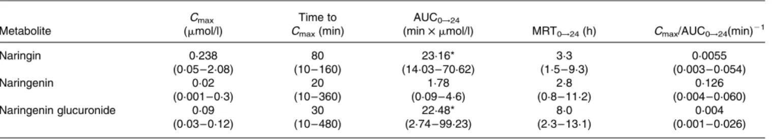
The following pharmacokinetic parameters corresponding to each of the flavanone metabolites (Cmax, AUC0!24, MRT0!t, time to Cmax and Cmax/AUC0!24) are summarised in Table 1. Values are expressed as median and range, together with the results of the statistical analysis carried out. There were no significant statistical differences between most of the pharmacokinetic parameters corresponding to nar-ingin, naringenin and naringenin glucuronide. This was not the case, however, with AUC0!24, whose values showed signifi-cant differences between naringenin and naringin, and between naringenin and naringenin glucuronide. Naringenin had the lowest extended exposure (AUC0!24) in plasma, whereas naringin in its native form presented the highest maximum plasma concentration (Cmax) values, as well as the highest extended exposure (AUC0!24). As shown in Table 1, naringenin glucuronide presented the highest MRT (MRT0!24; 8 h), followed by naringin (3.3 h) and naringenin (2.8 h). However, interindividual variations in the pharmaco-kinetic parameters values were observed.
Discussion
The metabolic forms that reach the peripheral circulation and tissues may be different from those present in foods, and their biological activity is consequently likely to be different (Kroon et al. 2004). The identification and measurement of flavonoid conjugates are key prerequisites to understand the role of these compounds, since these are the forms that will reach tissues and exert their biological effect. Previous studies of naringin (naringenin-7-O-rhamnoglucoside) metabolism have suggested that sugar moiety cleavage, by gut microflora a-rhamonosidases, is the first step of this pathway, leading to the formation of naringenin, which undergoes rapid glucuroni-dation or sulphatation in the intestine or liver (Fuhr & Kum-mert, 1995; Felgines et al. 2000; Scalbert & Williamson, 2000). Most studies have applied enzymatic hydrolysis with sulphatase and glucuronidase in order to identify the aglycone naringenin, and thus the individual metabolic profiles are lost during the hydrolysis procedure (Fuhr & Kummert, 1995; Ishii et al. 1996, 1997; Hollman et al. 1999; Erlund et al. 2001; Bugianesi et al. 2002; Manach et al. 2003; Zhang & Brodbelt, 2004).
In the present study, a method without prior sample hydrolysis and based on LC-MS/MS technique has been developed. The method is capable of identifying non-trans-formed naringin and flavanone metabolites in dog plasma after the oral administration of 70 mg citrus flavanone con-tained in a grapefruit extract. The results of this study corro-borate the suggestion that both flavonols and flavanone glycosides can be absorbed as glycosides. However, narirutin (naringenin-7-O-rutinoside), a flavonoid rutinoside also pres-ent in the grapefruit extract, could not be detected, probably because of its sugar moiety, which, as previously reported, affects flavonoid absorption (Erlund et al. 2000; Olthof et al. 2000; Rowland et al. 2000; Arts et al. 2004; Manach et al. 2004; Nielsen et al. 2006).
In recent years, a greater understanding of flavonoid absorp-tion and metabolism has been achieved. Flavonoid glycosides are thought to reach the small intestine intact, and it is believed that they may require deglycosidation for absorption across the intestine (Scalbert & Williamson, 2000; Manach et al. 2004). The presence of naringin in the plasma demon-strates that the deglycosidation of naringin is not always necessary for its absorption. Previous studies (Fang et al. 2006) have administered naringin as a pure compound, whereas in the present study citrus flavanones were adminis-tered in the form of a grapefruit extract (as it occurs in nature), in a dose equivalent to that of half a grapefruit. The influence of food matrices must always be considered when interpreting results; it should be borne in mind that they could have been different had pure compounds been used. Other compounds present in the extract could affect the mech-anisms involved in the absorption, distribution and elimination of the flavanones studied.
Previous studies using enzymatic hydrolysis have reported plasma concentrations of 1·3 – 2·2 mmol/l hesperitin metab-olites with an intake of 130 – 220 mg given as orange juice (Manach et al. 2003) and up to 6 mmol/l naringenin metab-olites with 200 mg ingested as grapefruit juice (Erlund et al. 2001). Fang et al. (2006) have reported plasma concentrations of 3·8, 0·23 and 43·58 mg/ml for naringin, naringenin and nar-ingenin glucuronide respectively after an oral administration of 746·7 mg/kg naringin as a pure compound. In the present study, 70 mg flavanones given as a grapefruit extract were orally administered, and several pharmacokinetic parameters were calculated for naringin and for each of the flavanone metabolites that have been found in dog plasma. The AUC
Table 1. Pharmacokinetic parameters of the grapefruit flavanone naringin and its metabolites (naringenin and naringenin glucuronide) in beagles after an oral intake of grapefruit extract
(Median values and ranges for ten determinations)
Metabolite Cmax (mmol/l) Time to Cmax(min) AUC0!24
(min £ mmol/l) MRT0!24(h) Cmax/AUC0!24(min)21
Naringin 0·238 80 23·16* 3·3 0·0055 (0·05 – 2·08) (10 – 160) (14·03 – 70·62) (1·5 – 9·3) (0·003 – 0·054) Naringenin 0·02 20 1·78 2·8 0·126 (0·001 – 0·3) (10 – 360) (0·09 – 4·6) (0·8 – 11·2) (0·004 – 0·060) Naringenin glucuronide 0·09 30 22·48* 8·0 0·004 (0·03 – 0·12) (10 – 480) (2·74 – 99·23) (2·3 – 13·1) (0·001 – 0·026)
MRT, mean residence time.
parameter until the final experimental time (AUC0!24) was used to compare the pharmacokinetic parameters because AUCextvalues were not less than 20 % in all cases.
The differences between naringenin and naringin and between naringenin and naringenin glucuronide in terms of AUC0!24 values suggest that the aglycone naringenin had the lower extended exposure. As shown in Table 1, naringenin glucuronide presented the highest MRT (MRT0!24), which indicates that this metabolite remains in the body for a longer period of time.
Similar interindividual variations have previously been reported, suggesting that these variations were caused by differ-ences in the gastrointestinal microbiota responsible for the hydrolysis of naringin (Erlund et al. 2000; Rowland et al. 2000). Interindividual variation is an important factor that must always be taken into consideration when performing diet-ary assessment studies.
The ratio Cmax/AUC0!24 represents the rate of absorption, and, as expected, the aglycone naringenin was the most rapidly absorbed, probably owing to its greater ability to cross the lipid cell membrane (Mohsen et al. 2004). In contrast, naringin and naringenin glucuronide reached their peak concentration at 80 and 30 min, respectively, whereas naringenin reached its Cmax at 20 min. Although aglycones are known to be absorbed more rapidly, the aglycone absorption was detected here at a much earlier time (20 min) than that reported by Bugianesi et al. (2002), who found that Cmaxwas reached 2 h after the ingestion of tomato paste (which contains naringenin aglycone) in men. This result could be due to differences in the species and to the influence of the food matrix.
Three different flavanone forms were found in dog plasma, thus demonstrating grapefruit flavanone absorption after an oral intake of grapefruit extract: naringin in its native form, naringenin and naringenin glucuronide. These results confirm the bioavailability of grapefruit flavanones and their metab-olites in beagles after the oral administration of 70 mg grape-fruit flavanone. The aglycone naringenin showed the highest rate of absorption but the lowest extended exposure and lowest MRT in the body. Both naringin and naringenin glucur-onide showed high extended exposure values, whereas narin-genin glucuronide presented the highest values for MRT, remaining in the body for approximately 8 h. This study demonstrates the presence of grapefruit flavanone and its metabolites in dog plasma, and the data could provide a model for further studies, although a greater number of sub-jects would be necessary to support these results.
Acknowledgements
The authors thank the R&D Department, Affinity Pet-care, Barcelona, without whose support this project would not have been possible. M. L. M. B. is also grateful to the Danone Institute for its partial contribution to the study through her pre-doctoral fellowship.
References
Arts ICW, Sesink ALA, Faassen-Peters M & Hollman PCH (2004) The type of sugar moiety is a major determinant of the small intes-tinal uptake and subsequent biliary excretion of dietary quercetin glycosides. Br J Nut 91, 841 – 847.
Benavente-Garcı´a O, Castillo J, Marin F, Ortun˜o A & Del Rı´o J (1997) Uses and properties of citrus flavonoids. J Agric Food Chem 45, 4505 – 4514.
Bugianesi R, Catasta G, Spigno P, D’Uva A & Maiani G (2002) Naringenin from cooked tomato paste is bioavailable in men. J Nutr 132, 3349 – 3352.
Committee on Care and use of Laboratory Animals (1985) Guide for the Care and Use of Laboratory Animals. Washington, DC: Institute of Laboratory Animals Resources, National Research Council.
Dahan A & Altman H (2004) Food-drug interaction: grapefruit juice augments drug bioavailability-mechanism, extent and relevance. Eur J Clin Nutr 58, 1 – 9.
Day AJ, Mellon F, Barron D, Sarrazin G, Morgan MRA & William-son G (2001) Human metabolism of dietary flavonoids: identifi-cation of plasma metabolites of quercetin. Free Radic Res 35, 941 – 952.
Day AJ & Williamson G (2001) Biomarkers of exposure to dietary flavonoids: a review of the current evidence for the identification of quercetin glycosides in plasma. Br J Nutr 86, S105 – S110. Erlund I, Kosonen T, Alfthan G, Ma¨enpa¨a¨ J, Perttunen K, Kenraali
J, Parantainen J & Aro A (2000) Pharmacokinetics of quercetin from quercetin aglycone and rutin in healthy volunteers. Eur J Clin Pharmacol 56, 545 – 553.
Erlund I, Meririnne E, Alfthan G & Aro A (2001) Plasma kinetics and urinary excretion of the flavanones naringenin and hesperetin in humans after ingestion of orange juice and grapefruit juice. J Nutr 131, 235 – 241.
Fang T, Wang Y, Ma Y, Su W, Bai Y & Zhao P (2006) A rapid LC/ MS/MS quantitation assay for naringin and its two metabolites in rat’s plasma. J Pharm Biomed Anal 40, 454 – 459.
Felgines C, Texier O, Morand C, Manach C, Scalbert A, Re´gerat F & Re´me´sy C (2000) Bioavailability of the flavanone naringenin and its glycosides in rats. Am J Physiol Gastrointest Liver Physiol 279, G1148 – G1154.
Fuhr U & Kummert AL (1995) The fate of naringin in humans: a key to grapefruit juice-drug interactions? Clin Pharmacol Ther 58, 365 – 373.
Fuhr U, Muller-Peltzer H, Kern R, Lopez-Rojas P, Junemann M, Harder S & Staib AH (2002) Effects of grapefruit juice and smok-ing on verapamil concentrations in steady state. Eur J Clin Phar-macol 58, 45 – 53.
Gao K, Henning SM, Niu YT, Youssefian AA, Seeram NP, Xu AL & Heber D (2006) The citrus flavonoid naringenin stimulates DNA repair in prostate cancer cells. J Nutr Biochem 17, 89 – 95. Graefe EU, Wittig J, Mueller S, Riethling AK, Uehleke B, Drewelow
B, Pforte H, Jacobasch G, Derendorf H & Veit M (2001) Pharma-cokinetics and bioavailability of quercetin glycosides in humans. J Clin Pharmacol 41, 492 – 499.
Harada M, Kan Y, Naoki H, Fukui Y, Kageyama N, Nakai M, Miki W & Kiso Y (1999) Identification of the major antioxidative metabolites in biological fluids of the rat with ingested (þ )-cate-chin and (2 )-epicate)-cate-chin. Biosci Biotechnology Biochem 63, 973 – 977.
Hollman PC, Bijsman MN, van Gameren Y, Cnossen EP, de Vries J & Katan MB (1999) The sugar moiety is a major determinant of the absorption of dietary flavonoid glycosides in man. Free Rad Res 31, 569 – 573.
Hollman PCH, Devries JHM, Vanleeuwen D, Mengelers MJB & Katan MB (1995) Absorption of dietary quercetin glycosides and quercetin in healthy ileostomy volunteers. Am J Clin Nutr 62, 1276 – 1282.
Hollman PCH & Katan MB (1997) Absorption, metabolism and health effects of dietary flavonoids in man. Biomed Pharmacother 51, 305 – 310.
Ishii K, Furuta T & Kasuya Y (1996) Determination of naringin and naringenin in human plasma by high-performance liquid
chromatography. J Chrom B Analyt Technol Biomed Life Sci 683, 225 – 229.
Ishii K, Furuta T & Kasuya Y (1997) Determination of naringin and naringenin in human urine by high-performance liquid chromatog-raphy utilizing solid-phase extraction. J Chrom B Analyt Technol Biomed Life Sci 704, 299 – 305.
Ishii K, Furuta T & Kasuya Y (2000) Mass spectrometric identifi-cation and high-performance liquid chromatographic determination of a flavonoid glycoside naringin in human urine. J Agric Food Chem 48, 56 – 59.
Kiser M & Dolan JW (2004) Selecting the best curve fit. LC-GC Europe 17, 138 – 143.
Kroon PA, Clifford MN, Crozier A, Day AJ, Donovan JL, Manach C & Williamson G (2004) How should we assess the effects of exposure to dietary polyphenols in vitro? Am J Clin Nutr 80, 15 – 21.
Lilja JJ, Neuvonen M & Neuvonen PJ (2004) Effects of regular con-sumption of grapefruit juice on the pharmacokinetics of simvasta-tin. Br J Clin Pharmacol 58, 56 – 60.
Macheix JJ, Fleuriet A & Billot J (1990) Fruits’ Flavonoids. Boca Raton, FL: CRC Press.
Manach C, Morand C, Gil-Izquierdo A, Bouteloup-Damange C & Remesy C (2003) Bioavailability in humans of the flavanones hesperidin and narirutin after the ingestion of two doses of orange juice. Eur J Clin Nutr 57, 235 – 242.
Manach C, Scalbert A, Morand C, Remesy C & Jimenez L (2004) Polyphenols: food sources and bioavailability. Am J Clin Nutr 79, 727 – 747.
Mata-Bilbao ML, Andre´s-Lacueva C, Ja´uregui O & Lamuela-Raven-to´s RM (2007) Determination of flavonoids in a citrus fruit extract by LC-DAD and LC-MS. Food Chem 101, 1742 – 1747.
Middleton E & Kandaswami C (1994) The impact of plant flavonoids on mammalian biology: implications for immunity, inflammation and cancer. In The Flavonoids: Advances in Research Since 1986, pp. 619 – 652 [JB Harborne, editor]. London: Chapman & Hall. Mohsen MA, Marks J, Kuhnle G, Rice-Evans C, Moore K, Gibson G,
Debnam E & Srai K (2004) The differential tissue distribution of the citrus flavanone naringenin following gastric instillation. Free Rad Res 38, 1329 – 1340.
Montanari A, Chen J & Widmer W (1998) Citrus flavonoids: a review of past biological activity against disease. Discovery of new flavo-noids from Dancy tangerine cold pressed peel oil solids and leaves. In Flavonoids in the Living System, pp. 103 – 116 [J Manthey and B Busling, editors]. New York: Plenum Press.
Murphy AT, Bonate PL, Kasper SC, Gillespie TA & Delong AF (1994) Determination of xanomeline in human plasma by ionspray tandem mass-spectrometry. Mass Spectrom 23, 621 – 625. Natsume M, Osakabe N, Oyama M, Sasaki M, Baba S, Nakamura
Y, Osawa T & Terao J (2003) Structures of (2 )-epicatechin glu-curonide identified from plasma and urine after oral ingestion of (2 )-epicatechin: differences between human and rat. Free Radic Biol Med 34, 840 – 849.
Nielsen ILF, Chee WSS, Poulsen L, Offord-Cavin E, Rasmussen SE, Frederiksen H, Enslen M, Barron D, Horcajada MN & Williamson G (2006) Bioavailability is improved by enzymatic modification of the citrus flavonoid hesperidin in humans: a randomized, double-blind, crossover trial. J Nutr 136, 404 – 408.
Okushio K, Suzuki M, Matsumoto N, Nanjo F & Hara Y (1999) Identification of (2 )-epicatechin metabolites and their metabolic fate in the rat. Drug Metab Dispos 27, 309 – 316.
O’Leary KA, Day AJ, Needs PW, Sly WS, O’Brien NM & Williamson G (2001) Flavonoid glucuronides are substrates for human liver [beta]-glucuronidase. FEBS Lett 503, 103 – 106.
Olthof MR, Hollman PCH, Vree TB & Katan MB (2000) Bioavail-abilities of quercetin-3-glucoside and quercetin-40-glucoside do not differ in humans. J Nutr 130, 1200 – 1203.
Paine MF, Criss AB & Watkins PB (2004) Two major grapefruit juice components differ in intestinal CYP3A4 inhibition kinetic and binding properties. Drug Metab Dispos 32, 1146 – 1153.
Paine MF, Criss AB & Watkins PB (2005) Two major grapefruit juice components differ in time to onset of intestinal CYP3A4 inhibition. J Pharm Exp Ther 312, 1151 – 1160.
Roura E, Andres-Lacueva C, Jauregui O, Badia E, Estruch R, Izquierdo-Pulido M & Lamuela-Raventos RM (2005) Rapid liquid chromatography tandem mass spectrometry assay to quantify plasma (2 )-epicatechin metabolites after ingestion of a standard portion of cocoa beverage in humans. J Agric Food Chem 53, 6190 – 6194.
Rowland IR, Wiseman H, Sanders TAB, Adlercreutz H & Bowey EA (2000) Interindividual variation in metabolism of soy isoflavones and lignans: influence of habitual diet on equal production by the gut microflora. Nutr Cancer 36, 27 – 32.
Scalbert A & Williamson G (2000) Dietary intake and bioavailability of polyphenols. J Nutr 130, 2073S – 2085S.
Schindler R & Mentlein R (2006) Flavonoids and vitamin E reduce the release of the angiogenic peptide vascular endothelial growth factor from human tumor cells. J Nutr 136, 1477 – 1482. Shimoi K, Okada H, Furugori M, Goda T, Takase S, Suzuki M, Hara
Y, Yamamoto H & Kinae N (1998) Intestinal absorption of luteolin and luteolin 7-O-beta-glucoside in rats and humans. FEBS Lett 438, 220 – 224.
Simio K, Saka N, Nozawa R, Sato M, Amano I, Nakayama T & Kinae N (2001) Deglucuronidation of a flavonoid, luteolin mono-glucuronide, during inflammation. Drug Metab Dispos 29, 1521 – 1524.
Spencer PE, Schroeter H, Crossthwaithe AJ, Kuhnle G, Williams RJ & Rice-Evans C (2001b) Contrasting influences of glucuronidation and O-methylation of epicatechin on hydrogen peroxide-induced cell death in neurons and fibroblasts. Free Radic Biol Med 31, 1139 – 1146.
Spencer JP, Schroeter H & Rice-Evans C (2001a) Epicatechin and its in vivo metabolite, 30-O-methyl epicatechin, protect human fibro-blasts from oxidative-stress-induced cell death involving caspase-3 activation. Biochem caspase-354, 49caspase-3 – 500.
Swezey R, Aldridge D, Le Valley E, Crowell J, Hara Y & Green C (2003) Absorption, tissue distribution and elimination of 4-[3H]-epigallocatechin gallate in beagle dogs. Int J Toxicol 22, 187 – 193. Urpı´-Sarda` M, Ja´uregui O, Lamuela-Ravento´s RM, Jaeger W, Miksits M, Covas MI & Andre´s-Lacueva C (2005) Uptake of diet resvera-trol into the human low-density lipoprotein. Identification and quantification of resveratrol metabolites by liquid chromatography coupled with tandem mass spectrometry. Anal Chem 77, 3149 – 3155.
US Department of Health and Human Services (2001) Guidance for Industry. Bioanalytical Method Validation. Washington, DC: US Department of Health and Human Services, Food and Drug Administration.
Zhang J & Brodbelt J (2004) Screening flavonoid metabolites of nar-ingin and narirutin in urine after human consumption of grapefruit juice by LC-MS and LC-MS/MS. Analyst 129, 1227 – 1233. Zhang Y, Hendrich S & Murphy PA (2003) Glucuronides are the
main isoflavone metabolites in women. J Nutr 133, 399 – 404. Zhang Y, Song TT, Cunnick JE, Murphy PA & Hendrich S (1999)
Daidzein and genistein glucuronides in vitro are weakly estrogenic and activate human natural killer cells at nutritionally relevant con-centrations. J Nutr 129, 399 – 495.