Abstract.
Background: Metastatic cancer is the most common
malignant disease of the skeletal system. Traditionally,
conventional fractionated external beam radiotherapy has been
the treatment of choice. Recently, minimally invasive surgical
techniques (MISS) have been added to the therapeutic
armamentarium. The purpose of our study was to assess the
effectiveness and safety of Radiofrequency Heat Ablation and
Vertebroplasty in the treatment of neoplastic Vertebral
Compressive Fractures (VCF). The aim of radiofrequency heat
ablation is to destroy the tumor tissue before stabilizing the
vertebra through the intrasomatic injection of cement. Patients
and Methods: We treated patients with unremitting pain over
spine, in absence of symptomatic spinal cord or roots compression
and refractory to conventional therapeutic options such as
radiation therapy, chemotherapy, surgery and use of analgesics.
Results: The method demonstrated swift pain relief associated
with an evident augmentation in the weight-bearing resistance.
Conclusion: The association of Radiofrequency Heat Ablation
and Vertebroplasty is an effective, simple and safe treatment of
vertebral collapse consequent to metastases.
Approximately 70% of patients with cancer have evidence of
metastatic disease at the time of their deaths (1,2). The spinal
column is the most common location among osseous sites for
metastatic deposits (3). Spinal involvement may occur in up to
40% of patients with cancer. Spinal cord compression from
epidural metastases occurs in 5-10% of cancer patients and in
up to 40% of patients with pre-existing non-spinal bone
metastases (4-9). Of those with body spinal disease, 10-20%
develop symptomatic spinal cord compression (8,10,11). The
thoracic spine is the most common site of disease (70%),
followed by the lumbar spine (20%) and cervical spine (10%)
(7,8,12). Metastatic spinal disease can arise from one of three
locations: the vertebral column (85%), the paravertebral region
(10-15%) and, rarely, the epidural or subarachnoid/
intramedullary space itself (<5%) (7,8,12). The posterior half
of the vertebral body is usually involved first, with the anterior
body, lamina and pedicles invaded later (13). Multiple lesions
at non-contiguous levels occur in 10-40% of cases (7,8,12,14).
Approximately 50% of metastases arise from one of these
primary types of cancer: breast, lung, prostate or melanoma
(7). These commonly cause spinal metastases in 74.3%, 44.9%,
90.5% and 54.5% of patients, respectively (4). These are
followed by renal cancer, gastrointestinal cancer, thyroid
cancer, sarcoma and the lymphoreticular malignancies:
lymphoma and multiple myeloma.
Painful bone metastases commonly occur in advanced
cancer patients. They are difficult to manage because of pain,
reduction in mobility and performance status. Possible
mechanisms that may cause pain from bone metastases
include: stimulation of nerve endings in the endosteum
resulting from the release of chemical agents from the
destroyed bone tissue such as prostaglandins, bradykinin,
substance P, or histamine; stretching of the periosteum by
increasing size of the tumor; fractures; tumor growth into
surrounding nerves and tissues. Few of these mechanisms are
supported by definitive data. Stimulation of nerve endings in
the endosteum by chemical agents released from the
destroyed bone tissue is probably the main cause of bone pain
from small metastases; as metastases enlarge, stretching of
the periosteum additionally contributes to the pain (15).
Vertebral fractures are generated when the combination
of the axial and rotational charges on the spine exceed the
resistance offered by the vertebral body (16). The Vertebral
Compressive Fracture (VCF) is defined as a reduction in
height, which must be at least 20% beyond its initial
dimensions. In relation to its severity, it is distinguished as
mild 20-25%, moderate 25-40%, or severe>40% (17). The
presence of a vertebral fracture puts the individual in the
Correspondence to: Salvatore Masala, MD, Department ofDiagnostic Radiology, " Tor Vergata " University General Hospital, 81 Oxford Street – 00133, Rome, Italy. Tel: +039-0620902401, Fax: +039-0620902404, e-mail: [email protected]
Key Words: Metastases, vertebral compression fracture, interventional radiology, radiofrequency heat ablation, vertebroplasty.
Radiofrequency Heat Ablation and Vertebroplasty
in the Treatment of Neoplastic Vertebral Body Fractures
SALVATORE MASALA
1, MARIO ROSELLI
2, FRANCESCO MASSARI
1, ROBERTO FIORI
1,
ANTONIO URSONE
1, EMANUELA FOSSILE
2, ANASTASIA LAUDISI
2and GIOVANNI SIMONETTI
1Departments of
1Interventional Radiology and
2Medical Oncology,
University of Rome " Tor Vergata ", 81 Oxford Street – 00133, Rome, Italy
following years at a 5 times greater risk of having a second
VCF, which in almost 20% of the cases occurs within a
one-year period (18-20).
Complications of VCF include, in more than a third of
these patients, chronic pain, lower quality of lifestyle,
progressive kyphosis of the spine and reduction of volumes
of the thoracic and abdominal cavities. In consequence to
this it generates pulmonary compression, with a reduction
of vital capacity (VC) to 9% for each level fractured and of
"forced expiratory volume to 1 second" (FEV1), as well as
gastrointestinal dysfunction. (18,21-24).
Patients and Methods
Indications. Vertebroplasty is a technique increasingly utilized in the field of VCF, based on primary and secondary neoplasm. It is extremely useful in the treatment of aggressive vertebral hemangiomas, bone osteolytic metastases and destructive multiple myeloma, which is associated with VCF in 55-70% of the cases (25). Contraindications. The contraindications of the combined procedure consist of osteoblastic tumors, fractures with retropulsion of the fragments within neural foramen, spread of tumor within the epidural space, local infection (osteomyelitis, discitis or epidural abscess), coagulative disorders, pain not related to vertebral collapse, steady asymptomatic fractures, and tumor involvement or missing integrity of pedicles or joint facets (26-29).
Technique. All the patients were subjected to a thorough clinical examination in order to determine the symptomatic vertebral level, which is revealed by acute pain and tenderness over the spine at or near the level of radiographic compression deformity (30).
Radiographic examinations and CT were preliminarily performed to evaluate the location, severity and the extension of the collapsed vertebra, as well as to ascertain the visibility of the vertebral pedicles and the integrity of the posterior wall. Magnetic resonance is fundamental in the pre-treatment diagnosis and in the follow-up to evaluate the success of the procedure. The presence of intraspongious edema, particularly in fat suppression sequences, testifies to a recent fracture.
The study was performed under the appropriate institutional ethics approvals and in accordance with the principles embodied in the Declaration of Helsinki. Written informed consent was obtained from each participating subject.
The whole procedure of Radiofrequency (RF) Heat Ablation and Vertebroplasty is performed under CT-fluoroscopy guidance and after administration of local anaesthesia. The patient is placed in prone position with two rolls of soft material inserted transversally, respectively under the chest and the pelvis, to obtain the maximum extension of the spine (17,21,26,30,31). After confining the vertebra and its corresponding pedicles to be treated, a small cutaneous incision is made in the dorsal or lumbar area. Then a bone biopsy needle of 11-Gauge is introduced through the posterior portion of the pedicles, sloping anteriorly, medially and caudally into the tumor (17,21,26,30,31). The access to the vertebral body is normally unilateral with transpedicular pathway, however one can opt for an inter-costo-vertebral entrance into the thoracic levels or posterolateral for the lumbar ones (32).
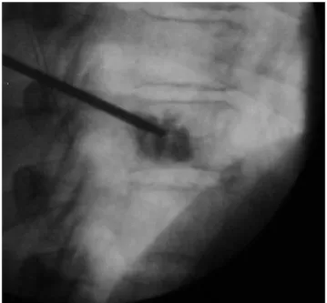
Once the exact position of the needle had been verified, a 19-Gauge needle electrode (MIRAS-RC, Invatec) and a thermocouple were introduced coaxially through the inserted Radiofrequency heat ablation and vertebroplasty, fluoroscopic images
(Figures 1-2):
Figure 1. Needle electrode tip deployed (white arrow) into the central part of the lesion, generates a well-defined area of tumor necrosis.
Figure 2. PMMA is injected through the needle into the vertebral body to stabilize the osteolytic lesion.
cannula into the central part of the lesion (Figure 1). After unsheathing the spiral electrode tine, which opened to a diameter of 9 mm and length of 10 mm in the metastasis, the needle was connected with a radiofrequency generator. The radiofrequency heat ablation started at an energy level of 15 W. The deployed energy was increased by 5W every 2 min, up to 25 W. Control CT scans revealed microbubble formation in the treated area, indicating tumor necrosis (33).
Meanwhile the cement was prepared by combining liquid monomer and powder cement polymer; everything was amalgamated until it formed into a paste with doughy viscosity. The Polymethylmethacrylate (PMMA) was charged into a dedicated device and injected through the vertebroplasty needle into the vertebral body under continuous fluoroscopic guidance (Figure 2). Control CT scans revealed a homogeneous distribution in the tumor necrosis. To complete the procedure, the needle was extracted, the cutaneous incision was sutured and the patient was instructed to remain in bed, positioned supine, for the following 4 h (34).
The most commonly encountered complication was localized pain and tenderness at the needle sites in the first 72 h after the procedure, usually caused by local bruising or hematoma, which can be resolved with mild analgesics (30). The length of the process for each vertebra was around 35-45 min. A traditional radiographic and CT inspection is performed immediately after the procedure to evaluate the results obtained (Figure 3). In general, the patient was dismissed on the following day.
Results
From January to May 2004, we treated 3 patients (F=2,
M=1; ages ranged from 63 to 82 years, mean age 72.3) with
metastatic vertebral collapse. Respectively, 2 (F=2) had
fractures consequent to breast cancer and 1 (M=1) due to
lung cancer. All patients underwent a single level treatment
(Dorsal D8 n= 1, D12 n=1; Lumbar L3 n=1).
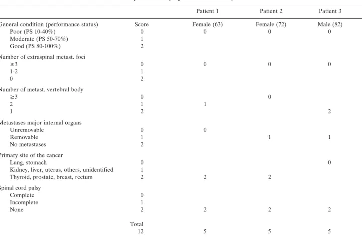
Tokuhashi et al. proposed a preoperative prognostic
scoring system. Six parameters employed in the assessment
system are: 1) general medical condition, 2) number of
extraspinal metastases, 3) number of vertebral metastases,
4) metastases to the major internal organs (lungs, liver,
kidneys and brain), 5) primary site of cancer and 6) severity
of spinal cord palsy. Each parameter ranged from 0 to 2
points. Patients who have a total score ≥ 9 survive an
average of 12 months or more and are indicated for
excisional procedure; whereas those with a score ≤ 5
generally die within 3 months or less and are candidates for
a palliative operation. No recommendations were made for
patients with a total score of 6 to 8 points (35) (Table I).
The procedure was successfully performed in our
patients, whose improvements were swift and persistent in
reducing all symptoms, decreasing from an average of 8.6
points of VAS to 2.6 (VAS of Huskisson = Visual analog
scale, pain score with points assigned subjectively from
patient pre- and post-procedure in a range between 0
absence of grief and 10 maximum pain). Resistance was
considerably increased. We did not find any condition of
extravasations of PMMA in the epidural or foraminal sites
with marrow or radicular compression.
Discussion
Traditional therapies to control pain and to treat bone
metastases include radiation therapy, chemotherapy,
hormonotherapy (prostate, breast), analgesics and, recently,
pamidronate (biphosphonate) has been recognized as useful in
osteolytic lesions (15). These conventional therapies, with their
well known drawbacks and side-effects, provide reasonable
pain relief, obtaining variable success rates. Furthermore,
radiotherapy and chemotherapy require a two- to four-week
delay to reach efficiency. In some cases, radiotherapy may not
be an option because of radiation insensitivity of the tumor or
high radiation doses previously delivered. Furthermore,
chemotherapy may not be recommended because of its
toxicity. Intolerable analgesic-related side-effects may develop
with increased doses (15,36,37). Surgical resection is
considered the only potentially curative option for secondary
malignant bone tumors. However, in secondary bone tumors
few patients are surgical candidates. Minimally invasive
techniques with quick pain relief can be an alternative option
to conventional treatments (15).
Radiofrequency ablation is one of the most promising
thermal ablation techniques for the treatment of
non-resectable tumors. The use of radiofrequency ablation was first
reported in 1990 for the treatment of hepatic tumors (15).
An alternating electric current operated in the range of
radiofrequency can produce a focal thermal injury in living
Figure 3. Post-procedure control of PMMA distribution, axial CT-image.tissue. Shielded needle electrodes are used to concentrate the
energy in selected tissue. The tip of the electrode conducts the
current, that oscillates in the range of high frequency (200-1.200
kHz), which causes local ionic agitation and subsequent
frictional heat, which leads to localized coagulation necrosis.
Schematically, a closed-loop circuit is created by placing a
generator, a large dispersive electrode (ground pad), a patient
and a needle electrode in series (15). The aim of performing
radiofrequency heat ablation before vertebroplasty was to
destroy tumor tissue and to thrombose the paravertebral and
intravertebral venous plexus and, thereby, minimize
procedure-related complications. Major benefits of radiofrequency heat
ablation are the immediate cell death and the accurate control
of lesion size with an imaging-guided procedure (33). The
purpose of vertebroplasty was then to stabilize the vertebra.
Necrotizing tumor tissue by radiofrequency heat ablation
optimizes cement distribution, facilitated by changes in tumor
consistency as a result of thermal alterations (33). Percutaneous
injection of PMMA bone cement into the vertebral body,
described for the first time by Galibert et al. in 1984 as
"Vertebroplasty", and successfully applied to the treatment of
C2 aggressive hemangiomas, is also used on VCF secondary to
osteolytic tumors and osteoporosis (38). Pulmonary embolism
and spinal compressions, that can derive from the extrusion of
cement, are rare potential complications.
In our study, although limited, vertebroplasty demonstrated
itself to be an effective, simple and safe method for the
treatment of vertebral compression fractures, providing a
precocious and long-lasting pain relief and an evident increase
in the resistance of the vertebral body.
Acknowledgements
The authors thank Invatec (Innovative Technologies) for providing the equipment and technical support for this study.
References
1 Harrington K: Metastatic tumors of the spine: diagnosis and treatment. J Am Acad Orthop Surg 1: 76-86, 1993.
2 Klimo P Jr and Schmidt MH: Surgical management of spinal metastases. Oncologist 9(2): 188-196, 2004.
3 Bohm P and Huber J: The surgical treatment of bony metastases of the spine and limbs. J Bone Joint Surg Br 84: 521-529, 2002.
Table I. Scoring system for preoperative evaluation of metastatic spine tumor prognosis (35).
Evaluation system for the prognosis of metastatic spine tumors
Patient 1 Patient 2 Patient 3
General condition (performance status) Score Female (63) Female (72) Male (82)
Poor (PS 10-40%) 0 0 0 0
Moderate (PS 50-70%) 1
Good (PS 80-100%) 2
Number of extraspinal metast. foci
≥3 0 0 0 0
1-2 1
0 2
Number of metast. vertebral body
≥3 0 0
2 1 1
1 2 2
Metastases major internal organs
Unremovable 0 0
Removable 1 1 1
No metastases 2
Primary site of the cancer
Lung, stomach 0 0
Kidney, liver, uterus, others, unidentified 1
Thyroid, prostate, breast, rectum 2 2 2
Spinal cord palsy
Complete 0
Incomplete 1
None 2 2 2 2
Total
4 Wong DA, Fornasier VL and MacNab I: Spinal metastases: the obvious, the occult, and the impostors. Spine 15(1): 1-4, 1990. 5 Healey JH and Brown HK: Complications of bone metastases:
surgical management. Cancer 88(12 Suppl): 2940-2951, 2000. 6 Bilsky MH, Lis E, Raizer J, Lee H and Boland P: The diagnosis
and treatment of metastatic spinal tumor. Oncologist 4(6): 459-469, 1999.
7 Byrne TN: Spinal cord compression from epidural metastases. N Engl J Med 327(9): 614-619, 1992.
8 Gerszten PC and Welch WC: Current surgical management of metastatic spinal disease. Oncology (Huntingt) 14(7): 1013-1024, 2000.
9 Barron Kd, Hirano A, Araki S and Terry Rd: Experiences with metastatic neoplasms involving the spinal cord. Neurology 9(2): 91-106, 1959.
10 Schaberg J and Gainor BJ: A profile of metastatic carcinoma of the spine. Spine 10(1): 19-20, 1985.
11 Lada R, Kaminski HJ and Ruff R: Metastatic spinal cord compression. In: Vecht C, ed. Neuro-oncology Part III. Neurological Disorders in Systemic Cancer. Amsterdam: Elsevier Biomedical Publishers, 1997:167-189.
12 Gilbert RW, Kim JH and Posner JB: Epidural spinal cord compression from metastatic tumor: diagnosis and treatment. Ann Neurol 3(1): 40-51, 1978.
13 Adams M and Sonntag VKH: Surgical treatment of metastatic cervical spine disease. Contemp Neurosurg 23: 1-5, 2001. 14 Cook AM, Lau TN, Tomlinson MJ et al: Magnetic resonance
imaging of the whole spine in suspected malignant spinal cord compression: impact on management. Clin Oncol (R Coll Radiol) 10: 39-43, 1998.
15 Gangi A, Guth S, Imbert JP, Marin H, Jeung MY and Wong LLS: Radiofrequency ablation of bone metastases. ECR 2003 Cum Laude.
16 Mathis JM, Barr JD, Belkoff SM, Barr MS, Jensen ME and Deramond H: Percutaneous vertebroplasty: a developing standard of care for vertebral compression fractures. Am J Neuroradiol 22(2): 373-381, 2001.
17 McKiernan F, Jensen R and Faciszewski T: The dynamic mobility of vertebral compression fractures. J Bone Miner Res 18(1): 24-29, 2003.
18 Zoarski GH, Snow P, Olan WJ, Stallmeyer MJ, Dick BW, Hebel JR and De Deyne M: Percutaneous vertebroplasty for osteoporotic compression fractures: quantitative prospective evaluation of long-term outcomes. J Vasc Interv Radiol 13(2 Pt 1): 139-148, 2002.
19 Belkoff SM, Mathis JM, Fenton DC, Scribner RM, Reiley ME and Talmadge K: An ex vivo biomechanical evaluation of an inflatable bone tamp used in the treatment of compression fracture. Spine 26(2): 151-156, 2001.
20 Higgins KB, Harten RD, Langrana NA and Reiter MF: Biomechanical effects of unipedicular vertebroplasty on intact vertebrae. Spine 28(14): 1540-1548, 2003.
21 Ahrar K, Schomer DF and Wallace MJ: Kyphoplasty for the treatment of vertebral compression fractures. Semin Intervent Radiol 19(3): 235-243, 2002.
22 Amar AP, Larsen DW, Esnaashari N, Albuquerque FC, Lavine SD and Teitelbaum GP: Percutaneous transpedicular polymethylmethacrylate vertebroplasty for the treatment of spinal compression fractures. Neurosurgery 49(5): 1105-1114, 2001.
23 Garfin SR, Yuan HA and Reiley MA: New technologies in spine: kyphoplasty and vertebroplasty for the treatment of painful osteoporotic compression fractures. Spine 26(14): 1511-1515, 2001. 24 Coumans JV, Reinhardt MK and Lieberman IH: Kyphoplasty for vertebral compression fractures: 1-year clinical outcomes from a prospective study. J Neurosurg 99(1 Suppl): 44-50, 2003. 25 Masala S, Cesaroni A, Sergiacomi G, Fiori R, Massari F, Manenti G, Nardi PV and Simonetti G: Percutaneous kyphoplasty: New treatment for painful vertebral body fractures. In Vivo 18(2): 149-153, 2004.
26 Fourney DR, Schomer DF, Nader R, Chlan-Fourney J, Suki D, Ahrar K, Rhines LD and Gokaslan ZL: Percutaneous vertebroplasty and kyphoplasty for painful vertebral body fractures in cancer patients. J Neurosurg 98(1 Suppl): 21-30, 2003. 27 Linville DA 2nd: Vertebroplasty and kyphoplasty. South Med
95(6): 583-587, 2002.
28 Ledlie JT and Renfro M: Balloon kyphoplasty: one-year outcomes in vertebral body height restoration, chronic pain, and activity levels. J Neurosurg 98(1 Suppl): 36-42, 2003.
29 Masala S, Fiori R, Massari F and Simonetti G: Kyphoplasty and vertebroplasty: new equipment for vertebral fractures treatment. J Exp Clin Cancer Res 22( 4 Suppl): 75-79, 2003. 30 Stallmeyer MJ, Zoarski GH and Obuchowski AM: Optimizing
patient selection in percutaneous vertebroplasty. J Vasc Interv Radiol 14(6): 683-696, 2003.
31 Hardouin P, Fayada P, Leclet H and Chopin D: Kyphoplasty. Joint Bone Spine 69(3): 256-261, 2002.
32 Gangi A, Wong LLS, Guth S and Dietemann JL: Percutaneous vertebroplasty: indications, technique, and results. Semin Intervent Radiol 19(3): 265-270, 2002.
33 Schaefer O, Lohrmann C, Markmiller M, Uhrmeister P and Langer M: Technical innovation. Combined treatment of a spinal metastasis with radiofrequency heat ablation and vertebroplasty. Am J Roentgenol 180(4): 1075-1077, 2003. 34 Dudeney S, Lieberman IH, Reinhardt MK and Hussein M:
Kyphoplasty in the treatment of osteolytic vertebral compression fractures as a result of multiple myeloma. J Clin Oncol 20(9): 2382-2387, 2002.
35 Tokuhashi Y, Matsuzaki H, Toriyama S, Kawano H and Ohsaka S: Scoring system for the preoperative evaluation of metastatic spine tumor prognosis. Spine 15(11): 1110-1113, 1990.
36 Callstrom MR, Charboneau JW, Goetz MP, Rubin J, Wong GY, Sloan JA, Novotny PJ, Lewis BD, Welch TJ, Farrell MA, Maus TP, Lee RA, Reading CC, Petersen IA and Pickett DD: Painful metastases involving bone: feasibility of percutaneous CT- and US-guided radio-frequency ablation. Radiology 224(1): 87-97, 2002.
37 Poggi G, Gatti C, Melazzini M, Bernardo G, Strada M, Teragni C, Delmonte A, Tagliaferri C, Bonezzi C, Barbieri M, Bernardo A and Fratino P: Percutaneous ultrasound-guided radiofrequency thermal ablation of malignant osteolyses. Anticancer Res 23(6D): 4977-4983, 2003.
38 Galibert P, Deramond H, Rosat P and Le Gars D: Preliminary note on the treatment of vertebral angioma by percutaneous acrylic vertebroplasty. Neurochirurgie 33(2): 166-168, 1987.