Activation of pro-survival pathways and apoptotic cell death escape are considered hallmarks of oncogenic cell transformation. Tissue microenvironment strongly influences tumorigenesis, redi-recting some pathways versus a persisting pro-survival state. Here, we report evidence on the role of interleukin 6 (IL-6) in affecting pro-survival pathways in colon cancer progression, modulating the expression and the molecular interactions among the pro-apoptotic factor Bax, the DNA repair proteins Ku70/86 and Clusterin isoforms.
In human colorectal carcinomas (n = 50) at different stages of disease, we found an increased IL-6 production, the loss of Ku86 and Clusterin 50–55 kDa pro-apoptotic isoform. Conversely, we observed the overexpression of Bax and the 40 kDa prosurvival sClusterin (sCLU) isoform. Bax co-localized with Ku70 that was found atypically expressed in the cytoplasm of advanced stage colon cancers (Dukes’C-D; n = 22).
IL-6 treatment of a colon cancer cell line, Caco-2, modulated the expression of genes involved in tumor invasion and apoptosis, as observed by microarrays. In particular, IL-6 downmodulated Bax expression at mRNA level. Concomitantly, IL-6 exposure influenced Bax also at protein level acting on the Bax-Ku70-sCLU physical interactions in the cytoplasm, by affecting the Ku70 acety-lation and phosphoryacety-lation state, thus leading to the inhibition of Bax pro-apoptotic activity. In addition, we found that IL-6 treat-ment induced a significant downregulation of Ku86 and a strong increase of sCLU, confirming tumor biopsies data. In contrast Somatostatin treatment of Caco-2 cells was able to restore apop-tosis, demonstrating that Ku70-Bax-CLU interactions could be dynamically modulated.
Hence, IL-6 could favor tumor expansion, promoting cell survival and apoptosis escape throughout the different stages of tumor evolution. Uncovering the molecular mechanisms of action of these factors may offer strategies for selectively manipulate the cancer cells sensitivity to therapy.
Introduction
During tumorigenesis, tissue microenvironment allows hetero-typic interactions that may act co-operatively to promote tumor growth, angiogenesis and cancer cell motility, through the elevation of secreted pleiotropic factors. Recent studies suggested a potential role of interleukin 6 (IL-6), a pro-inflammatory cytokine, and its soluble receptor (sIL-6R) in the pathogenesis of colon cancer.1-3 There is growing evidence that IL-6 may play a crucial role in the uncontrolled intestinal chronic inflammatory process, leading to colon cancer initiation. IL-6 regulates neoplastic cell growth in auto-crine and paraauto-crine fashion, although data on the possible relationship between IL-6 production and tumor progression are conflicting. An increased formation of IL-6-sIL-6R complexes that interact with gp130 on the cell membrane (trans-signaling) leads to the enhanced expression and nuclear translocation of STAT3,4,5 which can cause induction of anti-apoptotic genes, such as Bax antagonist, Bcl-xL. In this view, IL-6 seems to contribute to a key mechanism of tumor development and progression through the inhibition of cell pathways leading to apoptosis.1
A prominent step in caspase-dependent apoptotic cell death is the activation of Bax. Bax is localized in a physiologically inactive form in the cytoplasm of normal undamaged cells, where it heterodimerizes with the C terminus of Ku70,6 a protein that participates in the repair of DNA double strand breaks (DSBs),7,8 caused by V(D)J recombination,9 isotype switching, physiological oxidations, ionizing radiation and chemotherapeutic agents that target DNA.
The ability of Ku70 to sequester Bax is a main determinant in preventing this pro-apoptotic protein from homodimerizing, thereby abrogating key apoptotic initiation events. In fact the overexpression of Ku70 in vitro blocks Bax-induced apoptosis under a variety of stimuli in epithelial cells. After exposure of cells to DNA damaging agents that stimulate DNA DSBs, for example ionizing radiation (IR), Ku70 separates from Bax through post-translational modifica-tion of Ku70. Free Bax, in turn, translocates to mitochondria where it homodimerizes and stimulates apoptotic responses. The regulation of the ability of Ku70 to sequester Bax in the cytoplasm seems to be regulated by the lysines acetylation state within its C terminus region of the protein.6,10 Changes in subcellular localization of Ku can, apparently, be controlled by various external growth-regulating stimuli,11-13 suggesting new biological functions for each component
*Correspondence to: Sabina Pucci; Laboratory of Molecular Pathology; Department of Biopathology; University of Rome “Tor Vergata”; Via Montpellier 1; Rome 00133 Italy; Email: [email protected]
Submitted: 12/15/08; Accepted: 12/18/08
Previously published online as a Cell Cycle E-publication: http://www.landesbioscience.com/journals/cc/article/7652
Report
Interleukin-6 affects cell death escaping mechanisms acting on
Bax-Ku70-Clusterin interactions in human colon cancer progression
Sabina Pucci,1,* Paola Mazzarelli,1 Fabiola Sesti,1 David A. Boothman3 and Luigi G. Spagnoli1,21Department of Biopathology; University of Rome “Tor Vergata”; Rome, Italy; 2IRCCS San Raffaele; Rome, Italy; 3Department of Cell Stress and Cancer Nanomedicine; Simmons
Comprehensive Cancer Center; University of Texas Southwestern; Dallas, Texas USA
The expression of Bax, Ku70, Ku86 and CLU was analyzed by immunohistochemistry performed on the same samples. Bax showed only faint cytoplasmic staining in normal colonic mucosa (70% of controls, Table 1, Fig. 1A). Conversely, it was overexpressed in the cytoplasm of >70% of carcinomas (normal mucosae versus colon carcinomas: p = 0.04) (Fig. 1A and Table 1).
Ku70 staining was strongly positive in the nuclei of normal mucosa aside the neoplasia (Fig. 1A). In node negative carcinomas Ku70 expression slightly decreased and it localized in the nucleus, while 11 out 28 cases displayed also a cytoplasmic staining (Table 1, Fig. 1A). In node positive carcinomas Ku70 staining was not altered in total amount as compared to node negative tumors, but it was distributed between nucleus and cytoplasm (Fig. 1A and Table 1). In all cases Ku70 was positive in the cytoplasmic compartment. The expression of Ku86 was positive in the nuclei of normal mucosa of the nuclear Ku70/86 heterodimer, possibly driven
by microenvironment-soluble mediators. Thus, changes in the microenvironment play a central role in redirecting pathways involved in DNA repair and cell death, affecting tumorigenesis.14 In fact, in response to growth factors, also Ku86 translocate from the nucleus to the cell membrane, where it has been reported to behave as a natural receptor of Somatostatin (SST).15,16 Moreover, Ku86 activa-tion and translocaactiva-tion into the nucleus could be regulated or stimulated by the induction of nuclear Clusterin (nCLU)-Ku70 interactions.17,18 nCLU is a byproduct of alternative splicing18 and stimulates cell death,19 whereas the secretory form of CLU (sCLU) is a pro-survival protein.20-23 nCLU binds the Ku70 subunit after sub-lethal damage induc-tion allowing Bax to homodimerize.18 On the other hand sCLU seems to play an important role in cell survival pathways stabilizing the Ku70-Bax interaction.
In this scenario our purpose was to inves-tigate whether soluble mediators, in particular IL-6, released by the tumor and tumor-associated macrophages (TAMs), could actively affect colon cancer progression targeting the pro-survival path-ways of neoplastic cell.
In human colon cancer progression we found an increasing level of IL-6 correlated to a tumor specific modulation of Bax, Ku and CLU expression and sub cellular localization.
Further we provide evidence that the exposure to IL-6, in vitro, interferes with Bax-Ku-CLU physical interactions, affecting their production, acetyla-tion and intracellular distribuacetyla-tion. In addiacetyla-tion, we demonstrate that it is possible to re-modulate these interactions by the use of the growth regulatory hormone, Somatostatin, inducing Bax-dependent apoptosis.
Results
IL-6 increases in colon cancer progression. IL-6 expression was observed in 50 human colon cancer
tissues and in the normal mucosa (n = 50) of the same patients by immunohistochemistry (IHC). Colon carcinomas were characterized by grade and stage according to the Dukes’ classification.
In normal mucosa IL-6 expression was barely detectable only in the apical part of the criptas (Fig. 1A). Conversely, IL-6 was strongly expressed in tumors (90% of cases), and well correlated with their aggressiveness (Fig. 1A). In fact, in node positive carcinomas (C-D Duke’s stage) IL-6 was strongly increased compared to node negative tumors (A-B Duke’s stage) (Fig. 1A). In addition, a strong positive IL-6 immunostaining was detected in CD68 positive tumor-associated macrophages (TAMs), indicating a possible cross-talk between tumor and stromal cells, mediated by IL-6 in autocrine as well as paracrine fashion (Fig. 1B).
Tumor specific modulation of Bax, Ku70, Ku86 and CLU expression in human colon cancer. Bax and Ku 70 co-localization.
Figure 1. (A) Immunohistochemical analysis of Ku70, Ku86, Clusterin, Bax and IL-6 expression in normal colonic mucosa (H), node negative (N0) and node positive (N1) colon carcinomas. In normal mucosa, Ku70, Ku86 are exclusively detected in the nuclei, whereas Bax protein is weakly expressed in the cytoplasm. Clusterin is mostly localized in the nucleus. In node negative carcinomas, (N0) Ku70 localized almost exclusively in the nuclei and a weak cytoplasm stain-ing was also observed. Ku86 is lightly decreased in the nuclei, whereas Bax increases in the cytoplasm, as compared with normal mucosa. Clusterin is strongly increased in the cytoplasm. In node positive colorectal carcinomas Ku70 is detected in the nucleus and in the cytoplasm, whereas Ku 86 is not detectable. Bax and Clusterin are strongly upregulated in the cytoplasm and the nuclear staining for Clusterin is lost. IL-6 expression is weakly detectable in normal mucosa. Conversely, IL-6 staining increases in tumors (N0, N1) respect to the corresponding normal mucosae. The IL-6 increase is higher in the node positive carcinomas (N1). (B) (right) IL-6 expression detected in tumor associated macrophages (TAM) (IL-6), identified by CD68 immunostaining (CD68). (B) (left) Bax and Ku70 co-localization in colon carcinomas. Confocal microscopy analysis of Ku70 and Bax double staining in normal colon mucosa (H) and in node positive colon carcinomas (N1). Double fluorescence with anti-Ku70 (FITC-conjugated, green) and anti-Bax (Texas red-conjugate, red) antibodies was performed as described in Methods sec-tion. Yellow and/or light red staining were due to multiple positivity. Ku70 staining is detected in the nucleus of normal mucosa, whereas Bax faint staining is present in the cytoplasm. In the pT3N1 adenocarcinoma Ku70 co-localizes with Bax. Scale bar represents 100 μm.
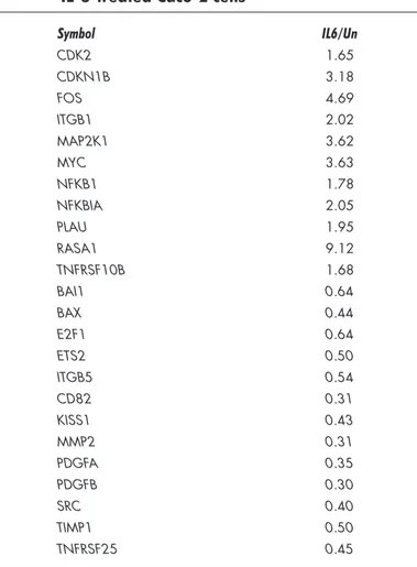
Superarray BioScience), able to detect the expression of 113 genes representative of six biological pathways involved in cell cycle control, apoptosis, tumor invasion and metastases. Results were obtained comparing the mRNA levels of 24 hrs IL-6-treated samples respect to mRNA levels of untreated cells. Only gene upregulation or downregulation by 1.5 fold or more was considered statistically significant. The analysis displayed that genes related to survival and tumor invasion were significantly upregulated (Table 2). Among these, cyclin-dependent kinase 2, Ras p21 protein activator (GTPase activating protein), c-myc and Fos were strongly increased by IL-6 treatment (Table 2). Conversely, genes involved in the suppression of metastases such as TIMP1 and KISS-1 metastasis suppressor were strongly inhibited (Table 2). Interestingly Bax was decreased of 2.2 fold (IL-6/Untreated = 0.44) indicating that IL-6 strongly influenced the apoptotic pathway at mRNA level.
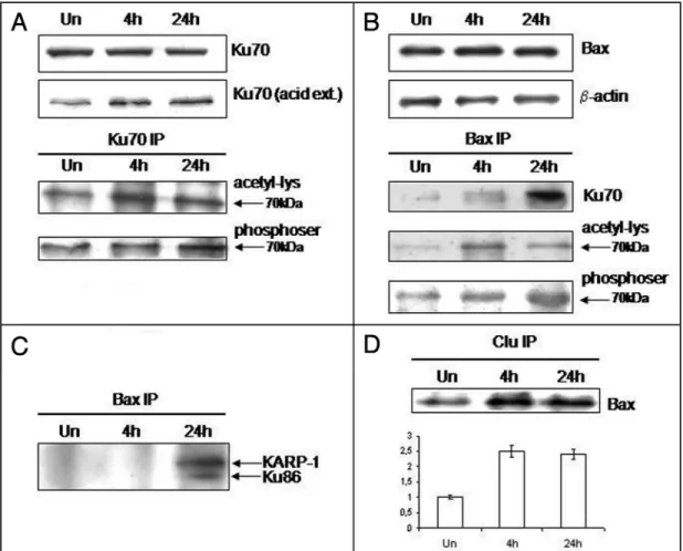
IL-6 increases Bax-Ku70 interaction affecting Ku70 acetyla-tion and phosphorylaacetyla-tion state. IL-6 treatment did not induce any significant change in Bax expression up to 24 hours. In addition, the Bax partner, Ku70 was not influenced by IL-6 treatment at protein level (Fig. 2A and B). To define if IL-6 could affect Ku70-Bax interaction by influencing Ku70 lysine acetylation pattern, nuclear and cytoplasmic acid extracts were prepared from IL-6 treated and untreated cells. Western blot analysis of cytoplasmic acid extracts, demonstrated a 1.8–1.5 fold increase of Ku70 acetylation pattern after 4 h and 24 h of IL-6 treatment, as compared to untreated cells (Fig. 2A). The IL-6-induced modulation of Ku70 acetylation pattern was confirmed by pan-acetyl lysine immunoblotting in co-immuno-precipitated proteins with anti-Ku70 antibody (Fig. 2A). Moreover, also Ku70 serin phosphorylation pattern was affected by IL-6 treat-ment, as shown by western blot analysis of cytoplasmic extracts co-immunoprecipitated with anti-Ku70 antibody and probed with anti-phosphoserin antibody (Fig. 2A).
In order to define if the Bax-Ku70 interaction was affected by IL-6 treatment, cytoplasmic proteins were co-immunoprecipitated with Bax monoclonal antibody 4 h and 24 h after IL-6 exposure (Fig. 2B). In control cells, Ku70 co-immunoprecipitated with Bax. Interestingly, Ku70-Bax complexes increased up to five-fold after 24 h of IL-6 treatment. The increased Ku70 fraction bound to Bax correlated to the Ku70 serin phosphorylated pattern raise (Fig. 2B). In addition an increase in the acetyl-lysine pattern was observed after 4 h–24 h of treatment. (2–1.8 times respectively) (Fig. 2B). The presence of Ku86 was also detected in Bax co-immunoprecipitated proteins after 24 h of IL-6 treatment (Fig. 2C). Moreover a specific and unexpected band at 96 kDa was detected. This band was specifi-cally recognized by the anti-Ku86 mAb raised against an epitope mapping at 433–732 amino acids of Ku86 protein, region that comprises KARP-1 (Fig. 2C). KARP-1 was firstly characterizated by Myung et al. as a second gene expressed from the Ku86 locus.24 It is not constitutively produced but it was demonstrated to be induced by oxidative stress and to be involved in survival and longevity in mammalians. In conclusion, the presence of Ku70 and Ku86 in Bax co-immunoprecipitates in response to IL-6 treatment, suggests the formation of a multimeric complex.
IL-6 modulates Ku86 and clusterin isoform expression. IL-6 treatment induced a significant decrease in Ku86 expression at both mRNA and protein level after 24 h of treatment (Fig. 3A). In partic-ular, Ku86 mRNA level was 4 times lower, as compared to untreated (Fig. 1A). Nuclear Ku86 expression was strongly decreased in A-B
stage tumors (Table 1, Fig. 1A). Importantly, no Ku86 staining was observed as in the nucleus as in the cytoplasm of node posi-tive carcinomas (C-D stages, Fig. 1A, Table 1). Interestingly, Ku86 expression was lost also in metastatic lymphonodes (not shown). CLU was expressed quite exclusively in the nuclei of normal mucosa (Table 1, Fig. 1A). Nuclear CLU staining was not detected in node positive carcinomas (stage C-D, Table 1, Fig. 1A). Conversely, cyto-plasmic CLU expression was strongly upregulated in tumors (normal mucosae versus carcinomas: p = 0.02), correlating with tumor aggres-siveness (A-B stages versus C-D stages, p = 0.04, Table 1).
Double immunofluorescence was performed to investigate whether the presence of Ku70 in the cytoplasm of aggressive tumors correlated to Ku70-Bax co-localization in this cell compartment. Normal colonic mucosa showed a strong nuclear Ku70 staining. Moreover, a very faint Bax immunofluorescence was detected in the cytoplasm (Fig. 1B). The observation on scarcely differentiated and node positive carcinomas (C-D stage) indicated increased levels of co-localized Ku70 and Bax in the cytoplasm (Fig. 1B).
IL-6 influences tumor invasion and proapoptotic gene expres-sion. To define if the increased IL-6 level found in colon cancer could influence or promote colon cancer progression we investigated, in vitro, the effect of IL-6 on Caco-2 cells, a p53-defective moderately differentiated human colon carcinoma cell line. Cells were treated with IL-6 for 24 h. No increase in cell death or cytotoxicity was observed. After 24 h of IL-6 treatment RNA was analyzed by oligo gene array human cancer pathway finder microarray (EHS033,
Table 1 Ku70, Ku86, clusterin and Bax protein expres-sion detected by immunohistochemistry, in human pathological and matched normal tissues°
Protein NM CRC A-B CRC C-D T-test§
(n = 50) (n = 28) (n = 22) Ku86* Pos cyt 0 % (0) 0% (0) 0% (0) NM vs. CRC: p ≤ 0.05 Pos nucleus 100% (++) 70 % (+/-) 0% (0) NM vs. CRC: p ≤ 0.03 Ku70* Pos cyt 0% (0) 40% (++) 100% (++) NM vs. CRC: p ≤ 0.02 CRC-AB vs. CRC-CD: p = 0.04 Pos nucleus 100% (+++) 70% (++) 30% (++) NM vs. CRC: p = 0.02 Clu* Pos cyt 10% (+) 82% (++) 100% (+++) NM vs. CRC: p = 0.02 CRC-AB vs. CRC-CD: p = 0.04 Pos nucleus 8% (++) 0% (0) 0% (0) NM vs. CRC: p ≤ 0.02 Bax* Pos cyt 70% (+/-) 75% (++) 80% (+++) NM vs. CRC: p = 0.04 °Number of cases deemed positive was shown as a percentage. In parentheses staining intensiy mean values were reported. *Cases were deemed positive when weak (+/-)-to-strong (+++) staining was present. §Mean values were compared using the two-tailed Student t-test, for independent samples (see
Materials and Methods). NM: Distant normal mucosa was tested for the colon samples. CRC A-B and CRC C-D: colorectal adenocarcinomas stages A or B (any T N0), and C or D (any T N1 and/or M1), according to the Dukes’ classification.25
and the nuclear form of the protein (Fig. 4C). After 4 h of SST treatment, the Ku70 interaction with nCLU was ten-fold increased, compared to untreated cells. nCLU and Ku70 interaction was still apparent in CLU co-immunoprecipitated proteins after 24 h of treat-ment (Fig. 4C).
Confocal analysis of Ku70, Bax and nCLU triple staining, performed on untreated (control) and SST-treated Caco-2 cells demonstrated the co-immunolocalization of these proteins in control cells. Conversely, after 24 h of SST treatment, Bax was released and nCLU and Ku70 co-localized in the cellular membrane and in the nucleus (Fig. 4D).
Discussion
The data reported in this study provide a novel link among tissue microenvironment and molecules involved in apoptosis cells (Fig. 3B). This inhibition was also confirmed at protein level by
immunocytochemistry and Western blot analysis (Fig. 3A). These data are consistent with the inverse correlation between Ku86 levels and IL-6 expression observed in node positive tumor samples (Fig. 1A). IL-6 exposure also affected Clusterin level (Fig. 3C and D). CLU mRNA was increased from 3-to-4-fold respect to untreated Caco-2 cells, 24 h after IL-6 treatment (Fig. 3C). Western blot analysis confirmed that the 40 kDa isoform of clusterin sCLU, was preferentially induced by IL-6 treatment (Fig. 3D). A similar increase in CLU expression was observed in human colon cancer progression (Fig. 1A).
IL-6 increases the stability of the Bax-Ku70 interaction: sCLu in the Bax-Ku70 complex. In order to evaluate the role of the 40 kDa isoform of Clusterin, sCLU, in the stabilization of the Ku70-Bax complex, cytoplasmic extracts were co-immunoprecipitated with an anti-Clusterin antibody raised against the processed β-chain, specific for the secretory form of CLU. In untreated cells, we found Bax in the CLU co-immunoprecipitated fraction (Fig. 2D). After 4 h and 24 h of IL-6 treatment, the binding between Bax and sCLU was strongly increased (2.5 times, as compared to untreated cells). As previously published in other normal cell systems,25 sCLU seems to prevent apoptosis by binding Bax and stabilizing the Ku70-Bax complex in the cytoplasm. Also Ku70 was found in CLU co-immu-noprecipitated proteins (data not shown).
In order to evaluate if these interactions are irreversible or if they could be modulated by a physiological regulator of cell growth, we treated Caco-2 cells with Somatostatin.
Somatostatin (SST) induces the release of Bax from the Ku70-nClu complex. Somatostatin (SST) is a natural ligand of Ku86,15 and it is a physiological anti-proliferative growth regulatory hormone. Caco-2 cells were treated with SST for 1, 4 and 24 h. A strong proliferative arrest was observed after 24 h, as we had previ-ously reported.16
The levels of Ku70, Ku86 and CLU proteins were then examined in whole cell lysates by Western blot. Ku70 level was not affected by SST treatment (Fig. 4A). Conversely, Western blot analysis showed an approximate two-fold increase in Ku86 level, 4 h after treatment. At 24 h, the expression of Ku86 was similar to that observed in untreated cells (Fig. 4A).
The expression of Clusterin isoforms was modulated in response to SST treatment. Increased levels of 55 kDa nCLU were observed after SST treatment. Moreover, a significant induction (2-to-5-fold) of Bax was detected after 1 h and 4 h of SST treatment (Fig. 4A). However, at later times (>4 h after treatment) Bax levels were compa-rable to those detected in untreated Caco2 cells.
In order to examine the dynamic interaction among Bax, Ku70 and nCLU in the cytoplasm, Ku70 co-immunoprecipitation was performed on cytoplasmic proteins extracted from SST-treated cells. In untreated cells, Ku70 was bound to Bax as previously observed. However, no interaction with Ku86 was found. After 4 h of pro-apoptotic SST treatment, Bax was partially released from Ku70; in particular, a two-fold decrease was observed as compared to untreated cells. Conversely, Ku70 interacted with Ku86, this binding being induced by SST treatment (Fig. 4B). Concomitantly, a significant increase in nCLU-Ku70 complex formation was evident 4 h after treatment. Cytoplasmic proteins were also co-immunoprecipitated with an anti-Clusterin oligoclonal antibody we have produced as previously described, directed against the αβ-unprocessed precursor
Table 2 Genes overexpressed and under-expressed in IL-6-treated Caco-2 cells
Symbol IL6/Un CDK2 1.65 CDKN1B 3.18 FOS 4.69 ITGB1 2.02 MAP2K1 3.62 MYC 3.63 NFKB1 1.78 NFKBIA 2.05 PLAU 1.95 RASA1 9.12 TNFRSF10B 1.68 BAI1 0.64 BAX 0.44 E2F1 0.64 ETS2 0.50 ITGB5 0.54 CD82 0.31 KISS1 0.43 MMP2 0.31 PDGFA 0.35 PDGFB 0.30 SRC 0.40 TIMP1 0.50 TNFRSF25 0.45
Cyclin-dependent kinase 2; Cyclin-dependent kinase inhibitor 1B (p27, Kip1); V-fos FBJ murine osteosar-coma viral oncogene homolog; Integrin, beta 1; Mitogen-activated protein kinase kinase 1; V-myc myelo-cytomatosis viral oncogene homolog (avian); Nuclear factor of kappa light polypeptide gene enhancer in B-cells 1 (p105); Nuclear factor of kappa light polypeptide gene enhancer in B-cells inhibitor, alpha; Plasminogen activator, urokinase; RAS p21 protein activator 1; Tumor necrosis factor receptor superfamily, member 10b; Brain-specific angiogenesis inhibitor 1; BCL2-associated X protein; E2F transcription factor 1; V-Ets erythroblastosis virus E26 oncogene homolog 2; Integrin, beta 5; CD82 molecule; KiSS-1 metastasis-suppressor; Matrix metallopeptidase 2 (gelatinase A, 72 kDa gelatinase, 72 kDa type IV collagenase); Platelet-derived growth factor alpha polypeptide; V-src sarcoma (Schmidt-Ruppin A-2) viral oncogene homolog (avian); TIMP metallopeptidase inhibitor 1; Tumor necrosis factor receptor superfamily, member 25. We compared normalized levels of IL-6-treated samples (IL6) with controls (Un). Only the genes that showed upregulation or downregulation (italics) by 1.5 fold or more were reported.
myc and NFκB. Conversely, IL-6 downmodulates the expression of genes that inhibit metastasis formation such as TIMP1 metallopepti-dase inhibitor 1 and KISS1.29,30 Moreover IL-6 downmodulates Bax expression at mRNA level. Concomitantly, IL-6 exposure influences Bax activity at protein level acting on the Bax-Ku70-sCLU physical interactions in the cytoplasm, affecting their intracellular distri-bution, production, phosphorylation and acetylation state. Data previously published demonstrated that the Bax release from Ku70 is regulated by the acetylation of K539 or K542, two of the seven putative KU70 acetylation sites.6 Our data show that Bax-Ku70 binding is significantly increased by IL-6 treatment and correlated to Ku70 lysine acetylation increase, suggesting a possible implication of other acetylation sites than K539 or K542 in the regulation of this interaction. The increased Bax expression found in tumor tissues could be strongly correlated to the Ku70 shifting to the cytoplasm. induction, DNA repair and cell cycle control in neoplastic cells,
highlighting the importance of their cooperative interaction in tumor progression. We found that the acquisition of an aggressive phenotype in colon carcinoma correlated with the over-production of IL-6, released by the tumor itself and by infiltrating tumor-associated macrophages (TAM). Concomitantly we observed a tumor specific modulation of Bax, Ku86 and CLU localization and expression.
The presence of IL-6 could influence cell proliferation, by activa-tion of STAT3 the AP-1 as previously demonstrated.28 Accordingly, our in vitro data demonstrated that IL-6 could influence tumor cell proliferation in a moderately differentiated colon carcinoma cell line and favour apoptosis escape. Microarray analysis showed that IL-6 could influence the proliferation not only by inducing AP1 formation, but also inducing an up regulation of RASp21 protein activator 1,
Figure 2. IL-6 influences Bax-Ku70 interaction. Ku70 and Bax protein levels in the cytoplasmic fraction of untreated (Un) and IL-6 treated Caco2 cells (A and B). (A) Ku70 level after 4 and 24 h of IL-6 treatment. The cytokine does not affect Ku70 cytoplasmic level. However, the acid extracti: Ku70u70 acid ext.) shows an increase of Ku70 presence in the cytoplasm of treated cells. In the lower gel, the cytoplasmic extracts immunoprecipitated with anti-Ku70 antibody (Ku70 IP) probed with anti-acetyl-lysine antibody to evidence the increase of the Ku70 acetylated fraction, following IL-6 treatment. (B) Bax level in untreated Caco-2 cells (Un) and after 4 h and 24 h of treatment with IL-6. The cytokine does not affect Bax cytoplasmic levels. β-actin levels were detected on the same filter to normalize the protein extracts. (C) Western blot analysis of Bax- and Ku70-immunoprecipitated cytoplasmic extracts from untreated (Un) and IL-6 treated Caco-2 cells. Bax co-immunoprecipitated fraction were probed with Ku70, acetyl-lysine and phosphoserine antibodies. IL-6 induces a significant increase of Ku70 fraction bound to Bax in the cytoplasm. This fraction is lysine-acetylated and serine-phosphorylated, as shown by the signal detected at 70 kDa molecular weight, in the corresponding lower filters. The lower panel shows the same extracts immunoprecipitated with anti-Ku70 antibody (Ku70 IP) and detected with anti-phosphoserine. IL-6 increases the Ku70 serine-phosphorylation at 24 hr (1.8 times) correlated to Ku70 phosphorylated fraction bound to Bax (BaxIP). (D) shows the same extracts from untreated (Un) or 4 h and 24 h IL-6 treated cells immunoprecipitated with Bax antibody (Bax IP) and probed with anti-Ku86 and Bax antibodies. Immunoblotting with anti-Ku86 MoAb shows a significant increase of Ku86 fraction bound to Bax after 24 h of IL-6 treatment. An additional and specific band of 96 kDa is evident in the gel. The Bax immunoblotting on the same IP filter is shown as loading control. The lower panel shows the cytoplasmic extracts from untreated (Un) or 4 h and 24 h treated cells immunoprecipitated with anti-Clusterin antibody (Clu IP) and probed with anti-Bax antibody. IL-6 treatment increases the sClu binding to Bax.
The overexpression of the secreted form of clusterin (sCLU), and the concomitant disappearance of the pro-apoptotic 55 kDa isoform (nCLU) in tumor cells, strongly associated to the inhi-bition of apoptosis and the loss of DNA repair activity of the Ku70/86 complex. Indeed, IL-6 influence may further explain Clusterin altered expression found in tumor biopsies. In fact we found that IL-6 induced predominantly the secreted pro-survival CLU form in vitro. We also demonstrated that sCLU isoform is bound to Bax in colon cancer cells and this binding is increased by IL-6. Hence, IL-6 confers apoptosis resistance by inducing the overexpression of sCLU and an increase of the Bax-Ku interaction. Our data suggest that the ambiguous role of the different isoforms of Clusterin could be defined by their partners and by the cell state. In fact the 55 kDa isoform that seems to be predominantly expressed in pro-cell death processes, was induced by SST treat-ment and it functions as a chaperon in DNA repair, binding Ku70. In contrast in pro-survival states the 40 kDa antagonistic isoform of Clusterin leads to apoptosis escape, stabilizing the Ku70-Bax bond. Our experimental data point out that these tumor-specific interac-tions are not caused by genetic changes within tumor cells, but they arise from altered localization and function of proteins, shifted by microenvironmental soluble factors in a reversible way. In fact, as In fact, as previously published by Amsel et al.27 Bax ubiquitylation
rate is finely regulated and correlates to Ku70 presence in the cyto-plasm, which prevents Bax ubiquitylation and thus its degradation. Therefore, the increased presence of Bax in tumors could be due, at least in part, to its improved de-ubiquitylated status induced by the abnormal high level of Ku70 in the cytoplasm, more than to an increased transcription. Furthermore, the co-localization of Bax and Ku70 in the cytoplasm demonstrated by confocal microscopy could indicate an anti-apoptotic condition, due to the Ku70-Bax physical interaction.
The decreased expression of Ku86 and increased expression of sCLU observed in colon carcinoma appeared to be a result of IL-6 exposure, as demonstrated by in vitro experiments. In fact, RT-PCR and immunocytochemistry performed on Caco-2 cells showed that Ku86 is downmodulated by IL-6 treatment. Ku86 is the limiting factor for non-homologous end-joining of DNA DSBs repair7,8 and its disappearance from the nucleus may suggest a decrease in the ability of cells to accurately repair DSBs in tumor progression, resulting in an increased mutational rate. These results could explain, almost in part, the incoming resistance to anti-neoplastic therapy, driven by the pharmacotherapy-selected clones that possess muta-tions, guaranteeing cell survival.
Figure 3. Effect of IL-6 treatment on Ku86 and Clusterin expression. (A) Immunocytochemistry of Ku86 expression in untreated (Un) and 24 h IL-6 treated Caco2 cells. IL-6 induces a strong inhibition of Ku86 protein expression at 24 h (upper). Western blot analysis with anti-Ku86 antibody on protein extracts from untreated (Un) and Caco2 cells treated with IL-6 for) and 24 h. The filter was reprobed with anti-β-actin to normalize the protein levels (lower). (B) Ku86 mRNA level after IL-6 treatment was determined by RT-PCR as described in the Methods section. M, molecular size marker; lane 1, untreated cells; lane 2, 24 h IL-6 treated Caco-2 cells. The optical densities (O.D.) obtained by scanning densitometric analysis of RT-PCR bands are reported by histograms. The values were normalized for β-2 microglobulin transcripts levels and are means (+/-SD) of three independent experiments. (C) Clusterin mRNA level after IL-6 treatment determined by RT-PCR as described in the Methods section. M, molecular size marker; lane 1, untreated cells; lanes 2 and 3, Caco2 cells treated with IL-6 for 4 h and 24 h. In the lower panel, the corresponding β-2 microglobulin transcript levels are shown. The optical densities (O.D.) obtained by scanning densitometric analysis of bands, normalized for β2-microglobulin levels, are reported by histograms. The values are means (+/-SD) of three independent experiments. (D) Western blot analysis with anti-Clusterin antibody on protein extracts from untreated (Un) and 24 h IL-6 treated Caco2 cells. The optical densities (O.D.) obtained by scanning densitometric analysis and normalized for β-actin protein levels, are reported by histograms. Values are means (+/-SD) of three independent experiments.
Materials and Methods
Patients. Fifty colorectal cancer patients undergone surgery at the “Tor Vergata” Hospital of Rome were enrolled in the study. Colon carcinomas were characterized by grade and stage according to the Dukes’ classifications.25 The corresponding normal mucosae adjacent to neoplasia of the colorectal cancer patients (n = 50) and five colorectal mucosae of human subjects without cancer or inflam-matory bowel disease were used as controls. Oral informed consent was obtained from all participating human subjects. This study was approved by the local Ethics Committee.
Immunohistochemical staining. Serial 5 μm thick sections from formalin-fixed and paraffin-embedded specimens were immu-nostained for Clusterin, Ku70, Ku86, Bax and IL-6 following the streptoavidin-biotin method, as previously described.31 Serial sections from colorectal cancer tissues were immunostained also we demonstrated with SST treatment, they could be redirected by
an antagonistic stimulus, acting on the same pathways stimulating cell death processes.
Overall, our findings suggest that Ku70-Bax-CLU interactions in human colon cancer are modulated by IL-6 that influences Bax-dependent cell death increasing Ku70 binding to Bax and influencing sCLU production that promotes tumor cell survival. Therefore, data suggest a significant role of the microenviron-ment and tumor-specific IL-6 expression in colon carcinogenesis. These molecular interactions underline the relevant role of IL-6 and other micro-environmental factors in the complicated cross talk among molecules that could effectively change the cell fate. Understanding the mechanisms that modulate the IL-6-dependent anti-apoptotic cascade, may lead to development of novel therapeutic strategies to selectively affect cell resistance to anti-neoplastic therapies.
Figure 4. (A) Western blot analysis on protein extracts from Caco-2 untreated (Un)or 1, 4 and 24 h somatostatin (SST) treated cells. The filters were probed with Ku86, Ku70, Clu and Bax antibodies. β-actin protein levels were detected as loading control. Ku86 protein is induced after 4 h of IL6 treatment, whereas Ku70 levels is not affected. Increased levels of nClu (50–55 kDa) and Bax are evident after 1 h, and 4 h of SST treatment. Bax decreases to the basal levels after 4 h of treatment. Cytoplasmic proteins were extracted from Caco-2 untreated or treated with SST for 4 h and 24 h and immunoprecipitated with anti-Ku70 (anti-Ku70 IP) (B) and anti-Clu Abs (Clu IP) (C). In (B), no interaction with Ku86 is found in untreated cells, whereas a strong binding with Bax are detected in Ku70 co-immunoprecipitated proteins from untreated (Un) cells. Moreover an interaction with nCLU was also evident. After 4 h of SST treatment, Bax is partially released (6 folds of reduction) from Ku70. Conversely, Ku70 interacts with Ku86 and a significant increase in nCLU-Ku70 binding is evident. After 24 h, Ku70-Ku86 complex decreases. In (C) the cytoplasmic extracts were co-immunoprecipitated with anti-Clusterin antibody directed (Clu IP). A significant increase of Ku70 protein is evident after 4 h of SST treatment. nCLU and Ku70 interaction is still evident 24 h after treatment. The non-immunoprecipitated levels of Ku70 corresponding cytoplasmic extracts are shown as Input. β-actin was detected on the Input filter, as loading control. (D) Confocal micros-copy analysis of Ku70, Clusterin and Bax staining in Caco-2 cells treated with somatostatin for 24 h. Panels show a 2D reconstruction triple stain for Bax (blue), Ku70 (red) and Clu (green) (2000x magn.), detected as described in Materials and Methods, in Caco-2 cells. Merging of triple stain in untreated Caco-2 cell (UnMerge) and after 24 h of SST treatment (SST Merge) is shown. Yellow, light blue and white areas are due to multiple positivity. The blue areas demonstrate the Bax release from Ku70 and CLU after SST treatment (arrow).
(NeoMarker, clone 2D2); anti-Clusterin-α (H330, Santa Cruz Biotechnologies, Inc.,), and β-actin (Sigma-Aldrich, USA) anti-bodies. Anti-nCLU oligoclonal antibody, N1-CLU, was obtained by the selection of very short antigenic epitopes specific for the unprocessed and nuclear CLU isoform. Short CLU-specific antigenic sequences having a length from 10 to 20 amino acids were synthe-sized on solid phase and selected for nCLU specificity and affinity (patent RM2004A000098).
The optical densities were obtained by scanning densitometry, after normalization for the β-actin gene product.
Immunoprecipitation assay (IP). Immunoprecipitation assay (IP) was performed on cytoplasmic extracts from Caco-2 cells, treated or not with IL-6 for 4 and 24 hours. 100 μg of extracts were used for immunoprecipitation experiments. In brief, cytoplasmic extracts were pre-cleared incubating them with Protein G-Agarose (Santa Cruz Biotechnology 50% slurry) for 30 min at 4°C with agitation. The Agarose was removed, then 2 μg of anti-Ku70 (A-9, Santa Cruz Biotechnology, Inc.,) or 4 μg of anti-Bax (clone 2D2, Neomarker) or 2 μg of anti-Clusterin-β (M-18, Santa Cruz Biotechnology, Inc.,) or 2 μg of anti-nCLU (N1-CLU) antibodies in 250 μl of reaction buffer (PBS + 0.05% NP-40) were added and incubated overnight at 4°C with rotation. IP negative control was performed without specific antibody. Each immunoprecipitate was electrophoresed on a 10% SDS-PAGE. Proteins were blotted onto PVDF membrane (Hybond-P, Amersham Biosciences), stained with Ponceau S-dye and then probed with the various antibodies reported in the Western Blot section.
RNA extraction and RT-PCR. Total RNA was isolated from treated and untreated cells using Tri Reagent (Ambion, Inc.,), according to the manufacturer’s instructions. RNA quantification was performed using spectrophotometry. Reverse transcription of total RNA (1 μg for each cell line) was performed with Gene Amp RNA PCR Kit (Applied BioSystems) using Random Examers as primers to cDNA synthesis. To amplify the cDNA, the following primers (Invitrogen) were used: forward 5' GTG CAA TGA GAC CAT GAT GG 3'; reverse 5' CAG GTA GTG GTA GGT ATC CT 3' for ApoJ34 and forward 5' AAG AAG GCC TTT GAG GA 3'; reverse 5' GCT TCC TCA GCT GTG ACA GA 3' for Ku86 transcripts. Ku86 amplification was performed under the following conditions: 30 cycles at 94°C 30 sec, 55°C 30 sec and 72°C 30 sec, obtaining an amplicon of 270 bp. β2-microglobulin housekeeping gene was ampli-fied as control. Ethidium-bromide stained 2% agarose gel was run at 100 V and picture of the gel was acquired by scanning system.
Gene expression analysis by oligo gene microarray. Total RNA extracted as above described from 24 h IL6-treated and untreated cells was quantified by spectrophotometry and checked on agarose gel for quality. Samples were analyzed with Gene Array Express Human Cancer Pathway Finder Microarray, EHS033, by Superarray BioScience (now renamed SABioscience Corporation). Values were normalized according to the Interquartile Normalization. All the data sets are corrected using minimum value for background subtraction before being compared to each other. The ratio between mRNA levels in IL-6 treated samples and in untreated controls was calcu-lated and only the upregulation or downregulation by 1.5 fold or more in gene expression was considered significant.
Statistical analysis. All values provided in the text and figures are means of three independent experiments ± standard deviations (SD). with anti-CD68 antibody (Dako Denmark A/S, clone KP-1) to
characterize the inflammatory infiltrate in the tumoral tissue. The primary antibodies used were: Ku70, Ku86 and anti-Clusterin-β (Santa Cruz Biotechnology, Inc., ImmunoCruz staining system M19, M20 and M18 respectively); anti-Clusterin-α (Upstate, clone 41D), anti-Bax (NeoMarker, clone 2D2) and anti-IL-6 (RD systems) antibodies. Negative controls were obtained incubating the sections without primary antibodies. Slides were examined by two pathologists, unaware of the clinical data and molecular results. Tissue staining was semi-quantitatively graded for intensity from negative (0) to strong (+++). Bax, Ku70, Ku86 and Clusterin protein expression was deemed positive when weak (+/-) to-strong (+++) cytoplasmic or nuclear staining was present.34
Immunofluorescence and confocal microscopy. A double immu-nostaining was performed on formalin-fixed and paraffin embedded specimens to study the co-localization of Ku70 and Bax proteins by immunofluorescence microscopy. The primary antibodies used were anti-Ku70 (Santa Cruz Biotechnology, Inc.,) and anti-Bax (NeoMarker, clone 2D2). Control sections were incubated with a mixture of isotype matched non-immune IgG. Biotinylated anti-goat and anti-mouse rabbit IgG (Dako A/S, Denmark) were used as secondary antibodies. Fluorescence was obtained with streptoavidin-FITC (for Ku70 detection) and streptoavidin-Texas Red (for Bax detection) conjugates (Ylem).
Triple staining was performed on Caco-2 cells treated or not with Somatostatin for 24 h with the same anti-Ku70 and anti-Bax antibodies and anti-Clusterin-α (Upstate, clone 41D) antibody. Fluorescence was obtained with streptoavidin-FITC (for Clusterin detection), streptoavidin-Texas Red (for Ku70 detection) and strep-toavidin-phycoerythrin (for Bax detection) conjugates (Ylem). Images were acquired by means of Noran confocal microscope at 60X/1.4 NA immersion oil lens. Three D stacks were acquired at resolution of 0.1 micron in X, Y, Z axis.
Cell culture and protein extraction. Human colon carcinoma Caco-2 (HTB-37, ATCC) cells were grown in complete culture medium, according to condition suggested by ATCC. For IL-6 treatment, 60,000 cells/cm2 were plated in 6 multiwells. After over-night culture, medium was removed and human recombinant IL-6 (Endogen) was added at a final concentration of 100 ng/ml for 4 and 24 hours. Protein extraction was performed according to Dignam method,33 modified as described by Pucci et al.16 For treatment of cells with somatostatin (SST) the protocol described by Pucci et al.16 was performed. Alternatively, cytoplasmic acid extracts were prepared according to the Upstate protocol for extraction of acety-lated proteins. The proteins were then precipitated for 1 h with 20% TCA on ice. The pellet obtained after 10 min of centrifugation at 14,000 rpm was washed once with a solution of acetone containing 0.1% HCl and once with acetone. Untreated cells were used as control. Alternatively, after medium withdrawal, treated and untreated cells were processed for RNA extraction (see specific section below).
Western blotting. Denatured cytoplasmic protein extracts (10 μg) were loaded on a 10% SDS-PAGE. Proteins were transferred to a PVDF membrane (Hybond-P, Amersham Biosciences). Membranes were probed with the following primary antibodies: anti-Ku70 (A9) and anti-Ku86 (B-19) (Santa Cruz Biotechnology, Inc.,); anti acetyl-lysine (Upstate); anti phosphor-serine (Sigma Aldrich); anti-Bax
25. Zhang H, Kim JK, Edwards CA, Xu Z, Taichman R, Wang CY. Clusterin inhibits apoptosis by interacting with activated Bax. Nat Cell Biol 2005; 7:909-15.
26. Gottlieb TM, Jackson SP. The DNA dependent protein kinase: requirement for DNA ends and association with Ku antigen. Cell 1993; 72:131-42.
27. Amsel AD, Rathaus M, Kronman N, Cohen HY. Regulation of the proapoptotic factor Bax by Ku70-dependent deubiquitylation. Proc Natl Acad Sci USA 2008; 105:5117-22. 28. Corvinus FM, Orth C, Moriggl R, Tsareva SA, Wagner S, Pfitzner EB, et al. Persistent
STAT3 activation in colon cancer is associated with enhanced cell proliferation and tumor growth. Neoplasia 2005; 7:545-55.
29. Sieuwerts AM, Usher PA, Meijer-van Gelder ME, Timmermans M, Martens JW, Brünner N, et al. Concentrations of TIMP1 mRNA splice variants and TIMP-1 protein are differen-tially associated with prognosis in primary breast cancer. Clin Chem 2007; 53:1280-8. 30. Nash KT, Phadke PA, Navenot JM, Hurst DR, Accavitti-Loper MA, Sztul E, et al.
Requirement of KISS1 secretion for multiple organ metastasis suppression and maintenance of tumor dormancy. J Natl Cancer Inst 2007; 99:309-21.
31. Hsu SM, Raine L, Fanger H. The use of avidin-biotin peroxidase complex (ABC) in immunoperoxidase technique: a comparison between ABC and unlabelled antibody (PAP) procedures. J Histochem Cytochem 1981; 29:577-86.
32. Visca P, Alò PL, Del Nonno F, Botti C, Trombetta G, Marandino F, et al. Immunohistochemical expression of fatty acid synthase, apoptotic-regulating genes, proliferating factors and ras protein product in colorectal adenomas, carcinomas and adjacent non neoplastic mucosa. Clin Cancer Res 1999; 5:4111-8.
33. Dignam JD, Lebovitz RM, Roeder RG. Accurate transcription initiation by RNA poly-merase II in a soluble extract from isolated mammalian nuclei. Nucleic Acids Res 1983; 1:1475-89.
34. Cervellera M, Raschella G, Santilli G, Tanno B, Ventura A, Mancini C, et al. Direct trans-activation of the anti-apoptotic gene apolipoprotein J (clusterin) by B-MYB. J Biol Chem 2000; 275:21055-60.
One or two-tailed unpaired t-test was utilized to assess inter-group differences. Differences were considered statistically significant for p < 0.05. Statistical significance was calculated comparing staining intensity values between different groups: e.g., Ku86 staining values in normal mucosae (NM) versus adenocarcinomas (CRC) cases.
Acknowledgements
Supported in part by Alleanza Contro il Cancro (ACC)—Istituto Superiore di Sanità (ISS). Art.3 DM 21 luglio 2006—Programma Straordinario di Ricerca Oncologica 2006. Programma 3 “Rete solidale e collaborazioni internazionali”; Ricerca Oncologica: Progetto Ordinario IRCCS.
References
1. Grivennikov S, Karin M. Autocrine IL-6 signaling: a key event in tumorigenesis? Cancer Cell 2008; 13:7-9.
2. Lawrence T, Hageman T, Balkwill F. Sex, cytokines and cancer. Science 2007; 317:51-2. 3. Mitsuyama K, Sata M, Rose-John S. Interleukin-6 trans-signaling in inflammatory bowel
disease. Cytokine Growth Factor Rev 2006; 17:451-61.
4. Mitsuyama K, Matsumoto S, Masuda J, Yamasakii H, Kuwaki K, Takedatsu H, et al. Therapeutic strategies for targeting the IL-6/STAT3 cytokine signaling pathway in inflam-matory bowel disease. Anticancer Re 2007; 27:3749-56.
5. Herber DL, Nagaraj S, Djeu JY, Gabrilovich DI. Mechanism and therapeutic reversal of immune suppression in cancer. Cancer Res 2007; 67:5067-9.
6. Cohen HY, Lavu S, Bitterman KJ, Hekking B, Imahiyerobo TA, Miller C, et al. Acetylation of the C terminus of Ku70 by CBP and PCAF controls Bax-mediated apoptosis. Mol Cell 2004; 13:627-38.
7. Wang J, Dong X, Reeves WH. A model for Ku heterodimer assembly and interaction with DNA. Implications for the function of Ku antigen. J Biol Chem 1998; 273:31068-74. 8. Ferguson DO, Sekiguchi JM, Chang S, Frank KM, Gao Y, DePinho RA, et al. The non
homologous end-joining pathway of DNA repair is required for genomic stability and the suppression of translocations. Proc Natl Acad Sci USA 2000; 97:6630-3.
9. Grawunder U, Finnie N, Jackson SP, Riwar B, Jessberger R. Expression of DNA-dependent protein kinase holoenzyme upon induction of lymphocyte differentiation and V(D)J recom-bination. Eur J Biochem 1996; 241:931-40.
10. Subramanian C, Opipari AW Jr, Bian X, Castle VP, Kwok RP. Ku70 acetylation mediates neuroblastoma cell death induced by histone deacetylase inhibitors. Proc Natl Acad Sci USA 2005; 102:4842-7.
11. Prabhakar BS, Allaway GP, Srinivasappa J, Notkins AL. Cell surface expression of the 70-kDa component of Ku, a DNA-binding nuclear autoantigen. J Clin Invest 1990; 86:1301-5.
12. Koike M, Awaji T, Kataoka M, Tsujimoto G, Kartasova T, Koike A, et al. Differential subcellular localization of DNA-dependent protein kinase components Ku and DNA-PKcs during mitosis. J Cell Sci 1999; 112:4031-9.
13. Fewell JW, Kuff EL. Intracellular redistribution of Ku immunoreactivity in response to cell-cell contact and growth modulating components in the medium. J Cell Sci 1996; 109:1937-46.
14. Pucci S, Mazzarelli P, Spagnoli LG. From normal to malignant phenotype. In: Wong D, ed. Tumorigenesis Reasearch Advances. Nova publishers 2007; 129-46.
15. Le Romancer M, Reyal-Desmars F, Cherifi Y, Pigeon C, Bottari S, et al. The 86-kD subunit of autoantigen Ku is a somatostatin receptor regulating protein phosphatase-2A activity. J Biol Chem 1994; 269:17464-8.
16. Pucci, S, Bonanno E, Pichiorri F, Mazzarelli P, Spagnoli LG. The expression and the nuclear activity of the caretaker gene ku86 are modulated by somatostatin. Eur J Histochem 2004; 48:103-10.
17. Yang CR, Yeh S, Leskov K, Odegaard E, Hsu HL, Chang C. Isolation of Ku70-binding proteins (KUBs). Nucleic Acids Res 1999; 27:2165-74.
18. Leskov KS, Klokov DY, Li J, Kinsella TJ and Boothman DA. Synthesis and functional analyses of nuclear clusterin, a cell death protein. J Biol Chem 2003; 278:11590-600. 19. Yang CR, Leskov K, Hosley-Eberlein K, Criswell T, Pink JJ, Kinsella TJ, et al. Nuclear
clusterin/XIP8, an x-ray-induced Ku70-binding protein that signals cell death. Proc Natl Acad Sci USA 2000; 97:5907-12.
20. Gleave M, Chi KN. Knock-down of the cytoprotective gene, clusterin, to enhance hormone and chemosensitivity in prostate and other cancers. Ann N Y Acad Sci 2005; 1058:1-15. 21. Pucci S, Bonanno E, Pichiorri F, Angeloni C, Spagnoli LG. Modulation of different
clus-terin isoforms in human colon tumorigenesis. Oncogene 2004; 23:2298-304.
22. Criswell T, Beman M, Araki S, Leskov K, Cataldo E, Mayo LD, et al. Delayed activation of insulin-like growth factor-1 receptor/Src/MAPK/Egr-1 signaling regulates clusterin expres-sion, a pro-survival factor. J Biol Chem 2005; 286:14212-21.
23. Sallman DA, Chen X, Zhong B, Gilvary DL, Zhou J, Wei S et al. Clusterin mediates TRAIL resistance in prostate tumor cells. Mol Cancer Ther 2007; 6:2938-47.
24. Myung K, He DM, Lee SE, Hendrickson EA. KARP-1. A novel leucine zipper protein expressed from the Ku86 autoantigen locus is implicated in the control of DNA-dependent protein kinase activity. EMBO J 1997; 16:3172-84.