Alma Mater Studiorum – Università di Bologna
DOTTORATO DI RICERCA IN
Bioingegneria
Ciclo XXIX
Settore Concorsuale: 05/E1- BIOCHIMICA GENERALE
Settore Scientifico Disciplinare: BIO/10 – BIOCHIMICA
TITOLO TESI
"INVESTIGATING THE MECHANOBIOLOGY OF
CANCER CELL-ECM INTERACTION: THE IMPACT
OF SUBSTRATE STIFFNESS IN BREAST CANCER
PROGRESSION"
Presentata da:
Chiara Liverani
Coordinatore Dottorato
Supervisore
Prof. Daniele Vigo
Prof. Emanuele Giordano
Co-Supervisore
Dott. Toni Ibrahim
2
TABLE OF CONTENTS
1. Abstract………...5
2. Introduction………...8
2.1 Breast cancer………...8
2.2 The extracellular matrix ………...9
2.3 The role of extracellular matrix in cancer...10
2.4 Extracellular collagen and its modification………...………..12
2.5 Collagen remodeling in breast cancer ...………...………..13
2.6 The role of the matrix-modifying enzyme LOX ...………...…14
2.7 Three-dimensional (3D) in vitro models to study cancer ………....17
2.8 Cancer cell response to matrix stiffness in 3D models...19
2.9 Determination of the mechanical properties of soft tissue biomaterials ………...21
2.10 Dynamic Mechanical Analysis………...23
2.11 Atomic Force Microscopy...25
2.12 Translational impact of these studies....………...………...28
3. Aims………...………....31
4. Material and methods………...33
4.1 Experimental in vitro model………...………...33
3
4.3 Cell seeding and culture………...35
4.4 Cell quantification………36
4.5 Histological analysis………...36
4.6 LOX inhibition………...37
4.7 Scaffold decellularization………...37
4.8 Development of a low-force mechanical testing device………...…………...37
4.9 Mechanical testing………39
4.9.1 Low-force testing device………...………39
4.9.2 Dynamic Mechanical Analysis (DMA) ………...……….39
4.9.3 Atomic Force Microscopy (AFM) ………..40
4.10 Microscopy analysis………...42
4.11 Quantitative Real-Time Reverse Transcription-PCR (qRT-PCR)………..42
4.12 MTT assay………..43 4.13 Drug testing……….43 4.14 Xenograft study………...44 4.15 Collagen staining………...44 4.16 Statistical analysis………...45
5. Results………46
5.1 Collagen remodeling in breast xenograft tumors….……….46
5.2 Collagen scaffold macro and micro properties ………..………..49
5.3 Phenotypes and behavior of breast cancer cells on the collagen matrix…………...51
5.4 Assessment of the low-force testing device repeatability……….59
5.5 Alteration of the compressive stiffness of collagen scaffolds by invasive breast cancer cells………...………..60
4
5.7 Collagen scaffold remodeling by breast cancer cells………67
5.8 LOX inhibition impairs MDA-MB-231 ability to increase collagen stiffness…….72
5.9 Atomic Force Microscopy analysis of single collagen fibers………...74
6. Discussion………..………78
7. Conclusions and future perspectives………...88
8. References………..90
9. Acknowledgments ………..101
5
1. Abstract
Rationale
The extracellular matrix (ECM) properties can be modified in multiple pathological processes either as a cause or as a result of the disease pathogenesis. In cancer, the loss of tissue homeostasis and mechanoreciprocity are considered one of the disease hallmarks. In particular, breast cancer has been defined as a disease of altered mechanobiology. It has been shown that an ECM gene signature can stratify breast cancer patients into subclasses that predict outcome. Increased breast tumor density in mammography has proven to be significantly correlated with reduced progression-free survival in metastatic patients. Studies on the tumor mechanobiology are thus expected to provide an insight into the disease pathogenesis as well as potentially useful biomarkers.
Type I collagen is among the major determinants of breast ECM structural and tensile properties, and the collagen modifications during tumor evolution drive a number of disease-related processes that favor cancer progression and invasion. Here we have applied a porous type I collagen-based three-dimensional (3D) scaffold to study how breast cancer cells interact and alter extracellular collagen. Combining this culturing technique with different methodologies to characterize the structural and mechanical properties of collagen-based scaffolds we assessed the modifications induced by tumor cells on the micro- and macro- characteristic of extracellular collagen and on its compressive stiffness.
Materials and methods
The human breast cancer cell lines MCF7 and MDA-MB-231 were cultured for 10 days on porous collagen scaffolds. The two cell lines belong to breast tumor subtypes
6
characterized by different clinical aggressiveness and distinct in vivo ECM characteristics. MCF7 belong to the luminal A subtype and have a weak in vitro invasiveness, while MDA-MB-231 are basal-like breast cancer cells that are associated with a highly aggressive behavior. At the end of the culturing time, we characterized the cell morphological and molecular phenotypes and their drug sensitiveness. We next tested, the changes induced by cancer cells in the stiffness property of the scaffold, and in its structural micro- and macro- architecture. Mechanical testing was conducted both with an in-house built low-force compressive device and by Dynamic Mechanical Analysis. The stiffness of single collagen fibers was also assessed by Atomic Force Microscopy. We next used orthotopic xenotransplantion of the two cell lines to evaluate the collagen content and the degree of macroscopic collagen remodeling in in vivo breast tumors.
Results
When cultured on collagen scaffolds, the two cell lines generated coherent tissue-like structures. MCF7 displayed an epithelial morphology with a tightly cohesive cobblestone appearance and high levels of E-cadherin expression, while MDA-MB-231 showed a mesenchymal phenotype with lower cell-to-cell contact, a spindly appearance and high vimentin expression. MDA-MB-231, which belongs to the aggressive basal-like subtype, increased scaffold stiffness from 46.9 ± 2.7 kPa, to 57.9 ± 3.6 kPa, and overexpressed the matrix-modifying enzyme, lysyl oxidase (LOX), whereas luminal A MCF-7 cells did not significantly alter the mechanical characteristics of extracellular collagen. These data were obtained either using an in-house built mechanical testing device, and a Dynamic Mechanical Analysis, a standard technique to measure the properties of biomaterials.
The phenotype observed in vitro replicates the behavior of in vivo tumors generated by MDA-MB-231, characterized by higher collagen content and LOX levels than MCF-7.
7
When the activity of LOX was blocked, MDA-MB-231 were unable to increase the scaffold stiffness: scaffold compressive modulus increased by only 8.9%, in contrast to the increase observed without LOX inhibition (23%).
No significant changes were observed between the Young’s modulus of fibers taken from control scaffolds, which was 1.08 ± 0.24 GPa, compared to the Young’s modulus of fibers taken from scaffolds decellularized after the culture with MDA-MB-231, which was 1.1 GPa ± 0.05.
Conclusions
Overall this work provides evidence that invasive, mesenchymal-like breast cancer
cells produce high levels of the collagen crosslinking enzyme LOX, and are able to increase the stiffness of extracellular type I collagen and to alter its structural characteristics. Measurements of the compressive modulus of these soft synthetic extracellular matrices were carried out using a prototypal mechanical testing instrument originally designed and developed. A causal relationship between LOX expression and the ability of cancer cells to alter the stiffness of extracellular collagen was also provided.
Our model offers a relevant in vitro tool to reproduce and investigate the biomechanical interplay subsisting between cancer cells and the surrounding ECM and its impact on the tumor phenotype and behavior.
8
2. Introduction
2.1 Breast cancer
Breast cancer is the most prevalent cancer diagnosed in women worldwide, and is the second leading cause of cancer death in women (Desantis et al., 2013 and Siegel et al., 2016). It is now the most common cancer both in developed and developing regions. Breast cancer ranks as the fifth cause of death from cancer overall (Ferlay J et al., 2012). The majority of breast cancers diagnosed are ductal invasive carcinomas that arise from luminal or basal epithelial cells. Breast cancer can be classified into four categories based on microarray gene expression profile analysis: luminal A and luminal B subtypes, characterized by the expression of the Estrogen Receptor (ER) and the Progesteron Receptor (PGR); the HER2-positive subtype, characterized by the overexpression of the human epidermal growth factor receptor 2 (HER2); and the basal-like subtype (Basal A and Basal B) characterized by triple negativity for ER and PGR expression and for HER2 overexpression (Sorlie et al., 2001). Expression of estrogen receptor (ER), progesterone receptor (PR), and HER2 receptor is used to determine the primary breast tumor subtype, the prognosis, and the targeted therapeutic regimen. Estrogen receptor (ER) and progesterone receptors (PR) are treated as predictive and prognostic markers (as per National Academy of Clinical Biochemistry guidelines). ER-α and PR should be measured in all patients with breast cancer and have clinical application. The presence of ER is an important discriminating factor: ER-positive tumors are associated with better prognosis, longer relapse-free survival, and improved overall survival compared to ER-negative tumors (Zhang et al., 2014 and Hoch et al., 1999). A number of targeted hormonal therapy are available for these patients. HER2-positive breast tumors show more aggressive features, but the treatment options have advanced through the use of monoclonal
9
antibodies to block HER2 activity (Dent et al., 2013). HER2 should be measured in all patients with invasive breast cancer. The primary purpose of measuring HER2 is to select patients with breast cancer that may be treated with trastuzumab.
Ten to twenty percent of all invasive breast cancers diagnosed are classified as triple negative breast cancer (TNBC), a subtype characterized by lack of expression of ER or PR, and lack of HER2 overexpression. TNBCs typically are diagnosed at higher tumor stage and grade, show an aggressive biology and are associated with poor prognosis. Treatment options for TNBC patients are restricted to the use of cytotoxic chemotherapies, since these tumors do not respond to anti-hormonal therapy (Tomao et al., 2015). Even if TNBC patients usually respond to chemotherapy, they have shorter relapse-free survival (Carey et al., 2007 and Hudis et al. 2011). TNBC tumors are also more likely to develop resistance to chemotherapies and present with distant recurrence and visceral metastases (Carey et al., Hudis et al. and Anders et al.). Consequently, in order to advance the development of targeted therapeutics in TNBC, a better understanding of the underlying molecular mechanisms distinguishing TNBC from other breast cancer subtypes is critical.
2.2 The extracellular matrix
The extracellular matrix (ECM) is the non-cellular component of all tissues and organs, that provides essential physical scaffolding for the cells, and regulates crucial biochemical and mechanical cues that are required for tissue morphogenesis, differentiation and homeostasis. The importance of the ECM is clearly demonstrated by the wide range of diseases that arise from genetic abnormalities in ECM proteins (Jarvelainen H et al., 2009). Essentially, the ECM is composed of two main classes of macromolecules: proteoglycans (PGs) and fibrous proteins. Each tissue has an ECM with a unique composition and topology that is generated during tissue development through a dynamic
10
interplay between the various cellular components (e.g. epithelial, fibroblast, adipocyte, endothelial elements) and the evolving cellular and protein microenvironment.
Cell adhesion to the ECM is mediated by ECM receptors, such as integrins, discoidin domain receptors and syndecans (Frantz C et al., 2010). Adhesion mediates cytoskeletal coupling to the ECM and is involved in cell migration through the ECM (Schmidt S. and Friedl P., 2010). The ECM generates the biochemical and mechanical properties of each organ, such as its tensile and compressive strength and its elasticity. In addition, the ECM directs essential morphological organization and physiological function by binding growth factors (GFs) and interacting with cell-surface receptors to modulate signal transduction and gene transcription (Hynes RO, 2009).
The ECM is a highly dynamic structure that is constantly being remodeled, either enzymatically or non-enzymatically, and its molecular components are subjected to a number of modifications. The physical, topological, and biochemical composition of the ECM is not only tissue-specific, but is also markedly heterogeneous (Frantz C et al., 2010). ECM biochemical and mechanical properties in a given tissue can change from one physiological state to another. Also the composition and the architecture of the ECM, its interactions with the cellular constituents, and its post-translational modifications can vary extremely in a pathological conditions (for examples in aged tissue, wounded or fibrotic tissue and tumors). This ECM remodeling, in turn, cause functional consequences on cellular behaviors including altered GF sensitivity elicited by changes in ECM tension (Frantz C et al., 2010).
2.3 The role of extracellular matrix in cancer
The ECM has a vital role in the regulation of various biological pathways contributing to development, tissue homeostasis and diseases (Bonnans et al., 2014). ECM is involved in
11
a number of different biological process as the maintenance of tissue polarity, the induction or blockade of cell migration, the creation of molecule concentration gradients and the exertion of direct intracellular signals (Figure 1).
Figure 1: schematic representation of the role of the ECM in different cellular processes (Pengfei et al., 2012).
In particular, forces are essential for normal tissue-specific organization, in which they regulates cell survival, growth and migration, coordinating tissue development and function.
ECM properties can be modified in multiple pathological processes either as a cause or as a result of the disease pathogenesis (Bonnans et al., 2014). Cancer is no longer merely considered as a disease of tumor cells: the imbalance of the tumor microenvironment also appears to play a crucial role. ECM, as the most abundant component in the tumor
12
microenvironment, can regulate cancer cell behavior and affect key tumor processes (Pengfei et al., 2012). Loss of tissue homeostasis and mechanoreciprocity are considered a hallmark of cancer.
In particular, breast cancer has been defined as a disease of altered mechanobiology: matrix density, structure and stiffness, together with interstitial fluid pressure and flow, change during tumor evolution (Dvorak et al., 2011). As an example, the collagen matrix of breast cancer stroma is significantly stiffer and more linearized than in normal tissue, due to a progressive enzymatic crosslinking mediated by cancer cells. Linearized collagen fibers can, in turn, increase growth factor-dependent cell migration (Levental et al., 2009) Two primary mechanisms are involved in migration of cancer cells: the cell physical rearrangement linked to the epithelial to mesenchymal transition (EMT) (Mierke et al., 2011 and Hoffman et al., 2011), and the reorientation of collagen by traction forces and by enzymatic cleavage (Wolf et al., 2007 and Gonzalez et al., 2010). Both can be regulated by tissue tension. Increased matrix cross-linking and ECM protein deposition or parallel reorientation of collagen can stiffen tissue locally and drive migration. Altered mechanical signaling can also affects cell survival, division and differentiation (Du Fort et al., 2011; Engler at al., 2006 and Janmey et al., 2011).ECM remodeling is thus capable of affecting a number of tumor processes such as invasion, progression(Levental et al., 2009), and metastasis initiation (Provenzano et al., 2006).
2.4 Extracellular collagen and its modification
Collagens comprise a large family of triple helical proteins and are the most abundant protein in human tissues, accounting for one-third of total proteins. There are now at least 29 genetically distinct types of collagen identified, that are encoded by at least 44 genes that can be divided into several subgroups, depending on the molecular structure and
13
assembly mode (Hulmes, 2008). Fibril-forming collagens comprise the largest subgroup, including types I, II, III, V, XI, XXIV and XXVII. Fibrillar type I collagen is the most abundant type providing most tissues and organs with form, stability and connectivity. In addition to the structural functions, type I and other collagens also function as ligands for specific cell receptors, such as integrins, discoidin domain receptors, glycoprotein VI and the mannose receptor family etc., to control cellular activities and extracellular matrix remodeling (Leitinger, 2011). One of the most important factors for the structural and biomechanical functions of type I collagen fibrils are the post-translational modifications of peptidyl lysine residues. These modifications are highly regulated processes that can take place either inside or outside the cell. In the cell, the peptidyl lysine residues can be hydroxylated both in the helical and non-helical (telopeptide) domains of the molecule. Specific hydroxylysine residues in the helical domain can then be glycosylated with the addition of galactose, some of which can be further glycosylated with the addition of glucose. Specific enzymes catalyse each of these sequential and domain-specific lysine modifications. Outside the cell, an enzymatic oxidative deamination occurs to the telopeptidyl lysine and hydroxylysine residues, producing reactive aldehydic residues. The aldehydes then initiate a series of chemical condensation reactions to form extensive covalent intra- and inter-molecular cross-links, which are critical for the biomechanical functions of the collagen fibrils (Yamauchi, 2008).
2.5 Collagen remodeling in breast cancer
Increased deposition of collagens I, III and IV and enhanced collagen matrix cross-linking are among the ECM structural changes that occur during tumor progression (Zhu et al., 1995; Lesniak et al., 2010 and Fang et al., 2014). Increased deposition and remodeling creates a reorganized microenvironment with significant impacts on tumor cell biology
14
including gene expression, cell differentiation, proliferation, migration and responses to treatments (Paszek et al., 2005).
Breast cancer is a typical example of these changes. Collagen is the major component of the breast connective tissue and is a key determinant of its tensile properties(Roeder et al., 2002) and of the macroscopic deformability of mammary tumors(Fenner et al., 2014) that are characterized by high collagen content and remodeling (Schedin et al., 2011). Clinicians have long recognized the connection between breast density and breast cancer risk (Wolfe, 1976). Collagen surrounding normal epithelial structures in breast tissue is typically curly and smooth. However, parallel with tumor development, collagen progressively thickens, linearizes and stiffens fostering cell migration into ECM. Intravital imaging shows that breast cancer cells and leukocytes migrate rapidly along collagen fibers (Wyckoff et al., 2007). Cancer cells might exploit these remodeled stiff collagens as invasion "highways".
2.6 The role of the matrix-modifying enzyme LOX
Extracellular collagen remodeling is predominantly catalyzed by enzymes such as lysyl oxidase (LOX) (Xiao and Ge, 2012). There are five lysine or hydroxylysine residues in the telopeptide domains of type I collagen, two in the C- and three in the N- telopeptide. These five lysine/hydroxylysine residues can be oxidatively deaminated by the copper (Cu2+)-dependent amine oxidase LOX to form the respective aldehydes which initiates a series of condensation reactions to form covalent intra- and inter-molecular cross-links (Figure 2) (Yamauchi and Sricholpech, 2012).
15
Figure 2. Lysine modifications in the telopeptide domains of type I collagen (Yamauchi and Sricholpech, 2012).
LOX has been found to be synthesized in response to hypoxic conditions: hypoxia promote HIF-1–α mediated endogenous LOX gene expression. Subsequently, the collagen maturation occurs through the extracellular modification of collagen fibers by LOX, resulting in the formation of covalent cross-links (Makris et al., 2014) (Figure 3).
16
Figure 3. Mechanistic description of HIF-1–mediated LOX transcription and subsequent collagen cross-linking that enhance the biomechanical properties of collagen-rich tissues (Makris et al., 2014).
In tumors, LOX can be synthesized by either stromal cells during early stages of carcinogenesis, or tumor cells during late stages of tumor progression. LOX, secreted by hypoxic tumor cells, crosslink collagens and elastin, thereby increasing insoluble matrix deposition and tissue stiffness (Erler et al., 2009).
This enzyme have demonstrated a crucial role in breast cancer progression and metastatic spread. LOX is essential in driving tumor cells escape from primary site, extravasation and growth at secondary sites during metastasis (Erler et al., 2006 and Erler et al., 2009). It is reported that LOX can also be disseminated into distal target organs via circulation to create pre-metastatic niche both at lung (Erler et al., 2009) and bone sites (Cox et al., 2015), as also evidenced by a consistent correlation between increased LOX expression and higher cancer metastasis risk (Erler et al., 2006). Finally, increased LOX expression is
17
associated with early stromal reaction in breast cancer, and reactive fibrosis at the invasive front of infiltrating tumors also releases high levels of LOX (Erler and Giaccia, 2006).
2.7 Three-dimensional (3D) in vitro models to study cancer
In vitro research offers several experimental methods and models to investigate the tumor
mechanobiology (Carey et al., 2012). However, studies examining the relationship between ECM mechanics and cell phenotype have been hampered by limitations in the experimental approaches used to manipulate the biophysical properties of the ECM. The systems predominantly applied are mechanically tunable two-dimensional (2D) polymer hydrogels conjugated with ECM ligands (Tse et al., 2010; Young et al., 2011). These 2D substrates, unfortunately, fail to model the heterogeneous and three-dimensional (3D) structure of native tissue. For this reason, a number of innovative 3D systems have recently been developed and applied to investigate how cancer cells interact with the ECM, or to address the impact of mechanical signaling in diverse tumor phenotypes and behaviors in a more physiological context (Cassereau et al., 2015; Paszek et al., 2005 and Wozniak et al., 2012). By employing 3D hydrogel substrates, Paszek and colleagues found that matrix compliance differs dramatically between 2D and 3D cultures, and between normal versus tumoral tissue in vivo. In this work the faithful recapitulation of tissue morphogenesis is favored by matrix conditions with an elastic modulus that corresponds with that of normal tissues in vivo. By examining the relationship between tissue rigidity and tumor behavior, Paszek and colleagues showed that tissue rigidity is linked to matrix stiffening and affects Rho GTPase-dependent cytoskeletal tension. They also found that matrix stiffness and cytoskeletal tension cooperate to modulate tissue behavior: matrix stiffness influences tissue growth and morphogenesis by modulating cell contractility, while tensional homeostasis appears to be necessary for normal tissue behavior. These
18
findings provided a new perspective on the role of the tissue microenvironment in tumorigenesis, providing new possible therapeutic targets (Paszek et al., 2005).
However, the use of hydrogel still presents some limitations, including the failure to control the pore size, and the gel inconsistencies. On the other hand, synthetic polymer scaffolds allow for the fine-tuning of ECM mechanical properties, but fail to recapitulate the architecture and composition of a natural ECM, not enabling cells to interact and remodel the scaffold matrix (Miroshnikova et al., 2011; Miller et al., 2010). 3D biomimetic matrices represent the ideal systems to efficiently recapitulate tissue dimensionality and the biomechanical and physical cues of tumor ECM (Infanger et al., 2013). Collagen-based scaffolds provide an efficient tool to mimic the ECM of soft tissues for in vitro research. These scaffold ideally show: i) high biocompatibility to allow cells to attach and proliferate; ii) a highly porous structure to allow cell and nutrient infiltration; iii) similar mechanical properties to the native tissue (Lamhamedi-Cherradi et al., 2014). Provenzano and colleagues, employed 3D collagen matrices that closely resemble the ECM architectures observed in vivo. They showed that utilizing in vivo-mimicking ECM conditions facilitates the acquisition of physiologically relevant information, while permitting manipulation of the system, and they used this matrix assays for studying contact guidance in vitro (Provenzano et al., 2010). Casserau and colleagues developed a novel tension bioreactor system that employs a native collagen I hydrogel and permits consistent manipulation of ECM stiffness in the absence of modifications to the structure, composition, or pore size of the gel. This system permitted precise mechanical tuning of collagen stiffness, while maintaining constant composition and pore size. Using this system Casserau and colleagues demonstrated that increasing ECM stiffness potentiates tumor cell migration, validating the use of this model to study malignant transformation and the invasive and migratory phenotype of tumor cells (Cassereau et al., 2015).
19
In conclusion natural and synthetic matrices can be used to recapitulate the interstitial ECM in in vitro culture with the aim to study tissue behavior or to deconstruct and analyze how specific ECM parameters (stiffness, fiber orientation, ligand presentation, dimensionality) provoke specific cellular behaviors (Figure 4).
Figure 4. Schematic of the in vitro models presently used to generate 3D cancer cultures, with different methods and materials and control of physical and biochemical factors (Asghar et al., 2015).
2.8 Cancer cell response to matrix stiffness in 3D models
Matrix stiffness is an important parameter when studying the cellular response of cancer cells in 3D microenvironment (Chauhan VP et al., 2014). The matrix stiffness of different organs and tissues can be quantified by the Young's modulus (E) that can be soft as the brain tissue (ranging from 250 to 500 Pa) or rigid as the bone tissue (ranging from 1 to 25
20
GPa). The cellular response in microenvironments with different degrees of ECM mechanical stiffness is different. The matrix stiffness of solid tumors changes during the course of cancer metastasis and progression (Paszek et al., 2005). For instance, the microenvironment of breast tumor show a matrix with a high degree of stiffness (4000 Pa) compared to the healthy breast tissue (200 Pa) (Butcher et al., 2009) (Figure 5).
Figure 5. Schematic representation of the stiffness properties of different human cells and tissues, and of the increasing stiffness observed in breast tumors (Butcher et al., 2009).
One of the most effective protein hydrogels that has been used to investigate cancer cell response to 3D matrices with different stiffness is collagen hydrogels, as previously described (Paszek et al., 2005). Stromal collagen deposition and crosslinking can define the stiffness of the tumor microenvironment and consequently can alter the cancer cell migration through the ECM (Carey et., 2012). In another report, a polyacrylamide
21
hydrogel substrate with varying stiffness was used to demonstrate that cells migrated from the soft substrate to the stiffer substrate (Levental et al., 2009). In another investigation, metastatic cancer cells were characterized by measuring the traction forces applied to 2D and 3D matrices by cancer cells on these substrates (Kraning-Rush et al., 2012). Breast, lung, and prostate cancer cells showed significantly stronger traction forces on the substrate at the late stage of the cancer disease compared to the normal healthy cells. Further, these results showed that cancer cells created stronger contractile forces on matrices with higher stiffness. The degrees of the traction and contraction forces generated by cancer cells are determined by the chemical and mechanical properties of the tumor microenvironment.
2.9 Determination of the mechanical properties of soft tissue biomaterials
The determination of mechanical properties of in vitro constructed ECM, as hydrogels or scaffolds, has becoming an important challenge for studying cell responses in these in 3D environments. Unlike hard tissue matrices, the mechanical properties of soft tissues can be challenging to be characterized. They usually can tolerate minimum stress, making it difficult to obtain precise measurement. Several experimental methods including three point bending, lateral and axial fiber vibration measurement, and direct axial tension tests have been exploited in recent years to characterize the mechanical properties of nanofibers (Edmondson et al, 2009). As an example, Florczyk and colleagues used dynamic mechanical compression to determine the stiffness of porous chytosan-hyaluronic acid matrices to recreate a scaffold with a Young’s modulus similar to that of the brain tissue and study human glioblastoma cells (Florczyk et al., 2013).
22
Other reported also the development of an in-house built instrument to measure the stiffness of small soft tissue samples. Fenner and colleagues developed a system to measure the nonlinear elastic behavior of soft tissues, using a cylindrical piston of known length and volume to indent tumor samples at steps of a known length (Figure 6), and recording changes in force for each successive step size that the cylindrical piston was applied to the specimen. The piston force, when knowing the degree of surface displacement, provided the nonlinear geometric stiffness characteristic for the given tumors. Using this system they were able to find an inverse correlation between the bulk modulus of freshly resected breast tumors generated by cancer cells xenotransplantation in murine models and the development of subsequent local recurrence and metastasis. In particular, the authors found that mice with compliant tumors were developing more frequent and larger local recurrences, and more extensive metastases than mice with relatively stiff tumors (Fenner et al., 2014).
Figure 6. Photo of the piston compression device system used by Fenner and colleagues to measure the bulk moduli of ex vivo breast tumors (Fenner et al., 2014)
Another recently developed solution is to determine matrices elastic modulus through nanoindentation (Zhu et al., 2011). Nanoindentation has been applied for characterizing
23
many soft materials' mechanical properties (Doube et al., 2010). In nanoindentation test, small loads can be used with an Atomic Force Microscope (AFM) (Barone et al., 2010). AFM can measure forces at the nN level (Clifford and Seah, 2006 and Darling et al., 2007) allowing measurement with a size-scale comparable to the cellular one, for a better understanding of the cell-substrate stiffness interaction. For example, Cavo and colleagues used AFM to measure the stiffness of an alginate hydrogel where breast cancer cells were cultured, demonstrating that stiffness directly influences cells fate (Cavo et al., 2016). Rheological testing has also been applied to study the mechanical properties of soft biological materials (Elango et al., 2016 and Yang et al., 2009). Although the storage moduli from rheological measurements is much different from tangent stiffness and, in rheological test, the movement of the interstitial fluid play a significant role in determining the overall mechanical properties of the samples (Xu et al., 2011).
Combining the recently developed 3D culture systems with devices that can measure the mechanical characteristics of small tissue, gel or scaffold samples might shed new light on the relation between the stiffness properties of ECM and tumor cells and its influence on pathological cancer processes. The use of different techniques to measure the diverse mechanical properties that tissues exhibit under specific loading conditions can further improve our understanding on the cancer mechanobiology.
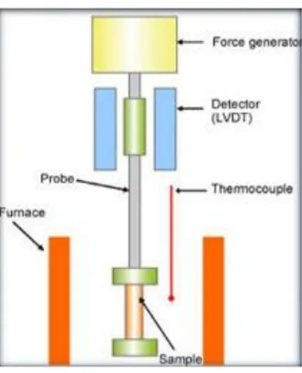
2.10 Dynamic Mechanical Analysis
Dynamic mechanical analysis (DMA) is a non-destructive technique widely used for the mechanical characterization of polymer-based systems, which is getting increasing importance in biomaterial research. Basically, it allows to characterize the viscoelastic properties of materials in a wide temperature and frequency ranges, by monitoring the
24
sample's response upon an imposed controlled cyclic load (with the corresponding development of stresses) or strain (Mano et al., 2002). The instrumentation of a DMA consists of a displacement sensor, such as a linear variable differential transformer (LVDT), a temperature control system or furnace, a force generator, a drive shaft support and guidance system to act as a guide for the force from the motor to the sample, and sample clamps in order to hold the sample being tested (Figure 7).
Figure 7. Schematic of the main component of a dynamic mechanical analysis instrument.
The samples is clamped into a frame and it is heated by the furnace. In the furnace the stress is applied from the force generator via a probe. The stress is applied as a sinusoidal force, to make the strain amplitude constant. The deformation of the sample is then detected. Mechanical properties such as elasticity or viscosity are derived from the stress applied and the strain, and plotted as a function of temperature or time. Several DMA equipments are now available, enabling studies in different mechanical configurations such as in tensile, flexural, compression or shear modes. Such studies are particularly relevant if the tests are performed at physiologically meaningful conditions, i.e., with the specimens completely incubated in solutions at 37 °C (Ghosh et al., 2008).
25
2.11 Atomic Force Microscopy
In order to improve the applicability of soft biomaterials, as collagen scaffolds, a characterization of their structure and properties at a nanoscale level is required. This characterization can be performed by Atomic Force Microscopy (AFM) (Stylianou and Yova, 2013). AFM is a scanning probe microscope that records interactions between a probe and the sample surface. This technique has been introduced in 1980s, by Binnig and colleagues (Binnig et al., 1986), and has now become a fundamental method in surface and biomedical sciences.
The AFM apparatus usually consists of a laser beam deflection system (optical lever) where a laser beam is reflected from the back of a cantilever (on which a very sharp tip is mounted to come into contact with a sample surface) and onto a position-sensitive detector (Stylianou A, 2017) (Figure 8).
Figure 8. Schematic of an AFM apparatus consisting of a probe (cantilever and tip), a laser source, a scanner, a photodetector, the electronics, and a computer (Stylianou A, 2017).
26
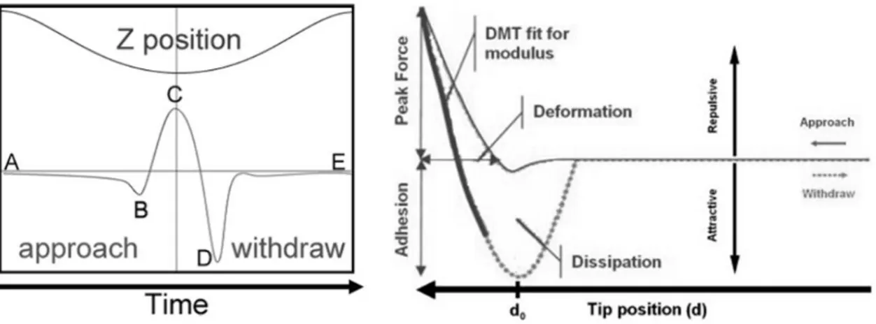
The AFM gathers information with the probe, which is the tip mounted on the cantilever. The probe scans the sample surface and measures the atomic forces among probe sample surfaces. Τhe accurate movement of the tip over the surface of a sample under the tip, depending on the system, is achieved with piezoelectric elements. The measurement of the tip deflection is obtained via a laser beam and then transferred to the system’s electronics which form 3D images of the samples and/or assess other sample’s properties, such as stiffness, roughness or adhesion. The AFM can perform force spectroscopy of force volume for measuring mechanical properties of the samples: indentation-force curves are formed and fitted using mathematical expressions to calculate values that are needed for measuring the sample’s Young modulus and for creating material’s properties maps (e.g., Young’s modulus maps). Various theoretical and empirical models can be used to analyse force-displacement curves: the Hertz theory (Hertz, 1981; Kurland et al., 2012), the Johnson-Kindall-Roberts (JKR) model (Johnson et al., 1971) or the Derjaguin-Müller-Toporov (DMT) model (Derjaguin et al., 1975). Recently, Bruker develop a new operating mode of AFM, called Peak Force Tapping. It provides high resolution images at a relatively high speed. When the tip is distant from the surface there is little or no force on the tip. As it approach the sample surface the cantilever is pulled down by attractive forces such as van der Waals, electrostatics or capillary forces. Then the tip touches the surface and stays on it until the Z position reaches its lowest position while the force is increasing. A constant force at this point is maintained by adjusting the extension of Z piezo through a feedback loop. After that the probe starts to withdraw and the force decreases until its minimum: at this point the surface forces of adhesion are determined. Finally, the tip leaves the surface and only long range forces affect it, and when the tip-sample separation is at its maximum there is a very small or zero force. Dependence of the force against the
27
Z-position can be compared with force-displacement curves that have usually been used in measuring mechanical properties of a sample. When the force curve is complete it is then analyzed to obtain the properties of the sample and the information is sent to one of the image data channel. Different material properties can be extracted, as elastic modulus, adhesion, energy dissipation and maximum deformation. The results are presented as maps of material properties in user-defined false colours (Kolářová et al., 2012).
Figure 9. Schematic representation of the indentation steps (left), and of the mechanical informations (right) of the peak force tapping operating model of AFM (Kolářová et al., 2012).
Compared to other techniques AFM has unique advantages, in particular over other microscopy techniques, such as Scanning or Transmission Electron Microscopy (SEM and TEM) and optical microscopy. First of all, AFM provides topological information at the nanoscale level that other microscopes cannot measure. Moreover, AFM can perform the characterization of the mechanical properties of samples at a nanoscale level, offering thus a combination of qualitative and quantitative information. Furthermore, AFM possesses the ability to perform nanoscale imaging/characterization without the need for presence of
28
vacuum conditions or any special treatment of the specimen, such as sample labeling or surface coating (Allison et al., 2010). AFM can be used for investigating a wide range of collagen-based structures, from collagen molecules to separated fibrils and fibers and collagen-based nanobiomaterials as well (Cisneros et al., 2007). The AFM characterization can be performed without destroying the fibrillar structure of collagen. As an example Zhu and colleagues successfully applied AFM nanoindentation in a liquid fashion to characterize the stiffness and the elastic modulus of collagen–chitosan hydrogel (Zhu et al., 2011).
2.12 Translation impact of these studies
Investigations into breast tumor biomechanics provided a new insight into the disease pathogenesis, leading to the identification of clinically useful biomarkers. It has been shown that an ECM gene signature can stratify breast cancer patients into subclasses that predict outcome (Plodinec et al., 2012 and Bergamaschi et al., 2008). Bergamaschi and colleagues proved that primary breast tumors can be stratified upon ECM composition and that this classification provides relevant information on the biology of breast carcinomas, further supporting the hypothesis that clinical outcome is strongly related to stromal characteristics (Bergamaschi et al., 2008).
Furthermore, molecular profile analyses carried out in other breast cancer cohorts as well as other tumor types, indicated that matrix molecules are present in expression signatures that classify disease progression (Perou et al., 2000; Sorlie et al., 2001; Van’t Veer and Weigelt, 2003; Sotiriou et al., 2003; Teschendorff et al., 2007 and Mintz et al., 2005). Increased breast tumor density in mammography has proven to be significantly correlated with reduced progression-free survival in metastatic patients (Elsamany et al., 2014) and
29
to be an excellent predictor of response to tamoxifen in the preventive setting (Cuzick et al., 2011). With an observational study on breast cancer patients with metastatic disease, Elsamany and colleagues demonstrated that progression-free survival (PFS) was correlated with breast density. PFS, stratified by different prognostic factors, was assessed in low- compared to high-density patients. Among the enrolled patients, median PFS in low-density patients was significantly better than those with high low-density. Breast low-density assessed at the time of diagnosis was significantly correlated with PFS of metastatic breast cancer patients, while survival outcome was improved in specific patients' subgroups with low breast density (Elsamany et al., 2014).
Finally, Cuzick and colleagues conducted a nested case-control study within the first International Breast Cancer Intervention Study, a randomized prevention trial of tamoxifen vs. placebo. Mammographic breast density was assessed visually and expressed as a percentage of the total breast area in 5% increments. Case subjects were 123 women diagnosed with breast cancer at or after their first follow-up mammogram, which took place 12-18 months after trial entry. Control subjects were 942 women without breast cancer. In the tamoxifen arm, 46% of women had 10% or greater reduction in breast density at their 12- to 18-month mammogram. Compared with all women in the placebo group, women in the tamoxifen group who experienced 10% or greater reduction in breast density had 63% reduction in breast cancer risk, whereas those who took tamoxifen but experienced less than 10% reduction in breast density had no risk reduction. In the placebo arm, there was no statistically significant difference in breast cancer risk between subjects who experienced less than 10% reduction in mammographic density and subjects who experienced a greater reduction. The 12- to 18-month change in mammographic breast density thus emerged as an excellent predictor of response to tamoxifen in the preventive setting (Cuzick et al., 2011).
30
All these examples show how the study of the tumor mechanobiology can provide new possible disease biomarkers or therapeutic targets to help clinicians in the diagnosis, prognosis and treatment of breast cancer patients.
31
3. Aims
In the present study we used an in vitro 3D cell culture model, based on porous type I collagen scaffolds, to investigate the ability of breast cancer cells to produce alterations in the structural and mechanical characteristics of extracellular collagen. This biomimetic matrix, providing a physiological 3D network, constitutes the model of choice to reproduce in vitro the cell-ECM interactions. We studied and applied different mechanical testing techniques and microscopy characterizations to assess the changes induced by cancer cells on the micro- and macro-structural properties of the collagen scaffolds, on the material median compressive stiffness and on the Young’s modulus of single collagen fibers.
Specifically we:
i. evaluated the phenotypes and behavior of two subtypes of breast cancer, characterized by different clinical aggressiveness, when cultured within the collagen scaffolds;
ii. developed a prototype low-force instrument to measure the compressive properties of the collagen scaffolds;
iii. evaluated the modifications induced by breast cancer cells on the compression-dependent viscoelastic properties of the scaffolds;
iv. evaluated the modifications induced by breast cancer cells on the micro properties of single collagen fibers;
v. evaluated the modifications induced by breast cancer cells on the micro/macro- structures and protein composition of the scaffolds;
32
The overall aim was to demonstrate whether this 3D in vitro model might offer a valid and informative tool to recapitulate and investigate the biomechanical interaction that occurs between breast cancer cells and the surrounding ECM, focusing on the ability of invasive cancer cells to modify the structural and stiffness properties of extracellular collagen.
33
4. Materials and Methods
4.1 Experimental in vitro model
Two different human breast cancer cell lines, MCF-7 and MDA-MB-231, were cultured for 10 days on 3D porous collagen scaffolds. MCF-7 cells belong to the luminal A subtype of breast cancer (ER positive) and are associated with weak in vitro invasiveness, whereas MDA-MB-231 belong to the basal-like subtype (triple negative breast cancer TNBC) and are associated with an invasive behavior(Holliday et al., 2011) and the ability to colonize different sites (e.g. bone, liver, lung and brain) after intravenous injection (Kang et al., 2003). From the clinical standpoint the luminal A subtype is often connected to indolent disease and a better prognosis, while the basal-like subtype is connected to high-grade and aggressive disease. At the end of the culture time, the phenotypes of the two cell lines were characterized by morphological and molecular analyses.
The scaffold samples were decellularized and their macro- and micro- mechanical and structural properties were tested to assess the modifications induced in the collagen matrix by either cell line (Liverani et al., 2017) (Figure 10).
34
Figure 10. Two human breast cancer cell lines, MCF-7 and MDA-MB-231, characterized by a diverse aggressive behavior, were cultured for 10 days on porous collagen-based 3D scaffolds. At the end of the culture time, cells were characterized for morphological and molecular phenotypes and the mechanical and structural properties of the scaffolds were analyzed (Liverani et al., 2017).
4.2 Synthesis of the porous collagen scaffolds
All chemicals were purchased from Sigma Aldrich (USA). The scaffolds were synthesized from type I bovine collagen as described elsewhere (Minardi et al., 2014). Briefly, a 1 wt% collagen suspension in acetate buffer (pH 3.5) was prepared. The collagen was then precipitated at pH 5.5 with NaOH 1M. The collagen was then washed three times with
35
deionized water and then crosslinked in an aqueous solution of 1,4-butanediol diglycidyl ether (BDDGE) (2.5 mM), for 24 h. This cross-linking step is essential to determine the stiffness of the collagen matrix: higher concentrations of the crosslinking reagent (BDDGE) can generate scaffolds with increasing stiffness properties. Finally, the collagen was washed three times and casted in Teflon molds (9 mm in diameter) and freeze dried through an optimized freezing and heating ramp to obtain the desired pore size and porosity (from 25 °C to -25 °C and from -25°C to 25 °C in 50 min, p = 0.20 mbar). The morphology of the scaffold was characterized by scanning electron microscopy (SEM) and the pore size determined by ImageJ (US National Institutes of Health); the swelling properties of the scaffold and overall porosity were determined through water and ethanol infiltration methods (Minardi et al., 2014). Mean diameters were determined by caliper measurement. All analyses was performed both on control scaffolds and on scaffolds after a 10 days culture with MCF-7 and MDA-MB-231 (after a decellularization step).
4.3 Cell seeding and culture
MCF-7 and MDA-MB-231 cell lines were obtained from the American Type Culture Collection (USA). All cells were maintained in DMEM medium supplemented with 10% fetal bovine serum, 1% penicillin-streptomycin and 1% glutamine (PAA, USA) at 37°C in a 5% CO2 atmosphere. Each scaffold was placed in a 6-multiwell plate and seeded with 5x10¹² cells by dropping 50 µl of the cell suspension on the scaffold. Cells were allowed to adhere for 1 h at 37°C, after which 4 ml of culture medium were gently added to each well. After a 24-h incubation, all scaffolds were carefully removed and placed in a new 6-multiwell plate. Medium was replaced daily. Uncellularized control scaffolds were maintained in cell-free culture medium at the same temperature and humidity conditions.
36
4.4 Cell quantification
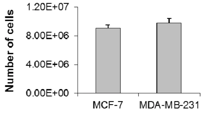
The number of cells that infiltrated the scaffold at day 10 was assessed by total DNA content quantification using the PicoGreen dsDNA assay (Invitrogen, Carlsbad, CA, USA). The total DNA was extracted using DNeasy Blood & Tissue Kit (Qiagen, Duesseldorf, Germany) following the manufacturer’s instructions and 100 µL of DNA mixture were then added to 100 µL of PicoGreen reagent working solution. Fluorescence of the samples was measured with a microplate reader (FLUOstar OPTIMA BMG LABTECH, Ortenberg, Germany) with excitation and emission wavelengths of 480 and 520 nm, respectively. The total number of cells was determined using the conversion factor of 7.7 pg DNA/cell.
4.5 Histological analysis
The collagen constructs were fixed in 10% neutral buffered formaldehyde and dehydrated in a graded series of ethanol. Samples were then embedded in paraffin, sliced at a thickness of 5 µm with a rotating microtome (Leica Biosystems, USA) and mounted on Superfrost Plus microslides (Thermo Fisher Scientific, USA). Hematoxylin and eosin (H&E) staining was performed to evaluate cell morphology and distribution in the scaffold matrix.
37
4.6 LOX inhibition
The activity of LOX was blocked by the irreversible LOX inhibitor, β-aminopropionitrile (BAPN) (Sigma Aldrich) (Nilsson et al., 2016; Yang et al., 2013 and Bondareva et al., 2009). MDA-MB-231 within the scaffold were treated with 500 µM BAPN after 24 hours from the seeding. Administration of BAPN was performed daily until the end of the experiment. At day 10 the culture was stopped, cell proliferation in the presence or absence of LOX inhibition was assessed by MTT assay and the mechanical test was performed.
4.7 Scaffold decellularization
The scaffolds were decellularized in 0.5% Triton X-100 for 1 h at room temperature and then washed 3 times in phosphate saline buffer (PBS). Uncellularized scaffolds were exposed to the same decellularization process to ensure that the experimental conditions did not contribute to modify the scaffold’s mechanical properties. Decellularization was confirmed by the absence of MTT metabolism. The remaining DNA content of the decellularized scaffolds was assessed as follows: total DNA was extracted using the DNeasy Blood & Tissue Kit (Qiagen, Germany) following the manufacturer’s instructions and quantified with a NanoDrop ND-1000 spectrophotometer.
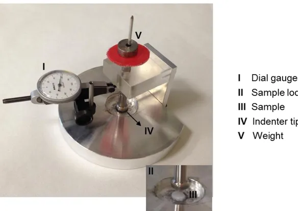
4.8 Development of the low-force testing device
We developed a prototype loading device to apply a state of unconfined uniaxial compression to the collagen scaffolds (Figure 11). This in-house built instrument consisted of a base support with a cylindrical hole (diameter 10 mm, depth 0.5 mm)
38
designed to lodge the scaffold specimen, and a cylindrical piston (diameter 10.1 mm) to indent the specimen with known force values. The applied stress (in kPa) could then be computed by dividing the applied force by the cross section of the specimen. The applied force consisted of a pre-load of 0.09 Newton (corresponding to a pre-stress of 1.8 kPa), which was held constant for 30 seconds; the full load of 0.38 Newton (corresponding to 7.6 kPa) was then constantly applied for 30 seconds. Such holding times were sufficient for the visco-elastic time-dependent deformation to reach an equilibrium. The piston was equipped with a dial gauge (Borletti, Milan, Italy) to measure the piston displacement while the specimen was compressed. The consequent strain (dimensionless) was computed by dividing the measured piston displacement by the height of the specimen. The dial gauge had a resolution of 0.002 mm (corresponding to a resolution of 0.1% for the strain).
Figure 11. Photo of the low-force mechanical testing device developed to measure the compressive modulus of the collagen scaffolds.
39
4.9 Mechanical testing
4.9.1 Low-force testing device
Prior to any measurements the scaffolds were decellularized in 0.5% Triton X-100 for 1 h at room temperature and then washed 3 times in phosphate saline buffer (PBS). Uncellularized scaffolds were exposed to the same decellularization process to ensure that the experimental conditions did not contribute to modify the scaffold’s mechanical properties. The specimens were tested in air immediately after having been removed from the PBS where they were soaked. For each sample, the pre-load of 0.09 Newton was constantly applied for 30 seconds, followed by the full load of 0.38 Newton, applied for other 30 seconds. The stiffness (compressive modulus, in kPa) of the collagen scaffold was computed as the ratio between the increment of stress and increment of strain from the pre-load to the full load values (Beer et al., 2011). The compressive modulus of collagen scaffolds decellularized after a 10-day culture with MCF-7 or MDA-MB-231 was compared with that of uncellularized scaffolds. Five specimens of each type were tested. Each specimen was tested 10 times to improve the quality of the measurement. The compressive modulus of each specimen was computed as the average of the 10 repeated measurements.
4.9.2 Dynamic Mechanical Analysis (DMA)
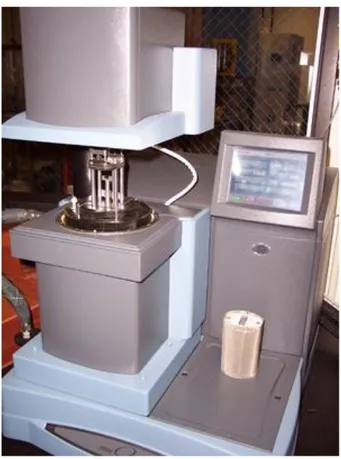
Compression tests were carried out also by dynamic mechanical analysis (DMA) using a Q800 instrument (Norleq, Portugal) (Figure 12), a standard methodology to characterize the mechanical properties of the scaffolds. As described before, prior to any measurements the scaffolds were decellularized in 0.5% Triton X-100 for 1 h at room temperature and then washed 3 times in phosphate saline buffer (PBS). Uncellularized scaffolds were exposed to the same decellularization process to ensure that the experimental conditions
40
did not contribute to modify the scaffold’s mechanical properties. Sample length and diameter were measured with a caliper before each testing. The measurements were performed under compression mode, following cycles of constantly increasing loads, ranging from 0 to 0.01N. The test were carried out at room temperature. For each sample measurements were performed in 5 technical replicates for two biologically independent experiment.
Figure 12. Photo of the Dynamic Mechanical Analysis instrument used to measure the compressive modulus of the collagen scaffolds.
4.9.3 Atomic Force Microscpy (AFM) analysis
The MultiModeNanoscope 8 (Bruker) atomic force microscope was used to perform the nanoindentation test (Figure 13). This instrument is an ultra-high resolution material
41
science microscope, equipped with ScanAsyst and PeakForce Quantitative Nanomechanical imaging modes and Peak Force Tapping technology. This microscope allowed for the determination of the mechanical characteristic of single collagen fibers. Fibers were removed from the bulky scaffold, suspended in MQ water and let dry on a mica surface. A sharp, rigid tip was used for probing the fiber mechanical properties. AFM images were collected using single-beam silicon cantilever probes (nominal tip radius of curvature 10 nm, force constant 0.65 N/m, tip half angle 18°C) from different samples and at random spot surface sampling (at least five areas per each sample). Quantitative mechanical characterization was determined under peak-force tapping mode with 1.0 Hz scan rates and a 200 mV amplitude set point. The DMT model (Derjaguin et al., 1975) was applied to calculate the Young's modulus. Several fibers were indented for each scaffold to calculate their strength.
Figure 13. The Atomic Force Microscope used to measure the mechanical properties of single collagen fibers.
42
4.10 Microscopy analysis
The collagen scaffolds were imaged by SEM and Laser Confocal Microscopy. For SEM imaging, samples were washed in 0.1 M sodium cacodylate buffer pH 7.4 and fixed in 2.5% glutaraldehyde in 0.1 M sodium cacodylate buffer pH 7.4 for 2 h at 4°C. Samples were then dehydrated in a graded series of ethanol, desiccated and sputter-coated with platinum. Images were acquired with a Nova NanoSEM (FEI, USA). For confocal imaging, cells were washed in 1% PBS, fixed with 4% paraformaldehyde for 20 min at room temperature and stained with 10 µM/ml DRAQ5 ™ (ImmunoChemistry Technology, Bloomington, MN, USA). Images where acquired with an A1 laser confocal microscope (Nikon Corporation, Tokyo, Japan), and analyzed with the NIS Elements software (Nikon Corporation, Tokyo, Japan).
4.11 Quantitative Real-Time Reverse Transcription-PCR (qRT-PCR)
Total mRNA was isolated using TRIzol Reagent (Invitrogen, USA) following the manufacturer’s instructions. Five hundred nanograms of RNA were reverse-transcribed using the iScript cDNA Synthesis Kit (BioRad, USA). The final mixture was incubated at 25°C for 5 min, 42°C for 20 min, 47°C for 20 min, 50°C for 15 min and finally a 85°C for 5 min. Real Time-PCR was performed in a 7500 Real-Time PCR System (Applied Biosystems, USA) using the TaqMan universal assay mix (Applied Biosystems). Amplification was performed in a final volume of 20 µl containing 2x Gene Expression Master Mix (Applied Biosystems) and 2 µl of cDNA. The reaction mixtures were all subjected to 2 min at 50°C, 10 min at 95°C followed by 40 PCR cycles at 95°C for 15 s and 60°C for 1 min. The stably expressed endogenous β-actin and HPRT were used as reference genes. Three target markers were analyzed: vimentin (VIM), E-cadherin
43
(CDH1) and lysyl oxidase (LOX). The amount of these transcripts was normalized to the endogenous reference genes and expressed as n-fold mRNA levels relative to a calibrator using the comparative threshold cycle (Ct) value method (ΔΔCt). For all analysis the calibrator used was a mix of the RNA extracted from MCF7 and MDA-MB-231 in standard monolayer culture.
4.12 MTT assay
Cell survival in MDA-MB-231 treated with the LOX inhibitor BAPN was assessed by MTT assay. Briefly, the scaffolds were incubated for 2 hours at 37°C in 0.5 mg/ml of MTT solution (Sigma Aldrich). After solubilization in acidic isopropanol, the absorbance was read at 550 nm. The cell survival was determined comparing the average absorbance of cells in the absence or presence of BAPN treatment.
4.13 Drug testing
Doxorubicin (DOXO) was administered in monolayer cultures or in the 3D scaffolds at the following concentrations: 0.8, 1.6 and 4 μg/ml. Cells were cultured for 24 h before exposure to the drug. Cell viability was assessed by MTT assay, as previously described. Survival percentages were calculated as the average absorbance of cells at each DOXO dose over the absorbance of untreated cells.
44
4.14 Xenograft study
To establish orthotopic breast tumors, MCF-7-Luc2 and MDA-MB-231-Luc2 cells, both marked with a luciferase probe, were suspended in 100 µl of matrigel (BD, USA) at a concentration of 5 x 10¹² cells and orthotopically injected into the right mammary fat pad of 8 week-old female immunodeficient nu/nu nude mice (n. 5 animals for each cell line). Tumor growth was followed by in vivo bioluminescence imaging and caliper measurement every 2-3 days after the injection. After 4 weeks, tumors were resected and embedded in Optimal Cutting Temperature (OCT) compound and cryo-sectioned. All experimental animal procedures were reviewed and approved by the Institutional Animal Care and Use Committee (IACUC) of the Houston Methodist Research Institute (HMRI) protocol number AUP 0614-0033.
4.15 Collagen staining
Trichrome staining and picrosirius red were performed on the OCT embedded frozen tissues to study the tumor collagen matrix and on the collagen scaffold sections. Trichrome staining (Abcam) was performed according to manufacturer’s instructions. For picrosirius red the frozen sections were stained with 0.1% (Direct Red 80) (Sigma Aldrich) sirius red in saturated aqueous solution of picric acid and counterstained with Weigert's hematoxilin. The RGB images were analyzed by ImageJ. At least n. 5 images per group were analyzed to quantify the collagen content.
45
4.16 Statistical analysis
At least three independent experiments were performed. Data are presented as mean ± standard error (SE), with n indicating the number of replicates. For in vitro and in vivo data, differences between groups were assessed by a two-tailed Student's t-test and accepted as significant at p < 0.05.
46
5. Results
5.1 Collagen remodeling in breast xenograft tumors
Orthotopic implantation of MCF-7 and MDA-MB-231 in female immunodeficient mice was performed to investigate the collagen content and architecture in the ECM of in vivo breast tumors. After 4 weeks both cell lines had developed detectable mammary tumors. Bioluminescence imaging and caliper measurement showed that MCF-7 grew considerably slower than MDA-MB-231 displaying a mean tumor volume of 70 ± 15 µm3 compared to 388 ± 68 µm3 for MDA-MB-231 (Figure 14a and 14b).
a
b
Figure 14. Bioluminescence imaging (a) of mammary tumors in nude mice (n=5 for each group) generated by orthotopic injection of MCF-7 or MDA-MB-231 and caliper measurement (b) of tumor volume after 4 weeks (µm3).
47
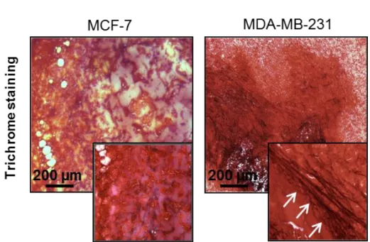
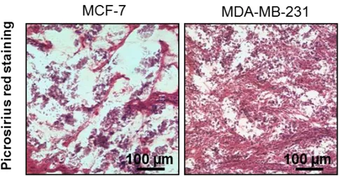
Trichrome staining of resected tumor sections revealed a higher presence of thick collagen fibers in MDA-MB-231 orthotopic tumors compared to those generated by MCF-7 (Figure 15a). Moreover, quantification of the collagen percentage in picrosirius
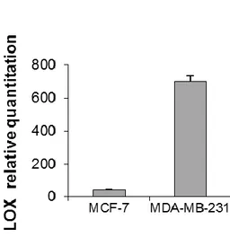
red-stained sections showed that MDA-MB-231 tumors displayed a significantly higher collagen content than that of MCF-7 ones (p = 0.0005) (Figure 15b). Finally, the in vivo expression of the matrix crosslinking enzyme LOX was markedly higher for MDA-MB-231 than for MCF-7 (Figure 15c).
a
Figure 15. (a) Trichrome staining of OCT-embedded xenograft breast tumors generated from orthotopic injection of MCF-7 and MDA-MB-231.
48
b
Figure 15. (b) Picrosirius red staining of OCT-embedded xenograft breast tumors generated from orthotopic injection of MCF-7 and MDA-MB-231, and quantification of the mean percentage of collagen to total area.
49
c
Figure 15. (c) relative quantitation values of LOX in MCF-7 and MDA-MB-231 orthotopic tumors. Data are mean ± SE. * p < 0.05.
5.2 Collagen scaffold macro- and micro- structural properties
Type I collagen scaffolds showed a mean diameter of 9 ± 0.5 mm (Figure 16a) and were 2 mm thick. The pH-driven method, through which the collagen scaffolds were synthetized, enabled the production of a biomimetic material with highly reproducible morphology and tunable macro- and micro-structure, as characterized by Scanning Electron Microscopy (SEM). The size of the pores ranged between 150 to 300 μm, with a mean pore size of 197 ± 25 μm (Figures 16b and 16c). Collagen fibers showed a high degree of structural assembling and preserved their typical D-bands (arrowhead), (Figure 16d) a characteristic axial D-periodic (67 nm) morphology derived from the self-assembly of single collagen molecules. The scaffolds displayed a mean porosity percentage of 87.8 ± 4.1 (Table 1). The average pore volume was 25 x 103 (±1300) μm3, the average pore diameter was 197 (±25) μm and the pore walls were 15 (±1.0) μm thick (Table 1).
50
Figure 16. (a) Pictures of the collagen-based scaffold; (b-d) SEM analysis of the scaffolds at different magnifications with the typical D-bands (d) identified by white arrows and a yellow dotted line to show their axial disposition.
Table 1.
Physical properties of the collagen scaffolds.
Average SD Scaffold Volume (µm 3 ) 57.7 · 10 3 ± 4.5 Pore area (µm 2 ) 24.7 · 10 3 ± 1.3 Volume of collagen (µm 3 ) 8.8 10 3 ± 0.5 Void space (µm 3 ) 48.9 10 3 ± 0.2 Porosity (%) 87.8 ± 4.1
Average porosity diameter (µm) 197 25