ORIGINAL ARTICLE
Predictive value of tender joints
compared to synovitis for structural
damage in rheumatoid arthritis
Peter P Cheung,1Karine Mari,2Valérie Devauchelle-Pensec,3,4
Sandrine Jousse-Joulin,3,4 Maria Antonietta D’Agostino,5Gérard Chalès,6 Philippe Gaudin,7Xavier Mariette,8Alain Saraux,3,4Maxime Dougados9 To cite: Cheung PP, Mari K,
Devauchelle-Pensec V,et al. Predictive value of tender joints compared to synovitis for structural damage in rheumatoid arthritis.RMD Open 2016;2:e000205. doi:10.1136/rmdopen-2015-000205
▸ Prepublication history and additional material is available. To view please visit the journal (http://dx.doi.org/ 10.1136/rmdopen-2015-000205).
Received 31 October 2015 Revised 6 January 2016 Accepted 7 January 2016
For numbered affiliations see end of article.
Correspondence to Dr Peter P Cheung; [email protected]
ABSTRACT
Objective:To evaluate the predictive value of tender joints compared to synovitis for structural damage in rheumatoid arthritis (RA).
Methods:A post hoc analysis was performed on a prospective 2-year study of 59 patients with active RA starting on antitumour necrosis factor (TNF). Tenderness and synovitis was assessed clinically at baseline, followed by blinded ultrasound assessment (B-mode and power Doppler ultrasound (PDUS)) on the hands and feet (2 wrists, 10 metacarpophalangeal, 10 proximal interphalangeal and 10
metatarsophalangeal (MTP) joints). Radiographs of these joints were performed at baseline and at 2 years. The risk of radiographic progression with respect to the presence of baseline tenderness or synovitis, as well as its persistence (after 4 months of anti-TNF), was estimated by OR (95% CI).
Results:Baseline tender joints were the least predictive for radiographic progression (OR=1.53 (95% CI 1.02 to 2.29) p<0.04), when compared to synovitis (clinical OR=2.08 (95% CI 1.39 to 3.11) p<0.001 or PDUS OR=1.80 (95% CI 1.20 to 2.71) p=0.005, respectively). Tender joints with the presence of synovitis were predictive of radiographic progression (OR=1.89 (95% CI 1.25 to 2.85) p=0.002), especially seen in the MTP joints. Non-tender joints with no synovitis were negatively predictive (OR=0.57 (95% CI 0.39 to 0.82) p=0.003). Persistence of tender joints was negatively predictive (OR=0.38 (95% CI 0.18 to 0.78) p=0.009) while persistence of synovitis was predictive (OR=2.41 (95% CI 1.24 to 4.67) p=0.01) of radiographic progression.
Conclusions:Synovitis is better than tenderness to predict for subsequent structural progression. However, coexistence of tenderness and synovitis at the level of an individual joint is predictive of structural damage, particularly in the MTP joints.
Trial registration number:NCT00444691.
INTRODUCTION
Rheumatoid arthritis (RA) is a chronic immune-mediated systemic inflammatory condition with synovitis as the pathological
hallmark, presenting with symptoms of joint pain, swelling and also systemic effects such as fatigue. Synovitis has been recognised as one of the most important predisposing factors for subsequent structural damage, which if untreated leads to significant phys-ical disability.1 2 Therefore, predicting RA outcomes such as structural damage is crucial for optimal clinical management, in order to effectively identify patients who would benefit from appropriate aggressive therapy at an early stage.3
Joint counts for tenderness and swelling have traditionally been used to assess for disease activity and remain as core data sets used in clinical practice and research.4 Initial disease activity measurements were largely based on evaluation of joint tender-ness, such as the Ritchie articular index,5 a
Key messages
What is already known about this subject? ▸ Synovitis ( joint swelling or
ultrasound-confirmed synovitis) can predict structural pro-gression in rheumatoid arthritis, assessed at the individual joint level, but evidence is conflicting for tender joints.
What does this study add?
▸ Tender joints were least predictive compared to synovitis to predict for structural progression.
▸ Coexistence of tender joints and synovitis at the individual level is most useful, particularly in the metatarsophalangeal joints.
How might this impact on clinical practice? ▸ Synovitis ( joint swelling or
ultrasound-confirmed synovitis) should be preferred over tender joints in assessing for potential structural progression.
▸ Tender joints should be interpreted together with synovitis when assessing for potential structural progression.
semiquantitative scale, later simplified by Hart et al.6 Tender joints continue to be an integral part of clinical assessment of disease activity by its inclusion as one of the core variables in both the American College of Rheumatology (ACR) response criteria as well as the disease activity score (DAS).7 8 The tender joint count (TJC) has better interobserver reliability compared to the swollen joint count (SJC); thus, it has a higher weighting than the SJC in the DAS calculation, for example.8–10 It is also more sensitive to change.11 Criteria for reimbursement for biological therapies, inclusion into clinical drug trials and measurement of treatment response usually include a tender joint evaluation.
Longitudinal studies indicate that synovitis, both clin-ical and by ultrasonography, can predict subsequent structural damage in RA both evaluated at the ‘patient’ level (ie, joint counts) and also at the ‘joint’ level.12–18 However, the predictive validity of tender joints for struc-tural damage is less clear. Conflicting results from previ-ous studies were seen with the ability of TJC to predict for radiographic progression. Wolfe and Sharp19 had found a positive correlation of TJC with subsequent radiographic progression, while Smolen et al20 found that TJC was unhelpful as compared to SJC. More importantly, no studies to date have evaluated tender joints at the ‘joint’ level, and neither has there been a comprehensive comparison with synovitis (evaluated clinically and also by ultrasonography). The aim was therefore to evaluate the predictive value of tenderness compared to synovitis for structural damage in RA.
METHODS Study design
This was a post hoc analysis on the original prospective multicentre 2-year study which evaluated the validity of ultrasound-confirmed synovitis scoring systems in RA.18 21 The appropriate ethical committees approved the study and all patients gave their written and verbal informed consent before study enrolment18 22 (NCT00444691).
The study had two phases:
1. Four months of follow-up in patients starting anti-TNF;
2. Assessment of radiographic progression (evaluation of hands and feet) at 2 years.
Patients
Adult patients fulfilling the 1987 ACR criteria for RA23 were included from March 2007 to August 2008 in the rheumatology outpatient clinics of nine French univer-sity teaching hospitals. Patients had to have active disease, enough to require anti-TNF therapy according to the opinion of the investigators. They also needed to have at least more than or equal to six tender and swollen joints on physical examination at time of inclusion.
Centres
Each centre required one dedicated clinician or metrol-ogist (either a rheumatolmetrol-ogist or research nurse) with experience in assessment of joint counts as well as one dedicated ultrasonographer (either a radiologist or rheumatologist) experienced in evaluating synovitis in RA (with at least 70 different examinations).
Collected data
Joints were evaluated clinically for tenderness and swel-ling (clinical synovitis), followed by a blinded assessment for ultrasound (US)-confirmed synovitis in B-mode and power Doppler ultrasound (PDUS) at baseline and 4 months after start of anti-TNF. These included a total of 32 joints for each patient: 10 metacarpophalangeal (MCP), 10 proximal interphalangeal (PIP), 2 wrists and 10 metatarsophalangeal (MTP) joints.
B-mode and PDUS examination was carried out according to a validated scanning method described pre-viously.18 21Evaluation of synovitis, both clinically and by ultrasonography, was standardised among investigators prior to the study during a 1½-day meeting in Brest, France.21 24 Interobserver agreement in clinical synovitis had a κ up to 0.62, while agreement in B-mode and PDUS synovitis ranged up to κ of 0.62 and 0.55, respectively.21 24
US-confirmed synovitis was defined according to the Outcome Measures in Rheumatology Clinical Trials.25–27 B-mode synovitis scoring was evaluated using a four-grade scale from 0 to 326 with the following subjective definitions for each category: grade 0, absence of syn-ovial thickening; grade 1, mild synsyn-ovial thickening; grade 2, moderate synovial thickening; grade 3, marked synovial thickening. PDUS synovitis scoring was also eval-uated using a four-grade scale from 0 to 327with the fol-lowing definitions for each category: grade 0: absence of signal, no intra-articular flow; grade 1: mild, one or two vessels signal (including one confluent vessel) for small joints and two or three signals for large joints (including two confluent vessels); grade 2: moderate confluent vessels (>grade 1) and less than 50% of normal area; grade 3: marked vessels signal in more than half of the synovial area.
For both B-mode and PDUS, a grading of more than or equal to 1 was considered as the presence of US-confirmed synovitis, respectively.
Structural damage
Structural damage of the corresponding joints, assessed clinically, was evaluated by X-rays of hands, wrists and feet; performed at baseline and at 2 years.
Radiographic progression/structural deterioration at the ‘joint’ level was defined as either the appearance or worsening of erosion or joint space narrowing ( JSN). This was recorded as a binary variable for each joint (scored 0=no change, or 1=worsening or appearance of either erosion or JSN) in radiographs performed at 2 years compared to baseline. The level of structural
damage was defined as the total number of joints with radiographic damage (ie, presence of either erosion or JSN) per patient (range, 0–32). A single experienced reader (VD-P) who was aware of the chronologies of the films but blinded to the clinical and US findings assessed all the X-rays, with a previous interobserver reliability (intraclass correlation coefficient) of 0.80–0.90.28–30 Statistical analysis
All analysis was performed using SAS V.9.2. The statis-tical analysis was performed on patients with a complete data set (clinical and US evaluation at baseline and after 4 months of follow–up, and radiological evaluation at baseline and after 2 years).
First, radiographic progression in specific joint regions was evaluated by calculating the percentage of joints with radiological progression. Then baseline structural damage (total number of joints with radiographic damage, ie, presence of either erosion or JSN) was eval-uated to see if there was any relationship with baseline TJC, SJC, US-confirmed B-mode synovitis count or US-confirmed PDUS synovitis count using Spearman’s correlation.
Risk of radiographic progression
The risk of radiographic progression, at the‘joint’ level, with respect to the presence of tenderness, clinical syno-vitis or US-confirmed synovitis at baseline, was estimated by OR (95% CI), using generalised estimating equations (GEE) with a logit link adjusting for within-patient cor-relation (exchangeable corcor-relation matrix), and also other cofactors such as age, gender, disease duration, baseline TJC, baseline SJC, ESR, joint type (PIP, MCP, wrists, MTP) and baseline joint structural damage. These cofactors were determined a priori, as for the pre-vious analysis reported on comparison of the predictive value of clinical and US-confirmed synovitis on radio-graphic progression.18 GEE allows correction for within-patient correlation between time points and produce estimates of time averaged ORs for binomial data.31
The probability of radiographic progression was ini-tially calculated independently on the joints assessed for tenderness, clinical synovitis and US-confirmed synovitis.
The second step of the analysis calculated the prob-ability of radiographic progression when joints were assessed together (tenderness and synovitis, clinically or by US). The findings in the joints were stratified under the following groups: (1) joints that were tender with no synovitis, (2) tender with synovitis, (3) not tender with no synovitis and (4) not tender but with synovitis. The respective groups were compared to the rest of the other joints that did not fulfil that particular definition so that they were coded as binary variables.
Third, to evaluate specifically whether tenderness in particular locations such as MTP joints would less likely be predictive of structural progression, a subgroup ana-lysis was performed by (1) evaluating only the MTP
joints as well as (2) only the joints of the hands (wrist, MCP and PIP joints).
Finally, we checked whether a positive short-term treat-ment effect on synovitis (either clinical or US) or ten-derness enabled prevention of structural progression. For this purpose, joints were divided into two categories: (1) joints that had either persistence of the abnormality at baseline or (2) joints that had normalisation of the abnormality after 4 months of anti-TNF. The probability of observing radiological progression after 2 years with respect to this treatment effect was calculated.
RESULTS
In all, 59 of the 77 recruited patients had completed the study with radiographic evaluation at baseline and 2 years. Table 1shows the clinical characteristics at base-line of patients with (1) complete basebase-line data, (2) 4-month data and (3) complete radiographic data at 2 years. There were no statistical differences. Of the 59 patients with complete radiographic data at 2 years, 81% were female, 73% positive for rheumatoid factor, with a mean age of 56 years and disease duration of 10 years. Patients had active disease with mean DAS in 28 joints of 5.1. Regarding anti-TNF therapy, 34 patients were started on etanercept, 23 on adalimumab and 2 on infliximab.
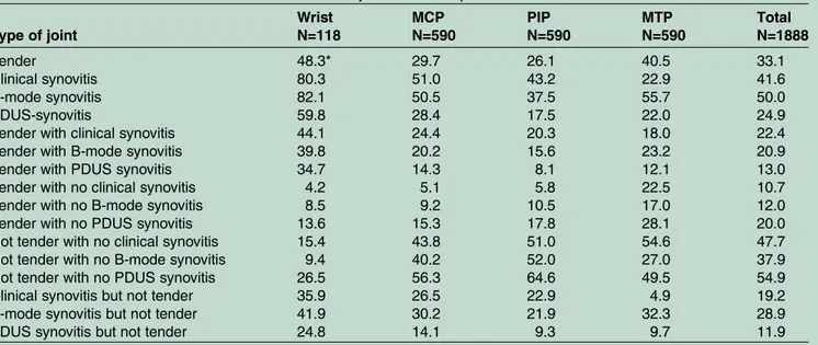
Radiographic progression was observed in 9% of the 1888 evaluated joints (16% of the 119 wrists, 7% of the 590 MCP, 8% of the 590 PIP and 11% of the 590 MTP joints; table 2). At baseline, 33% of 1888 joints were tender, with the wrist, followed by the MTP joints, being more commonly affected. The wrist was also more often accompanied with tenderness and US-confirmed PDUS synovitis. However, 28% of MTP joints were tender with no evidence of active inflammation (US-confirmed PDUS synovitis) at baseline.
Relationship of structural damage with joint counts at baseline
The relationship of TJC, SJC, US-confirmed B-mode synovitis count and US-confirmed PDUS synovitis count with existing structural damage was evaluated at the ‘patient’ level at baseline. SJC, US-confirmed B-mode synovitis count and US-confirmed PDUS synovitis count correlated positively with the degree of structural damage at baseline (r=0.39, 0.27 and 0.51), respectively. This correlation was not seen with TJC.
Value of baseline tender joints (‘joint’ level) to predict for radiographic progression
Tender joints versus clinical synovitis or US-confirmed synovitis
Structural deterioration, defined as either occurrence or worsening in either erosion or joint space narrowing after 2 years, was observed in 11.9% of the 625 tender joints. For joints that had synovitis, structural deterior-ation was observed in 12.1% (clinical synovitis), 12.1% (B-mode) and 15% (PDUS), respectively (table 3).
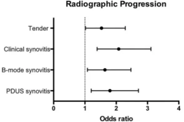
Although both tender joints and synovitis (both clinical and US) at baseline appeared to be predictive of radio-graphic progression at 2 years, baseline tender joints were the least predictive with OR=1.53 (95% CI 1.02 to 2.29) p<0.04. Synovitis, on the other hand, performed better, especially with clinical synovitis with OR=2.08 (95% CI 1.39 to 3.11) p<0.001 (figure 1).
Tender joints and clinical synovitis or US-confirmed synovitis analysed together
Subsequently, we explored the predictive value for radio-graphic progression when tender joints were assessed together with synovitis. Joints that were tender with the presence of synovitis (both clinical and US) appeared to be highly associated with radiographic progression. For
example, of the 422 tender joints with clinical synovitis at baseline, 14% vs 7.5% had radiographic progression with OR=1.89 (95% CI 1.25 to 2.85) p=0.002) (figure 2 and table 3). There was also a trend in favour of a pre-diction of radiographic progression in the case of joints that were not tender but had clinical synovitis.
On the other hand, for 899 joints that were not tender with no clinical synovitis at baseline, only 6.6% of joints progressed, and were negatively associated with radio-graphic progression (OR=0.57 (95% CI 0.39 to 0.82) p=0.003). This negative association was additionally observed with US-confirmed B-mode and PDUS synovitis (OR=0.45 (95% CI 0.29 to 0.72) p<0.001 and OR=0.52 (95% CI 0.35 to 0.77) p=0.001, respectively;figure 3and
table 3).
Table 1 Baseline characteristics of the patients with RA of the various subsets of patients Baseline characteristics
Complete joint and US evaluations
N=76
Complete joint and US evaluations at 4 months N=68 Complete radiographic evaluations N=59 Female, n (%) 64 (84) 56 (82) 48 (81)
Age, years, mean (SD) 55.0 (12.6) 55.7 (11.9) 56.1 (12.4)
Disease duration, years, mean (SD) 11.2 (8.9) 11.1 (9.0) 10.0 (7.7)
Rheumatoid factor-positive, n (%) 59 (78) 51 (75) 43 (73)
DAS28-ESR, mean (SD) 5.1 (1.3) 5.1 (1.3) 5.1 (1.3)
ESR, mm/h, mean (SD) 29.9 (20.8) 30.0 (20.2) 29.7 (20.0)
CRP, mg/L, mean (SD) 17.5 (19.0) 17.1 (15.5) 17.2 (15.7)
HAQ, mean (SD) 1.4 (0.7) 1.5 (0.6) 1.4 (0.6)
Number of DMARDs, mean (SD) 3.1 (1.9) 3.1 (1.9) 3.1 (1.9)
History of anti-TNF, n (%) 23 (30) 21 (31) 19 (32)
History of surgery for RA, n (%) 21 (28%) 18 (27%) 14 (24%)
CRP, C reactive protein; DAS28, disease activity score; DMARD, disease modifying antirheumatic drug; ESR, erythrocyte sedimentation rate; HAQ, Health Assessment Questionnaire; RA, rheumatoid arthritis; TNF, tumour necrosis factor; US, ultrasound.
Table 2 Baseline characteristics of the 1888 evaluated joints of the 59 patients with rheumatoid arthritis Type of joint Wrist N=118 MCP N=590 PIP N=590 MTP N=590 Total N=1888 Tender 48.3* 29.7 26.1 40.5 33.1 Clinical synovitis 80.3 51.0 43.2 22.9 41.6 B-mode synovitis 82.1 50.5 37.5 55.7 50.0 PDUS-synovitis 59.8 28.4 17.5 22.0 24.9
Tender with clinical synovitis 44.1 24.4 20.3 18.0 22.4
Tender with B-mode synovitis 39.8 20.2 15.6 23.2 20.9
Tender with PDUS synovitis 34.7 14.3 8.1 12.1 13.0
Tender with no clinical synovitis 4.2 5.1 5.8 22.5 10.7
Tender with no B-mode synovitis 8.5 9.2 10.5 17.0 12.0
Tender with no PDUS synovitis 13.6 15.3 17.8 28.1 20.0
Not tender with no clinical synovitis 15.4 43.8 51.0 54.6 47.7
Not tender with no B-mode synovitis 9.4 40.2 52.0 27.0 37.9
Not tender with no PDUS synovitis 26.5 56.3 64.6 49.5 54.9
Clinical synovitis but not tender 35.9 26.5 22.9 4.9 19.2
B-mode synovitis but not tender 41.9 30.2 21.9 32.3 28.9
PDUS synovitis but not tender 24.8 14.1 9.3 9.7 11.9
*Percentage proportion involved in the referred joint region
Subanalysis on tender joints in the feet
Radiographic progression was seen in 13% of the 239 tender MTP joints. However, tender joints were not asso-ciated with radiographic progression (OR=1.20 (95% CI 0.73 to 1.97) p=0.47). A higher proportion of joints with US-confirmed PDUS synovitis had radiographic progres-sion (17% of 128 joints) and was the most predictive of all the assessments (OR=2.65 (95% CI 1.33 to 5.27) p=0.005). Joints with US-confirmed B-mode synovitis also had a positive association with OR=2.24 (95% CI 1.23 to 4.10) p=0.009). There was also a trend in favour of prediction of progression with clinical synovitis (see online supplementary table S1).
When tender joints were assessed with synovitis, the predictive value improved. MTP joints which were tender
had more frequent structural progression if there was concurrent synovitis—clinical synovitis, US-confirmed B-mode synovitis or PDUS synovitis (19.2% vs 9.2%, 15.8% vs 9.6% and 21.4% vs 9.2%, respectively). Radiographic progression ranged from OR=1.96 (95% CI 1.15 to 3.36) p=0.014 for clinical assessment (tenderness and clinical synovitis) to OR=2.83 (95% CI 1.56 to 5.15) p<0.001 by US-confirmed synovitis (tenderness and US-confirmed PDUS synovitis; see online supplementary table S2). On the other hand, this clear positive associ-ation was not seen in tender joints without concurrent synovitis, or in the situation when joints had synovitis but were not tender.
When only the small joints of the hands were ana-lysed, joints which were not tender and without synovitis Table 3 Radiological progression of joints at 2 years according to presence of baseline tenderness or synovitis and when tenderness and synovitis analysed together
Radiological progression
Presence Number of joints Number of joints OR (95% CI) p Value Tenderness vs synovitis, analysed independently
Tender No 1263 88 (7.5%) 1.53 (1.02 to 2.29) 0.039 Yes 625 72 (11.9%) Clinical synovitis No 1101 68 (6.6%) 2.08 (1.39 to 3.11) <0.001 Yes 784 91 (12.1%) B-mode synovitis No 939 53 (5.9%) 1.64 (1.08 to 2.47) 0.019 Yes 939 106 (12.1%) PDUS synovitis No 1410 91 (6.9%) 1.80 (1.2 to 2.71) 0.005 Yes 468 68 (15%)
Tenderness and synovitis analysed together Tenderness with no synovitis
Tender+No clinical synovitis No 1685 145 (9.2%) 0.72 (0.42 to 1.22) 0.220
Yes 202 15 (7.6%)
Tender+No B-mode synovitis No 1657 138 (8.9%) 1.32 (0.69 to 2.55) 0.401
Yes 226 22 (10%)
Tender+No PDUS synovitis No 1507 126 (8.9%) 1.11 (0.67 to 1.84) 0.671
Yes 376 34 (9.3%)
Tenderness with synovitis
Tender+clinical synovitis No 1465 103 (7.5%) 1.89 (1.25 to 2.85) 0.002 Yes 422 57 (14%) Tender+B-mode synovitis No 1489 110 (7.9%) 1.44 (0.90 to 2.30) 0.124 Yes 394 50 (13.1%) Tender+PDUS synovitis No 1639 122 (7.9%) 1.73 (1.07 to 2.80) 0.026 Yes 244 38 (16%)
Absence of tenderness and synovitis
Not tender+no clinical synovitis No 987 106 (11.2%) 0.57 (0.39 to 0.82) 0.003
Yes 899 53 (6.4%)
Not tender+no B-mode synovitis No 1170 128 (11.6%) 0.45 (0.29 to 0.72) <0.001
Yes 713 31 (4.6%)
Not tender+no PDUS synovitis No 849 102 (12.4%) 0.52 (0.35 to 0.77) 0.001
Yes 1034 57 (6%)
Absence of tenderness but synovitis
Not tender+clinical synovitis No 1524 125 (8.7%) 1.32 (0.93 to 1.87) 0.116
Yes 362 34 (9.9%)
Not tender+B-mode synovitis No 1338 103 (8%) 1.21 (0.88 to 1.66) 0.241
Yes 545 56 (11.3%)
Not tender+PDUS synovitis No 1659 129 (8.3%) 1.31 (0.89 to 1.91) 0.168
Yes 224 30 (13.9%)
negatively predicted radiographic progression (see online supplementary tables S3 and 4).
Persistence of tenderness versus clinical synovitis or US-confirmed synovitis at 4 months
Finally, the persistence of either tender joints or synovitis with respect to subsequent radiographic progression was evaluated. This was carried out where persistence was compared to joints that had normalisation after 4 months of anti-TNF. Radiographic progression was observed less in joints that remained tender (9.8% vs 19.8%, OR=0.38 (95% CI 0.18 to 0.78) p=0.009)). On the other hand, radiographic progression was observed more frequently in the case when synovitis persisted (16.6% vs 8.9% for clinical synovitis (OR=1.26 (95% CI 0.79 to 2.02), p=0.336); 17.1% vs 5.9% for US-confirmed B-mode synovitis (OR=2.41 (95% CI 1.24 to 4.67), p=0.010) and 20.7% vs 11.9% for US-confirmed PDUS synovitis (OR=1.63 (95% CI 0.75 to 3.57), p=0.218) in joints with versus without persistent synovitis, respectively.
DISCUSSION
This study has confirmed that synovitis, especially detected clinically and also by US, is better than tender joints to predict for subsequent structural deterioration in patients with RA at the‘joint’ level, both at baseline
and its persistence, in the setting of patients who received anti-TNF. However, the data observed in the whole set of joints suggest that the predictive value is best interpreted when both tenderness and synovitis are considered together.
This study has a number of limitations. This was a post hoc analysis of a previous study with the initial primary objective of evaluating the validity of ultrasonography in RA, with a formal follow-up of disease activity up to 4 months. As such, a large proportion of patients from the original study were unable to be included, mainly due to absence of radiographs at 2 years for comparison, and hence contributing to a smaller sample size.
Although there are many known potential predictors of radiographic progression, this analysis (as well as the previous study evaluating synovitis alone18) was focused on the comparison of the variables of tender joints and synovitis, adjusted on cofactors defined a priori. Therefore, this was not a study to extensively explore predictive factors of radiological progression. The list of cofactors remained the same in both analyses to ensure that our results were comparable. In addition, disease activity throughout the total duration of the study was not available and would be an important cofactor when assessing structural progression.
This cohort included patients who had received anti-TNF, which may have reduced the incidence of radiographic progression. However, patients had active disease at inclusion and would have been the group of patients most at risk of getting structural progression. Although metrologists and sonographists participated at a training session, where reliability in clinical synovitis and US-confirmed synovitis was evaluated, there was no formal documentation of tender joint reliability.32 Even though the metrologists were experienced in joint counts, the absence of standardisation in tender joint should be considered in the overall interpretation of results. In addition, the use of a non-validated system for assessment for radiographic progression could have been perceived as a limitation. However, in essence, the analysis was at the ‘joint’ level, and therefore a binary scoring was applied. Although other validated scoring systems such as the Sharp-van der Heidje or even the Figure 1 Ability of baseline tenderness to predict for
radiographic progression at 2 years compared to synovitis.
Figure 2 Ability of tenderness and clinical synovitis assessed together, to predict for radiographic progression at 2 years.
Figure 3 Ability of baseline tenderness and various synovitis definitions (both clinical and ultrasound), assessed together to predict for radiographic progression at 2 years.
Larsen system of scoring would be desirable,33 34 it reflects radiographic damage at the ‘patient’ level and therefore would not be suitable for this analysis. Additionally, radiographic assessment rested only on the joints that were assessed both clinically and by US, thus omitting some of the joints that would be assessed with the aforementioned radiographic scoring methods.
Most of the studies evaluating the predictive validity of tender joints and radiological changes in the past were at the ‘patient’ level (the total number of joints with synovitis and a summed radiological scoring system), and only a few studies compared the predictive ability of tenderness and swelling in the same cohort.19 20 35 Although one study showed that both TJC and SJC were not predictive,35 two studies have indicated that SJC had better predictive validity than TJC.20 36We have focused our study by evaluating the risk of structural progression at the level of the joints. The statistical analysis per-formed also factored into the potential bias of clustering with analysis at the level of the joints by applying GEE. Although GEE is theoretically an analysis for longitu-dinal studies with repeated measures,31 it is still an appropriate way to factor in potential bias of clustering, especially in studies evaluating multiple repeated mea-sures such as joint count assessments.
An important strength of this study is that tender joints were compared with different forms of synovitis. Considering the increasing use of US in the assessment of synovitis, this study is important in order to further validate the role of US in daily clinical disease activity assessment. As in our previous study, there was no differ-ence between clinical and US assessments of synovitis in terms of its predictive validity for structural progres-sion.18 However, this is a cohort of patients who had active disease, while previous evidence on the superiority of US to predict radiographic progression as compared with clinical synovitis assessment was predominantly in patients with low disease activity or in clinical remis-sion.15 41 42 In an active group of patients like our cohort, all forms of synovitis assessment were better than tenderness.
In this study, results suggest that the persistence of tender joints was unlikely to result in structural progres-sion. Although further evaluations are required, one possible explanation would be that these tender joints could exist in the context of the ‘fibromyalgic rheuma-toid’, where joints no longer had active inflammation despite reported joint pain. The small sample size pre-vented further subanalysis.
Despite the fact that tender joints were not as predict-ive as synovitis at baseline for structural progression, the combination of tender joints and synovitis could be clin-ically useful. This may seem obvious, however, has not been formally reported with US and clinical evaluations concurrently at the‘joint’ level. Felson et al37 had evalu-ated combinations of various assessments for predicting structural damage; however, the components included other measures besides the TJC and SJC, evaluated at
the ‘patient’ level. These components were predictive with positive likelihood ratios ranging from 3.2 (95% CI 1.9 to 5.3) up to 8.0 (95% CI 3.6 to 17.8).
In early RA, radiographic damage involves the fifth MTP joint early on, suggesting that the joints of the feet are important to assess,29 38with good interobserver reli-ability.39 It has also been demonstrated that expected joint damage of patients with early RA predominantly occurs in the feet.40 Moreover, in patients who were pre-dominantly foot progressors, the TJC and SJC were lon-gitudinally related to radiographic progression.40 Although we included patients largely with active estab-lished RA, a significant number of MTP joints had radio-graphic progression. Hence, a subanalysis was performed, demonstrating that tender MTP joints are best interpreted in association with synovitis. A tender joint was likely to have radiographic progression only if it was associated with synovitis. One potential limitation was that we did not have information on osteoarthritic changes in the feet, which could have been a potential form of bias.
CONCLUSION
In conclusion, synovitis is better than tender joints to predict structural progression. The coexistence of both tender joints and synovitis is highly predictive of progres-sion. The persistence of tender joints and its negative predictive ability for structural progression should be further evaluated in other cohorts.
Author affiliations
1Division of Rheumatology, National University Hospital, Singapore, Singapore 2RCTs, Lyon, France
3Department of Rheumatology, CHU de la Cavale Blanche, Boulevard Tanguy
Prigent, Brest, France
4EA 2216, INSERM ESPRI, ERI29 Université Bretagne Occidentale, Brest,
France
5Department of Rheumatology, APHP, Ambrose-Paré, Versailles-Saint Quentin
en Yvelines University, Boulogne-Billancourt, France
6Department of Rheumatology, Hôpital Sud, CHU Rennes, Rennes, France 7Department of Rheumatology, University Hospital, Grenoble, Hôpital Sud,
Echirolles, France
8Department of Rheumatology, Hôpitaux Universitaires Paris-Sud; Université
Paris-Sud; INSERM U1012; Le Kremlin Bicêtre, Paris, France
9Department of Rheumatology, Hôpital Cochin, Paris, France
Twitter Follow Alain Saraux at @alain.saraux
Acknowledgements The authors thank Abbvie France for financial assistance with the conduct of this study.
Contributors MD, AS, VD-P, SJ-J, MAD, GC, PG and XM participated in the design of the study and in the collection of data. PPC, KM and MD carried out the design, statistical analysis and drafting of the manuscript. All authors read and approved the final manuscript.
Funding AbbVie France.
Competing interests None declared. Patient consent Obtained.
Ethics approval Assistance Publique Hôpitaux de Paris.
Data sharing statement Additional unpublished data are available from the study on request to [email protected].
Open Access This is an Open Access article distributed in accordance with the Creative Commons Attribution Non Commercial (CC BY-NC 4.0) license, which permits others to distribute, remix, adapt, build upon this work non-commercially, and license their derivative works on different terms, provided the original work is properly cited and the use is non-commercial. See: http:// creativecommons.org/licenses/by-nc/4.0/
REFERENCES
1. McInnes IB, O’Dell JR. State of the art: rheumatoid arthritis. Ann Rheum Dis2010;69:1889–906.
2. Welsing PM, van Gestel AM, Swinkels HL, et al. The relationship between disease activity, joint destruction, and functional capacity over the course of rheumatoid arthritis.Arthritis Rheum
2001;44:2009–17.
3. Soubrier M, Dougados M. How to assess early rheumatoid arthritis in daily clinical practice.Best Pract Res Clin Rheumatol
2005;19:73–89.
4. Scott DL, Antoni C, Choy EH, et al. Joint counts in routine practice. Rheumatology (Oxford)2003;42:919–23.
5. Ritchie DM, Boyle JA, McInnes JM, et al. Clinical studies with an articular index for the assessment of joint tenderness in patients with rheumatoid arthritis. Q J Med 1968;37:393–406.
6. Hart LE, Tugwell P, Buchanan W, et al. Grading of tenderness as a source of interrater error in the Ritchie articular index. J Rheumatol 1985;12:716–17.
7. Felson DT, Anderson JJ, Boers M, et al. The American College of Rheumatology preliminary core set of disease activity measures for rheumatoid arthritis clinical trials. The Committee on outcome measures in rheumatoid arthritis clinical trials.Arthritis Rheum 1993;36:729–40.
8. van der Heijde DM, van’t Hof M, van Riel PL, et al. Development of a disease activity score based on judgment in clinical practice by rheumatologists. J Rheumatol 1993;20:579–81.
9. Cheung PP, Ruyssen-Witrand A, Gossec L, et al. Reliability of patient self-evaluation of swollen and tender joints in rheumatoid arthritis: a comparison study with ultrasonography, physician, and nurse assessments.Arthritis Care Res (Hoboken)2010;62:1112–19. 10. Cheung PP, Gossec L, Mak A, et al. Reliability of joint counts
assessment in rheumatoid arthritis: a systematic literature review. Seminars Arthritis Rheum2014;43:721–9.
11. Anderson JJ, Felson DT, Meenan RF, et al. Which traditional measures should be used in rheumatoid arthritis clinical trials? Arthritis Rheum1989;32:1093–9.
12. Bøyesen P, Haavardsholm EA, van der Heijde D, et al. Prediction of MRI erosive progression: a comparison of modern imaging modalities in early rheumatoid arthritis patients.Ann Rheum Dis 2011;70:176–9.
13. Lillegraven S, Bøyeson P, Hammer HB, et al. Tenosynovitis of the extensor carpi ulnaris tendon predicts erosive progression in early rheumatoid arthritis.Ann Rheum Dis2011;70:2049–50.
14. Aletaha D, Smolen JS. Joint damage in rheumatoid arthritis progresses in remission according to the disease activity score in 28 joints and is driven by residual swollen joints.Arthritis Rheum 2011;63:3702–11.
15. Foltz V, Gandjbakhch F, Etchepare F, et al. Power Doppler ultrasound, but not low-field magnetic resonance imaging, predicts relapse and radiographic disease progression in rheumatoid arthritis patients with low levels of disease activity.Arthritis Rheum 2012;64:67–76.
16. Naredo E, Collado P, Cruz A, et al. Longitudinal power Doppler ultrasonographic assessment of joint inflammatory activity in early rheumatoid arthritis: predictive value in disease activity and radiologic progression.Arthritis Rheum2007;57:116–24.
17. Molenaar ET, Voskuyl AE, Dinant HJ, et al. Progression of radiologic damage in patients with rheumatoid arthritis in clinical remission. Arthritis Rheum2004;50:36–42.
18. Dougados M, Devauchelle-Pensec V, Ferlet JF, et al. The ability of synovitis to predict structural damage in rheumatoid arthritis: a comparative study between clinical examination and ultrasound. Ann Rheum Dis2013;72:665–71.
19. Wolfe F, Sharp JT. Radiographic outcome of recent-onset rheumatoid arthritis: a 19-year study of radiographic progression. Arthritis Rheum1998;41:1571–82.
20. Smolen JS, Van Der Heijde DM, St Clair EW, et al. Predictors of joint damage in patients with early rheumatoid arthritis treated with
high-dose methotrexate with or without concomitant infliximab: results from the ASPIRE Trial.Arthritis Rheum2006;54:702–10. 21. Dougados M, Jousse-Joulin S, Mistretta F, et al. Evaluation of
several ultrasonography scoring systems for synovitis and comparison to clinical examination: results from a prospective multicentre study of rheumatoid arthritis.Ann Rheum Dis 2010;69:828–33.
22. Association de Recherche Clinique en Rhumatologie. Sensitivity of Echography in Arthritis (SEA). In: ClinicalTrials.gov. Bethesda, MD: National Library of Medicine (US), 2000 [cited 2015 Dec 27]. http:// clinicaltrials.gov/show/NCT00444691 NLM Identifier: NCT00444691. 23. Arnett FC, Edworthy SM, Bloch DA, et al. The American
Rheumatism Association 1987 revised criteria for the classification of rheumatoid arthritis.Arthritis Rheum1988;31:315–24.
24. Jousse-Joulin S, d’Agostino MA, Marhadour T, et al. Reproducibility of joint swelling assessment by sonography in patients with long-standing rheumatoid arthritis (SEA-Repro study Part II). J Rheumatol2010;37:938–45.
25. Wakefield RJ, Balint PV, Szkudlarek M, et al. Musculoskeletal ultrasound including definitions for ultrasonographic pathology. J Rheumatol 2005;32:2485–7.
26. Scheel AK, Hermann KG, Kahler E, et al. A novel ultrasonographic synovitis scoring system suitable for analyzing finger joint inflammation in rheumatoid arthritis.Arthritis Rheum 2005;52:733–43.
27. Szkudlarek M, Court-Payen M, Jacobsen S, et al. Interobserver agreement in ultrasonography of the finger and toe
joints in rheumatoid arthritis.Arthritis Rheum2003; 48:955–62.
28. Devauchelle-Pensec V, Saraux A, Jousse S, et al. Performance of hand radiographs in predicting the diagnosis in patients with early arthritis. J Rheumatol 2006;33:1511–15.
29. Devauchelle-Pensec V, Saraux A, Berthelot JM, et al. Ability of foot radiographs to predict rheumatoid arthritis in patients with early arthritis. J Rheumatol 2004;31:66–70.
30. Devauchelle-Pensec V, Saraux A, Berthelot JM, et al. Ability of hand radiographs to predict a further diagnosis of
rheumatoid arthritis in patients with early arthritis. J Rheumatol 2001;28:2603–7.
31. Zegler SL, Liang KY. Longitudinal data analysis for discrete and continuous outcomes.Biometrics1986;42:121–30.
32. Marhadour T, Jousse-Joulin S, Chalès G, et al. Reproducibility of joint swelling assessment in long-lasting rheumatoid arthritis: influence on disease activity score-28 values (SEA-Repro study part I).J Rheumatol2010;37:932–7.
33. Van der Heijde DM, van Riel PL, Nuver-Zwart HH, et al. Effects of hydroxychloroquine and sulphasalazine on progression of joint damage in rheumatoid arthritis.Lancet1989;1:1036–8. 34. Larsen A, Dale K, Eek M. Radiographic evaluation of rheumatoid
arthritis and related conditions by standard reference films. Acta Radiol Diagn1977;18:481–91.
35. Combe B, Dougados M, Goupille P, et al. Prognostic factors for radiographic damage in early rheumatoid arthritis: a multiparameter prospective study.Arthritis Rheum2001;44:1736–43.
36. Weinblatt ME, Keystone EC, Cohen MD, et al. Factors associated with radiographic progression in patients with rheumatoid arthritis who were treated with methotrexate.J Rheumatol 2011;38:242–6.
37. Felson DT, Smolen JS, Wells G, et al. American College of Rheumatology/European League Against Rheumatism Provisional definition of remission in rheumatoid arthritis for clinical trials. Arthritis Rheum2011;63:573–86.
38. Hulsmans HM, Jacobs JW, van der Heijde DM, et al. The course of radiologic damage during the first six years of rheumatoid arthritis. Arthritis Rheum2000;43:1927–40.
39. Devauchelle-Pensec V, Josseaume T, Samjee I, et al. Ability of oblique radiographs to detect erosions in early arthritis: results in the ESPOIR cohort.Arthritis Rheum2008;59:1739–4.
40. Bakker MF, Jacobs JW, Kruize AA, et al. Misclassification of disease activity when assessing individual patients with early rheumatoid arthritis using disease activity indices that do not include joints of feet.Ann Rheum Dis2012;71:830–5.
41. Brown AK, Conaghan PG, Karim Z, et al. An explanation for the apparent dissociation between clinical remission and continued structural deterioration in rheumatoid arthritis.Arthritis Rheum 2008;58:2958–67.
42. Saleem B, Brown AK, Keen H, et al. Disease remission state in patients treated with the combination of tumor necrosis factor blockade and methotrexate or with disease-modifying antirheumatic drugs: a clinical and imaging comparative study.Arthritis Rheum 2009;60:1915–22.