Research Article
Clinicopathological and Molecular Findings in a Case of Canine
Anaplasma phagocytophilum Infection in Northern Italy
Francesco Dondi,
1Samanta Russo,
1Chiara Agnoli,
1Nicola Mengoli,
2Andrea Balboni,
1Alberto Alberti,
3and Mara Battilani
11Department of Veterinary Medical Sciences, Alma Mater Studiorum, University of Bologna, Via Tolara di Sopra 50, 40064 Ozzano dell’Emilia, Italy
2“Dr Nicola Mengoli” Veterinary Clinic, Via Di Mezzo 51, 40060 Toscanella di Dozza, Italy 3Department of Veterinary Medicine, University of Sassari, Via Vienna 2, 07100 Sassari, Italy
Correspondence should be addressed to Francesco Dondi; [email protected] Received 8 April 2014; Accepted 22 May 2014; Published 5 June 2014 Academic Editor: Hiromi Nishida
Copyright © 2014 Francesco Dondi et al. This is an open access article distributed under the Creative Commons Attribution License, which permits unrestricted use, distribution, and reproduction in any medium, provided the original work is properly cited.
A documented case of canine granulocytic anaplasmosis coupled with the molecular characterization of the etiological agent is reported for the first time in Northern Italy. The patient showed nonspecific clinical signs such as fever and weight loss. The most relevant clinicopathological findings were thrombocytopenia, hypoalbuminemia, and normal azotemic proteinuria consistent with glomerular diseases. Blood smear examination revealed the presence of intracytoplasmatic inclusions in neutrophils associated with high positive serology for Anaplasma phagocytophilum. PCR analysis and sequencing of the amplicon confirm serological diagnosis of A. phagocytophilum. Phylogenetic analysis evidenced that the detected bacterial strain belongs to the A. phagocytophilum Europe 1 lineage. Data indicates that A. phagocytophilum circulates in natural environments of Emilia-Romagna region (Northern Italy) and its prevalence in dogs could be underestimated because the clinical signs are frequently nonspecific and a certain diagnosis requires the combination of clinicopathological and molecular assays. Pets living in this area should be regularly monitored and treated for ectoparasites to minimize health risks for humans and pets. Also, surveillance of A. phagocytophilum should be improved in Northern Italy and canine anaplasmosis should be considered in differential diagnosis of persistent proteinuria.
1. Introduction
Tick-borne rickettsiae in the genera Ehrlichia and Anaplasma are emerging pathogens with both veterinary and human health implications [1]. Recent taxonomic changes have reclassified the families Rickettsiaceae and Anaplasmataceae in the order Rickettsiales with the unification of some species of Ehrlichia under the unique species Anaplasma
phagocy-tophilum [2]. A. phagocytophilum is maintained in nature
in an enzootic cycle including Ixodes spp. ticks as the main competent vector and a wide range of mammalian species acts as reservoir and source of infection for domestic animals and humans [1]. In Italy, A. phagocytophilum has been widely detected in ticks [3,4], wild ungulates [5], domestic animals including pets [6–8], and humans [9]. A. phagocytophilum in dogs causes nonspecific clinical and clinicopathological find-ings as fever, anorexia, weight loss, and thrombocytopenia
[10]. Additional diagnostic procedures in clinical practice are frequently necessary in order to reach a correct diagnosis of canine anaplasmosis.
In this study, we report a case of canine granulocytic anaplasmosis documented by complete clinical and clinico-pathological description and by molecular investigation of the etiological agent, in Northern Italy. Furthermore, two asymptomatic dogs sharing the same household with the reported case and showing serological evidences of anaplas-mosis were evaluated.
2. Materials and Methods
2.1. Hematology, Chemistry, Urinalysis, Serology, and Vector-Borne Pathogens Screening. Hematology and chemistry were Volume 2014, Article ID 810587, 6 pages
performed at days 0, 3, 10, and 30 of illness using an auto-mated hematology system (ADVIA 2120, Siemens Healthcare Diagnostics, Tarrytown NY, USA) and a chemistry analyzer (AU 400, Olympus/Beckman Coulter, Munich, Germany), respectively. Blood smear Romanowsky staining and micro-scopic evaluation were performed. C-reactive protein (CRP) (CRP OSR6147, Olympus/Beckman Coulter, Munich, Ger-many) and urinary protein to creatinine ratio (UPC) and urinary albumin to creatinine ratio (UAC, Microalbumin OSR6167, Olympus/Beckman Coulter, Munich, Germany) were performed as previously reported [11, 12]. Indirect immunofluorescent antibody (IFA) titers for A.
phagocy-tophilum, Ehrlichia canis, and Leishmania spp. were
quan-tified (MegaScreen FLUOANAPLASMA ph, MegaScreen FLUOEHRLICHIA c., MegaCor Diagnostik, H¨oerbranz, Austria; FLUOLEISH, Virbac, Carros, France). Titers were considered as indicative of infection if>1 : 40. Microscopic agglutination test (MAT) for Leptospira spp. was performed at the Animal National Leptospirosis Referral Laboratory (Istituto Zooprofilattico Sperimentale della Lombardia e dell’Emilia-Romagna, Bologna, Italy); antibody titers were determined against 8 serogroups (Australis, Ballum, Cani-cola, Grippotyphosa, Icterohemorrhagiae, Pomona, Sejroe, and Tarassovi). The dog was also tested for Dirofilaria immitis antigen (SNAP Heartworm RT Test, IDEXX laboratories Inc., Westbrook, USA).
2.2. Molecular and Phylogenetic Analysis. Genomic DNA
extraction from EDTA-blood samples was performed using a commercial kit (NucleoSpin Tissue Mini Kit, Macherey-Nagel, D¨uren, DE). DNA amplification was implemented with conventional PCR as previously described [13]: a couple of degenerate primers, targeting a fragment of the heat shock protein (groEL), was used to detect DNA from all known Ehrlichia and Anaplasma spp. A recombinant plasmid containing a portion of the groEL gene of A. phagocytophilum was used as positive control [8]. Amplified DNA product was purified and directly sequenced. The nucleotide sequences obtained were assembled and analyzed by BLAST web inter-face (http://blast.ncbi.nlm.nih.gov/Blast.cgi). A nucleotide-nucleotide search (BLASTN), performed with the default settings, has allowed us to reassemble the sequence obtained in this study to A. phagocytophilum. The sequence was submitted to the GenBank database with accession number KF778380 (strain 393/2013). Multiple alignments with ref-erence sequences available from the GenBank nucleotide database were generated using the CLUSTAL W method [14] and phylogenetic analysis was performed using maximum likelihood (ML) methods implemented on MEGA version 5.2.2 [15].
3. Results
3.1. Case Report. A 12-year-old spayed female English Setter
dog (Case 1) was presented to the authors’ veterinary teach-ing hospital (VTH) followteach-ing a 4-day history of anorexia, weakness, and polyuria/polydipsia. The referring practitioner reported no previous signs of illness or recent treatments
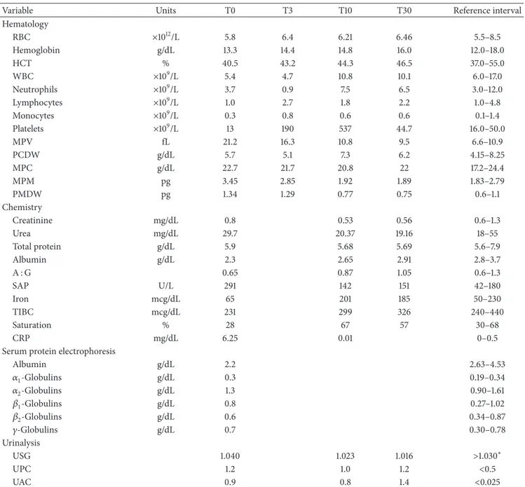
and a past occasional exposure to ticks. Routine vaccina-tions (including Leptospira Canicola and Icterohemorrha-giae serovars), heartworm, and flea/tick chemoprophylaxis were current. The dog was mostly an outdoor pet and was usually used for hunting activities; however, no trav-els outside the region (Emilia-Romagna, Northern Italy) were reported. Physical examination revealed hyperthermia (39.8∘C), tachypnea (40 breathe/min), and tachycardia (80 pulse/min). Thoracic radiographs and abdominal ultrasound were unremarkable. Laboratory variables results are reported in Table1. At the blood smear examination a mean of 10% of neutrophils presented cytoplasmic inclusions that were characterized by blue-violet aggregates of punctiform bodies, coherent with morulae of A. phagocytophilum (Figure1(b)). Final interpretation of the blood smear was mild to mod-erate leukopenia, severe thrombocytopenia, and suspected granulocytic anaplasmosis. All serological tests as well as the
Dirofilaria immitis antigen test resulted in negative except for
A. phagocytophilum IFA titer (≥1 : 1280).
The dog was hospitalized and immediately treated with oral doxycycline (Vibravet, 10 mg/kg q24, for 28 days). No bleeding tendency was detected and the platelet count returned within the reference interval (WRI) 48 hours after starting the therapy. Clinical signs and clinicopathological abnormalities disappeared completely at day 10, with the exception of proteinuria and albuminuria. At day 30 protein-uria persisted and oral enalapril (Enacard, 0.5 mg/kg q12) was started. No renal biopsy was performed. To date (day 305) the dog is completely asymptomatic; however, it is still presents mild persistent proteinuria and albuminuria.
Further two asymptomatic dogs (cases 2 and 3), both English Setter, female, 7-year-old, sharing the same house-hold of the reported clinical case, were referred to the authors’ VTH and the same diagnostic protocol reported above was applied. They showed only high A. phagocytophilum IFA titer (≥1 : 1280) and were treated with oral doxycycline (Vibravet, 10 mg/Kg q24, for 28 days). Other clinicopathological vari-ables were WRI.
3.2. Molecular and Phylogenetic Analysis. Positive PCR
prod-uct of the expected size of 600 bp, corresponding to a frag-ment of the heat shock protein (groEL) gene was observed for Case 1. Cases 2 and 3 resulted in negative. A nucleotide sequence of 467 bp was obtained from the amplicon detected in Case 1. The partial groEL gene sequence obtained was ana-lyzed by BLAST web interface and it resulted in having 100% of identity with analogous sequences of A. phagocytophilum present in GenBank.
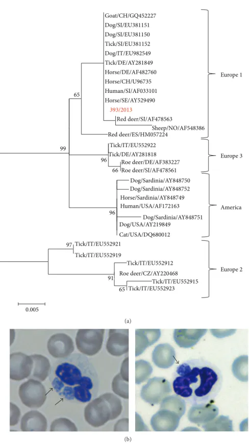
The nucleotide alignment showed complete identity between strain 393/2013 and several A. phagocytophilum strains detected in various hosts and countries (Horse/ SE/AY529490, Human/SI/AF033101, Horse/CH/U96735, Horse/DE/AF482760, Tick/DE/AY281849, Dog/IT/ EU982549, Tick/SI/EU381152, Dog/SI/EU381150, Dog/SI/ EU381151, and Goat/CH/GQ452227). Identity values among the 393/2013 strain and the reference strains ranged from 92 (Dog/Sardinia/AY848751) to 99,7% (Red deer/ SI/AF478563). Phylogenetic tree showed four main clusters, supported by significant bootstrap values, consistent with
America Goat/CH/GQ452227 Dog/SI/EU381151 Dog/SI/EU381150 Tick/SI/EU381152 Dog/IT/EU982549 Tick/DE/AY281849 Horse/DE/AF482760 Horse/CH/U96735 Human/SI/AF033101 Horse/SE/AY529490 393/2013 Red deer/SI/AF478563 Sheep/NO/AF548386 Red deer/ES/HM057224 Tick/IT/EU552922 Tick/DE/AY281818 Roe deer/DE/AF383227 Roe deer/SI/AF478561 Dog/Sardinia/AY848750 Dog/Sardinia/AY848752 Horse/Sardinia/AY848749 Human/USA/AF172163 Dog/Sardinia/AY848751 Dog/USA/AY219849 Cat/USA/DQ680012 Tick/IT/EU552921 Tick/IT/EU552919 Tick/IT/EU552912 Roe deer/CZ/AY220468 Tick/IT/EU552915 Tick/IT/EU552923 97 91 65 96 66 96 99 0.005 65 Europe1 Europe3 Europe2 (a) (b)
Figure 1: (a) ML tree based on the GroEL alignment. The following reference A. phagocytophilum strains detected in several hosts from various parts of the world were obtained from GenBank and included in the molecular analysis: America lineage, accession numbers: AY848750; AY848752; AY848749; AF172163; AY848751; AY219849; DQ680012; Europe 1 lineage, accession numbers: AF033101; GQ452227; EU381151; EU381150; EU381152; EU982549; AY281849; AF482760; U96735; AY529490; AF478563; AF548386; HM057224; Europe 2 lineage, accession numbers: EU552921; EU552919; EU552912; AY220468; EU552915; EU552923; and Europe 3 lineage, accession numbers: EU552922; AY281818; AF383227; AF478561. To assess support for individual nodes, bootstrap resampling values were estimated with 1000 replicates. (b) Blood smear (May-Grunwald Giemsa staining) showing A. phagocytophilum morulae (black arrows) in the cytoplasm of neutrophil granulocytes of Case 1.
Table 1: Pertinent clinicopathological findings of Case 1.
Variable Units T0 T3 T10 T30 Reference interval
Hematology RBC ×1012/L 5.8 6.4 6.21 6.46 5.5–8.5 Hemoglobin g/dL 13.3 14.4 14.8 16.0 12.0–18.0 HCT % 40.5 43.2 44.3 46.5 37.0–55.0 WBC ×109/L 5.4 4.7 10.8 10.1 6.0–17.0 Neutrophils ×109/L 3.7 0.9 7.5 6.5 3.0–12.0 Lymphocytes ×109/L 1.0 2.7 1.8 2.2 1.0–4.8 Monocytes ×109/L 0.3 0.8 0.6 0.6 0.1–1.4 Platelets ×109/L 13 190 537 44.7 16.0–50.0 MPV fL 21.2 16.3 10.8 9.5 6.6–10.9 PCDW g/dL 5.7 5.1 7.3 6.2 4.15–8.25 MPC g/dL 22.7 21.7 20.8 22 17.2–24.4 MPM pg 3.45 2.85 1.92 1.89 1.83–2.79 PMDW pg 1.34 1.29 0.77 0.75 0.6–1.1 Chemistry Creatinine mg/dL 0.8 0.53 0.56 0.6–1.3 Urea mg/dL 29.7 20.37 19.16 18–55 Total protein g/dL 5.9 5.68 5.69 5.6–7.9 Albumin g/dL 2.3 2.65 2.91 2.8–3.7 A : G 0.65 0.87 1.05 0.6–1.3 SAP U/L 291 142 151 42–180 Iron mcg/dL 65 201 185 50–230 TIBC mcg/dL 231 299 326 240–440 Saturation % 28 67 57 30–68 CRP mg/dL 6.25 0.01 0–0.5
Serum protein electrophoresis
Albumin g/dL 2.2 2.63–4.53 𝛼1-Globulins g/dL 0.3 0.19–0.34 𝛼2-Globulins g/dL 1.3 0.90–1.61 𝛽1-Globulins g/dL 0.8 0.27–1.02 𝛽2-Globulins g/dL 0.6 0.34–0.87 𝛾-Globulins g/dL 0.7 0.30–0.78 Urinalysis USG 1.040 1.023 1.016 >1.030∗ UPC 1.2 1.0 1.2 <0.5 UAC 0.9 0.8 1.4 <0.025
RBC: red blood cells; HCT: hematocrit value; WBC: white blood cells; MPV: mean platelet volume; MPC: mean platelet component; PCDW: platelet concentration distribution width; MPM: mean platelet mass; PMDW: platelet mass distribution width; A : G: albumin to globulin ratio; SAP: serum alkaline phosphatase; TIBC: total iron binding capacity; CRP: C-reactive protein; USG: urine specific gravity; UPC: urine protein to creatinine ratio; UAC: urine albumin to creatinine ratio.
∗Canine adequate USG.
previous observation and with the accepted nomenclature (Figure1(a)) [5, 8]. The 393/2013 strain is included in the cluster Europe 1, containing A. phagocytophilum strains detected in various hosts, human included. The mean distance calculated with MEGA software between Europe 1 versus other groups ranged from 0.063 (Europe 1 and Europe 2) to 0.017 (Europe 1 and America).
4. Discussion
In Italy, A. phagocytophilum infection was detected in humans and wild and domestic animals [5–7,9]; a few reports have documented the evidence of A. phagocytophilum infection in nonruminant domestic animals such as cats, dogs, and horses [8,16]. Most cases of canine granulocytic anaplasmosis were reported in Europe [17–19]; however, in Italy just one
case was clinically described (Sicily, Southern Italy) and documented by molecular characterization of the bacterial strain [20]. Natural infection by Anaplasma spp. in pet animals frequently goes undetected, because the disease may be subclinical or clinical findings are nonspecific: often, the only signs are fever, depression, and weight loss, and the most common laboratory findings is thrombocytopenia [10]. In clinical practice, diagnosis of anaplasmosis in dogs should be accomplished by combining history, clinical signs, and clinicopathological analysis, including identification of morulae-containing granulocytes on blood smear, serology, and PCR. Diagnostic assays to detect Anaplasma spp. infec-tion, however, present some limitations mainly due to short duration of bacteremia and chronic phase of infection.
In this report, we documented clinical and clinicopatho-logical manifestations of A. phagocytophilum infection as well as molecular characterization of the bacterial strain detected in a dog. The clinical signs reported in Case 1 supported the diagnosis of infectious tick-borne disease; however, they were nonspecific. Clinicopathological findings in combination with granulocytic morulae, high IFA titer against A. phagocytophilum, and even PCR results, allowed clinicians to confirm the etiology. Interestingly, clinical pre-sentation of Case 1 was compatible with a subclinical non-azotemic proteinuric renal disease probably sustained by an infection-associated glomerulopathy as previously suggested [21]. Molecular characterization of the A. phagocytophilum strain 393/2013 showed that it belongs to Europe 1 lineage, according to the nomenclature introduced by other authors [8]. Phylogenetic tree showed a clear separation of the strains in European and American lineages, as described previously [5,8], with a strong statistical support for a partitioning of strains based on sampling location. The nucleotide sequence of 393/2013, in the fragment of groEL gene sequenced, is identical to A. phagocytophilum strains detected in various hosts (humans, horses and ticks) and geographical areas, as well as it is identical to the sequence EU982549 available on GenBank, an A. phagocytophilum isolate detected in the pleural fluid of dog in Emilia-Romagna region (Northern Italy). The epidemiological significance of genetic variants of
A. phagocytophilum is poorly understood, but previous
stud-ies showed that the Europe 2 genotype was associated with roe deer, whereas the Europe 1 genotype was associated with a wider host range, including both domestic and wild animals [5]. The genetic characteristics do not seem to be clinically or ecologically meaningful and multiple unique strains of A.
phagocytophilum with distinct host tropisms can circulate in
the same geographic area [22]. In order to understand the risk factors associated with transmission of a tick-borne pathogen to humans and domestic animals, several studies of molec-ular epidemiology were carried out also in Northern Italy. Previous molecular surveys in the Emilia Romagna region have been performed through collecting ticks after having been dragged and removed from wild and domestic animals, dogs included. These surveys have shown the presence of A.
phagocytophilum [4,23]. This data, in addition to our case
report, demonstrated that A. phagocytophilum circulates in the natural environment; therefore, pets living in this area should be regularly monitored and treated for ectoparasites
to minimize health risks for humans and pets as well the surveillance of A. phagocytophilum should be increased in Northern Italy.
5. Conclusion
At the knowledge of the authors, this is the first docu-ment case of canine granulocytic anaplasmosis reported in Northern Italy. Our data indicates that A. phagocytophilum prevalence in dogs could be underestimated because the clin-ical signs are frequently nonspecific and a certain diagnosis requires the combination of clinicopathological and molec-ular assays. Surveillance for A. phagocytophilum could be increased also in Northern Italy and canine anaplasmosis should be considered in differential diagnosis of persistent proteinuria.
Conflict of Interests
The authors declare that there is no conflict of interests regarding the publication of this paper.
References
[1] Z. Woldehiwet, “The natural history of Anaplasma
phagocy-tophilum,” Veterinary Parasitology, vol. 167, no. 2–4, pp. 108–122,
2010.
[2] J. S. Dumler, A. F. Barbet, C. P. J. Bekker et al., “Reorganization of genera in the families Rickettsiaceae and Anaplasmataceae in the order Rickettsiales: unification of some species of Ehrlichia with Anaplasma, Cowdria with Ehrlichia and Ehrlichia with
Neorickettsia, descriptions of six new species combinations and
designation of Ehrlichia equi and “HGE agent” as subjective synonyms of Ehrlichia phagocytophila,” International Journal of
Systematic and Evolutionary Microbiology, vol. 51, no. 6, pp.
2145–2165, 2001.
[3] Y. O. Sanogo, P. Parola, S. Shpynov et al., “Genetic diversity of bacterial agents detected in ticks removed from asymptomatic patients in Northeastern Italy,” Annals of the New York Academy
of Sciences, vol. 990, pp. 182–190, 2003.
[4] S. Aureli, J. E. Foley, R. Galuppi, D. Rejmanek, C. Bonoli, and M. P. Tampieri, “Anaplasma phagocytophilum in ticks from parks in the Emilia-Romagna region of northern Italy,” Veterinaria
Italiana, vol. 48, no. 4, pp. 413–423, 2012.
[5] G. Carpi, L. Bertolotti, E. Pecchioli, F. Cagnacci, and A. Rizzoli, “Anaplasma phagocytophilum groEL gene heterogeneity in
Ixodes ricinus larvae feeding on roe deer in Northeastern Italy,” Vector-Borne and Zoonotic Diseases, vol. 9, no. 2, pp. 179–184,
2009.
[6] R. Zobba, A. G. Anfossi, M. L. P. Parpaglia et al., “Molecular investigation and phylogeny of Anaplasma spp. in Mediter-ranean ruminants reveal the presence of neutrophil-tropic strains closely related to A. platys,” Applied and Environmental
Microbiology, vol. 80, no. 1, pp. 271–280, 2014.
[7] A. Alberti, M. F. Addis, O. Sparagano et al., “Anaplasma
phago-cytophilum, Sardinia, Italy,” Emerging Infectious Diseases, vol.
11, no. 8, pp. 1322–1324, 2005.
[8] A. Alberti, R. Zobba, B. Chessa et al., “Equine and canine
Anaplasma phagocytophilum strains isolated on the island of
strains from the United States,” Applied and Environmental
Microbiology, vol. 71, no. 10, pp. 6418–6422, 2005.
[9] M. Ruscio and M. Cinco, “Human granulocytic ehrlichiosis in Italy: first report on two confirmed cases,” Annals of the New
York Academy of Sciences, vol. 990, pp. 350–352, 2003.
[10] A. M. Clark, G. F. Hopkins, and I. A. MacLean, “Tick-borne fever in dogs,” The Veterinary Record, vol. 139, no. 11, p. 268, 1996. [11] F. Gentilini, D. Mancini, F. Dondi et al., “Validation of a human immunoturbidimetric assay for measuring canine C-reactive protein,” Veterinary Clinical Pathology, vol. 34, supplement 318, 2005.
[12] F. Gentilini, F. Dondi, C. Mastrorilli et al., “Validation of a human immunoturbidimetric assay to measure canine albumin in urine and cerebrospinal fluid,” Journal of Veterinary
Diagnos-tic Investigation, vol. 17, no. 2, pp. 179–183, 2005.
[13] R. M. Barber, Q. Li, P. P. Diniz et al., “Evaluation of brain tissue or cerebrospinal fluid with broadly reactive polymerase chain reaction for Ehrlichia, Anaplasma, spotted fever group
Rick-ettsia, Bartonella, and Borrelia species in canine neurological
diseases (109 cases),” Journal of Veterinary Internal Medicine, vol. 24, no. 2, pp. 372–378, 2010.
[14] J. D. Thompson, D. G. Higgins, and T. J. Gibson, “CLUSTAL W: improving the sensitivity of progressive multiple sequence alignment through sequence weighting, position-specific gap penalties and weight matrix choice,” Nucleic Acids Research, vol. 22, no. 22, pp. 4673–4680, 1994.
[15] K. Tamura, D. Peterson, N. Peterson, G. Stecher, M. Nei, and S. Kumar, “MEGA5: molecular evolutionary genetics analysis using maximum likelihood, evolutionary distance, and max-imum parsimony methods,” Molecular Biology and Evolution, vol. 28, no. 10, pp. 2731–2739, 2011.
[16] E. Spada, D. Proverbio, P. Galluzzo et al., “Molecular study on selected vector-borne infections in urban stray colony cats in Northern Italy,” Journal of Feline Medicine Surgery, 2013. [17] M. Canelas Domingos, M. Trotta, A. Briend-Marchal, and C.
Medaille, “Anaplasmosis in two dogs in France and molecu-lar and phylogenetic characterization of Anaplasma
phagocy-tophilum,” Veterinary Clinical Pathology, vol. 40, no. 2, pp. 215–
221, 2011.
[18] N. Tozon, M. Petrovec, and T. Avˇsiˇc- ˇZupanc, “Clinical and
laboratory features of the first detected cases of A.
phagocy-tophila infections in dogs from Slovenia,” Annals of the New York Academy of Sciences, vol. 990, pp. 424–428, 2003.
[19] A. E. Egenvall, A. A. Hedhammar, and A. I. Bj¨oersdorff, “Clin-ical features and serology of 14 dogs affected by granulocytic ehrlichiosis in Sweden,” The Veterinary Record, vol. 140, no. 9, pp. 222–226, 1997.
[20] L. Manna, A. Alberti, L. M. Pavone, A. Scibelli, N. Staiano, and A. E. Gravino, “First molecular characterization of a granulocytic Ehrlichia strain isolated from a dog in South Italy,”
Veterinary Journal, vol. 167, no. 3, pp. 224–227, 2004.
[21] U. Ravnik, N. Tozon, K. S. Smrdel, and T. A. Zupanc, “Anaplas-mosis in dogs: the relation of haematological, biochemical and clinical alterations to antibody titre and PCR confirmed infection,” Veterinary Microbiology, vol. 149, no. 1-2, pp. 172–176, 2011.
[22] D. Rejmanek, G. Bradburd, and J. Foley, “Molecular charac-terization reveals distinct genospecies of Anaplasma
phago-cytophilum from diverse North American hosts,” Journal of Medical Microbiology, vol. 61, no. 2, pp. 204–212, 2012.
[23] G. Maioli, D. Pistone, P. Bonilauri et al., “Ethiological agents of rickettsiosis and anaplasmosis in ticks collected in Emilia-Romagna region (Italy) during 2008 and 2009,” Experimental
Submit your manuscripts at
http://www.hindawi.com
Veterinary MedicineJournal of
Hindawi Publishing Corporation
http://www.hindawi.com Volume 2014
Veterinary Medicine International
Hindawi Publishing Corporation
http://www.hindawi.com Volume 2014
Hindawi Publishing Corporation
http://www.hindawi.com Volume 2014
International Journal of
Microbiology
Hindawi Publishing Corporation
http://www.hindawi.com Volume 2014
Animals
Journal ofEcology
Hindawi Publishing Corporation
http://www.hindawi.com Volume 2014
Psyche
Hindawi Publishing Corporation
http://www.hindawi.com Volume 2014
Evolutionary Biology International Journal of Hindawi Publishing Corporation
http://www.hindawi.com Volume 2014
Hindawi Publishing Corporation http://www.hindawi.com
Applied &
Environmental
Soil Science
Volume 2014 Biotechnology Research InternationalHindawi Publishing Corporation
http://www.hindawi.com Volume 2014 Hindawi Publishing Corporation
http://www.hindawi.com Volume 2014
Hindawi Publishing Corporation
http://www.hindawi.com Volume 2014 Journal of
Parasitology Research
Hindawi Publishing Corporation http://www.hindawi.com
International Journal of
Volume 2014
Zoology
Hindawi Publishing Corporation http://www.hindawi.com
Genomics
International Journal ofVolume 2014
Insects
Journal ofHindawi Publishing Corporation
http://www.hindawi.com Volume 2014
The Scientific
World Journal
Hindawi Publishing Corporation
http://www.hindawi.com Volume 2014
Hindawi Publishing Corporation
http://www.hindawi.com Volume 2014
Viruses
Journal ofScientifica
Hindawi Publishing Corporation
http://www.hindawi.com Volume 2014
Cell Biology
International Journal of
Hindawi Publishing Corporation
http://www.hindawi.com Volume 2014
Hindawi Publishing Corporation
http://www.hindawi.com Volume 2014
Case Reports in