R E S E A R C H
Open Access
Discrepant alterations in main candidate genes
among multiple primary melanomas
Maria Colombino
1, MariaCristina Sini
1, Amelia Lissia
2, Vincenzo De Giorgi
3, Ignazio Stanganelli
4, Fabrizio Ayala
5,
Daniela Massi
6, Corrado Rubino
7, Antonella Manca
1, Panagiotis Paliogiannis
2, Susanna Rossari
3, Serena Magi
4,
Laura Mazzoni
4, Gerardo Botti
5, Mariaelena Capone
5, Marco Palla
5, Paolo A Ascierto
5, Antonio Cossu
2†,
Giuseppe Palmieri
1*†and on behalf of the Italian Melanoma Intergroup (IMI)
Abstract
Background: Alterations in key-regulator genes of disease pathogenesis (BRAF, cKIT, CyclinD1) have been evaluated in patients with multiple primary melanoma (MPM).
Methods: One hundred twelve MPM patients (96 cases with two primary melanomas, 15 with three, and 1 with four) were included into the study. Paired synchronous/asynchronous MPM tissues (N = 229) were analyzed for BRAF mutations and cKIT/CyclynD1 gene amplifications.
Results: BRAF mutations were identified in 109/229 (48%) primary melanomas, whereas cKIT and CyclinD1
amplifications were observed in 10/216 (5%) and 29/214 (14%) tumor tissues, respectively. While frequency rates of BRAF mutations were quite identical across the different MPM lesions, a significant increase of cKIT (p < 0.001) and CyclinD1 (p = 0.002) amplification rates was observed between first and subsequent primary melanomas. Among the 107 patients with paired melanoma samples, 53 (49.5%) presented consistent alteration patterns between first and subsequent primary tumors. About one third (40/122; 32.8%) of subsequent melanomas presented a discrepant pattern of BRAF mutations as compared to incident primary tumors.
Conclusions: The low consistency in somatic mutation patterns among MPM lesions from same patients provides further evidence that melanomagenesis is heterogeneous and different cell types may be involved. This may have implications in clinical practice due to the difficulties in molecularly classifying patients with discrepant primary melanomas.
Keywords: Multiple melanoma, Mutation analysis, Gene amplification, Melanomagenesis, Molecular classification Introduction
Incidence of cutaneous melanoma has increased during last decades in Western population [1,2]. Several risk factors have been reported. A light phototype (especially when associated with excessive sun exposure and/or increased incidence of sunburns), a large number of ac-quired common nevi, and the occurrence of atypical nevi have been associated with a higher risk of melanoma [3,4]. Among others, family history of melanoma (pres-ence of two or, mainly, three or more affected relatives)
confers the highest risk for the development of the disease [3,5]. Nevertheless, patients with cutaneous melanoma present a higher incidence of second or even additional melanomas (risk seems to be highest in the first years after diagnosis of the first melanoma and decreases pro-gressively with time) [6,7]. However, subsequent primary melanomas have been found to be significantly thinner than index lesions [8], possibly due to increased surveil-lance and not to differences in tumor biology [9-11]. In patients with multiple primary melanoma (MPM), the disease staging is based on the melanoma with the worst prognostic features [12].
From the pathogenetic point of view, the mitogen-acti-vated protein kinase(MAPK) signal transduction pathway (including the cascade of NRAS, BRAF, MEK1/2, and
* Correspondence:[email protected] †Equal contributors
1Unit of Cancer Genetics, Institute of Biomolecular Chemistry (ICB), National
Research Council (CNR) - Traversa La Crucca 3, Baldinca Li Punti, 07100 Sassari, Italy
Full list of author information is available at the end of the article
© 2014 Colombino et al.; licensee BioMed Central Ltd. This is an Open Access article distributed under the terms of the Creative Commons Attribution License (http://creativecommons.org/licenses/by/4.0), which permits unrestricted use, distribution, and reproduction in any medium, provided the original work is properly credited. The Creative Commons Public Domain Dedication waiver (http://creativecommons.org/publicdomain/zero/1.0/) applies to the data made available in this article, unless otherwise stated.
ERK1/2 proteins) has been reported to play a major role in both the development and progression of melanoma [13,14]. The increased activity of ERK1/2 proteins, which is constitutively activated in melanomas mostly as a con-sequence of mutations in upstream components of the pathway, has been implicated in rapid melanoma cell growth, enhanced cell survival and resistance to apoptosis [15,16]. Oncogenic mutations of BRAF, all constituted by single amino acid substitutions, have been found in approximately 8% of all types of human cancer, including colorectal, ovarian, thyroid, and lung cancers as well as in cholangiocarcinoma and hepatocellular carcinoma [15,17,18], but their highest rates remain those observed in melanoma. Overall, slightly less than half of melanomas carry activating mutations in the BRAF gene [19,20], regardless of the mutation screening approach used [21]. The affirmation of new drugs inhibiting some mediators of the MAPK pathway, including mutated BRAF and activated MEK, has led to major advances in the treatment of patients with melanoma [22].
A less common primary pathway which stimulates cell proliferation, without MAPK activation, seems to be the reduction of RB (retinoblastoma protein family) activity by CyclinD1 or CDK4 amplification or RB mutation (impaired RB activity through increased CDK4/cyclin D1 could substitute for the MAPK activation and initiate clonal expansion) [23]. Nevertheless, impairment of the p16CDKN2A protein, which acts as an inhibitor of melanocytic proliferation by binding the CDK4/6 ki-nases and blocking phosphorylation of the RB protein, may also lead to uncontrolled cell growth as well as to increased aggressiveness of transformed melanocytic cells [23,24].
It has been reported that melanomas on skin not chronically exposed to sun usually carry a mutated BRAF whereas those arising from chronically sun-damaged (CSD) skin infrequently have BRAF mutations but present an increased copy number of the proliferation-controlling CyclinD1 (CCND1) or cKIT genes, with subsequent in-creased expression of the correspondent proteins [25-28]. Overexpression of the CyclinD1 gene is commonly obser-ved in several human cancers, including breast, head and neck, and bladder cancers [29]. In melanoma, the elevated intracellular concentration of CyclinD1, related to the amplification of the gene locus at chromosomal level, has been implicated into the resistance to both BRAF and MEK inhibitors since it promotes a MAPK-independent cell proliferation [27,30]. With no stratification for ana-tomical location, amplification of cKIT has been reported in about 7% of all cutaneous melanomas [25,31]; its frequency increase up to 30% or more in acral and CSD melanomas as well as in melanomas carrying a cKIT mu-tation (prevalence is even higher in Chinese population [32]) [25,31,33].
In this study, we aimed at assessing the frequency and distribution of alterations in candidate genes (BRAF, cKIT, CyclinD1) involved in pathogenesis of melanoma in a large series of patients with synchronous or asyn-chronous MPM lesions.
Methods
Patients
One-hundred twelve patients with histologically-proven diagnosis of multiple melanoma (96 cases with two primary melanomas, 15 with three, and 1 with four) were included into the study. Among them, 229 tissue samples of synchronous (N = 40; 17%) or asynchronous (N = 189; 83%) primary melanomas (93 cases with two paired tumor tissues, 13 with three, and 1 case with 4) were available and addressed to somatic molecular analysis. Melanomas were considered as synchronous when a second melan-oma was diagnosed during the same first observation or, at the most, within one month from the first diagnosis, as previously stated [34,35]. Among the 189 patients with asynchronous multiple tumors, the subsequent melano-mas were diagnosed at a median time from the first diag-nosis of 34 months (range, 6-173 months). In particular, intervals between the first diagnosis and the subsequent melanomas were:≤ 2 years (84 cases; 44%), > 2 to ≤ 4 years (37; 20%), > 4 to≤ 6 years (34; 18%), > 6 to ≤ 8 years (13; 7%), > 8 to≤ 10 years (7; 4%), and > 10 years (14; 7%).
Patients were enrolled consecutively between January 2009 and October 2012 from centers in Italy, after evalu-ation of a collection of 1893 patients with diagnosis of cutaneous melanoma (our series of 112 MPM patients thus represents the 5.9% of the total amount of screened cases). To avoid bias, patients were included regardless of age of onset, cancer family history, and disease charac-teristics. Familial recurrence of melanoma was ascertained by using a questionnaire to interview patients about their first- and second-degree relatives. Melanoma families were identified according to standardized criteria [36].
Patients were informed about aims and limits of the study and a written consent was obtained for tissue sam-pling. The study was approved by the ethical review board at the University of Sassari.
Samples
Paired samples of incident primary melanomas and synchronous or asynchronous subsequent primary mela-nomas from the same patient were collected. Paraffin-embedded tumor tissues were taken from pathological archives. Using light microscopy, the neoplastic portion of each tissue section was isolated in order to obtain tumor samples with at least 80% neoplastic cells (improving sensitivity of nucleotide sequencing, which may detect a mutation when the mutant alleles are at least 15%-20% of the analyzed DNA sample). Histologic classification and
disease stage at diagnosis were confirmed by medical re-cords, pathology reports, and/or review of pathologic material.
Molecular analysis
For mutation analysis, genomic DNA was isolated from tumor tissues, using standard methods. The coding sequence and splice junctions of the exon 15 in BRAF gene were screened by directly sequencing the amplified PCR products, using an automated fluorescence-cycle sequencer (ABIPRISM 3130, Life Technologies, CA). Sequencing analysis was conducted in duplicate (two PCR assays from two different tumor sections) and in both directions (forward and reverse) for all samples. A nucleotide sequence was considered as valid when the quality value (QV) was higher than 20 (<1/100 error probability); in this study, the QV average was 40 (range, 30-45; <1/1000-1/10,000 error probability).
For fluorescence in situ hybridization (FISH) analysis, probes specific for CyclinD1 and cKIT genes or control centromeres were labelled with Spectrum Orange or Green (Vysis, Des Plaines, IL), respectively. Three distinct experiments were performed for each case. To be sure that FISH results were exclusively from tumor cells, histo-logic examination using conventional hematoxylin-eosin staining was systematically carried out on adjacent sec-tions from paraffin-embedded tissues. Digital images were captured using an Olympus BX-61 epifluorescence micro-scope equipped with the appropriate filters for excitation of DAPI, Cy3 (orange) or FluorX (green), and with a COHU video and Cytovision software. Hybridization signals on at least 200 intact, well-preserved, and non-overlapping nuclei were evaluated by at least two inves-tigators. The CyclinD1 or cKIT gene amplification was defined by the presence of at least a tetrasomic signal (≥2.0 gene copies per control centromere) in more than one tenth (>10%) of cells.
Statistical analysis
Univariate analysis of the presence of BRAF, CyclinD1, or cKIT alterations versus the various clinical character-istics of the multiple primary melanomas was performed by Pearson’s Chi-Square test, using the statistical package SPSS/7.5 for Windows.
Results
Patients and samples
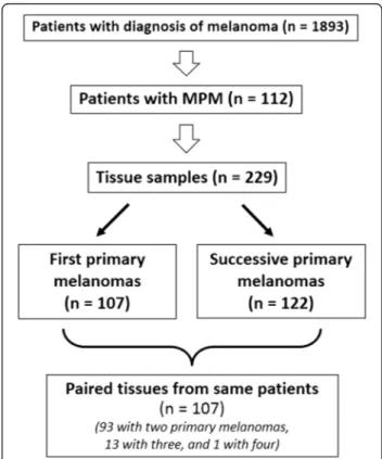
A total of 112 patients with multiple primary melanoma (96 cases with two primary tumors, 15 with three, and 1 with four) were enrolled. Paired samples of synchronous or asynchronous primary melanomas (N = 229; 93 cases with two paired tumor tissues, 13 with three, 1 with four) underwent molecular analysis at somatic level. Overall, a total of 341 samples were screened for mutations in
candidate genes, as summarized in Figure 1. Median age of the 112 enrolled patients was 59 years (range, 23-87 years); 59 (53%) were women. Considering the 102 first primary melanomas, trunk was the most frequent location (trunk, 57 [51%]; limbs, 41 [37%]; head and neck, 14 [12%]); median Breslow thickness was 1.7 mm (range, 0.35-5.8 mm).
Somatic alteration frequencies
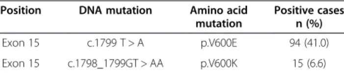
BRAF mutations were detected in 109 (47.6%) of 229 primary melanomas. All BRAF mutations across samples were located in codon 600 of the gene and were of two subtypes only: V600E (94/109; 86.2%) and V600K (15/109; 13.8%) (Table 1). Both mutations are reported in the Human Gene Mutation Database at http://www.hgmd.cf. ac.uk/ac/index.php and the Catalogue Of Somatic Muta-tions In Cancer (COSMIC) at http://www.sanger.ac.uk/ genetics/CGP/cosmic/.
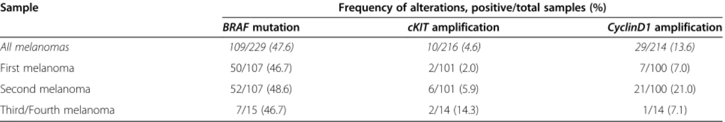
No association between BRAF mutations and any clinicopathological parameters was observed (Table 2). Frequency rates of BRAF mutations were quite identical across the different types of MPM lesions (first vs. second vs. subsequent melanoma; Table 3).
Paraffin-embedded nuclei from available tissue sections of primary melanomas were investigated by a two-colour FISH analysis, using genomic subclones corresponding to either CyclinD1 or cKIT gene loci as well as to the relative chromosome centromeres as controls. Gene amplification,
as inferred by the presence of a tetrasomic signal in more than one tenth of cells (see Methods), were observed in cancer cells only. No karyotypic alteration was found in cells from normal tissues surrounding the tumours (diploid signals were consistently detected).
Overall, 10/216 (4.6%) and 29/214 (13.6%) primary melanomas were found to carry cKIT and/or CyclinD1 gene amplification, respectively. As shown in Table 3, a significant increase of cKIT amplification rates was observed moving from first to subsequent primary mela-nomas (p < 0.001); analogously, the rate of CyclinD1
amplification was significantly higher in subsequent melanomas (22/114; 19.3%) than first primary melanomas (7/100; 7%) (p = 0.002). Again, no correlation between CyclinD1or cKIT amplification status and any clinicopath-ological parameters was found (not shown).
Distribution of somatic alterations into the three candidate genes is summarized in Table 4. Among the 229 multiple melanomas analyzed, majority of them (127; 55.5%) presented a genetic alteration in at least one of such genes; no sample was found to carry all three genes affected.
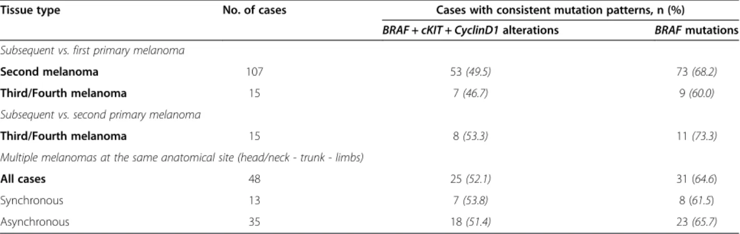
Considering the 107 patients who had paired samples of primary melanomas, about half of them showed consistent alteration patterns between either first and second primary tumors (53; 49.5%) or first and third/ fourth primary tumors (7/15; 46.7%) (Table 5). Focusing on BRAF mutations only, about one third of patients presented discrepant mutation patterns between first and second primary melanomas (34/107; 31.8%); such a discrepancy was even higher when comparing first and third or fourth primary tumors (6/15; 40%) (Table 5). Since differences in genetic alterations underlying mel-anoma pathogenesis may depend on the anatomical site of the primary lesion [18,25], consistency was evaluated among multiple melanomas arisen into the same body district. Among the 48 (42.9%) patients satisfying such a criterion, again roughly half of them (25; 52.1%) presented consistency in all somatic alteration patterns as well as about one third of cases (17; 35.4%) showed discrepant distribution of BRAF mutations (Table 5). No difference in consistency rates was observed between the two subsets of synchronous and asynchronous multiple melanomas (Table 5).
Among the 62 paired samples (54/107 [50.5%] patients) with discrepancies in BRAF/cKIT/CyclinD1 mutation patterns between first and subsequent primary melano-mas, majority of them (40; 64.5%) displayed differences in BRAF mutation distribution (19 with a wild-type first tumor and a mutated subsequent tumor, 19 with a muta-ted first tumor and a wild-type subsequent tumor, and 2 with a change in mutation variants between the two tumor lesions) (Additional file 1: Table S1). The remaining 22 (35.5%) discrepant paired samples showed differences in cKIT and/or CyclinD1 gene amplification status (Additional file 1: Table S1). A quite similar distribution of genetic alterations into the three candidate genes was observed when comparing subsequent versus second pri-mary melanomas (Additional file 2: Table S2).
The BRAF/cKIT/CyclinD1 mutation status was not eval-uated for association with clinical outcome in our series.
Discussion
Melanoma development and progression have been reported to occur by sequential accumulation of genetic
Table 1BRAF mutations in 229 tumor tissues from MPM patients
Position DNA mutation Amino acid mutation
Positive cases n (%) Exon 15 c.1799 T > A p.V600E 94 (41.0) Exon 15 c.1798_1799GT > AA p.V600K 15 (6.6)
Table 2 Frequency ofBRAF somatic mutations according to patients’ characteristics
Subgroups (No. of samples) Cases positive to BRAF mutations No. % P All samples (229) 109 47.6 Sex Male (107) 50 46.7 0.809 Female (122) 59 48.4
Site of primary melanoma
Head-neck (20) 9 45.0
0.623
Trunk (129) 58 44.9
Limbs (86) 42 48.8
Number of primary tumors
2 melanomas (186) 86 46.2 0.172 ≥ 3 melanomas (43) 23 53.5 Type of melanoma Synchronous (40) 18 45.0 0.208 Asynchronous (189) 91 48.1
AJCC disease stage
≤ I (161) 72 44.7 0.276 ≥ II (78) 37 47.4 Age at diagnosis < 40 years (34) 18 52.9 0.089 40-50 years (52) 25 48.1 > 50 years (143) 66 46.2
Family history of melanoma
1 affected members (193) 92 47.7 0.962 ≥ 2 affected members (36) 17 47.2
Disease stage was defined according to the recent American Joint Committee on Cancer (AJCC) guidelines. P: chi-squared test; two tailed; 95% confidence interval.
and molecular alterations [18,37]. Two main genetic net-works have been demonstrated to play a crucial role in the control of growth, proliferation, and survival of the melanocyte cells: the CDKN2A-driven pathway and the mitogen-activated protein kinase (MAPK) signal trans-duction cascade [38,39]. Genetic alterations in different members of these pathways have been associated with the pathogenesis of distinct types of primary melanomas: high frequency of BRAF or NRAS mutations (which are mutually exclusive) is mostly frequent in melanoma on skin without chronic sun-damage, whereas CyclinD1 or cKITamplifications are prevalent in CSD or acral melan-oma, respectively. In our study, we investigated the prevalence and distribution of such genetic alterations in MPM patients.
A high prevalence of somatic mutations in BRAF gene was detected in incident and subsequent melanomas. The frequency of BRAF mutations in primary melanomas (47%) was consistent with that observed in our previous study on 451 Italian patients with single melanoma (49%) [40] and slightly higher than that reported in a meta-analysis on 2521 patients with cutaneous melanomas (41%) [41]. In our series, two BRAFV600mutation subtypes were detected: V600E and V600K (in 41% and 7% of cases, respectively). Such two variants represent the most preva-lent BRAF mutations (our frequencies were consistent with most of those reported in literature [41]) and are able to constitutively activate BRAF kinase [21]. Amplification of CyclinD1 and cKIT genes, as determined by FISH analysis, was found in about 14% and 5% of melanoma tis-sues from our series, respectively (see Table 3). Again, such frequencies were consistent with those reported in
literature (ranging from 12% to 19% for CyclinD1 amplification [27,42-44] and calculated in about 7% of all cutaneous melanomas for cKIT amplification [25,31]). One (0.4%) out of 229 melanoma samples presented a coexistence of BRAF mutation and cKIT amplification (see Table 4), confirming that aberrations in these two genes can be considered as mutually exclusive [26].
A markedly higher rate of either BRAF mutations (59%) or CyclinD1 (38%) or cKIT (13%) amplifications was previously observed in 32 melanoma cell lines as controls by our group ([45] and unpublished data). As reported [45], these control cell lines were established as primary cell cultures from tumor samples obtained from donor patients with documented diagnosis of melanoma. Since cultured melanomas are thought to represent cells with the most malignant phenotype, one could speculate that genetic alterations in these three candidate genes play a role in tumor progression.
Sixty-two paired samples from 54 (51%) patients showed discrepancies in BRAF/cKIT/CyclinD1 mutation patterns between first and subsequent primary melano-mas (see Table 5). In the discrepant cases, we observed 20 (37%) patients with a wild-type first tumor and a mutated subsequent tumor, 14 (26%) with a mutated first tumor and a wild-type subsequent tumor, 8 (15%) with change in alteration variants between the two tumor lesions, and 12 (22%) with an additional gene amplifica-tion in the two BRAF-mutated tumors (3 cases in first but not in subsequent tumors and 9 with an opposite condi-tion). In majority of cases (29/54; 53%), gene alterations seem to be acquired in subsequent melanomas. Moreover, while BRAF mutations were equally distributed among discrepant multiple melanomas (47.5% wild-type first tumors and mutated subsequent tumors, 47.5% mutated first tumor and wild-type subsequent tumors), rates of cKITand CyclinD1 amplification were found to signifi-cantly increase moving from incident to subsequent primary melanomas (p values, <0.001 and 0.002, re-spectively). Such discrepancies were also confirmed among paired primary melanomas located at the same anatomical site as well as in synchronous primary melanomas (see Table 5). Overall, these observations provide evidence about the heterogeneity of the mo-lecular mechanisms underlying the development of MPM in the same patients. The knowledge that molecularly
Table 3 Distribution of somatic alterations in multiple melanomas from our series
Sample Frequency of alterations, positive/total samples (%)
BRAF mutation cKIT amplification CyclinD1 amplification
All melanomas 109/229 (47.6) 10/216 (4.6) 29/214 (13.6)
First melanoma 50/107 (46.7) 2/101 (2.0) 7/100 (7.0)
Second melanoma 52/107 (48.6) 6/101 (5.9) 21/100 (21.0)
Third/Fourth melanoma 7/15 (46.7) 2/14 (14.3) 1/14 (7.1)
Table 4 Somatic alterations in 229 tumor tissues from patients with multiple melanoma
Alteration type No. of samples %
BRAF mutation only 91 39.7
cKIT amplification only 6 2.6
CyclinD1 amplification only 9 3.9
BRAF mutation + CyclinD1 amplification 17 7.4 BRAF mutation + cKIT amplification 1 0.4 cKIT + CyclinD1 amplifications 3 1.3
heterogeneous cell types may coexist in primary melanomas [45,46] is a further confirmation that complex pathogenetic scenarios exist in melanomagenesis.
About one third of patients presented a discrepant pattern of BRAF mutations between incident and subse-quent primary melanomas (overall, 40/122; 32.8%). The introduction into the clinical practice of vemurafenib and dabrafenib, potent inhibitors of BRAFV600 mutants, makes the assessment of BRAF mutations as a crucial step toward the appropriate use of a targeted melanoma treatment. The low consistency in BRAF mutation pat-terns among MPM lesions from the same patients arises the practical question on how cases with coexistence of BRAFwild-type and BRAFmutant primary melanomas (and, to a less extent, those carrying different BRAF variants -which may present a different degree of responsiveness to BRAF inhibitors) should be molecularly classified. Nevertheless, progression of disease in patients with such discrepancies in primary melanomas may suggest taking into consideration all developing metastases for BRAFmutation analysis cucaccording to the recent indi-cations provided by the National Comprehensive Cancer Network (NCCN; at http://www.nccn.org/professionals/ physician_gls/f_guidelines.asp) guidelines, most recent melanoma tissue samples should be considered as ad-equate for BRAF mutation screening].
In our study, we contributed to provide additional clues about the prevalence of alterations in some candi-date genes (with particular attention to BRAF mutations) among synchronous or asynchronous multiple primary melanomas. Our findings further support evidence that molecular events underlying development and progression of melanoma are really complex. A better comprehension of the factors crucially involved in activating one or the other pathogenetic molecular mechanism, even in the same individual, might have an impact on the disease management. Since the future of melanoma therapy is likely to focus on targeting multiple pathways, advancing
technologies (i.e., deep-sequencing approaches) will permit to simultaneously investigate multiple genes and targets toward more accurate correlations between mo-lecular signatures and clinical outcome.
Additional files
Additional file 1: Table S1. Mutation patterns in patients presenting discrepancies in tumor lesions (54 second and 8 third/fourth vs. first primary melanomas) for BRAF/cKIT/CyclinD1 alterations.
Additional file 2: Table S2. Mutation patterns in patients presenting discrepancies in tumor lesions (7 subsequent vs. second primary melanomas) for BRAF/cKIT/CyclinD1 alterations.
Abbreviations
MPM:Multiple primary melanoma; MAPK: Mitogen-activated protein kinase; PCR: Polymerase chain reaction; FISH: Fluorescence in situ hybridization.
Competing interests
PAA is consultant of Bristol Myers Squibb, MSD, and Roche-Genentech. He participated into the Advisory Board from Bristol Myers Squibb, MSD, Roche-Genentech, GSK, Amgen, Celgene, Medimmune, and Novartis. He received honoraria from Brystol Myers Squibb, MSD, and Roche-Genentech. All remaining authors declare the absence of any Competing Interest.
Authors’ contributions
MCo, performed mutation analysis and data interpretation, helped to draft the manuscript; MCS, performed FISH analysis and data interpretation; AL, performed quality control of pathological data; VDG, IS, FA, DM, CR, SR, SM, LM, GB, MP, and PAA participated in patients’ collection and data acquisition; AM, PP, and MCa, performed data analysis; AC, performed pathological review and participated into the design of the study; GP, performed data interpretation, conceived of the study, drafted the manuscript. All authors read and approved the final manuscript.
Acknowledgments
Authors would like to thank all the other members of the Italian Melanoma Intergroup (IMI): C. Caracò, V. Chiarion Sileni, N. Mozzillo, P. Queirolo, C.R. Rossi, and A. Testori. Authors are grateful to patients for their important contribution to this study.
Work was partially supported by the Italian Ministry of Health“Progetto Ricerca Finalizzata” and Sardinia Regional Government (Regione Autonoma della Sardegna).
Table 5 Consistency betweenBRAF/cKIT/CyclinD1 alterations in paired samples from patients with multiple melanoma
Tissue type No. of cases Cases with consistent mutation patterns, n (%)
BRAF + cKIT + CyclinD1 alterations BRAF mutations Subsequent vs. first primary melanoma
Second melanoma 107 53 (49.5) 73 (68.2)
Third/Fourth melanoma 15 7 (46.7) 9 (60.0)
Subsequent vs. second primary melanoma
Third/Fourth melanoma 15 8 (53.3) 11 (73.3)
Multiple melanomas at the same anatomical site (head/neck - trunk - limbs)
All cases 48 25 (52.1) 31 (64.6)
Synchronous 13 7 (53.8) 8 (61.5)
Author details 1
Unit of Cancer Genetics, Institute of Biomolecular Chemistry (ICB), National Research Council (CNR) - Traversa La Crucca 3, Baldinca Li Punti, 07100 Sassari, Italy.2Dipartimento di Scienze Chirurgiche, Microchirurgiche e Mediche, University of Sassari, Sassari, Italy.3Department of Dermatology,
University of Firenze, Firenze, Italy.4Skin Cancer Unit, Istituto Scientifico Romagnolo per Studio e Cura dei Tumori, Meldola, Italy.5Istituto Nazionale
Tumori Fondazione Pascale, Napoli, Italy.6Department of Pathology, University of Firenze, Firenze, Italy.7Unit of Plastic Surgery, University of
Salerno, Salerno, Italy.
Received: 2 April 2014 Accepted: 2 May 2014 Published: 8 May 2014
References
1. Ferlay J, Shin HR, Bray F, Forman D, Mathers C, Parkin DM: Estimates of worldwide burden of cancer in 2008: GLOBOCAN 2008. Int J Cancer 2010, 127:2893–2917.
2. Dummer R, Hauschild A, Guggenheim M, Keilholz U, Pentheroudakis G: ESMO Guidelines Working Group: Cutaneous melanoma: ESMO Clinical Practice Guidelines for diagnosis, treatment and follow-up. Ann Oncol 2012, 23(Suppl 7):vii86–vii91.
3. Gandini S, Sera F, Cattaruzza MS, Pasquini P, Zanetti R, Masini C, Boyle P, Melchi CF: Meta-analysis of risk factors for cutaneous melanoma: III. Family history, actinic damage and phenotypic factors. Eur J Cancer 2005, 41:2040–2059.
4. Whiteman DC, Stickley M, Watt P, Hughes MC, Davis MB, Green AC: Anatomic site, sun exposure, and risk of cutaneous melanoma. J Clin Oncol 2006, 24:3172–3177.
5. Chaudru V, Chompret A, Bressac-de Paillerets B, Spatz A, Avril MF, Demenais F: Influence of genes, nevi, and sun sensitivity on melanoma risk in a family sample unselected by family history and in
melanoma-prone families. J Natl Cancer Inst 2004, 96:785–795. 6. Ferrone CR, Ben Porat L, Panageas KS, Berwick M, Halpern AC, Patel A,
Coit DG: Clinicopathological features of and risk factors for multiple primary melanomas. JAMA 2005, 294:1647–1654.
7. Murali R, Goumas C, Kricker A, From L, Busam KJ, Begg CB, Dwyer T, Gruber SB, Kanetsky PA, Orlow I, Rosso S, Thomas NE, Berwick M, Scolyer RA, Armstrong BK, GEM Study Group: Clinicopathologic features of incident and subsequent tumors in patients with multiple primary cutaneous melanomas. Ann Surg Oncol 2012, 19:1024–1033.
8. Bower MR, Scoggins CR, Martin RC 2nd, Mays MP, Edwards MJ, Reintgen DS, Ross MI, Urist MM, Noyes RD, Sussman JJ, Hagendoorn LJ, Stromberg AJ, McMasters K: Second primary melanomas: incidence and outcome. Am Surg 2010, 76:675–681.
9. De Giorgi V, Rossari S, Papi F, Gori A, Alfaioli B, Grazzini M, Crocetti E, Verdelli A, Foo CW, Lotti T: Multiple primary melanoma: the impact of atypical naevi and follow up. Br J Dermatol 2010, 163:1319–1322. 10. Hwa C, Price LS, Belitskaya-Levy I, Ma MW, Shapiro RL, Berman RS,
Kamino H, Darvishian F, Osman I, Stein JA: Single versus multiple primary melanomas: old questions and new answers. Cancer 2012, 118:4184–4192. 11. Moscarella E, Rabinovitz H, Puig S, Zalaudek I, Oliviero MC, Brown L, Alarcon
I, Malvehy J, Longo C, Formisano D, Carrera C, Badenas C, Piana S, Albertini G, Pellacani G, Argenziano G: Multiple primary melanomas: do they look the same? Br J Dermatol 2013, 168:1267–1272.
12. Balch CM, Gershenwald JE, Soong SJ, Thompson JF, Atkins MB, Byrd DR, Buzaid AC, Cochran AJ, Coit DG, Ding S, Eggermont AM, Flaherty KT, Gimotty PA, Kirkwood JM, McMasters KM, Mihm MC Jr, Morton DL, Ross MI, Sober AJ, Sondak VK: Final version of 2009 AJCC melanoma staging and classification. J Clin Oncol 2009, 27:6199–6206.
13. Fecher LA, Amaravadi RK, Flaherty KT: The MAPK pathway in melanoma. Curr Opin Oncol 2008, 20:183–189.
14. Pritchard AL, Hayward NK: Molecular pathways: mitogen-activated protein kinase pathway mutations and drug resistance. Clin Cancer Res 2013, 19:2301–2309.
15. Davies H, Bignell GR, Cox C, Stephens P, Edkins S, Clegg S, Teague J, Woffendin H, Garnett MJ, Bottomley W, Davis N, Dicks E, Ewing R, Floyd Y, Gray K, Hall S, Hawes R, Hughes J, Kosmidou V, Menzies A, Mould C, Parker A, Stevens C, Watt S, Hooper S, Wilson R, Jayatilake H, Gusterson BA, Cooper C, Shipley J, et al: Mutations of the BRAF gene in human cancer. Nature 2002, 417:949–954.
16. Lopez-Bergami P: The role of mitogen- and stress-activated protein kinase pathways in melanoma. Pigment Cell Melanoma Res 2011, 24:902–921.
17. Tannapfel A, Sommerer F, Benicke M, Katalinic A, Uhlmann D, Witzigmann H, Hauss J, Wittekind C: Mutation of the BRAF gene in cholangiocarcinoma but not in hepatocellular carcinoma. Gut 2003, 52:706–712.
18. Colombino M, Sperlongano P, Izzo F, Tatangelo F, Botti G, Lombardi A, Accardo M, Taran-tino L, Sordelli I, Agresti M, Abbruzzese A, Caraglia M, Palmieri G: BRAF and PIK3CA genes are somatically mutated in hepatocellular carcinoma among patients from South Italy. Cell Death Dis 2012, 3:e259.
19. Greaves WO, Verma S, Patel KP, Davies MA, Barkoh BA, Galbincea JM, Yao H, Lazar AJ, Aldape KD, Medeiros LJ, Luthra R: Frequency and spectrum of BRAF mutations in a retrospective, single-institution study of 1112 cases of melanoma. J Mol Diagn 2013, 15:220–226.
20. Heinzerling L, Baiter M, Kühnapfel S, Schuler G, Keikavoussi P, Agaimy A, Kiesewetter F, Hartmann A, Schneider-Stock R: Mutation landscape in melanoma patients clinical implications of heterogeneity of BRAF mutations. Br J Cancer 2013, 109:2833–2841.
21. Emile JF, Tisserand J, Bergougnoux L, Nowak F, Faucher G, Surel S, Lamy A, Lecorre D, Helias-Rodzewicz Z, Hofman P, Sabourin JC, Laurent-Puig P, BRAF EQA Group: Improvement of the quality of BRAF testing in melanomas with nationwide external quality assessment, for the BRAF EQA group. BMC Cancer 2013, 13:472.
22. Sullivan RJ, Flaherty K: MAP kinase signaling and inhibition in melanoma. Oncogene 2013, 32:2373–2379.
23. Palmieri G, Capone ME, Ascierto ML, Gentilcore G, Stroncek DF, Casula M, Sini MC, Palla M, Mozzillo N, Ascierto PA: Main roads to melanoma. J Transl Med 2009, 7:86.
24. Sharpless E, Chin L: The INK4a/ARF locus and melanoma. Oncogene 2003, 22:3092–3098.
25. Curtin JA, Fridlyand J, Kageshita T, Patel HN, Busam KJ, Kutzner H, Cho KH, Aiba S, Bröcker EB, LeBoit PE, Pinkel D, Bastian BC: Distinct sets of genetic alterations in melanoma. N Engl J Med 2005, 353:2135–2147.
26. Curtin JA, Busam K, Pinkel D, Bastian BC: Somatic activation of KIT in distinct subtypes of melanoma. J Clin Oncol 2006, 24:4340–4346. 27. Smalley KS, Lioni M, Dalla Palma M, Xiao M, Desai B, Egyhazi S,
Hansson J, Wu H, King AJ, Van Belle P, Elder DE, Flaherty KT, Herlyn M, Nathanson KL: Increased cyclin D1 expression can mediate BRAF inhibitor resistance in BRAF V600E-mutated melanomas. Mol Cancer Ther 2008, 7:2876–2883.
28. Ascierto PA, Grimaldi AM, Acquavella N, Borgognoni L, Calabrò L, Cascinelli N, Cesano A, Del Vecchio M, Eggermont AM, Faries M, Ferrone S, Fox BA, Gajewski TF, Galon J, Gnjatic S, Gogas H, Kashani-Sabet M, Kaufman HL, Larkin J, Lo RS, Mantovani A, Margolin K, Melief C, McArthur G, Palmieri G, Puzanov I, Ribas A, Seliger B, Sosman J, Suenaert P, et al: Future
perspectives in melanoma research Meeting report from the“Melanoma Bridge Napoli, December 2nd-4th 2012”. J Transl Med 2013, 11:137. 29. Lázár V, Ecsedi S, Szöllosi AG, Tóth R, Vízkeleti L, Rákosy Z, Bégány A,
Adány R, Balázs M: Characterization of candidate gene copy number alterations in the 11q13 region along with BRAF and NRAS mutations in human melanoma. Mod Pathol 2009, 22:1367–1378.
30. Carlino MS, Gowrishankar K, Saunders CA, Pupo GM, Snoyman S, Zhang XD, Saw R, Becker TM, Kefford RF, Long GV, Rizos H: Antiproliferative effects of continued mitogen-activated protein kinase pathway inhibition following acquired resistance to BRAF and/or MEK inhibition in melanoma. Mol Cancer Ther 2013, 12:1332–1342.
31. Beadling C, Jacobson-Dunlop E, Hodi FS, Le C, Warrick A, Patterson J, Town A, Harlow A, Cruz F 3rd, Azar S, Rubin BP, Muller S, West R, Heinrich MC, Corless CL: KIT gene mutations and copy number in melanoma subtypes. Clin Cancer Res 2008, 14:6821–6828.
32. Kong Y, Si L, Zhu Y, Xu X, Corless CL, Flaherty KT, Li L, Li H, Sheng X, Cui C, Chi Z, Li S, Han M, Mao L, Lu A, Guo J: Large-scale analysis of KIT aberrations in Chinese patients with melanoma. Clin Cancer Res 2011, 17:1684–1691.
33. Hodi FS, Corless CL, Giobbie-Hurder A, Fletcher JA, Zhu M, Marino-Enriquez A, Friedlander P, Gonzalez R, Weber JS, Gajewski TF, O’Day SJ, Kim KB, Lawrence D, Flaherty KT, Luke JJ, Collichio FA, Ernstoff MS, Heinrich MC, Beadling C, Zukotynski KA, Yap JT, Van den Abbeele AD, Demetri GD, Fisher DE: Imatinib for melanomas harboring mutationally activated or
amplified KIT arising on mucosal, acral, and chronically sun-damaged skin. J Clin Oncol 2013, 31:3182–3190.
34. Johnson TM, Hamilton T, Lowe L: Multiple primary melanomas. J Am Acad Dermatol 1998, 39:422–427.
35. Carli P, De Giorgi V, Chiarugi A, Stante M, Giannotti B: Multiple synchronous cutaneous melanomas: implications for prevention. Int J Dermatol 2002, 41:583–585.
36. Haluska FG, Hodi FS: Molecular genetics of familial cutaneous melanoma. J Clin Oncol 1998, 16:670–682.
37. Miller AJ, Mihm MC: Melanoma. N Engl J Med 2006, 355:51–65. 38. Thompson JF, Scolyer RA, Kefford RF: Cutaneous melanoma in the era of
molecular profiling. Lancet 2009, 374:362–365.
39. Wagle N, Van Allen EM, Treacy DJ, Frederick DT, Cooper ZA, Taylor-Weiner A, Rosenberg M, Goetz EM, Sullivan RJ, Farlow DN, Friedrich DC, Anderka K, Perrin D, Johannessen CM, McKenna A, Cibulskis K, Kryukov G, Hodis E, Lawrence DP, Fisher S, Getz G, Gabriel SB, Carter SL, Flaherty KT, Wargo JA, Garraway LA: MAP kinase pathway alterations in BRAF-mutant melanoma patients with acquired resistance to combined RAF/MEK inhibition. Cancer Discov 2014, 4:61–68.
40. Colombino M, Lissia A, Capone M, De Giorgi V, Massi D, Stanganelli I, Fonsatti E, Maio M, Botti G, Caracò C, Mozzillo N, Ascierto PA, Cossu A, Palmieri G: Heterogeneous distribution of BRAF/NRAS mutations among Italian patients with advanced melanoma. J Transl Med 2013, 11:202. 41. Lee JH, Choi JW, Kim YS: Frequencies of BRAF and NRAS mutations are
different in histological types and sites of origin of cutaneous melanoma: a meta-analysis. Br J Dermatol 2011, 164:776–784.
42. Sauter ER, Yeo UC, von Stemm A, Zhu W, Litwin S, Tichansky DS, Pistritto G, Nesbit M, Pinkel D, Herlyn M, Bastian BC: Cyclin D1 is a candidate oncogene in cutaneous melanoma. Cancer Res 2002, 62:3200–3206. 43. Glatz-Krieger K, Pache M, Tapia C, Fuchs A, Savic S, Glatz D, Mihatsch M,
Meyer P: Anatomic site-specific patterns of gene copy number gains in skin, mucosal, and uveal melanomas detected by fluorescence in situ hybridization. Virchows Arch 2006, 449:328–333.
44. Gast A, Scherer D, Chen B, Bloethner S, Melchert S, Sucker A, Hemminki K, Schadendorf D, Kumar R: Somatic alterations in the melanoma genome: a high-resolution array-based comparative genomic hybridization study. Genes Chromosomes Cancer 2010, 49:733–745.
45. Colombino M, Capone M, Lissia A, Cossu A, Rubino C, De Giorgi V, Massi D, Fonsatti E, Staibano S, Nappi O, Pagani E, Casula M, Manca A, Sini MC, Franco R, Botti G, Caracò C, Mozzillo N, Ascierto PA, Palmieri G: BRAF/NRAS mutation frequencies among primary tumors and metastases in patients with melanoma. J Clin Oncol 2012, 30:2522–2529.
46. Lin J, Goto Y, Murata H, Sakaizawa K, Uchiyama A, Saida T, Takata M: Polyclonality of BRAF mutations in primary melanoma and the selection of mutant alleles during progression. Br J Cancer 2011, 104:464–468.
doi:10.1186/1479-5876-12-117
Cite this article as: Colombino et al.: Discrepant alterations in main candidate genes among multiple primary melanomas. Journal of Translational Medicine 2014 12:117.
Submit your next manuscript to BioMed Central and take full advantage of:
• Convenient online submission
• Thorough peer review
• No space constraints or color figure charges
• Immediate publication on acceptance
• Inclusion in PubMed, CAS, Scopus and Google Scholar
• Research which is freely available for redistribution
Submit your manuscript at www.biomedcentral.com/submit