UNIVERSITÀ DEGLI STUDI DI URBINO
CARLO BO
Department of Biomolecular Sciences (DiSB)
Ph.D. Course in Life Sciences, Health and Biotechnologies
XXX cycle
The biological role of the IGF-1 pool
in breast cancer
SSD: BIO/13
Supervisor:
Ph.D. student:
Prof. Elena Barbieri
Dr. Serena Contarelli
Co-Advisor:
Ph.D. Giosuè Annibalini
A
BSTRACT
The insulin-like growth factor-1 (IGF-1) is a polypeptide growth factor that is essential for normal body growth and development of several tissues. IGF-1 is implicated in the progression and risk of several malignancies, including breast cancer (BC). Alternative splicing of terminal exon 5 of the igf-1 gene results in multiple isoforms possessing distinct carboxy-terminal extensions, called the Ea-, Eb- and Ec-domains.
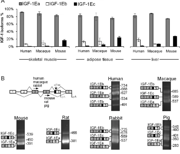
The first chapter of the Thesis provides evidence for an evolutionary mechanism generating the diversity of igf-1 splicing and expression across species. Our study highlights how the igf-1 exon 5 originates from exonization of a Mammalian interspersed repetitive-b (MIR-b) element in mammals. The acquisition of exon 5 alters the splicing pattern of igf-1 in mammals by generating two new isoforms: IGF-1Eb and IGF-1Ec. The evolutionary analysis of mammalian IGF-1 domains showed that E-domains are subjected to a strong evolutionary constraint on the synonymous sites, and they are enriched in disorder-promoting amino acids (i.e. intrinsically disordered), suggesting an important and novel regulatory role for these domains, not previously described.
In the second chapter, we highlighted that IGF-1 pro-hormones are not a simple inactive precursor of mature IGF-1, but are stable intermediates of their posttranslational processing. The IGF-1 pro-hormones can induce BC cell proliferation via the IGF-1 receptor, independently from the mature IGF-1 form. These results underline the importance of an accurate assessment of the presence of IGF-1 pro-hormones within the BC microenvironment.
The third chapter describes the mechanisms, which control the IGF-1 pro-hormones biosynthesis. We demonstrated that N-linked glycosylation regulates the stability and secretion of IGF-1Ea pro-hormone, probably ensuring proper pro-hormone folding and favoring its passage through the secretory pathway. The alternative Eb- and Ec-domains lack N-terminal glycosylation sites hence IGF-1Eb and IGF-1Ec pro-hormones were insensitive to glycosylation status of the cells. Moreover, the Eb- and Ec-domains regulate the subcellular localizations of IGF-1Eb and IGF-1Ec pro-hormones, promoting their nuclear accumulation. Thus, disordered E-domains play an important role in the structure, regulation and functioning of IGF-1.
The final chapter of the Thesis describes the data deriving from DIANA-5, and focuses on the effectiveness of modification in dietary change-associated with moderate physical activity in the prevention of BC recurrence, highlighting the importance of the lifestyle modification in the modulation of the circulating levels of IGF-1.
INTRODUCTION ... 1
The Complexity of the IGF-1 Pool: Gene Splicing, Regulation and Function ... 2
THE GENE STRUCTURE OF IGF-1 AND ALTERNATIVE SPLICE VARIANTS ...4
THE PROCESSING OF THE IGF-1PRE-PRO-HORMONE ...7
THE BIOLOGICAL ACTIVITY OF PRO-IGF-1 AND E-PEPTIDES ...9
IGF-1 AND BINDING PROTEINS (IGFBPS) ...14
THE IGF-1 RECEPTOR (IGF-1R) AND INTRACELLULAR SIGNALING ...16
IGF-1 AND CANCER ...19
IGF-1 AND BREAST CANCER ...21
AIMS OF THE THESIS ... 24
CHAPTER 1 ... 26
MIR retroposon exonization promotes evolutionary variability and generates species-specific expression of IGF-1 splice variants...27
INTRODUCTION ...31
MATERIALS AND METHODS ...33
RESULTS...38
DISCUSSION ...50
REFERENCES ...71
CHAPTER 2 ... 77
Human IGF-1 pro-forms induce breast cancer cell proliferation via the IGF-1 receptor ...78
INTRODUCTION ...81
MATERIALS AND METHODS ...82
RESULTS...86
DISCUSSION ...93
REFERENCES ...101
CHAPTER 3 ... 104
Regulation of IGF-1 stability, localization and secretion by intrinsically disordered E-domain tails ...105
INTRODUCTION ...107
MATERIALS AND METHODS ...108
RESULTS...111
DISCUSSION ...120
REFERENCES ...124
The Insulin-like Growth Factor Pool in Breast Cancer and the Effect of Exercise
and Lifestyle ...127
INTRODUCTION ...127
MATERIALS AND METHODS ...130
RESULTS...133
DISCUSSION ...139
REFERENCES ...144
CONCLUSIONS ... 147
O
RIGINAL
P
APERS
This Thesis is based on the following original research articles, which will be referred to by their Roman numerals.
I. Annibalini G, Bielli P, De Santi M, Agostini D, Guescini M, Sisti D, Contarelli S, Brandi G, Villarini A, Stocchi V, Sette C, Barbieri E. MIR retroposon exonization promotes evolutionary variability and generates species-specific expression of IGF-1 splice variants. Biochim Biophys Acta (BBA) - Gene Regulatory Mechanisms, 2016 May; 1859(5):757-68. doi: 10.1016/j.bbagrm.2016.03.014. Epub 2016 Apr 19.
II. De Santi M, Annibalini G, Barbieri E, Villarini A, Vallorani L, Contarelli S, Berrino F, Stocchi V, Brandi G. Human IGF-1 pro-forms induce breast cancer cell proliferation via the IGF-1 receptor. Cellular Oncology, (Dordr) 2016 Apr; 39(2):149-159. doi: 10.1007/s13402-015-0263-3. Epub 2015 Dec 23.
III. Glycosylation stabilizes IGF-1Ea pro-hormone and regulates its secretion - In preparation.
IV. Italian Diana5 clinical trial: lifestyles diary food-free diet and/or Mediterranean principles and moderate physical activity influence circulating levels of unbound IGF-1 - In preparation.
V. Circulating IGF-1 and early muscle adaptive responses to an acute isoinertial exercise - In preparation.
1
2
T
HE
C
OMPLEXITY OF THE
IGF-1
P
OOL
:
G
ENE
S
PLICING
,
R
EGULATION AND
F
UNCTION
The insulin-like growth factor 1 (IGF-1), also called somatomedin C, is a polypeptide growth factor, which is essential for normal body growth and development [1]. A variety of cellular responses are induced by IGF-1, including cell proliferation, differentiation and survival [2].
Initially, in the early 1970s, the ―somatomedin hypothesis‖ was proposed as a model for the actions of IGF-1 on the skeleton [3]. This hypothesis postulated that the growth hormone (GH), secreted by the pituitary gland, stimulated IGF-1 synthesis in the liver and its release into the blood stream to target organs acting in an endocrine manner. This hypothesis was later challenged by subsequent findings, including a seminal study examining skeletal development in mice with liver-specific deletion of the Igf-1 gene (LiverIGF-1−/−) [4]. In this experiment, the circulating IGF-1 level was reduced to less than 25% of normal. Despite this severe reduction, the knockout mice developed and grew normally, and their skeletal changes were minimal, indicating that local IGF-1 production is enough to guarantee general growth and skeletal development. The local IGF-1 production extended this hypothesis and included the autocrine-paracrine manner of IGF-1 action. Investigators have recognized that the physio-pathologic mechanisms, through which IGF-1 is regulated and secreted, are more complicated than originally believed. In most of the cellular-animal models, it was proposed that GH stabilized serum IGF-1 by promoting the formation of the ternary complex composed of IGF-1, IGF binding protein (IGFBP) 3 (IGFBP-3) and the acid-labile subunit (ALS) [3]. The bioavailability of circulating IGF-1 modulated by the IGFBPs is further described in the paragraph ―IGF-1
and binding proteins (IGFBPs)‖.
In physiological conditions, circulating IGF-1 is mostly synthesized in the liver and acts as an endocrine factor. IGF-1 levels are relatively low at birth, increase during childhood, reaching peak levels in adolescence and begin to decline during the third decade of life [5]. It plays an important role in the first decades of life in normal development and
3 growth as a key regulator of cell proliferation and differentiation and as an apoptosis and necrosis inhibitor.
Several factors may affect the hepatic synthesis of IGF-1, including insulin, GH, age and nutrition. The growth hormone, produced by the pituitary gland, is the most important hormone involved in regulating body growth and development as well as carbohydrate and lipid metabolism, and its action can be directed to target tissues that possess specific receptors or indirectly through other factors that enhance and complement its effects. The most important mediator of GH effects is IGF-1 [6]. The production and concentration of the two hormones are positively related to each other. The connective tissue cell types that synthesize IGF-1 contain GH receptors and an increase in GH secretion stimulates IGF-1 synthesis. At the same time, an increase in IGF-1 blood concentration suppresses GH synthesis in the pituitary gland though a negative-feedback regulation that represents an important homeostatic mechanism for the maintenance of normal plasma IGF-1 concentration [3].
Other hormones participate with GH in regulating hepatic IGF-1 synthesis, including thyroxin, cortisol, estradiol and testosterone. Thyroxin enhances sensitivity to GH and can increase IGF-1 concentration in hyperthyroidism. Cortisol acts to inhibit IGF-1 synthesis, and high cortisol concentrations can lead to growth attenuation. Etradiol inhibits IGF-1 secretion in the liver by constraining GH stimulated signal transduction [7]. Testosterone enhances hepatic IGF-1 synthesis, but also alters the sensitivity of the pituitary gland to negative-feedback regulation of GH secretion, leading to an increase in GH synthesis and thus an increment in IGF-1 secretion.
Nutrient intake is another variable regulating plasma IGF-1 concentrations. The IGF-1 plasma concentrations are markedly reduced in low protein or calorie-restricted diets [8]. In adults, total caloric intake is more important than protein intake. In fact, in the presence of an adequate caloric intake, even with a low protein intake regime, there can be an increase in IGF-1 levels. Conversely, there is a threshold of caloric intake below which protein intake cannot increase the levels of IGF-1 after fasting. When caloric intake is severely reduced, the dietary content of carbohydrates and essential amino acids is critical for an optimal recovery of IGF-1 levels after fasting [9]. These conditions are associated with a marked decrease in the number of somatotropic receptors supporting the role of a receptor deficiency in the decline of the circulating IGF-1.
4 In the last decade, many in vitro and in vivo studies have investigated the igf-1 gene conservative structure. Different mRNA transcripts are produced as a result of the alternative splicing of the igf-1 gene, encoding for several IGF-1 precursor proteins also called isoforms. These IGF-1 protein isoforms can be distinguished by the structure of their extension peptides, or E-peptides, on the carboxy-terminal end and by the length of their amino-terminal signal peptides. Interestingly, it has been proposed that these pro-hormones might possess bioactivities that are distinct from those of mature IGF-1 [10].
T
HE GENE STRUCTURE OFIGF-1
AND ALTERNATIVE SPLICEVARIANTS
The igf-1 gene is highly conserved among mammals and primates [11]. It is located on the long arm of chromosome 12 in humans and consists of six exons and five introns that cover about 90 kb of DNA with different promoter regions. It is widely believed that all IGF-1 biological actions are mediated by mature IGF-1, but the igf-1 gene encodes multiple mRNA variants that differ in terms of the presence of an alternative leader sequence and polyadenylation signal [2, 12]. The gene transcription of igf-1 is very complex due to many transcriptional and post-transcriptional modifications that give rise to several isoforms, of which six are known in the literature.
In particular, exons 1 and 2 encode for the sequence that determines the class of the protein deriving from different splicing of these two exons to the common exon 3. Transcripts starting with exon 1 are referred to as class 1, whereas class 2 transcripts use exon 2 as their leader exon. These exons form two different non-coding 5 'UTR sequences and a sequence that contains the information for a portion of the signal peptide. The expression of these two exons seems to be dependent on two different promoters that are regulated in a tissue specific manner [13]. In particular, class 2 transcripts are expressed mainly in the liver and represent the circulating IGF-1 forms. It has been shown that these forms are dispensable for fetal and postnatal growth [14], which are thought to be more GH dependent [15], whereas transcripts initiating at promoter 1 are widely expressed in many tissues representing local tissue forms [16].
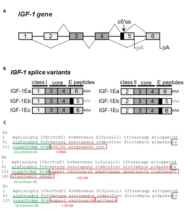
5 The alternative splicing at the 3' end of the gene involving exons 4, 5 and 6 forms three different extension peptides in humans, called E-peptides, at the carboxy-teminal end (Fig. 1). However, all of them contain exon 3 and exon 4, which encode the mature IGF-1 peptide sequence. This sequence is composed of a total of 70 amino acids, 25 of which derive from exon 3 and 45 from exon 4, forming four domains named for their homology to insulin as B amino-terminal domain, C and A domain and D carboxy-terminal domain. This sequence represents the mature invariant peptide present in all transcripts [17]. The final part of exon 4 encodes for the first 16 amino acids of the amino-terminal portion of the IGF-1 peptide domain, which are common to all the three different E-peptides. The alternative splicing involving exons 4, 5 and 6, producing three different IGF-1 E-peptides in humans, produces the remaining portion of the E-peptide.
The IGF-1Ea transcripts from the splicing of exon 4 and exon 6 excluding exon 5, which represents the main isoform for the formation of mature IGF-1 produced by the liver and also in most other tissues and the most conserved isoform across species [11]. This mRNA splicing produces the Ea-peptide, which is composed of 35 amino acids. The first 16 are common peptides deriving from exon 4, whereas the remaining 19 amino acids are encoded by exon 6.
The variant, called IGF-1Eb isoform, contains exon 4 and splices with exon 5, whereas exon 6 is excluded. This transcript is rarely expressed in other species, so this isoform is often considered a human-specific splice variant [12]. It was first detected in human liver [18] and then found in other tissues such as lung carcinoma cells [10], skeletal muscle [19], prostate [20] and endometrium [21]. This variant yields the Eb-peptide, which contain 16 common peptides and 61 additional amino acids deriving from exon 5, resulting in a total of 77 amino acids.
Finally, the alternative spicing of the igf-1 gene also generates a third transcript, human IGF-1Ec, which corresponds to IGF-1Eb in rodents. In this variant, exon 4 is joined to a partial sequence of exon 5, which in turn is joined to exon 6. It was first detected in the liver and its expression accounts for about 10% of the IGF-1 transcripts. This transcript differs from IGF-1Ea for the presence of the first 49 base pairs from exon 5 and a premature stop codon within exon 6. IGF-1Ec is generated through a cryptic IGF633 donor
splice site located 49 bp downstream from the 5' end of exon 5, which in turn splices with the acceptor site in the intron preceding exon 6. When this cryptic IGF633 donor splice
6 produced [22]. It results in a different C-terminal peptide sequence due to a read frame shift leading to an Ec-peptide composed of 16 common amino acids, 16 encoded by exon 5 and 8 by exon 6. This splice variant is also referred to as mechano-growth factor (MGF) because it has been shown to be up-regulated in response to muscle exercise and damage [23].
Figure 1. Schematic representation of the igf-1 gene (A) and its splice variants (B-C). (A) Map of the
igf-1 gene showing exons (boxes), introns (solid lines), splicing options (dashed lines), cryptic 5' splice site
(c5'ss) in exon 5 and poly(A) sites (pA). (B) Splice variants of the igf-1 gene. Exons 1 and 2 encode for the sequence that determines the class of the protein. The mature IGF-1 is encoded by exons 3 and 4, and the three different E-peptides in humans constitute the carboxy-teminal end, and the three different E-peptides
Human
Ea
1 mgkisslptq lfkccfcdfl kvkmhtmsss hlfylalcll tftssatagp etlcgaelvd 61 alqfvcgdrg fyfnkptgyg sssrrapqtg ivdeccfrsc dlrrlemyca plkpaksars 121 vraqrhtdmp ktqkevhlkn asrgsagnkn yrm
Eb
1 mgkisslptq lfkccfcdfl kvkmhtmsss hlfylalcll tftssatagp etlcgaelvd 61 alqfvcgdrg fyfnkptgyg sssrrapqtg ivdeccfrsc dlrrlemyca plkpaksars 121 vraqrhtdmp ktqkyqppst nkntksqrrk gwpkthpgge qkegteaslq irgkkkeqrr 181 eigsrnaecr gkkgk
Ec
1 mgkisslptq lfkccfcdfl kvkmhtmsss hlfylalcll tftssatagp etlcgaelvd 61 alqfvcgdrg fyfnkptgyg sssrrapqtg ivdeccfrsc dlrrlemyca plkpaksars 121 vraqrhtdmp ktqkyqppst nkntksqrrk gstfeerk
+19AA
Human
Ea
1 mgkisslptq lfkccfcdfl kvkmhtmsss hlfylalcll tftssatagp etlcgaelvd 61 alqfvcgdrg fyfnkptgyg sssrrapqtg ivdeccfrsc dlrrlemyca plkpaksars 121 vraqrhtdmp ktqkevhlkn asrgsagnkn yrm
Eb
1 mgkisslptq lfkccfcdfl kvkmhtmsss hlfylalcll tftssatagp etlcgaelvd 61 alqfvcgdrg fyfnkptgyg sssrrapqtg ivdeccfrsc dlrrlemyca plkpaksars 121 vraqrhtdmp ktqkyqppst nkntksqrrk gwpkthpgge qkegteaslq irgkkkeqrr 181 eigsrnaecr gkkgk
Ec
1 mgkisslptq lfkccfcdfl kvkmhtmsss hlfylalcll tftssatagp etlcgaelvd 61 alqfvcgdrg fyfnkptgyg sssrrapqtg ivdeccfrsc dlrrlemyca plkpaksars 121 vraqrhtdmp ktqkyqppst nkntksqrrk gstfeerk
+ 61 AA
Human
Ea
1 mgkisslptq lfkccfcdfl kvkmhtmsss hlfylalcll tftssatagp etlcgaelvd 61 alqfvcgdrg fyfnkptgyg sssrrapqtg ivdeccfrsc dlrrlemyca plkpaksars 121 vraqrhtdmp ktqkevhlkn asrgsagnkn yrm
Eb
1 mgkisslptq lfkccfcdfl kvkmhtmsss hlfylalcll tftssatagp etlcgaelvd 61 alqfvcgdrg fyfnkptgyg sssrrapqtg ivdeccfrsc dlrrlemyca plkpaksars 121 vraqrhtdmp ktqkyqppst nkntksqrrk gwpkthpgge qkegteaslq irgkkkeqrr 181 eigsrnaecr gkkgk
Ec
1 mgkisslptq lfkccfcdfl kvkmhtmsss hlfylalcll tftssatagp etlcgaelvd 61 alqfvcgdrg fyfnkptgyg sssrrapqtg ivdeccfrsc dlrrlemyca plkpaksars 121 vraqrhtdmp ktqkyqppst nkntksqrrk gstfeerk
+ 24 AA 16 common AA 16 common AA 16 common AA A B C
7
are produced by the alternative splicing at the 3' end of the gene involving exons 4, 5 and 6. (C) Of the amino acid sequences of the three E-peptides, the first 16 common peptides deriving from exon 4 are in green and the different Ea, Eb and Ec amino acids are in red.
T
HE PROCESSING OF THEIGF-1
P
RE-P
RO-
HORMONEThe translation of the igf-1 gene gives rise to an immature IGF-1 peptide, the pre-pro-IGF-1. This precursor of the mature IGF-1, contains a signal peptide at the 5‘ end of the gene, the mature IGF-1 and a C-terminal E-peptide extension at the 3‘ end. Pre-pro-IGF-1 is subject to numerous post-translational modifications leading to mature peptide production composed of four domains and 70 amino acids. The mature sequence is highly conserved among primate species, whereas it has been shown that the sequences of both the signal peptides and the E-peptides are less strongly conserved compared to the mature IGF-1 peptide [11].
The first cleavage leads to N-terminal signal peptide removal by intracellular serine proteases facilitating the passage of the polypeptide into the endoplasmic reticulum. The resulting molecule is the pro-IGF-1 composed of the mature IGF-1 plus the E-peptide. The pro-IGF-1 can be subject to additional processing prior to secretion, including the cleavage of the carboxy-terminal E domain resulting in the release of free mature IGF-1 and E-peptide [24] (Fig. 2). All the classes of pro-IGF-1 contain a highly conserved and unique pentabasic motif K65-X-X-K68-X-X-R71-XX-R74-X-X-R77. All the E-peptides begin with amino acid 71; thus, cleavage occurs at Arg71 [17]. Proproteins can be processed at this specific motif by serine protease from the subtilisin-related proprotein convertase family (SPCs), a major family of endoproteolytic processing enzymes of the secretory pathway in mammals. Seven mammalian PCs have been identified, namely, PC1, PC2, furin, PC4, PC5, PACE4 and PC7, and they have been proposed as predictors of general cleavage sites. Furin appears to have a more rigorous specificity recognizing sites that contain the sequence motif R-X-[R/K]-R, whereas R-X-X-R is its minimal cleavage sequence [25].
Because SPCs are located in the secretory pathway, the process that leads to the formation of mature IGF-1 by the cleavage of the E-peptides has been shown to occur intracellularly, as expected for intracellular convertases, such as furin [24]. However, the
8 unprocessed pro-IGF-1 can be secreted and has been detected in conditioned media and in vivo serum [26]. This finding shows that the E domains are not cleaved intracellularly and suggests the presence of potential proprotein convertases that could process pro-IGF-1 extracellularly [27].
There is evidence of possible candidate proteases that could release the mature peptide outside of the cell when needed. The proprotein convertase subtilisin/kexin type 6, commonly known as PACE4, is expressed constitutively in muscle cells [28] and can be found in the Golgi as well as extracellularly; hence, it is a likely candidate to cleave pro-IGF-1 in both areas performing the same intracellular reaction [24].
Figure 2. Processing of IGF-1 leading to the mature peptide. The igf-1 gene is translated into the
pre-pro-IGF-1, which contains a signal peptide, mature IGF-1 and a C-terminal E-peptide extension. During translation the N-terminal signal peptide is removed and the resulting molecule is the Pro-IGF-1. An additional protease cleavage separates the mature IGF-1 from the E-peptides. The Y represents the glycosylation site present within the Ea-peptide.
IGF-1Ea IGF-1Eb IGF-1Ec/MGF 1 or 2 3 4 6 5 1 or 2 3 4 6 1 or 2 3 4 AAA AAA AAA IGF-1 prepropeptide IGF-1 propeptide mature IGF-1 Signal peptide Translation E-peptide Mature 70 aa Ea-peptide (35 aa) Eb-peptide (77 aa) Ec-peptide (40 aa) Class I (48 aa) Class II (32 aa) Mature 70 aa Ea-peptide (35 aa) Eb-peptide (77 aa) Ec-peptide (40 aa) Mature 70 aa Ea-peptide (35 aa) Eb-peptide (77 aa) Ec-peptide (40 aa) E-peptides Y Y Y
9 In addition, the secretion of unprocessed pro-IGF-1Ea isoform, both glycosylated and nonglycosylated, has been reported [27, 29].
In rodents there are two potential N-glycosylation sites at Asn92 and Asn100, whereas the human Ea domain contains one N-linked glycosylation site at Asn92 [30] based on the consensus sequence Asn-X-Ser/Thr, where X represents any encoded amino acid except proline [31]. This glycosylation site is not present in the human Eb- and Ec-peptides because of the reading frame shift caused by the insertion of exon 5 in these two IGF-1 isoforms.
Considering the unique role of glycosylation in the protein biosynthesis process [31], it is possible that Ea-peptide glycosylation might play a role in interactions regarding the regulation of the bioavailability of the different species of this IGF-1 isoform (i.e., pro-IGF-1Ea, mature IGF-1 or Ea-peptide). Although glycosylation has been shown to be a critical step in the protein biosynthesis process, its significance to IGF-1 function has yet to be determined. The presence of a human N-linked glycosylation site only in Ea-peptide and not in the Ec and Eb domains might play a role in the regulation of the bioavailability of this isoform relating to its secretion and stability and possibly reflecting a different biological role of the IGF-1Ea isoform [27].
It is traditionally believed that IGF-1 post-transcriptional processing leads to the formation of the mature protein, which is the main mediator of IGF-1 actions binding IGF-1R. On the contrary, the complexity introduced by the post-transcriptional regulation and post-translational modification of the igf-1 gene leads to the production of different IGF-1 forms that can be secreted. In particular, three forms of the IGF-1 protein could exist in the extracellular environment: mature IGF-1, non-glycosylated pro-IGF-1, and glycosylated IGF-1 [27]. To date, however, it remains to be determined whether pro-IGF-1 is bioactive or simply an inactive precursor of mature pro-IGF-1 and/or E-peptide [32].
T
HE BIOLOGICAL ACTIVITY OFP
RO-IGF-1
ANDE-
PEPTIDESThe biological significance of each IGF-1 splice variant is currently unknown, and the physiological and molecular mechanisms that regulate their expression and their circulating levels are unclear [2]. It is generally assumed that the biological actions of
10
IGF-1 are inferred through the mature peptide, whereas different biological effects have been reported for the different IGF-1 pro-forms or for their E-peptides exogenously administrated or over-expressed in various model systems. In particular, recent studies in humans have shown a differential expression profile of the splice variants in response to various conditions and pathologies, such as skeletal muscle damage [19], endometriosis [21], prostate [20], cervix [33] and colorectal cancer [34]. Therefore, it is not just the mature IGF-1 that possesses bioactivity, and differential expression of igf-1 gene could indicate distinct regulatory mechanisms and biological roles of the different pro-IGF-1 forms, implying that the E-peptides may promote biological effects.
A divergent action of the IGF-1 isoforms has been reported after viral-mediated expression in mouse skeletal muscle of the two different rodent pro-1 forms, IGF-1Ea and IGF-1Eb, as well as the mature IGF-1 without the E-peptides. Interestingly, it has been shown that overexpression of mature IGF-1 in skeletal muscle does not promote muscle hypertrophy in young mice, whereas both the pro-IGF-1 forms caused hypertrophy. These results suggest that the pro-IGF-1 forms are required for IGF-1 action leading to muscle hypertrophy, and the presence of E-peptides is necessary for IGF-1 action in muscle [35]. Although the two splice variants increased phosphorylation of IGF-1R, the murine IGF-1Ea overexpression resulted in increased Akt phosphorylation only, whereas the overexpression of murine IGF-1Eb in skeletal muscle activated both the PI3K/Akt and MAPK pathways [36]. These results suggest variable IGF-1 isoform– specific actions on the signaling pathways involved after IGF-1R phosphorylation. Considering that IGF-1 isoforms differ only in terms of E-peptides, it has been suggested that E-peptides of human IGF-1 precursors may act as independent growth factors with their own biological activity [37]. Curiously, few studies have explored the activity of the Ea-peptide, whereas many investigations have focused on the other two peptides. Since it is more highly expressed than other isoforms, the Ea-peptide probably has an essential biological function also because its sequence is highly conserved in many species, while the other splice variants diverge within primates [11].
Initial studies focused on the role of human Eb-peptides and reported mitogenic activity in human bronchial epithelial cells as a result of exposure to a specific region of the human Eb domain. Furthermore, it was observed that this peptide could still induce proliferation even after IGF-1R neutralization with a specific antibody [10]. It was first suggested that the Eb-peptide mediates its effect through a specific receptor. Conversely,
11 it was found that the Eb-peptide was not cleaved and the pro-IGF-1Eb was not secreted but was accumulated in the nucleolus [38]. Thus, this human IGF-1Eb pro-peptide may have biological roles that are independent of the mature IGF-1 effects.
Interestingly, the role of the Ec-peptide has been the focus of several studies, mainly because of its action in skeletal muscle. Studies began at the end of the 1990s in the Goldspink Laboratory and revealed that IGF-1Ea was the only isoform expressed in resting rabbit muscles, whereas exon 5 inclusive transcripts were found to increase in rabbit muscles subjected to stretch and electrical stimulation. [39]. It was suggested that the IGF-1Eb splice variant, corresponding to the human IGF-1Ec, was responsible for the stretch induced hypertrophy. It was named Mechano-Growth Factor (MGF) because it was identified in muscle tissue in response to mechanical damage and to distinguish it from the liver forms of IGF-1 [40].
Numerous studies have investigated the role of MGF in muscle repair and survival, leading to the so-called ―MGF hypothesis‖ (Fig. 3). The theory is based on the following findings. After skeletal muscle injury, it was found that both IGF-1Ea and MGF were produced with differential expression regulation and different time course. In particular, there was a transient increase in the splice variant containing the exon 5, which corresponds to IGF-1Eb in rodents and IGF-1Ec in humans. After some days, levels of this transcript decreased, and an up-regulation of the IGF-1Ea splice variant associated with the decline of the IGF-1Ec/MGF mRNA levels was found [23, 41]. Because of this varied expression regulation, distinct roles of these isoforms in muscle remodelling were suggested. The temporal expression of IGF-1Ec/MGF was postulated to be responsible for activating quiescent satellite cells after muscle damage and promoting myoblast proliferation. In contrast, the increase in IGF-1Ea expression appeared to correlate with myoblast differentiation and to promote the fusion of myogenic cells leading to tissue repair [23, 41]. However, it should be noted that the expression levels of MGF are normally vastly lower than those of IGF-1Ea mRNA in skeletal muscle and in other extra-hepatic tissues and the absolute mRNA levels of IGF-1Ea were always 10-fold greater than the MGF transcript levels, even when those transcript levels were elevated. In addition, there is no evidence that the mature IGF-1 and IGF-1Ea are not present in elevated amounts even during the early phase of muscle repair, when MGF is predicted to be up-regulated. Taking all these data into account, the MGF hypothesis should be viewed with extreme caution [42, 43].
12
Figure 3. Schematic representation of the MGF hypothesis. The MGF hypothesis suggests that after
muscle injury or exercise, the igf-1 gene transient increases the IGF-1Eb splice variant in rabbit muscles, the so-called MGF. This specific isoform is postulated to be responsible for activating quiescent satellite cells to enter the cell cycle and become mononucleated myoblasts. Thus, MGF promotes myoblast proliferation. On the contrary, during the myoblast proliferative stage, splicing is increasingly shifted towards the IGF-1Ea splice variant, which promotes myoblast proliferation and differentiation into multinucleated fibers called myotubes. The MGF hypothesis therefore suggests that MGF is responsible for satellite cell activation and myoblast proliferation, whereas IGF-1Ea is responsible for myoblast differentiation. Although the graph shows cellular repair events that coincide with the postulated IGF-1 splice variant mRNA levels, it does not reflect the absolute mRNA levels of the two isoforms. In particular, it is essential to note that levels of IGF-1Eb transcripts are far lower than those of IGF-1Ea mRNA. All the mRNA data extrapolated should be examined with extreme caution [42].
Recently, actions regarding the bioactivity of E-peptides have been established. In particular, the biological actions of synthetic E-peptides, corresponding to the rodent Ea and Eb sequences, were compared to test the effects of these E-peptides on IGF-1R signaling. To determine whether peptides activate IGF-1R directly, the treatment of E-peptides alone or with mature IGF-1 in the activation of IGF-1R was tested. It was shown that E-peptides do not induce IGF-1R phosphorylation directly, but they amplify IGF-1R activation in an IGF-1-dependent manner but not when IGF-1R was inhibited. In addition, when myoblasts were treated with both mature IGF-1 and E-peptides, there was an increase in the phosphorylation of ERK1/2, but not of phospho-AKT. It was therefore proposed that E-peptides might modulate IGF-1 signaling by modulating IGF-1R downstream signaling [44].
13 The biological activity of pro-IGF-1 has been investigated in a recent study on murine skeletal muscle cells, particularly focused on the IGF-1Ea isoform. It was shown that the untreated cells secreted IGF-1 predominantly in the form of pro-IGF-1Ea. Moreover, both the glycosylated and non-glycosylated forms were present at high levels, but only a small portion of mature 1 was present. It was shown that pro-1Ea could activate IGF-1R as well as mature IGF-1, and pro-IGF-1Ea was more potent than the mature peptide by about 20% of receptor phosphorylation when compared in an IGF-1R activation assay. On the contrary, glycosylated pro-IGF-1Ea was less efficient at receptor activation than pro-IGF-1Ea and mature IGF-1 was about 2-fold less potent than mature IGF-1. Because of its differential ability to activate IGF-1R, it was suggested that glycosylated pro-IGF-1Ea might serve as a reservoir for IGF-1, which can be stored until needed [27].
In this regard, it is known that the extracellular matrix (ECM) interacts with a range of growth factors and cytokines likely mediated by positively charged amino acid sequence motifs present in these peptides [45]. The E-peptides contain a high proportion of basic amino acids conferring a high positive charge at physiological pH and therefore pro-IGF-1 might bind to negatively charged molecules in the ECM. It has been shown that both mouse pro-IGF-1Ea and pro-IGF-1Eb bind ECM with significantly higher affinity than does mature IGF-1. Therefore, the C-terminal IGF-1 E-peptides could function by tethering pro-IGF-1 to the ECM, which presumably plays a biological role in retaining high local concentrations of IGF-1 in tissues for subsequent cleavage and receptor activation [46].
It is generally accepted that the post-translational processing of IGF-1 leads to the formation of mature protein, which is the main mediator of the IGF-1 action activating the IGF-1R. In contrast, collectively these studies suggest that E-peptides are actually translated and secreted and exist as part of pro-IGF-1. To date, however, the biological significance of the IGF-1 isoforms remains unclear.
Although much research has aimed to distinguish the E-peptides activity from that of mature peptides, it has been suggested that the E-peptides possess IGF-1 dependent activity. Recent evidence supports this hypothesis, suggesting that the E-peptides may modulate IGF-1 activity through multiple mechanisms. Thus, the retention of these sequences could control the bioavailability of IGF-1 by altering the IGF-1secretion or its association with IGFBs, modulating its power in receptor activation and improving its stabilization and localization in tissues.
14
IGF-1
AND BINDING PROTEINS(IGFBP
S)
A family of specific proteins modulates the bioavailability and the biological activity of circulating IGF-1: the binding proteins (IGFBPs) (Fig. 4). At least six IGFBPs have been identified and well characterized [47, 48] as well as nine IGFBP-related proteins (IGFBP-rPs) [49]. These proteins belong to a family that shares the same cysteine pattern in the amino-terminal and carboxy-terminal, which is indispensable in the binding affinity of the IGF-1. Most of the circulating IGF-1 is in the form of a ternary complex of 150 KDa composed of IGFBP-3, the most abundant IGFBPs in blood, and the ALS subunit, a glycoprotein acid-labile [50]. This complex does not pass through the endothelium and acts as an inactive IGF-1 reserve, accounting for 75-80% of the total carrying capacity. It is hypothesized that binding with IGFBPs increases the half-life of IGF-1. In fact, the half-life of free IGF-1 is less than 15 minutes, whereas the ternary complex persists in circulation for a half-life of about 12-14 hours protecting the growth factor from proteolytic degradation and modulating peptide interaction with its receptor, increasing or decreasing the binding affinity [51]. The formation of this ternary complex results in most of the IGF-1 in the blood representing a stable IGF-1 reserve, while the free IGF-1 concentration in normal subjects is less than 1% compared to that of total IGF-1. The GH stimulates the secretion of IGFBP-3 and ALS and this helps to stabilize IGF-1 levels. IGFBP-3 goes through proteolysis during various catabolic processes or dysmetabolic conditions such as diabetes, and as a result, with a lower concentration of this binding protein, IGF-1 will tend to be more degraded [52]. Additionally, IGFBPs compete with receptor binding and normally have higher binding affinity to IGF-1 than the receptor does. Consequently, the binding of IGFBPs to IGF-1 prevents the ligand from interacting with the receptor and suppresses the growth factor actions [51]. The exact mechanism through which IGF-1 is released from the ternary complex into the tissue is not completely known; however, it seems that an important role is played by proteases that degrade IGFBPs by allowing the release of IGF-1 and the binding to its receptor.
It is known that IGFBP-2 is the second most abundant IGFBP. This IGFBP is not linked to ALS, and the IGF-1/IGFBP-2 complex has a rather short half-life of about 90 minutes. The serum IGFBP-2 is unsaturated and represents a reservoir capable of binding and thus carrying IGF-1. In contrast, IGFBP-1 carries only a small percentage of IGF-1. Like IGFBP-2, IGFBP-1 is generally unsaturated, it represents a potential regulator of free
15 IGF-1 level. The expression of this IGFBP is inhibited by insulin, whereas in fasting conditions there is a four- to five-fold increase in their expression. Following eating, the levels of these binding proteins decrease rapidly resulting in increased availability of free IGF-1 in peripheral tissues [52].
The last three binding proteins, IGFBP 4-5-6, are present in low concentrations and appear to be less important in controlling free IGF-1 blood concentration. The IGFBPs are mainly produced in the liver, and their concentrations are modulated in response to GH and by changes in nutrition, but peripheral tissues and interstitial fluids also synthesize them, which often involve additional IGF-1 transport.
Although the IGFBP family was recently expanded to include nine IGFBP-rPs that can bind IGF-1 and IGF2 [49], some investigators have challenged their inclusion due to the absence of clear phylogenetic relationships between the IGFBP-rPs and the IGFBPs [53] and the limited understanding of IGFBP-rP function [54].
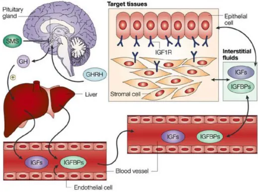
Figure 4. Regulation of circulating IGF-1 levels. Circulating IGF-1 is mostly produced by the liver acting
as an endocrine factor and by extra hepatic tissues acting as autocrine and paracrine mechanisms. Growth hormone, which is produced in the pituitary gland under the control of hypothalamic factors, is the main hormone that regulates IGF-1 and IGFBPs production in the liver. The bioavailability of IGF-1 is influenced by the presence of IGFBPs, which are found in circulation and in extravascular fluid, and modulate the interactions between IGF-1 and the receptors present on the cell surfaces [55].
16
T
HEIGF-1
RECEPTOR(IGF-1R)
AND SIGNALING PATHWAYSIGF-1 mediates its biological actions on cell proliferation, differentiation and survival by binding to specific receptors present on cell surfaces. IGF-1 can interact with different receptors such as IGF-1R (or type 1 IGF receptor) and IGF-2R (or type 2 IGF receptor), insulin receptor (IR), and some atypical receptors such as the hybrid IR/IGF-1R [56]. Mature IGF-1, which is responsible for binding to the receptors, binds with high affinity to IGF-1R, which has a high degree of homology to the insulin receptor [57]. It is also able to interact with lower affinity IGF-2R, which has been shown to be identical to the cation–independent mannose 6-phosphate receptor and to IR. The IGF-1 receptor pathway shares multiple intracellular mediators with the insulin-signaling cascade stimulating glucose intake and protein synthesis in skeletal muscle. Many tissues, including skeletal muscle, express hybrid receptors, but the functional importance of these receptors remains poorly understood.
The tyrosine-kinase receptor IGF-1R is expressed in many types of cells and is a key mediator of cell growth and proliferation. The IGF-1R is a hetero-tetramic protein composed of two extracellular -subunits specific for binding to the hormone and two transmembrane β-subunits containing the tyrosine kinase domain activity with a cluster presenting three tyrosine residues at positions 1131, 1135, and 1136 [1].
The binding of circulating IGF-1 to the cysteine-rich domain contained in the -subunits of the receptor causes a structural rearrangement in the transmembrane β-subunits. This, in turn, induces the activation of tyrosine kinase activity and leads to the autophosphorylation of the cytoplasmic tyrosine kinase domain of the receptor, as one kinase domain phosphorylates the other.
These autophosphorylation events and conformational changes permit unrestricted access for a variety of protein substrates, including members of the insulin receptor substrate (IRS) proteins, whose function is to activate a complex signal transduction network. Transduction involves the PI3K and AKT pathways, leading to protein synthesis, cell survival and inhibition of apoptosis, as well as the pathway of MAP kinases that stimulates cell proliferation and differentiation (Fig. 5).
Following phosphorylation, the IRS interacts with the SH2 domains of the PI3K (phosphoinositide 3-kinase) cytoplasmic protein, which actively catalyzes the phosphorylation of PIP2 (phosphatidylinositol 4,5-bisphosphate) leading to the synthesis
17 of PIP3 (phosphatidylinositol 3,4,5-triphosphate). The PIP3 accumulates at high concentrations and can recruit and activate PDK-1 (phosphoinositide-dependent kinases). Then PDK-1 phosphorylates another protein kinase: the AKT at Thr308 residue [58]. The activated AKT has a variety of substrates that are important to bone and muscle. It increases protein synthesis, promoting the activation of mTOR, which phosphorylates other protein substrates: p70S6K and 4E-BP (eukaryotic initiation factor 4E-binding protein) [6].
An additional pathway activated by AKT is the inhibition of GSK3 (glycogen synthase kinase-3), which results from its phosphorylation in an N-terminal serine residue. In response to IGF-1, GSK3 inhibition promotes dephosphorylation and activation of glycogen synthase, contributing to the stimulation of glycogen synthesis. Activated AKT also phosphorylates PKC (kinase-C protein). The PKC, together with AKT, increase the cell‘s glucose intake by facilitating the translocation of GLUT4, glucose transporters, from the intracellular vesicle to the membrane.
The AKT pathways plays a critical role in apoptosis by inhibiting BAD, which is a protein involved in the apoptosis process. When BAD proteins are not phosphorylated, they remain on the mitochondrial membrane and interact with BCL2 (B-Cell Lymphoma-2) preventing its anti-apoptotic action. When, on the other hand, BAD is phosphorylated by AKT, it is associated with a cytoplasmic protein, and is unable to interfere with the action of BCL2. In addition, AKT phosphorylates FOXO family members, promoting the export from nucleus to cytoplasm, thus reducing the induction of genes such as atrogin-1 and MuRF1, which are ubiquitin ligases involved in protein degradation [59].
Activated AKT also phosphorylates several pro-apoptotic members of the forkhead family (transcription factor), FKHRL1, FKHR, and prevents their activity. The actions of AKT diminish the expression of FasL (Fas Ligand), thus decreasing Fas-mediated apoptosis. In addition to inhibition of pro-apoptotic transcription factors, AKT activity also increases levels of anti-apoptotic proteins, including BCL2 and BCL-X and various adhesion molecules of the extracellular matrix. Activity induced by AKT also involves the expression of the anti-apoptotic transcription factor NF-kB, which enters into the nucleus and activates the transcription of anti-apoptotic genes.
The activation of the PI3K/AKT pathway leads to the transduction of multiple IGF-1 effects, such as increased glucose transport and inhibition of apoptosis through the activation of different proteins.
18 Another pathway activated by IGF-1R/IRS1 is the MAP kinase pathway, which is more involved in cell proliferation and migration. In this signaling, the activated IRS interacts with Shc that binds to the SH2 domain of Grb2, which in turn forms a complex with Sos, a guanine nucleotide exchange factor. This leads to activation of the small G-protein Ras and continues with a sequence of cascade phosphorylation. Subsequently, Ras activates the protein serine kinase Raf, which phosphorylates and activates MEK, leading to the phosphorylation and activation of ERK1/2 (MAPK). This complex results in the translocation of ERK1/2 to the nucleus, which phosphorylates and activates transcription factors such as elk-1 and c-jun, stimulating cell proliferation and cell cycle progression. These nuclear transcriptional factors lead to increase cyclin D1 and reduced p21 and p27 expression, stimulating cell cycle progression from G1 to S and promoting proliferation.
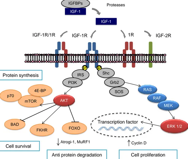
Figure 5. IGF-1 intracellular signaling. The IGF-1R comprises two extracellular -subunits and two
transmembrane β-subunits containing the tyrosine kinase domain. The binding of circulating IGF-1 induces the activation of tyrosine kinase activity of the cytoplasmic portion of the β subunits, forming binding sites for a number of signaling molecules. Transduction involves the activation of the PI3K and AKT pathway.
IGF-1 IGF-1R IGF-1R/1R 1R IGFBPs IGF-2R P P IRS PI3K Shc Grb2 SOS BAD Cell survival Protein synthesis RAS RAF MEK ERK 1/2 IGF-1 mTOR p70 AKT FOXO FKHR Cell proliferation Transcription factor 4E-BP
Anti protein degradation
Atrogi-1, MuRF1 Cyclin D
19
This leads to protein synthesis through the activation of mTOR, which stimulates p70sk6, cell survival and inhibition of apoptosis phosphorylating BAD and FOXO. When phosphorylated, FOXO is prevented from entering the nucleus and stimulating various ubiquitin ligases such as atrogin-1/MAFbx. The Shc/Grb2/SOS complex activates the MAP kinase pathway leading to cell proliferation by activating ERK1/2, which enter the nucleus to activate various transcription factors.
The signaling of IGF-1R plays an important role in both physiological and pathophysiological conditions. Under normal physiological conditions, IGF-1 increases the synthesis of DNA and proteins in cardiomyocytes, promotes myofibrenia development, and is necessary for cellular entry into the S phase. An important role is played by IGF-1 in the development of the hypertrophic response, when the expression of contractile proteins such as actin, myosin and troponin increases. Through its tyrosine kinase, with PI3K and MAPK signaling pathways, IGF-1R can also reduce the risk of heart failure by preventing apoptosis. In addition, IGF protects cells from apoptosis induced by a variety of conditions, including chemotherapy and the expression of oncogenes.
The ability to regulate apoptosis may have an impact on various types of serious illnesses in humans, including several human cancers. Hence, it is of crucial importance to gain a fuller understanding of the different activity of the pro-IGF-1s on the IGF-1R intracellular pathways and subsequently to develop strategies for cancer prevention. Chapter 2 will focus on the results of our study indicating how IGF-1 pro-hormones can activate the IGF-1R independently of the mature IGF-1 form. These results underline the importance of an accurate assessment of the presence of IGF-1 pro-forms within the physio-pathologic context under study.
IGF-1
AND CANCERIGF-1 is a cell growth factor that stimulates proliferation, differentiation and inhibits cellular apoptosis, all key factors associated with tumor. The circulating levels of IGF-1 were evaluated in different pathological conditions. Growth factor is the main regulator of IGF-1 plasma levels, whereas transport proteins, IGFBPs, which bind most of the circulating IGF-1, regulate bioavailability. Studies have revealed that high level of IGF-1
20 or altered levels of IGFBPs are associated with an increased risk of cancers such as lung [60], colon [61], prostate and breast [62]. A prospective study has shown the association with high circulation IGF-1 and lower plasma IGFBP3 concentrations and an increased risk of colorectal adenoma [63]. The cleavage from IGFBPs can results in and increased released of free IGF-1, which can then bind the receptor and promote cell proliferation. Moreover, the overexpression of the IGF-1R is evaluated in different cancer cells [64] and can be implicated in the acquisition of the transformed phenotype [65]. The concentrations of IGF-1 and its transport proteins are extremely variable between individuals and this could affect the distribution of cancer risk in the population.
The IGF-1R can regulate cell-cycle progression that may be important for acquisition of the malignant phenotype. It can promote G1-S transition by increasing cyclin D1 and cyclin-dependent kinase 4 (CDK4) leading to retinoblastoma (RB) phosphorylation and releasing the transcription factor E2F [66]. In addition, it down regulates the transcriptional inhibitor p27 and phosphatases the tumor suppressor PTEN, that has been deregulated in cancer [67]. Studies have shown an increasing expression of IGF-1 or of IGF-1R in different cancer such as breast, lung, thyroid, prostate, glioblastoma, neuroblastoma, meningioma and rhabdomyosarcoma. IGF-1 is also implicated in the development and progression of angiogenesis and can modulate the expression of the vascular endothelial growth factor (VEGF), a potent angiogenic factor [68]. Another IGF-1 action that is implicated in the development of tumor is the inhibition of apoptosis by inhibiting pro-apoptotic proteins such as BAD and inducing the expression of anti-apoptotic proteins such as BCL2. Multiple mechanisms have been shown to modulate tumor cell sensitivity to IGF-1 including increase in IGF-1 synthesis, increased IGF-1R expression, release of proteases that cleave IGFBPs.
Recent studies have investigated the biological action induced by IGF-1 isoforms in tumor tissues. In particular, a differential expression profile of IGF-1 isoforms has been documented in the development and progression of human prostate cancer (PCa). It was shown that the IGF-1Ec isoform is overexpressed in human prostate cancer tissues and in human cancer PC-3 and LNCaP cell lines not only at expression level, but also at protein level. In addition, the transcription of this specific isoform was significantly higher in PCa and in prostatic intraepithelial neoplasia (PIN) than in normal prostate tissues, whereas the normal prostate epithelial cells (HPrEC) did not express IGF-1Ec [20].
21 A differential expression profile of IGF-1 transcript variants between normal and tumor tissues has also been observed in other in vivo human cancers, such as endometriosis [21], cervix [33], colorectal cancer [34] and in osteosarcoma cells in vitro [69].
The expression analysis of IGF-1 isoforms in eutopic and ectopic endometrium suggest that the IGF-1Ec splice variant may be involved in the pathophysiology of endometriosis. In particular, it was revealed that all the three splice variants were expressed in both eutopic and ectopic endometrium, though it is significantly lower in endometriotic cysts both at the transcript and protein level. Different expression level was also analysed between the glandular cells of the eutopic and the ectopic endometrium, where the first not express any IGF-1 isoforms whereas the second express the IGF-1Ec isoform [21]. In addiction the analysis of the expression profile during human papillomavirus (HPV) dependent cervical carcinogenesis revealed that all the IGF-1 splice variant were up-regulated in pre-cancerous cells and in particular the IGF-1Eb expression was very high in the cancer samples [33]. Furthermore, testing the exogenous administration of the synthetic E-peptide of the pro-IGF-1Ec, consisting on the C-terminal 24 amino acids sequence of the human Ec-peptide, has been revealed in an increase in proliferation not only in PCa, PC-3 and LNCaP cells [20], in endometrial KLE cells [21] and in MG-63 osteosarcoma cells [69]. The stimulation of PCa and LNCaP cells with the synthetic MGF led to a differential activation of intracellular signaling, with greater phosphorylation of ERK 1/2 without affecting the AKT activation, suggesting, suggesting its independent mode of action of this E-peptice [20].
These data suggest the potentially different roles of different pro-IGF-1 in the pathophysiology of all those conditions. Interestingly, IGF-1 splice profile appears to be different between tumors and the state of the disease, showing a differential regulation of its splice variants. The functional meaning of IGF-1 different splice patter and how the pro-peptides are connected to different autocrine, paracrine and endocrine roles in tumor genesis remains to be clarified.
IGF-1
AND BREAST CANCERThe IGF-1 plays a significant role in human physiology, particularly in the development of many tissues, including the mammary gland. In particular IGF-1 is a key mediator of
22 mammary gland terminal end bud formation and ductal morphogenesis during development [70]. In contrast, many studies have been associated to the aberrant expression of the IGF-1 system in the development of several malignancies including breast cancer (BC). This is the most common malignant neoplasm in women. BC recurrences and survival are influenced by the presence of many prognostic factors, including hormone factors. IGF-1 is mainly expressed by stromal and only rarely by epithelial cells both in normal and BC tissues but its mitogenic actions are mainly expressed in the epithelium [71]. Various studies have been conducted to evaluate whether mature IGF-1 blood levels are associated with the development of BC or recurrence. Many epidemiological and prospective studies have tried to confirm the positive correlation between plasma IGF-1 levels and BC risk. A pooled data analysis of seventeen prospective studies from twelve countries by the Endogenous Hormones and BC Collaborative Group showed a positively association between circulating IGF-1 and BC risk independently by IGFBP3 levels and menopausal status in estrogen-receptor (ER+) positive tumors [72]. This result is also supported by the data analysis from the European Prospective Investigation into Cancer and Nutrition cohort [73]. Furthermore in an Italian cohort study, serum IGF-1 levels have been positively associated with increased disease risk among BRCA gene mutation (hereditary BC) [74].
Numerous studies have investigated the role of IGF-1 levels and altered circulating IGFBPs levels, in particular IGFBP3, in association with BC risk and prognosis. It has been shown that low levels of IGF-1 are associated with improved survival in BC patients [75, 76]. In addition, it has been shown that low levels of IGFBP3 increase the amount of free IGF-1 that is associated with increased mortality. This study also confirmed a better prognosis in low-level IGF-1 BC patients [76].
IGF-1R mediates the mitotic and anti-apoptotic effects of IGF-1 and has been indicated a correlation between IGF-1R expression and disease development [77]. Another study revealed that IGF-1 receptor is over-expressed in about 90% of BC cases and its level are higher in malignant cells than in normal tissue [78]. Moreover, this over-expression has been related with poor prognosis in patients with early BC [79].
In addition, IGF-1R has been involved in the metastatic progression in BC [80] and another study indicated the relation between IGF-1 and tumor progression. It favors the expression of angiogenic factors in synergy with other growth factors such as vascular endothelial growth factor (VEGF) and platelet-derived factor (PDGF). In particular,
IGF-23 1 promotes tumor growth and lymphatic metastases by inducing VEGF and it has been indicated a relation between high levels of IGF-1, IGFBP3 and VEGF and lymph node metastasis in patients affected by BC [81].
Although much has been investigated regarding serum IGF-1 expression, it was recently revealed by microarray analysis that the increase of IGF-1 mRNA levels within tissue samples was associated with better prognosis [82]. This finding suggests a contradictory role of circulating and tissue IGF-1 and it can be explained by the lack correlation between circulating and tissue IGF-1 levels, thus local IGF-1 expression may be a better marker of tumor compared to circulating IGF-1 levels.
Despite the large amount of studies that have investigated the role of mature IGF-1 in the BC development and recurrences, only few studies have considered the biological action of each IGF-1 isoforms involved in this tumor. Many tissues such as muscle, liver and adipose tissue synthesize IGF-1 specific isoforms, which act locally with autocrine/paracrine mechanisms and play a key role in repairing cellular damage. Not completely clear is the local contribution to the systemic IGF-1 and the potential environmental stimuli that might be involved to favour their local or systemic release. Data on survival of patients with BC indicate that subjects with a high tissue expression of one of the transcripts of IGF-1, IGF-1Ea, have a lower risk of recurrence and mortality than patients with a lower expression of the same transcript [83].
The biological role of different IGF-1 isoforms and how they are related to different autocrine, paracrine and endocrine roles in tumor genesis is not entirely explained. Indeed, the different isoforms correlate with numerous cellular responses, thus knowing which IGF-1 transcriptional variant is involved in BC could help to elucidate the effect of the IGF-1 pool in the BC development and recurrence.
24
25 The primary aim of this Thesis was to investigate the regulation of IGF-1 alternative splicing and the biological role of IGF-1 pro-hormones in human breast cancer (BC). The study objectives include:
a) The evaluation of the evolutionary pressure within the different coding regions of the mammalian igf-1 gene.
b) The comparison of the mechanisms regulating the IGF-1 exon 5 transcription and splicing between human and mouse.
c) The characterization of the IGF-1 pool and their biological activity on BC cell lines. d) The investigation of the cellular mechanisms controlling IGF-1 pro-hormones production and secretion.
e) The determination of the effects of lifestyle modification program on circulating IGF-1 level in BC patients.
26
27
MIR
R
ETROPOSON
E
XONIZATION
P
ROMOTES
E
VOLUTIONARY
V
ARIABILITY AND
G
ENERATES
S
PECIES
-S
PECIFIC
E
XPRESSION OF
IGF-1
S
PLICE
V
ARIANTS
Giosuè Annibalini1, Pamela Bielli2, Mauro De Santi1, Deborah Agostini1, Michele Guescini1, Davide Sisti1, Serena Contarelli1, Giorgio Brandi1, Anna Villarini3, Vilberto Stocchi1, Claudio Sette2 and Elena Barbieri1^
1
Department of Biomolecular Sciences, University of Urbino Carlo Bo, 61029 Urbino, Italy.
2
Department of Biomedicine and Prevention, University of Rome Tor Vergata, 00133 Rome, Italy; Laboratory of Neuroembryology, Fondazione Santa Lucia, 00133 Rome, Italy.
3
Department of Preventive & Predictive Medicine, Fondazione IRCCS Istituto Nazionale dei Tumori, 20133 Milan, Italy.
^IIM, Interuniversity Institute of Myology.
Published in Biochimica Biophysica Acta, Gene Regulatory Mechanisms 1859(5):757-68, 2016.
28
MIR retroposon exonization promotes evolutionary variability and
generates species-specific expression of IGF-1 splice variants
Giosuè Annibalini1*ϯ, Pamela Bielli2ϯ, Mauro De Santi1, Deborah Agostini1, Michele Guescini1, Davide Sisti1, Serena Contarelli1, Giorgio Brandi1, Anna Villarini3, Vilberto Stocchi1, Claudio Sette2 and Elena Barbieri1^
1
Department of Biomolecular Sciences, University of Urbino Carlo Bo, 61029 Urbino, Italy.
2
Department of Biomedicine and Prevention, University of Rome Tor Vergata, 00133 Rome, Italy; Laboratory of Neuroembryology, Fondazione Santa Lucia, 00133 Rome, Italy.
3
Department of Preventive & Predictive Medicine, Fondazione IRCCS Istituto Nazionale dei Tumori, 20133 Milan, Italy.
^IIM, Interuniversity Institute of Myology
* To whom correspondence should be addressed. Tel:+39 303418 Fax:+39 0722-303401; Email: [email protected]
ϯ
Giosuè Annibalini and Pamela Bielli equally contributed to this work.
Abstract
Insulin-like growth factor (IGF-1) -1 is a pleiotropic hormone exerting mitogenic and anti-apoptotic effects. Inclusion or exclusion of exon 5 into the IGF-1 mRNA gives rise to three transcripts, IGF-1Ea, IGF-1Eb and IGF-1Ec, which yield three different C-terminal extensions called Ea, Eb and Ec peptides. The biological significance of the IGF-1 splice variants and how the E-peptides affect the actions of mature IGF-1 are largely unknown. In this study we investigated the origin and conservation of the IGF-1 E-peptides and we compared the pattern of expression of the IGF-1 isoforms in vivo, in nine mammalian species, and in vitro using human and mouse IGF-1 minigenes.
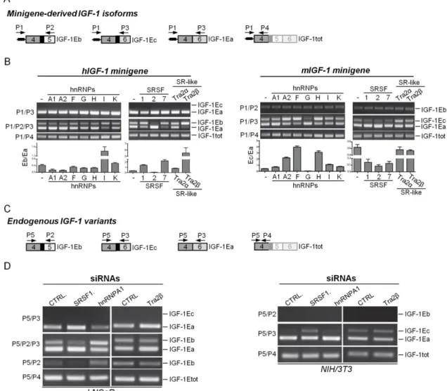
29 Our analysis showed that only 1Ea is conserved among all vertebrates, whereas IGF-1Eb and IGF-1Ec are an evolutionary novelty originated from the exonization of a mammalian interspersed repetitive-b (MIR-b) element. Both IGF-1Eb and IGF-1Ec mRNAs were constitutively expressed in all mammalian species analyzed but their expression ratio varies greatly among species. Using IGF-1 minigenes we demonstrated that divergence in cis-acting regulatory elements between human and mouse conferred species-specific features to the exon 5 region. Finally, the protein-coding sequences of exon 5 showed low rate of synonymous mutations and contain disorder-promoting amino acids, suggesting a regulatory role for these domains.
In conclusion, exonization of a MIR-b element in the IGF-1 gene determined gain of exon 5 during mammalian evolution. Alternative splicing of this novel exon added new regulatory elements at the mRNA and protein level potentially able to regulate the mature IGF-1 across tissues and species.
Keywords
IGF-1 isoforms, alternative splicing, retroposon exonization, synonymous sites, intrinsically disordered regions
30
31
1. Introduction
The mammalian insulin-like growth factor (IGF) -1 gene is a single copy gene composed of six exons and five introns, which gives rise to an immature IGF-1 peptide (IGF-1 pro-peptide) containing a signal peptide at the 5' end of the gene, a core region and an E-peptide at the 3' end (Figs. 1A and 1B). Both signal E-peptide and E-E-peptide are then removed by protease cleavage to form the 70 amino acid-long mature IGF-1 peptide (IGF-1 core), which displays growth-promoting and metabolic functions.
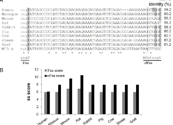
Four of the six IGF-1 exons are subjected to alternative splicing (Figs. 1A and 1B) [1]. Exon 1 and exon 2 are mutually exclusive first exons and generate different signal peptides. Transcripts containing exon 1 are referred to as Class I transcripts whereas those containing exon 2 are referred to as Class II transcripts. The Class II IGF-1 knockout mice indicated that class II isoforms are dispensable for fetal and postnatal growth, and the significance of the alternate signal peptides encoded by the first two IGF-1 exons was discussed in [2-3]. At the 3‘ end of the gene, alternative splicing yields three different mRNA transcripts, each encoding distinct carboxyl-terminal portions of E-peptide followed by the 3'-untranslated region (3'UTR) (Figs. 1A and 1B). Thus, although alternative splicing generates different precursor peptides, it does not alter the sequence of the mature IGF-1 peptide.
Splicing of exon 4 with exon 6 yields the most common IGF-1Ea variant, which encodes the 35 amino acids-long Ea peptide. The first 16 amino acids of the Ea peptide are encoded by exon 4 and are common in all E-peptides, whereas the remaining 19 amino acids are encoded by exon 6 and are unique to this isoform. The IGF-1Eb variant is produced when exon 4 is spliced with exon 5 and encodes the Eb peptide, which contains the 16 common amino acids and 61 additional amino acids encoded by exon 5. IGF-1Eb transcript terminates in exon 5 and excludes exon 6 from the mRNA, hence it has a completely different 3‘UTR compared to IGF-1Ea and IGF-1Ec. The third variant, named IGF-1Ec in humans, is generated by usage of a cryptic 5' splice site (c5‘ss) (named IGF633) [4] present in exon 5, which is in turn spliced with exon 6. The c5‘ss of IGF-1 exon 5 deviates from the vertebrate consensus and is commonly used in rodents and rabbits [5-7] but rarely and in a tissue-specific manner in human [1, 4]. Notably, although this variant is named IGF-1Eb in rodents, for clarity we will use the human IGF-1Ec nomenclature throughout this manuscript, regardless of the species. The human Ec