Calcium Sensing Receptor Expression in Ovine Amniotic
Fluid Mesenchymal Stem Cells and the Potential Role of
R-568 during Osteogenic Differentiation
Pamela Di Tomo1,2,3., Caterina Pipino1,2,3., Paola Lanuti2,3,4, Caterina Morabito2,3,5, Laura Pierdomenico2,3,4, Vittorio Sirolli4, Mario Bonomini4, Sebastiano Miscia2,3,4, Maria Addolorata Mariggio`2,3,5, Marco Marchisio2,3,4, Barbara Barboni3,6, Assunta Pandolfi1,2,3*
1 Department of Experimental and Clinical Sciences, University ‘‘G. d’Annunzio’’ Chieti-Pescara, Chieti, Italy, 2 Aging Research Center, Ce.S.I., ‘‘University G. d’Annunzio’’ Foundation Chieti, University ‘‘G. d’Annunzio’’ Chieti-Pescara, Chieti, Italy,3 StemTeCh Group Chieti, University ‘‘G. d’Annunzio’’ Chieti-Pescara, Chieti, Italy, 4 Department of Medicine and Aging Science, University ‘‘G. d’Annunzio’’ Chieti-Pescara, Chieti, Italy,5 Department of Neuroscience and Imaging, University ‘‘G. d’Annunzio’’ Chieti-Pescara, Chieti, Italy,6 Department of Comparative Biomedical Science, University of Teramo, Teramo, Italy
Abstract
Amniotic fluid-derived stem (AFS) cells have been identified as a promising source for cell therapy applications in bone traumatic and degenerative damage. Calcium Sensing Receptor (CaSR), a G protein-coupled receptor able to bind calcium ions, plays a physiological role in regulating bone metabolism. It is expressed in different kinds of cells, as well as in some stem cells. The bone CaSR could potentially be targeted by allosteric modulators, in particular by agonists such as calcimimetic R-568, which may potentially be helpful for the treatment of bone disease. The aim of our study was first to investigate the presence of CaSR in ovine Amniotic Fluid Mesenchymal Stem Cells (oAFMSCs) and then the potential role of calcimimetics in in vitro osteogenesis. oAFMSCs were isolated, characterized and analyzed to examine the possible presence of CaSR by western blotting and flow cytometry analysis. Once we had demonstrated CaSR expression, we worked out that 1mM R-568 was the optimal and effective concentration by cell viability test (MTT), cell number, Alkaline Phosphatase (ALP) and Alizarin Red S (ARS) assays. Interestingly, we observed that basal diffuse CaSR expression in oAFMSCs increased at the membrane when cells were treated with R-568 (1mM), potentially resulting in activation of the receptor. This was associated with significantly increased cell mineralization (ALP and ARS staining) and augmented intracellular calcium and Inositol trisphosphate (IP3) levels, thus demonstrating a potential role for calcimimetics during osteogenic differentiation. Calhex-231, a CaSR allosteric inhibitor, totally reversed R-568 induced mineralization. Taken together, our results demonstrate for the first time that CaSR is expressed in oAFMSCs and that calcimimetic R-568, possibly through CaSR activation, can significantly improve the osteogenic process. Hence, our study may provide useful information on the mechanisms regulating osteogenesis in oAFMSCs, perhaps prompting the use of calcimimetics in bone regenerative medicine.
Citation: Di Tomo P, Pipino C, Lanuti P, Morabito C, Pierdomenico L, et al. (2013) Calcium Sensing Receptor Expression in Ovine Amniotic Fluid Mesenchymal Stem Cells and the Potential Role of R-568 during Osteogenic Differentiation. PLoS ONE 8(9): e73816. doi:10.1371/journal.pone.0073816
Editor: Irene Georgakoudi, Tufts University, United States of America
Received February 7, 2013; Accepted July 24, 2013; Published September 9, 2013
Copyright: ß 2013 Di Tomo et al. This is an open-access article distributed under the terms of the Creative Commons Attribution License, which permits unrestricted use, distribution, and reproduction in any medium, provided the original author and source are credited.
Funding: This study was supported by CariChieti Foundation and ERA-Amgen (Contract#2010547324). The authors received funding from a commercial source (Era-Amgen, USA), this does not alter the authors’ adherence to all the PLOS ONE policies on sharing data and materials. The funders had no role in study design, data collection and analysis, decision to publish, or preparation of the manuscript.
Competing Interests: The authors have declared that no competing interests exist. * E-mail: [email protected]
.These authors contributed equally to this work.
Introduction
Amniotic fluid stem (AFS) cells, isolated during pregnancy for prenatal genetic tests, have been recognized as an efficient source of cells with therapeutic potential [1]. AFS cells are widely multipotent, express some pluripotency markers and can be differentiated within the tissues of the three germ layers [1,2]. Their properties such as low immunogenicity, the inability to form tumors, easy accessibility, and the absence of ethical problems associated with their use, make them ideal candidates for regenerative medicine [3–5].
Amniotic fluid-derived mesenchymal stem cells from sheep (oAFMSCs) have recently been isolated and characterized [6]. Particularly, it was demonstrated that these cells can differentiate
into osteogenic, adipogenic [7] and smooth muscle lineages [6]. Moreover, sheep are considered a good animal model as they are similar to humans in size and some physiological properties. Large animals form an optimal preclinical model on which to study various diseases, such as bone disease. In this context, oAFMSCs used in allotransplantation of injured Achilles tendon led to matrix organization and tissue regeneration [8]. Furthermore, oAFMSCs have been used in tissue renovation such as the repair of diaphragmatic tendon [9] and prenatal tracheal reconstruction [10].
Again, Shaw et al. have demonstrated that oAFMSCs may be employed for autologous in utero stem cell gene therapy. Ovine AFMSCs obtained from sheep were transduced with GFP lentiviral vector and reinjected into the peritoneal cavity of the
fetal donor. The results obtained showed the presence of GFP positive cells in many fetal organs detected by PCR, immuno-staining and cytofluorimetric analysis [7].
More recently, oAFMSCs in combination with a collagen-based scaffold were used in an experimental animal study of sinus augmentation, leading to bone regeneration in vivo [11].
Although several studies have demonstrated the ability of AFS cells to differentiate within the injury areas, additional studies are required to increase the efficiency of osteogenic differentiation. In this connection CaSR, a G protein-coupled receptor able to bind calcium ions, may play a potentially active role. This receptor, initially cloned from bovine parathyroid, controls parathyroid hormone (PTH) secretion from the parathyroid glands in response to extracellular calcium signals [12].
Recently CaSR was found to be expressed in several tissues, such as kidney [13], the gastrointestinal tract [14], blood vessels [15,16], bone marrow [17], mouse embryonic stem cells [18] and osteoblast cells [19]. Additionally, CaSR exercises an important role in controlling osteoblasts as well as osteoclast recruitment, differentiation and survival via multiple intracellular signals [20,21]. Moreover, it is known that mice with cell type–specific knockout of CaSR display postnatal skeletal defects [22]. Thus, agonists that bind the bone CaSR may be advantageous for the treatment of bone diseases [21,23].
Calcimimetics, such as R-568, are thought to activate G protein-linked CaSR by allosterically increasing the affinity of the receptor for Ca2+ leading to efficient control of uremic hyperparathyroidism [24]. Several recent studies have suggested they also possess the ability to modulate bone cell metabolism via CaSR, being therefore, of potential interest in the treatment of bone disease [23].
Today, nothing is known regarding the expression of CaSR in the model of oAFMSCs. Hence, the first aim of this work is to investigate the possible expression of CaSR in these cells. Secondly, taking into account the role of calcimimetics in bone development, our experiments were designed to test the optimal concentration of calcimimetic R-568 and its enantiomer S-568 and then investigate their selective effect through CaSR on osteogenic in vitro differentiation of oAFMSCs.
Materials and Methods Chemicals
Powders R-568-HCl and S-568-HCl were provided by Amgen (Amgen, Inc., Thousand Oaks, CA, USA), resuspended in Dimethyl sulfoxide (DMSO) at 10 mM concentration and stored at 220uC. Calhex2231 (Santa Cruz, sc-207394) was resuspended in Ethanol at 10 mM concentration and stored at 220uC.
Ethics Statement
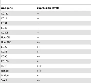
All cells and tissues were collected from slaughtered animals, and this did not require an ethic statement. The uterus were collected from the slaughterhouse of the Teramo province as material used for teaching and research activity of Veterinary University course. The collection from the slaughterhouse of biological material is normed by specific conventions subscribed between the Faculty of Veterinary Medicine of Teramo and the Table 1. Flow cytometry analysis of the oAFMSCs phenotype.
Antigens Expression levels
CD117 2 CD14 2 CD31 2 CD45 2 CD49f 2 HLA-DR 2 HLA-ABC ++ CD29 ++ CD58 ++ CD90 ++ CD166 + TERT +++ Nanog +++ Oct3/4 + Sox 2 ++ – negative expression;
+/2 low expression;+moderate expression; ++positive;
+++high expression.
doi:10.1371/journal.pone.0073816.t001
Figure 1. CaSR identification in oAFMSCs. (A) Western blot analysis shows expression of CaSR in oAFMSCs comparable to sheep fetal calvarial osteoblast (OBs) used as a positive control. b-actin was used as an internal control. Figure (B) shows the shift of mean fluorescence intensity of CaSR in oAFMSCs (filled histogram) as compared to the mean fluorescence intensity of secondary antibody alone (open histogram). (C) A representative cell in a bright field and the diffuse expression of CaSR obtained by IDEAS software (Amnis). Magnification 40x.
doi:10.1371/journal.pone.0073816.g001
ASL, organ of the Ministry of Health vacated to the control and safety of slaughterhouses. In each slaughterhouse the collection of tissues is controlled by an inspector which is a Veterinary that follow the regularity of all the procedures.
Cell Cultures
The oAFMSCs were isolated and cultured as previously described [8]. Briefly, sheep fetuses were collected at a local abattoir by removing the whole pregnant uterus and bringing it at 30uC to the laboratory. Only fetuses of 25–35 cm length (approximately 3 months’ gestation) were used. Once the uterus wall was opened, ,100 ml of amniotic fluid (AF) was collected, centrifuged at 800 g for 20 min and the resulting pellets seeded in 250 ml flasks in growth medium containing 5 ng/ml Fibroblast Growth Factor (b-FGF) as described in Colosimo et al. [8].
An aliquot of oAFMSCs was analysed, as previously described [8] for surface and intracellular stem cell markers by flow cytometry and the remaining cells were cryopreserved until use.
Cells were cultured in medium composed of Dulbecco’s Modified Eagle Medium (DMEM, PAA) with 10% Fetal Bovine Serum (FBS, PAA), 1% Penicillin/Streptomicin and 1% L-Glutamine (PAA). The medium was changed every other day. Cells were expanded when they reached 80% confluence, using
0.05% Trypsin-EDTA. All experiments were performed on cells between passages V and VI.
Flow Cytometry
Antibodies. FITC-conjugated anti-CD14 and FITC-conju-gated anti-CD58 were obtained from LifeSpan Bioscences (Seattle, WA, USA); anti-CD29, anti-HLA-ABC and anti HLA-DR were obtained from VMRD (Pullman, WA, USA); FITC-conjugated anti-CD31 and FITC-conjugated anti-CD45 were purchased from AbD Serotec (Oxford, UK); anti-CD49f was purchased from Beckman Coulter (Fullerton, CA, USA); CD117 and anti-SOX2 were obtained from abcam (Cambridge, UK); FITC-conjugated anti-CD166 (CD166-FITC) was obtained from Ancell (MN, USA); PE-conjugated anti-Thy-1 (CD90) was obtained from Bioss (Woburn, MA, USA); PE-conjugated anti-OCT3/4 (OCT3/ 4-PE) was obtained from Becton Dickinson (BD, San Jose, CA); anti-TERT was purchased from Calbiochem (Gibbstown, NJ); anti-Nanog was obtained from Chemicon International (Billerica, MA). Primary antibodies were labeled using the appropriate FITC secondary antibodies (Jackson ImmunoResearch Laboratories).
Cell staining for flow cytometry. Staining of surface and intracellular antigens was performed as previously reported [25].
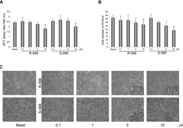
Figure 2. Evaluation of the optimum concentration of calcimimetics on oAFMSCs treated for 14 days. (A) MTT assay performed at day 14 on oAFMSCs shows a reduction in cell viability upon increasing the concentrations of R-568 or S-568 (0.1–10 mM) (*p,0.01 vs Basal). (B) Decreasing cell number trend on day 14 at enhanced concentrations of calcimimetics R-568 and S-568 (0.1–10 mM) (*p,0.001 vs Basal). The number of cells is normalized per cm2. All histograms represent the mean 6 SD of three independent experiments. (C) Bright field images of oAFMSCs at increased concentrations of calcimimetics showing the cell morphology and the accumulations of vacuoles at high concentrations (enlargement in the right corner). Magnification 10x.
doi:10.1371/journal.pone.0073816.g002
Cells were analyzed on a FACSCalibur flow cytometer (BD), using CellQuestTMsoftware 3.2.1.f1 (BD) [26].
Flow cytometry measurement. Quality control included regular check-ups with Rainbow Calibration Particles (6 peaks) and CaliBRITE beads (both from BD Biosciences). Debris was excluded from the analysis by gating on morphological parameters (lymphocyte gate); 20,000 non-debris events in the morphological gate were recorded for each sample. All antibodies were titrated under assay conditions and optimal photomultiplier (PMT) gains were established for each channel. Data were analyzed using FlowJoTM v.8.8.6 software (TreeStar, Ashland, OR). The Mean Fluorescence Intensity Ratio (MFI Ratio) was calculated dividing the MFI of positive events by the MFI of negative events [27].
Imaging flow cytometry. Cell staining for surface or intracellular CaSR was performed as previously described for conventional flow cytometry analysis [25]. Cells were stained in both cases by a primary antibody against CaSR (1:100, Thermo Scientific), followed by incubation with the specific FITC-labeled secondary antibody (1:250, Jackson ImmunoResearch). Dead cells were excluded by 7-Aminoactinomycin D (7-AAD) staining (BD Biosciences), a fluorescent chemical compound that can be kept by cells characterized by damaged membranes. Sample acquisitions were performed by ImageStream (Amnis, Seattle, WA, USA; one
laser, six-color configuration). To assess nonspecific fluorescence, samples stained with the respective secondary antibody alone were used. Analyses were performed by the IDEAS software (Amnis) examining 5,000 cells for each samples. Data are indicated as a percentage of CaSR positive cells in live cells whether permea-bilized or not.
Western Blotting Analysis
CaSR protein levels and b-actin were evaluated by Western blot analysis on oAFMSCs as previously described [16]. Confluent monolayer cells were cultured in 100 mm2dishes, then rinsed with ice-cold PBS and scraped on ice into lysis buffer containing 50 mM Tris-HCl, pH 7.4, NP 40 1%, 150 mM NaCl and a cocktail of protease and phosphatase inhibitors (1mg/ml each of aprotinin, leupeptin and pepstatin, 1 mM each of PMSF, Na3VO4
and NaF). Total cell lysate was either used directly for SDS-PAGE or stored at 280uC. In denaturing and reducing conditions, 80mg of protein was used and resolved electrophoretically on linear 8% gels.
After incubation with blocking solution (5% dry milk in TBS Tween 20 0.1%) for 1h at room temperature, the blots were incubated overnight at 4uC with primary monoclonal anti-CaSR (1:1000, Thermo Scientific) and also with a secondary goat
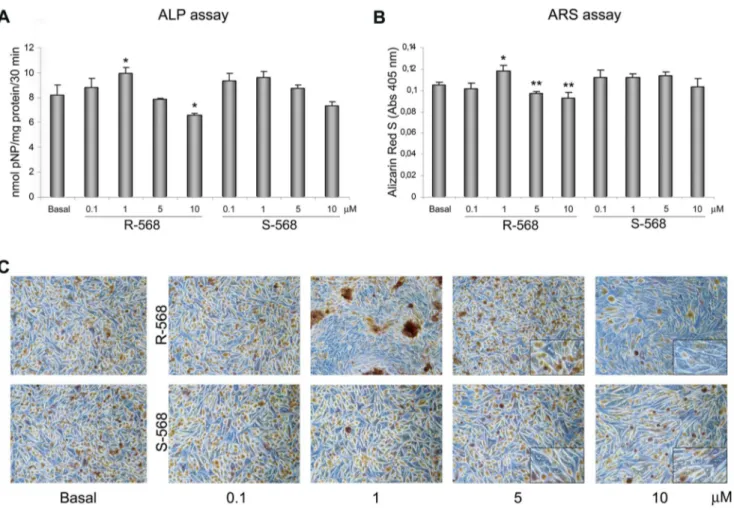
anti-Figure 3. Estimation of osteogenic differentiation by Alkaline Phosphatase (ALP) and Alizarin Red S (ARS) staining. (A) ALP activity performed on day 7 of oAFMSC culture at increased concentrations of R-568 and S-568 (0.1–10 mM) (*p,0.05 vs Basal; **p,0.01 vs Basal). ALP activities were calculated with p-nitrophenol standard and normalized per mg of proteins. (B) ARS staining performed on day 14 of oAFMSC culture at increased concentrations of R-568 and S-568 (0.1–10 mM) (*p,0.01 vs Basal; **p,0.01 vs Basal). All histograms represent the mean 6 SD of three independent experiments. The results are normalized per total cell number/well. (C) Representative images of ARS assay after 14 days of treatment with calcimimetics. Note the presence of mineral deposits after treatment with R-568 (1 mM). High concentrations (5 and 10 mM) of calcimimetics lead to a change in cell morphology with the presence of vacuoles (enlargement in the right corner). Magnification 10x.
doi:10.1371/journal.pone.0073816.g003
mouse IgG antibody (1:10000, Calbiochem). The blots were finally washed and protein bands were detected using ECL Prime Western Blotting Detection Reagent (GE Healthcare). Sheep fetal calvarial derived cells (OBs) were used as positive controls.
In vitro Treatment with Calcimimetics
Ovine AFMSCs were seeded at a density of 3000 cells/cm2in 6 well plates and treated for 14 days in the presence of increasing concentrations of calcimimetics R-568 and S-568 (0.1–10mM). In some experiments, the inhibitor Calhex-231 was pre-incubated 30 minutes before the addition of calcimimetics. To test the appropriate concentration of calcimimetics, MTT, cell number, Alkaline Phosphatase (ALP) and Alizarin Red S (ARS) assays were performed.
MTT Assay and Cell Number
The effect of calcimimetics on oAFMSC viability was assessed by the 3-[4,5-dimethylthiazol-2-yl]-2,5-diphenyltetrazolium bro-mide (MTT) method. After treatment with calcimimetics as described above, a solution of MTT in phosphate buffered saline (PBS) was added to each well to a final concentration of 0.5 mg/ ml. After a further 3 hours’ incubation the cells were washed and 200ml DMSO was added for crystal solubilization. The plates were put in the dark at 37uC, and continuous gentle shaking was performed for 30 min to thoroughly dissolve the MTT dye. The spectrometric absorbance at 540 nm (for formazan dye) and 690 nm (as a background level) was read using a microplate reader (SpectraMAX 190, Molecular Devices). The effect of increased doses of calcimimetics on the number of cells was also evaluated. Cells were washed at least three times with PBS w/o Ca++and Mg++. Then, the cells were incubated with Trypsin-EDTA at 37uC for 10 minutes and gently detached with cell scraper, centrifuged, resuspended in PBS and counted by dilution (1:1) in Trypan blue using a Burker’s chamber. Cell counts were normalized per cm2.
ALP and Alizarin Red S Assays
After treatment with different concentrations of calcimimetics (0.1–10mM), osteogenic differentiation was assessed by ALP and Alizarin Red S staining (Sigma) at 7 and 14 days respectively using adapted published protocols [28,29]. Briefly, for ALP activity the cells were washed twice with PBS, lysed with TRIS/HCl 10 mM pH 7.5+0.1% Triton and then incubated with a p-Nitrophenyl Phosphate Substrate (Sigma) for 30 min at 37uC in the dark. To stop the reaction NaOH 2N was used. ALP activity was measured by absorbance at 405 nm on a plate reader and absolute amounts of enzyme activity were calculated with p-nitrophenol (pNP, Sigma) standard. Total enzyme activity of each well was expressed as nM of pNP/mg protein/30 min.
For ARS assay, the cells were fixed in paraformaldehyde 4% for 15 minutes. Following a rinse with distilled water, the cells were stained in 40 mM Alizarin Red S (pH4.2) for 20 minutes at room temperature. For quantification of staining, 800mL 10% (v/v) acetic acid was added to each well, and the plate was incubated at room temperature for 30 min with gentle shaking. The monolayer was then scraped from the plate with a cell scraper, transferred in 1.5 mL microcentrifuge tube and then 10% (v/v) acetic acid was added. Subsequently, 500mL mineral oil (Sigma Aldrich) was added and samples were heated to 85uC for 10 min and then 5 min on ice. The samples were then centrifuged at 20,000 g for 15 min and 500mL of the supernatant was transferred to a new 1.5 mL microcentrifuge tube. Then 200mL of 10% (v/v) ammonium hydroxide was added to neutralize the acid and aliquots (150mL) of the supernatant were read in triplicate at 405 nm in 96 well plate and normalized with respect to total cell number for each sample.
Intracellular Calcium Measurements
Following the addition of 1mM R- or S-568, in presence or absence of the inhibitor Calhex-231, the intracellular calcium levels in oAFMSC was measured using Fluo-4/AM (Molecular
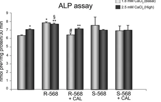
Figure 4. Effect of calcimimetics on ALP activity. ALP activity, detected at day 7, on oAFMSCs treated with calcimimetics pre-incubated (30 minutes) or not with Calhex-231 (1 mM, *p,0.0001 2.5 mM CaCl2vs Basal 1.8 mM CaCl2and R-568 vs Basal 1.8 mM CaCl2;
1
p,0.01 R-568+2.5 mM CaCl2vs S-568+2.5 mM CaCl2; **p,0.001 R-568+2.5 mM CaCl2vs 2.5 mM CaCl2and R-568+Calhex+2.5 mM CaCl2vs R-568+2.5 mM CaCl2;
# p,0.002 R-568+Calhex vs R-568+1.8 mM CaCl2). ALP activities were calculated with p-nitrophenol standard and normalized per mg of proteins.
doi:10.1371/journal.pone.0073816.g004
Probes, Life technologies Italia, Monza, Italy), and an inverted Olympus microscope connected to a high-speed wavelength switcher (Polychrome II; Till Photonics, Germany) and a cooled charge coupled device (CCD) camera (C6790 model; Hamamatsu Photonics, Hamamatsu, Japan). The cells were loaded with 5mM Fluo-4/AM for 40 min at 37uC in normal external solution (NES: 140 mM NaCl, 2.8 mM KCl, 2 mM CaCl2, 2 mM MgCl2,
10 mM glucose, 10 mM Hepes, pH 7.3) supplemented with 1% (w/v) bovine serum albumin. Fluo-4/AM loaded cells were excited at 488 nm; emitted fluorescence was collected at 522 nm (one image per second). Ca2+levels were reported as traces calculated as f/f0, where f is the fluorescence emission of a single
Fluo-4-loaded cell at times ranging from 0.0 to 8 min, and f0is the mean
fluorescence intensity value of the same cell calculated during the first minute [30].
IP3 Immunoassay
In the same experimental conditions described above, we quantified R-568 stimulated Inositol trisphosphate (IP3) levels in oAFMSCs using Sheep IP3 ELISA kit (BlueGene Biotech, Shangai, China). Briefly, the cells lysed (500,000 cells) were incubated for one hour together with IP3-HRP conjugate in a monoclonal anti-IP3 antibody pre-coated plate. Then, after five washing, the wells were incubated with a substrate for HRP
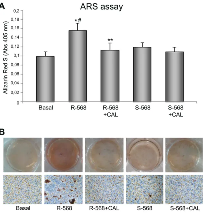
Figure 5. Effect of calcimimetics on ARS assay. (A) Ovine AFMSCs were treated with calcimimetics (1 mM) for 14 days and the mineralization was quantified at the end of treatment by reading the absorbance. The cells showed higher mineralization in response to CaSR activator R2568 (1 mM, *p,0.01), while S2568 did not lead to any mineralization. The mineral deposits produced by the culture with R2568 were significantly reduced in the presence of Calhex2231 (**p,0.05) and R-568 significantly increased ARS compared to S2568 (#p,0.05 R-568 vs S-568). The results are normalized per total cell number/well. (B) Representative full plate and microscope images of ARS assay after 14 days of treatment. Magnification 10x.
doi:10.1371/journal.pone.0073816.g005
enzyme. Finally, a stop solution was added to stop the reaction and the intensity of the color was measured spectrophotometrically at 450 nm in a microplate reader. The IP3 concentration in each sample was calculated from the standard curve.
Statistical Analysis of Data
Data are reported as means 6SD. Statistical comparisons were made using Student’s t test. An analysis of variance was used when multiple comparisons were performed. A difference was consid-ered significant at p,0.05.
Results
Ovine AFMSC Isolation and Characterization
The oAFMSCs were harvested from the amniotic cavity of sheep at mid gestation. After 5–6 days of culture the cells adhered to the plastic dish and, confirming our previous observation [6], two distinct cell populations, differing in size and shape, were recorded: small cells showing a fibroblast-like morphology and large polyhedral cells, resembling epithelial cells (data not shown). During in vitro expansion the fibroblast-like cells became prevalent and after the third passage of culturing the cell population appeared morphologically homogeneous with an average doubling time comprised between 12–18 hours which was not affected by cryopreservation.
Thereafter, oAFMSCs were characterized by Flow Cytometry analysis (Table 1). Confirming our previous data [8], the results indicated that oAFMSCs did not display any hematopoietic molecular markers (CD14, 31 and 45), while they expressed several surface adhesion molecules (CD29, CD58, CD166 and CD90) and intracellular stemness markers (TERT, SOX2 and Nanog). The OCT4 expression was lower than other stemness markers, while CD117 proved to be unexpressed. The absence of HLA-DR and presence of HLA-ABC suggested that these cells
may be applicable in clinical transplantation settings under auto/ allogenic conditions.
Ovine AFMSCs Express CaSR
Ovine AFMSCs were studied for CaSR expression by western blotting and cytometry analysis. As shown in figure 1A, western blot analysis performed on four different oAFMSC samples under reducing and denaturing conditions demonstrated a pattern of bands (,70, ,100, ,140 and ,165 kDa) comparable to what was obtained in OBs, used as a positive control. These data are consistent with the results obtained from other studies reporting the expression of CaSR in various tissues [16,19,31].
Imaging flow cytometry in permeabilized conditions confirmed the presence of CaSR in oAFMSCs (Fig. 1B), as is also shown in a representative image (Fig. 1C) where widespread intracellular distribution of the receptor is evident.
Effect of Calcimimetics on oAFMSCs
To evaluate the optimal concentration of calcimimetics, oAFMSCs were cultured in the presence or absence of increasing concentrations (0.1–10mM) of R-568 and its enantiomer S-568. After 14 days of treatment, an MTT assay (Fig. 2A) and cell count (Fig. 2B) were performed. As evident from the histograms, calcimimetic concentrations of 0.1 and 1mM were comparable to the basal condition and therefore not toxic to cells, while 5mM induced a tendency to reduce cell viability, reaching statistical significance at 10mM.
In addition, microscope analysis (Fig. 2C) confirmed the results obtained by MTT and cell counting assays. As shown by the magnification in the right-hand corners of figure 2C, we observed changes in cell morphology exclusively using 5 and 10mM R- or S-568: oAFMSCs appeared increased in size and changed in morphology in that they were full of vacuoles. Since in these experimental conditions we also performed Oil-red O staining, this allows us to affirm that calcimimetics do not induce
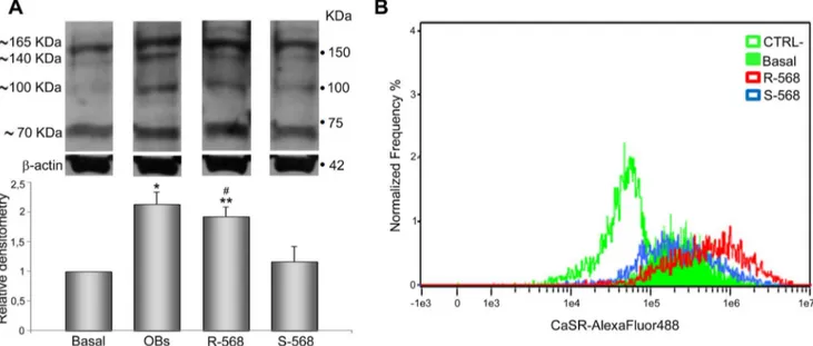
Figure 6. CaSR protein expression in oAFMSCs treated with calcimimetics. (A) Western blot analysis of oAFMSCs cultured with R-568 or S-568. Whole cell lysates were separated by SDS-PAGE, immunoblotted, and revealed with anti-CaR antibody. Equivalent amounts (40 mg) of total protein were loaded in each lane. b-actin was used as an internal control. *p,0.001 OBs vs Basal; **p,0.001 R-568 vs Basal;#p,0.01 R2568 vs S2568. (B) Representative graph derived from Imaging flow cytometry (Amnis) shows the shift of mean fluorescence intensity (MFI) of CaSR in oAFMSCs treated with R2568 (red curve) versus basal condition (green filled curve), while treatment with S-568 did not cause any shift (blue curve). The MFI of secondary antibody alone (Alexa-488, CTRL-) is indicated by the empty green curve.
doi:10.1371/journal.pone.0073816.g006
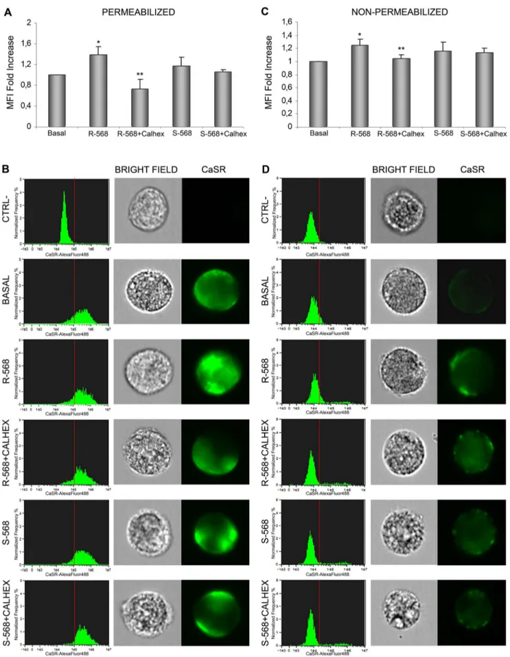
Figure 7. Imaging flow cytometry analysis of CaSR expression in oAFMSCs evaluated intracellularly and at membrane level. Shown here in a permeabilizing condition (A–B) is the CaSR expression after treatment for 14 days with R-568 or S-568 in the presence or absence of Calhex-231 (*p,0.05 R-568 vs Basal; **p,0.02 R-568+Calhex vs R-568). The histograms in B (left), related to the MFI bars in (A), show the CaSR distribution CaSR in Ovine Amniotic Fluid Stem Cells and R568
adipogenesis (data not shown). Thus, at high doses, these molecules can induce effective cytotoxic effects, as proven by the presence of vacuoles. Moreover, as expected, the oAFMSCs were over confluent because they had been cultured for 14 days.
Osteogenic Effect of Calcimimetics on oAFMSCs
To evaluate the potential role of R- and S-568 in osteogenesis, we performed ALP and ARS assays to assess the mineralization at different concentrations of calcimimetics (Fig. 3). As demonstrated by both ALP (Fig. 3A) and ARS assays (Fig. 3B,C), 1mM R-568 led to a significant increase in mineral deposits (p,0.05 and p,0.01, respectively), while the enantiomer S-568 did not lead to any mineralization increase versus the basal condition. Again, we observed that both the concentrations of 5mM and 10mM were unhealthy to cells. All these data suggest that 1mM R-568 may direct oAFMSCs toward osteogenic differentiation.
To better assess the potential calcimimetic CaSR-mediated pathway in osteogenic differentiation, we showed in Figure 4 that 1mM R-568 increased ALP activity significantly greater than both 1.8 or 2.5 mM calcium, which is a principal agonist of CaSR receptor and thus considered a current gold standard in osteogenic process. Notably, R-568 induced ALP activity was significantly reduced by Calhex-231, a CaSR allosteric inhibitor. On the other hand, the stimulation of oAFMSCs with enantiomer S-568 (in presence or absence of Calhex-231) did not lead to any increase in ALP activity.
To further demonstrate the role of CaSR in R-568-induced osteogenesis in oAFMSCs, we evaluated the mineralization levels in cells treated with the CaSR inhibitor, Calhex2231 (Fig. 5A, B). As expected, the assay showed a significant R2568 increased ARS level, which significantly decreased when the cells were pre-treated with Calhex2231. Again, we did not observe any significant effects by the inhibitor on oAFMSCs treated with S-568.
Taken together these data demonstrate that the calcimimetic differentiation capabilities may be actually mediated by CaSR-pathway activation.
Effect of Calcimimetics on CaSR Expression
To evaluate whether the osteogenesis induced by calcimimetics is associated with increased CaSR protein levels, we performed western blotting analysis (Fig. 6A). Interestingly, as compared to basal level, we observed that in our cell model calcimimetic R-568 significantly augmented CaSR protein expression (p,0.05), which was comparable with CaSR protein levels in OBs (positive control). On the contrary, enantiomer S-568 did not show any significant effect. These data were confirmed by Imaging flow cytometry (Fig. 6B), where there is an evident shift in the mean fluorescence intensity (MFI) of CaSR in oAFMSCs treated with R2568 (red curve) compared to basal condition (green filled curve), while treatment with S-568 did not cause any such shift (blue curve).
As we observed that R-568 was able to increase both CaSR protein levels and osteogenic differentiation of oAFMSCs, we investigated its potential role in the modification of CaSR cellular localization in the presence or absence of CaSR inhibitor, Calhex2231. As shown in figure 7, cells were treated in permeabilizing or non-permeabilizing conditions. In both
exper-imental protocols we again confirmed that R2568 significantly increased CaSR levels (p,0.05), and this was reduced by Calhex2231. Treatment with enantiomer S2568, in the presence or absence of Calhex-231, did not lead to any effect on the CaSR protein expression level.
In particular, as confirmed in figure 7A, incubation with R-568 significantly increased CaSR intracellular levels (p,0.05) in a permeabilizing condition. Moreover, data shown in histograms (Fig. 7B, left) confirmed that incubation with R-568 significantly increased CaSR intracellular levels (p,0.05). Following R-568 incubation in a non-permeabilizing condition (Fig. 7C, D), we observed increased cell membrane localization of CaSR, which was significantly reversed by pre-incubation with Calhex-231. In this case too, treatment with enantiomer S-568 (in the presence or absence of Calhex-231) failed to induce any effect.
All these data suggest that calcimimetic R-568 may promote osteogenic differentiation of oAFMSCs, increasing the inner cell localization of CaSR and notably its membrane exposure, which may indicate that it has some functional role in the osteogenic process.
Effect of Calcimimetics on Intracellular Calcium Variations and IP3 Levels
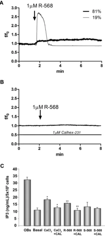
To study the calcimimetic effects on CaSR functional activa-tion, we evaluated the potential role of R-568 on intracellular calcium and IP3 level variations. As shown in Fig. 8A, R-568 (1mM) induced an intracellular calcium increase in 1963% of cells (28 cells out of 146 total tested cells in three indipendent fields). The cells responsive to 1mM R-568, showed a high and transient calcium increase (19% of cells) or a low and sustained ion increase (81% of cells). When the cells were treated with 1mM Calhex-231, no response was observed after R-568 addition (Fig. 8B). At the same concentration S-568 (1mM, in presence or absence of Calhex-231) was not able to influence intracellular calcium levels (data not shown). Interestingly, we observed that acute stimulation (2 min) of cells with R-568 induced a significant increase of intracellular IP3 levels (p,0.05). This effect was comparable to that obtained when the cells were stimulated with the high extracellular calcium concentration, 2.5 mM. Calhex-231 (1mM) totally abolished the R-568 effect; S-568 (1mM, in presence or absence of Calhex-231) was not able to influence intracellular calcium levels (Fig. 8C).
Discussion
Various different sources of mesenchymal stem cells have previously been considered attractive for orthopedic regenerative medicine, such as adipose tissue, dental pulp, or bone marrow [32], and these cells expressed a prompt osteogenic phenotype when incubated under adequate in vitro conditions [32,33].
However, amniotic fluid-derived cells may have some additional biological properties that could be particularly useful toward developing a new protocol in regenerative medicine [1]. Amniotic fluid, rich in mesenchymal-like cells [34], can be easily collected during prenatal diagnosis amniocentesis without raising any ethical problems [35]. In addition AFMSCs display a high differentiation capacity toward all three germ layers demonstrated
in 56103-acquired-oAFMSCs/sample. Representative single cell images inB (right). The same experiments were performed in non-permeabilizing conditions to detect CaSR membrane localization (*p,0.01 R-568 vs Basal; **p,0.05 R-568+Calhex vs R-568) (C–D). Dead cells were excluded by 7-Aminoactinomycin D (7-AAD) staining. Data analyses were performed by IDEAS software (Amnis). The results shown by the bars (A–C) are expressed as a fold increase and are calculated by subtracting the MFI value of negative events (MFI of secondary antibody alone, CTRL-) from that of positive events. Data represent three separate experiments.
doi:10.1371/journal.pone.0073816.g007
under both in vitro and in vivo conditions [1]. Due to this multi/ pluripotency and to the early embryonic origin, amniotic-derived
cells are considered as fetal cells, showing a degree of stemness that is typical of embryonic stem cells, even though they lack both tumorigenicity and immunogenicity [35,36].
Our recent data demonstrated that oAFMSCs can differentiate in vitro into osteoblast-like cells [6,11], as previously reported for human and rodent AFMSCs [1,2,37]. The fact that osteogenesis occurred was here clearly confirmed after 3 weeks’ culture in an osteogenic medium by the extensive mineralization of the extracellular matrix (data not shown).
Interestingly, in view of their therapeutic potential for bone tissue engineering, the present study shows for the first time that CaSR is expressed in oAFMSCs. It is known that CaSR is a physiological regulator of bone cell metabolism through modula-tion of the recruitment, differentiamodula-tion and survival of osteoblasts and osteoclasts by activation of different pathways in bone cells [38].
In particular, unlike mature OB cells which are known to express CaSR [39] in totally or partially glycosylated monomers (,140 and ,165 kDa, considered the mature form which reaches the cell surface as a dimer of ,300 kDa) and unlike immune-reactive polypetides of ,70 and ,100 kDa, we here consistently found the presence of immuno-reactive polypeptides of about 70, 100, 140 and 165 kDa in four different oAFMSC cultures (Fig. 1A).
There is substantial evidence for the implication of CaSR in the bone [20,38] and the presence of this receptor in different bone cells including osteoblasts, osteoclasts, stromal cells, monocytes, macrophages and chondrocytes [17,20,40,41]. In addition, recent works have suggested that this important receptor is expressed in stem cells such as size-sieved umbilical cord matrix equine stem cells [42], bone marrow hematopoietic stem cells [43] and mouse embryo stem cells [18].
We report here, for the first time, the presence of this functional receptor in oAFMSCs, cells known for their ability to differentiate into osteoblasts and hence be of potential use for therapeutic use in vivo [6,11].
In this respect, the development of drugs able to target CaSR in bone cells may lead to an improvement in bone and cartilage regeneration. Recent studies do indeed indicate the possibility of treating bone diseases such as osteoporosis by CaSR allosteric modulators (calcimimetics and calcilytics) [21]. Interestingly, and confirming previous observations in different cellular models [39], we demonstrated by both western blot and cytometry (Fig. 6) that CaSR expression in oAFMSCs increased when the cells were treated with R-568 (1mM). In the same experimental condition CaSR localization increased at the membrane level, potentially resulting in activation of the receptor (Fig. 7C,D). Moreover, western blot analysis (Fig. 6A) substantiated these results and gave further demonstration of what has been found up to now, namely that only R-568 treatment increased band of about 140 and 165 kDa, which may correspond to the partially and totally glycosylated monomeric form of the receptor. This represents its mature form which, when exposed on the membrane cell surface, takes on the nature of a dimer.
The calcimimetic effect on CaSR levels and cellular localization was associated with a significant increase in sheep AFMSC mineralization (ALP and ARS staining, Figs. 4 and 5), thus demonstrating a potential role for calcimimetics during osteogenic differentiation.
To confirm the potential role of CaSR in mediating the R-568 osteogenic effect we tested the effect of Calhex-231, a CaSR allosteric inhibitor; this totally reversed R-568 induced mineral-ization. One notes that treatment with enantiomer S-568, in the presence or absence of Calhex-231, had no effect on either
Figure 8. Intracellular Ca2+ and IP3 variations in oAFMSCs stimulated with R-568. Temporal changes in intracellular Ca2+levels, expressed as f/f0ratio (see Material and Methods), were measured in oAFMSCs. In panel (A) the representative traces showed calcium variations in cells treated with 1 mM R-568. In panel (B) the traces showed the cell response in presence of 1 mM Calhex-231. In panel (C) the IP3 levels were expressed as ng/ml/56105cells. CaCl2was used at 2.5 mM; Basal indicates 1.8 mM CaCl2. *p,0.02 CaCl2vs Basal and CaCl2+Calhex-231 vs CaCl2; **p,0.05 R-568 vs Basal and R-568+Calhex-231 vs R-568.
doi:10.1371/journal.pone.0073816.g008
mineralization or the CaSR protein expression level (Figs. 5 and 6). Moreover, we studied the R-568 effects on CaSR functional activation evaluating pathways usually activated by extracellular high calcium level. Interestingly, we observed that acute stimula-tion of cells with R-568 induced a significant increase of both intracellular calcium and IP3 levels (Fig. 8).
Taken all together, these results suggest that not only is CaSR more expressed in R-568 treated cells, but its augmented expression at a membrane level is associated to its activation, indicating an increased functionality: thus it may actively contribute to the process of osteogenesis.
This finding is in agreement with reports on other cells, such as the human osteoblastic MG-63 cell line [19] in which it has been observed that CaSR is not only expressed but is functionally active in osteoblast plasma membrane.
Our results strongly suggest that agonists binding bone CaSR may be advantageous for the treatment of bone injury [21,23].
Although Barradas et al. have recently shown that calcium-enhanced osteogenic differentiation in bone marrow-derived mesenchymal stromal cells is not mediated by CaSR activation [44], our results are in accordance with part of the literature in demonstrating that an elevation of Ca2+oconcentration, as well as
the use of calcimimetics, strongly activates functional CaSR in several cell lines [39,42,45].
One should note that calcimimetics may have a favorable effect on bone cells expressing CaSR: they are not able to potentiate the effects of extracellular calcium on osteoblasts and osteoclasts in the
absence of CaSR [46] but are efficacious in cells expressing the receptor [47].
Another noteworthy point we have demonstrated is that at a low and not cytotoxic concentration of 1mM, calcimimetic R-568 significantly increases osteogenic differentiation in control culture conditions. Different data were obtained when S-568 was examined at the same concentration, showing that there is no effect of S-568 in the osteogenic differentiation process. The effect of R-568 on osteogenesis is specific.
In conclusion, we have shown for the first time that not only is CaSR expressed in oAFMSCs, but its expression and function are significantly increased by calcimimetic R-568, which is also able to induce polarization of the receptor up to the membrane; this may indicate a helpful role by this calcimimetic in CaSR activation and hence possible implication of it in osteogenic differentiation. Further studies will be necessary to evaluate the potential basal and stimulated role of CaSR in human AFMSCs, since drugs targeting CaSR may be used in addition to boost movement of transplanted stem cells to the site of injury, entailing significant clinical implications for stem cell transplantation.
Author Contributions
Conceived and designed the experiments: PDT CP AP. Performed the experiments: PDT CP PL CM LP. Analyzed the data: PDT CP CM VS MM AP BB. Contributed reagents/materials/analysis tools: MM BB MAM MB SM. Wrote the paper: PDT CP AP.
References
1. De Coppi P, Bartsch G, Siddiqui MM, Xu T, Santos CC, et al. (2007) Isolation of amniotic stem cell lines with potential for therapy. Nature biotechnology 25: 100–106.
2. D’Alimonte I, Lannutti A, Pipino C, Di Tomo P, Pierdomenico L, et al. (2013) Wnt Signaling Behaves as a ‘‘Master Regulator’’ in the Osteogenic and Adipogenic Commitment of Human Amniotic Fluid Mesenchymal Stem Cells. Stem cell reviews. Available: http://www.ncbi.nlm.nih.gov/pubmed/23605563. doi 10.1007/s12015-013-9436-5.
3. Piccoli M, Franzin C, Bertin E, Urbani L, Blaauw B, et al. (2012) Amniotic Fluid Stem Cells Restore the Muscle Cell Niche in a HSA-Cre, SmnF7/F7 Mouse Model. Stem Cells 30: 1675–1684.
4. Sedrakyan S, Angelow S, De Filippo R, Perin L (2012) Stem cells as a therapeutic approach to chronic kidney diseases. Curr Urol Rep 13: 47–54. 5. Bollini S, Cheung K, Riegler J, Dong X, Smart N, et al. (2011) Amniotic fluid
stem cells are cardioprotective following acute myocardial infarction. Stem Cells Dev 20: 1985–1994.
6. Mauro A, Turriani M, Ioannoni A, Russo V, Martelli A, et al. (2010) Isolation, characterization, and in vitro differentiation of ovine amniotic stem cells. Veterinary research communications 34 Suppl 1: S25–8. doi: 10.1007/s11259-010-9393-2.
7. Shaw SWS, Bollini S, Nader KA, Gastadello A, Mehta V, et al. (2011) Autologous transplantation of amniotic fluid-derived mesenchymal stem cells into sheep fetuses. Cell transplantation 20: 1015–1031.
8. Colosimo A, Curini V, Russo V, Mauro A, Bernabo` N, et al. (2012) Characterization, GFP gene nucleofection and allotransplantation in injured tendons of ovine amniotic fluid-derived stem cells. Cell Transplantation 22: 99– 117.
9. Turner CG, Klein JD, Steigman S A, Armant M, Nicksa G A, et al. (2011) Preclinical regulatory validation of an engineered diaphragmatic tendon made with amniotic mesenchymal stem cells. Journal of pediatric surgery 46: 57–61. 10. Gray FL, Turner CG, Ahmed A, Calvert CE, Zurakowski D, et al. (2012) Prenatal tracheal reconstruction with a hybrid amniotic mesenchymal stem cells-engineered construct derived from decellularized airway. Journal of pediatric surgery 47: 1072–1079.
11. Berardinelli P, Valbonetti L, Muttini A, Martelli A, Peli R, et al. (2012) Role of amniotic fluid mesenchymal cells engineered on MgHA/collagen-based scaffold allotransplanted on an experimental animal study of sinus augmentation. Clinical oral investigations. doi: 10.1007/s00784-012-0857-3.
12. Garrett JE, Capuano IV, Hammerland LG, Hung BC, Brown EM, et al. (1995) Molecular cloning and functional expression of human parathyroid calcium receptor cDNAs. J Biol Chem 270: 12919–12925.
13. Riccardi D, Park J, Lee WS, Gamba G, Brown EM, et al. (1995) Cloning and functional expression of a rat kidney extracellular calcium/polyvalent cation-sensing receptor. Proceedings of the National Academy of Sciences of the United States of America 92: 131–135.
14. Hebert SC, Cheng S, Geibel J (2004) Functions and roles of the extracellular Ca2+-sensing receptor in the gastrointestinal tract. Cell Calcium 35: 239–247. 15. Brown EM (2007) The calcium-sensing receptor: physiology, pathophysiology
and CaR-based therapeutics. Subcell Biochem 45: 139–167.
16. Bonomini M, Giardinelli A, Morabito C, Di Silvestre S, Di Cesare M, et al. (2012) Calcimimetic R-568 and its enantiomer S-568 increase nitric oxide release in human endothelial cells. PloS one 7: e30682. doi: 10.1371/ journal.pone.0030682.
17. House MG, Kohlmeier L, Chattopadhyay N, Kifor O, Yamaguchi T, et al. (1997) Expression of an extracellular calcium-sensing receptor in human and mouse bone marrow cells. Journal of bone and mineral research 12: 1959–1970. 18. Sun J, He W, Bai S-Z, Peng X, Zhang N, et al. (2013) The expression of calcium-sensing receptor in mouse embryonic stem cells (mESCs) and its influence on differentiation of mESC into cardiomyocytes. Differentiation 85: 32–40. Available: http://www.ncbi.nlm.nih.gov/pubmed/23314289. 19. Yamaguchi T, Chattopadhyay N, Kifor O, Ye C, Peter M, et al. (2001)
Expression of extracellular calcium-sensing receptor in human osteoblastic MG-63 cell line. Am J Physiol Cell Physiol 280: C382–C393.
20. Yamaguchi T (2008) The calcium-sensing receptor in bone. Journal of bone and mineral metabolism 26: 301–311.
21. Kameda T, Mano H, Yamada Y, Takai H, Amizuka N, et al. (1998) Calcium-Sensing Receptor in Mature Osteoclasts, Which Are Bone Resorbing Cells. Biochem Biophys Res Commun 422: 419–422.
22. Chang W, Tu C, Chen TH, Bikle D and Shoback D (2013) The Extracellular Calcium-Sensing Receptor (CaSR) Is a Critical Modulator of Skeletal Development. Sci Signal 1: 1–23.
23. Marie PJ (2010) The calcium-sensing receptor in bone cells: a potential therapeutic target in osteoporosis. Bone 46: 571–576.
24. Dru¨eke TB, Ritz E (2009) Treatment of secondary hyperparathyroidism in CKD patients with cinacalcet and/or vitamin D derivatives. Clinical journal of the American Society of Nephrology?: CJASN 4: 234–241.
25. Lanuti P, Santilli F, Marchisio M, Pierdomenico L, Vitacolonna E, et al. (2012) A novel flow cytometric approach to distinguish circulating endothelial cells from endothelial microparticles: relevance for the evaluation of endothelial dysfunction. Journal of immunological methods 380: 16–22.
26. Lanuti P, Marchisio M, Cantilena S, Paludi M, Bascelli A, et al. (2006) A flow cytometry procedure for simultaneous characterization of cell DNA content and expression of intracellular protein kinase C-zeta. Journal of immunological methods 315: 37–48.
27. Miscia S, Ciccocioppo F, Lanuti P, Velluto L, Bascelli A, et al. (2009) Abeta(1– 42) stimulated T cells express P-PKC-delta and P-PKC-zeta in Alzheimer disease. Neurobiology of aging 30: 394–406.
28. Gregory CA, Gunn WG, Peister A, Prockop DJ (2004) An Alizarin red-based assay of mineralization by adherent cells in culture: comparison with cetylpyridinium chloride extraction. Analytical biochemistry 329: 77–84.
29. Song I-H, Caplan AI, Dennis JE (2009) In vitro dexamethasone pretreatment enhances bone formation of human mesenchymal stem cells in vivo. Journal of orthopaedic research 27: 916–921.
30. Orciani M, Morabito C, Emanuelli M, Guarnieri S, Sartini D, et al. (2011) Neurogenic potential of mesenchymal-like stem cells from human amniotic fluid: the influence of extracellular growth factors. J Biol Regul Homeost Agents 25: 115–130.
31. Bai M, Trivedi S, Brown EM (1998) Dimerization of the extracellular calcium-sensing receptor (CaR) on the cell surface of CaR-transfected HEK293 cells. The Journal of biological chemistry 273: 23605–23610.
32. Gutwald R, Haberstroh J, Kuschnierz J, Kister C, Lysek DA, et al. (2010) Mesenchymal stem cells and inorganic bovine bone mineral in sinus augmentation: comparison with augmentation by autologous bone in adult sheep. Br J Oral Maxillofac Surg 48: 285–290.
33. D’Aquino R, Graziano A, Sampaolesi M, Laino G, Pirozzi G, et al. (2007) Human postnatal dental pulp cells co-differentiate into osteoblasts and endotheliocytes: a pivotal synergy leading to adult bone tissue formation. Cell death and differentiation 14: 1162–1171.
34. Antonucci I, Stuppia L, Kaneko Y, Yu S, Tajiri N, et al. (2011) Amniotic fluid as a rich source of mesenchymal stromal cells for transplantation therapy. Cell transplantation 20: 789–795.
35. Parolini O, Soncini M, Evangelista M Schmidt D (2009) Amniotic membrane and amniotic fluid derived cells; potential tools for regenerative medicine? Regen Med 4: 275–291.
36. Pipino C, Shangaris P, Resca E, Zia S, Deprest J, et al. (2012) Placenta as a reservoir of stem cells: an underutilized resource? British medical bulletin 105: 43–68. doi:10.1093/bmb/lds033.
37. Antonucci I, Iezzi I, Morizio E, Mastrangelo F, Pantalone A, et al. (2009) Isolation of osteogenic progenitors from human amniotic fluid using a single step culture protocol. BMC biotechnology 9: 9. doi:10.1186/1472-6750-9-9. 38. Brown EM, Chattopadhyay N, Yano S (2004) Calcium-sensing receptors in
bone cells. Journal of musculoskeletal & neuronal interactions 4: 412–413.
39. Yamauchi M, Yamaguchi T, Kaji H, Sugimoto T, Chihara K (2005) Involvement of calcium-sensing receptor in osteoblastic differentiation of mouse MC3T3-E1 cells. American journal of physiology Endocrinology and metab-olism 288: E608–16.
40. Miller S, Strewler G, Shoback D (1999) Calcium Sensing in Cultured Chondrogenic RCJ3.1C5.18 Cells*. 140: 1911–1919.
41. Chattopadhyay N, Yano S, Tfelt-Hansen J, Rooney P, Kanuparthi D, et al. (2004) Mitogenic action of calcium-sensing receptor on rat calvarial osteoblasts. Endocrinology 145: 3451–3462.
42. Martino NA, Lange-Consiglio A, Cremonesi F, Valentini L, Caira M, et al. (2011) Functional expression of the extracellular calcium sensing receptor (CaSR) in equine umbilical cord matrix size-sieved stem cells. PloS one 6: e17714. doi:10.1371/journal.pone.0017714.
43. Lam BS, Cunningham C, Adams GB (2011) Pharmacologic modulation of the calcium-sensing receptor enhances hematopoietic stem cell lodgment in the adult bone marrow. Blood 117: 1167–1175.
44. Barradas AMC, Fernandes H A M, Groen N, Chai YC, Schrooten J, et al. (2012) A calcium-induced signaling cascade leading to osteogenic differentiation of human bone marrow-derived mesenchymal stromal cells. Biomaterials 33: 3205–3215.
45. Dvorak MM, Siddiqua A, Ward DT, Carter DH, Dallas SL, et al. (2004) Physiological changes in extracellular calcium concentration directly control osteoblast function in the absence of calciotropic hormones. Proceedings of the National Academy of Sciences of the United States of America 101: 5140–5145. 46. Hebert SC (2006) Therapeutic use of calcimimetics. Annual review of medicine
57: 349–364.
47. Kanatani M, Sugimoto T, Kanzawa M, Yano S, Chihara K (1999) High extracellular calcium inhibits osteoclast-like cell formation by directly acting on the calcium-sensing receptor existing in osteoclast precursor cells. Biochemical and biophysical research communications 261: 144–148.