Prognostic significance of left ventricular non-compaction: A systematic review and meta-analysis of observational studies
Brief title: Meta-analysis of prognosis in LVNC
Nay Aung, MD1,2; Sara Doimo, MD3; Fabrizio Ricci, MD, PhD4; Mihir M Sanghvi, MD1,2;
Cesar Pedrosa, MD1; Simon P Woodbridge, BMedSci1; Amer Al-Balah, BSC5; Filip Zemrak,
MD, PhD1,2; Mohammed Y Khanji, MD, PhD1,2; Patricia B Munroe, PhD1,2,6; Huseyin Naci,
PhD7; Steffen E Petersen, MD, DPhil, MPH1,2
1William Harvey Research Institute, NIHR Cardiovascular Biomedical Research Centre at Barts, Queen
Mary University of London, Charterhouse Square, London, UK
2Barts Heart Centre, St Bartholomew’s Hospital, Barts Health NHS Trust, West Smithfield, London, UK 3Cardiovascular Department, Azienda Sanitaria Universitaria Integrata, University of Trieste, Trieste,
Italy
4Institute of Advanced Biomedical Technologies, Department of Neuroscience, Imaging and Clinical
Sciences, “G. d’Annunzio” University, Chieti, Italy
5Imperial College London, Kensington, London, UK
6Clinical Pharmacology, William Harvey Research Institute, Barts and The London School of Medicine
and Dentistry, Queen Mary University of London, London, UK.
7Department of Health Policy, London School of Economics and Political Science, London, UK.
Address for correspondence
Professor Steffen E. Petersen, MD DPhil MSc MPH FRCP FSCMR FESC FACC
William Harvey Research Institute, NIHR Cardiovascular Biomedical Research Centre at Barts, Queen Mary University of London, Charterhouse Square, London EC1M 6BQ, UK.
Manuscript word count: 4381
ABSTRACT
Background: Although left ventricular non-compaction (LVNC) has been associated
with an increased risk of adverse cardiovascular events, the accurate incidence of cardiovascular morbidity and mortality is unknown. We therefore aimed to assess the incidence rate of LVNC-related cardiovascular events.
Methods: We systematically searched observational studies reporting the adverse
outcomes related to LVNC. The primary end-point was cardiovascular mortality.
Results: We identified 28 eligible studies enrolling 2501 LVNC patients (mean age: 46
years, male/female ratio: 1.7). After a median follow-up of 2.9 years, the pooled event rate for cardiovascular mortality was 1.92 (95% CI: 1.54 – 2.30) per 100 person-years. LVNC patients had a similar risk of cardiovascular mortality compared to a DCM control group (odds ratio: 1.10, 95% CI: 0.18 – 6.67). The incidence rates of all-cause mortality, stroke and systemic emboli, heart failure admission, cardiac transplantation, ventricular arrhythmias and cardiac device implantation were 2.16, 1.54, 3.53, 1.24, 2.17, and 2.66, respectively, per 100 person-years. Meta-regression and subgroup analyses revealed that left ventricular ejection fraction (LVEF), not the extent of left ventricular trabeculation, had an important influence on the variability of incidence rates. The risks of thromboembolism and ventricular arrhythmias in LVNC patients were similar to dilated cardiomyopathy (DCM) patients. However, LVNC patients had a higher incidence of heart failure hospitalization than DCM patients.
Conclusions: Patients with LVNC carry a similar cardiovascular risk when compared
with DCM patients. LVEF, a conventional indicator of heart failure severity, not the extent of trabeculation, appears to be an important determinant of adverse outcomes in LVNC patients.
PORSPERO registration ID: CRD42018096313
Abbreviations
CMR, cardiovascular magnetic resonance DCM, Dilated cardiomyopathy
HCM, Hypertrophic cardiomyopathy ICD, Implantable cardioverter defibrillator LVEDD, Left ventricular end-diastolic diameter LVEF, Left ventricular ejection fraction
LVNC, Left ventricular non-compaction
LVNC:C, Left-ventricular non-compaction to compaction ratio NYHA, New York Heart Association classification
QUIPS, Quality In Prognosis Studies
Keywords
CLINICAL PERSEPCTIVE
In this large meta-analysis of adult patients with left ventricular non-compaction (LVNC) identified by currently accepted imaging criteria, the incidences of objective cardiovascular outcomes appear comparable to those observed in dilated
cardiomyopathy. The frequency of adverse outcomes is mostly driven by left ventricular systolic impairment rather than the burden of trabeculation. The diversity of current imaging diagnostic criteria for LVNC creates significant challenges for accurate phenotyping. Further prospective clinical registries with access to individual-level data are required to standardize the LVNC diagnostic criteria, co-morbidities and outcome measures to fully evaluate the prognostic markers of this poorly understood condition.
INTRODUCTION
Left ventricular non-compaction (LVNC) cardiomyopathy is characterized by prominent left ventricular (LV) trabeculations, deep intertrabecular recesses
communicating with the ventricular cavity, and a thin and compacted epicardial layer. While LVNC is considered a genetic cardiomyopathy by The American Heart
Association 1, the European Society of Cardiology categorizes it as an unclassified
cardiomyopathy 2. Multiple etiologies of the LVNC phenotype have been proposed: it
may be familial (inherited) or non-familial (sporadic and proven absent in relatives), and may occur as an isolated disease or in association with genetic diseases and
congenital defects 3. Non-familial and sporadic forms have been described in
highly-trained athletes 4, sickle cell anemia 5 and pregnancy 6. The genetic basis of familial
in the same genes associated with other types of inherited cardiomyopathies (Figure 1A)
7.
The diagnosis of LVNC has conventionally been made by imaging the left ventricle and demonstrating the presence of specific criteria based mostly upon the relative thickness of the compacted myocardial wall and the mesh of trabeculated (“non-compacted”) layer of cardiac muscle using either echocardiography or cardiovascular magnetic resonance (CMR) imaging (Figure 1B). All current methodologies used to establish a diagnosis have strengths and weaknesses in how they are derived, their ease of use, the time to acquire the relevant images and their diagnostic accuracy, but there is no evidence to suggest that any particular criteria or imaging modality is superior. However, as image quality and awareness of diagnostic criteria have improved, the LVNC phenotype has emerged as an increasingly-recognized finding with the inherent
risk of over-diagnosis noted as a significant concern 8.
The clinical outcomes of LVNC vary widely in the reported literature which perhaps reflects the underlying diversity of study cohorts. In view of the continued uncertainty, we conducted a systematic review of observational cohort studies to explore the clinical outcomes of patients considered to be affected by LVNC.
METHODS
The data, analytic methods and study materials can be obtained from the corresponding author for purposes of reproducing the results or replicating the results. Since this is a meta-analysis of aggregate data from the published literature, no informed consent was
required. Likewise, since we have not recruited new patients, an institutional review board’s approval was not necessary.
We aimed to explore the adverse outcomes of patients with LVNC through a systematic review of the literature including prospective longitudinal and retrospective
observational studies. The complete study protocol was registered on PROSPERO – an international database of prospectively registered systematic reviews – and can be accessed at
www.crd.york.ac.uk/PROSPERO/display_record.php?ID=CRD42018096313. We recognized the challenges associated with meta-analyses of observational studies due to variable study designs and inherent biases. Therefore, we conducted this systematic review following the recommendations by the Meta-analysis of
Observational Studies in Epidemiology group 9 and the PRISMA guidelines 10.
Search strategy
We searched PubMed and Embase databases, the Cochrane Database of Systematic Reviews, the PROSPERO database (www.crd.york.ac.uk/prospero), and the Clinical Trials Registry (www.clinicaltrials.gov), as well as abstracts from major cardiological societies for potentially relevant articles using a combination of keywords related to
trabeculation or LVNC and the cardiovascular outcomes for the period from 1st January
1966 to 3rd July 2019 without any language restriction. Details of the search terms are
Selection criteria
Inclusion criteria were: (i) patients over 18 years old; (ii) a diagnosis of LVNC by echocardiographic or CMR criteria; (iii) crude and/or adjusted event rates of all-cause mortality, cardiovascular (CV) mortality, ventricular arrhythmias, sudden cardiac death, heart failure hospitalization, myocardial infarction, stroke, systemic embolic events, new cardiac implantable electronic device and heart transplantation. Definitions of
excessive trabeculation according to cardiac imaging were defined by Petersen 11, Chin
12, Jenni 13, Jacquier 14, Grothoff 15, Stacey 16, Stöllberger 17 or Captur 18 criteria. We
excluded case reports, non-outcome studies and reviews.
Data extraction
Two authors (F.R., S.D.) performed the screening of titles and abstracts, reviewed the full-text articles, and determined their eligibility. Divergences were solved by consensus and/or involving the third author (N.A.). We also hand-searched the reference list of all eligible articles for additional relevant studies.
We collated study-level covariates and events reported in original publications, using a standardized data extraction form. We translated relevant non-English articles into English. We contacted the authors of studies where clarification of data was required. In studies with overlapping cohorts, we used the data from the most recent study and/or the study with the largest sample size.
We assessed the individual study-level quality by the Quality In Prognosis Studies
(QUIPS) tool 19 which evaluates 32 key considerations across six bias domains: (i)
Study Participation, (ii) Study Attrition, (iii) Prognostic Factor Measurement, (iv) Outcome Measurement, (v) Study Confounding, and (vi) Statistical Analysis and
Reporting. An overall quality grade (high quality, intermediate quality, low quality) was assigned to each study after considering all six bias domains. Two authors (M.K. and A.A.) independently rated the quality items and disagreements were resolved by another author (N.A.).
Outcomes
The primary end-point was the incidence of CV mortality. Secondary end-points included incidences of all-cause mortality, stroke and systemic embolic events, heart failure requiring hospitalization, cardiac transplantation, ventricular arrhythmias (ventricular tachycardia or ventricular fibrillation) and cardiac device implantation defined as insertion of implantable cardioverter defibrillator (ICD) or cardiac synchronization therapy with ICD.
Statistical analysis
Dichotomous variables were reported as percentages, with continuous variables reported as mean±standard deviation (SD) or median (interquartile range [IQR]), based on data distribution. For each included study, we calculated an event rate with its 95%
confidence interval (CI) for every predefined outcome. Event rates were computed as the ratio between the number of events and the person-time in years at risk, in order to
performed Freeman-Tukey transformation 20 of the number of events for variance
stabilization. We added 0.5 to the count in studies with zero event to achieve numerical stability. For studies reporting the event rates in both LVNC subjects and non-LVNC controls, we calculated odds ratio (OR) and 95% CI for each outcome.
We used random-effects models to estimate the summary pooled event rates or odd ratios of pre-specified outcomes using the DerSimonian and Laird method. We graphically presented the results in forest plots, with point estimates of the effect size and 95% CI for each study and the combined estimate. The area of squares and diamonds in the forest plots are proportional to each study weight.
We assessed funnel plot asymmetry which could result from publication bias. We additionally used the Egger’s regression asymmetry test for end-points with asymmetric funnel plots. We also performed the non-parametric ‘trim-and-fill’ procedure which adjusts for funnel plot asymmetry by computing hypothetical missing studies. We formally assessed statistical heterogeneity by a chi-squared test, and quantified it using
the inconsistency index (I2) statistic, which ranges from 0 to 100% and is defined as the
percentage of observed between-trial variability that is due to heterogeneity rather than
chance. A lack of homogeneity was considered to be significant with an I2 ≥ 50%. We
anticipated a high degree of heterogeneity across individual studies due to the
multiplicity of LVNC diagnostic criteria and the variability of inclusion and exclusion criteria used by individual studies. Accordingly, we used random-effects models to account for the between-study variabilities in the effect estimates. To explore the possible reasons of heterogeneity, we performed the following secondary analyses: (i)
univariate meta-regression assessing the mediating effect of age, sex (percentage of men), New York Heart Association (NYHA) classification, left ventricular end-diastolic diameter (LVEDD), and left-ventricular non-compaction to compaction ratio (LV NC:C) (for thromboembolic endpoint, we additionally investigated the mediating effects of the percentage of prevalent atrial fibrillation); (ii) subgroup analyses according to person-time at risk in years (sample size multiplied by mean follow-up years), presence of moderate-to-severe left ventricular systolic dysfunction (LVEF < 45%) at the time of recruitment, and overall quality of included studies. We also sought to compare the event rates of the LVNC patients in our study with a recently published
meta-analysis of non-ischemic dilated cardiomyopathy (DCM) patients 21, which
reported the incidences of cardiovascular mortality, heart failure hospitalization and ventricular arrhythmias. We extracted the sample size, absolute number of events and follow-up duration of individual studies from this DCM meta-analysis to calculate the incidence rate per 100 person-years. The difference in effect estimates between the disease groups and subgroups were assessed with Z-test. We evaluated the impact of a single study on the overall pooled estimate in meta-analysis by removing one study at a time and recomputing the pooled result – this procedure is known as ‘leave-one-out’ analysis. Additional details on the statistical tests were outlined in Supplemental material online.
A two-sided p-value < 0.05 was considered statistically significant. We performed all
analyses and constructed graphs using the ‘metafor’ package 22 in R version (3.5.0) 23.
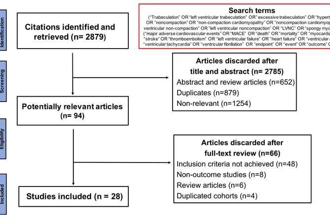
Our search strategy yielded 2879 studies, of which 94 full texts were relevant for evaluation (Figure 2). Exclusion of non-relevant studies, review articles and studies with duplicated cohorts resulted in 28 publications related to outcomes in LVNC. Searches of the Clinical Trials Registry identified one ongoing study titled “Prognosis of Isolated Left Ventricular Non-compaction in Adults” in France.
The final list (28 studies) consisted of 13 prospective and 15 retrospective observational studies. The studies were published between 1997 and 2019. A total of 2501 patients were included (mean±SD age: 46±7 years, male/female ratio: 1.7) with an overall median follow-up of 2.8 (IQR: 2.3 – 4.1) years. Although the diagnosis of LVNC was based mainly on quantification of excessive trabeculation, the majority of included studies (18 out of 28) comprised cohorts with significantly impaired LV systolic function (mean LVEF < 45%). The main characteristics of included studies are
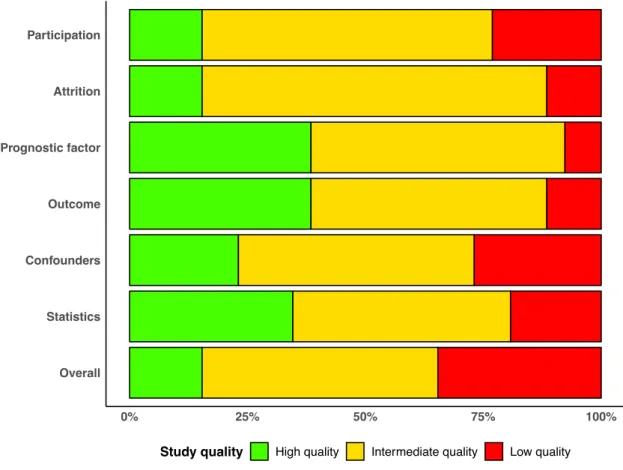
presented in Table 1 24–51. Among 28 studies, the distribution of overall study quality
was 18%, 50% and 32% for high, intermediate and low quality, respectively (Figure 3).
Primary outcome
Out of 28 included studies, 22 studies provided data on CV mortality in a total of 1822 patients who were followed up for a median (IQR) duration of 2.9 (2.4 – 4.4) years. The pooled incidence rate of CV death was 1.92 (95% CI: 1.54 – 2.30) per 100 person-years
(Figure 4 25–31,33–38,40–44,47–50). The funnel plot for the primary outcome appeared
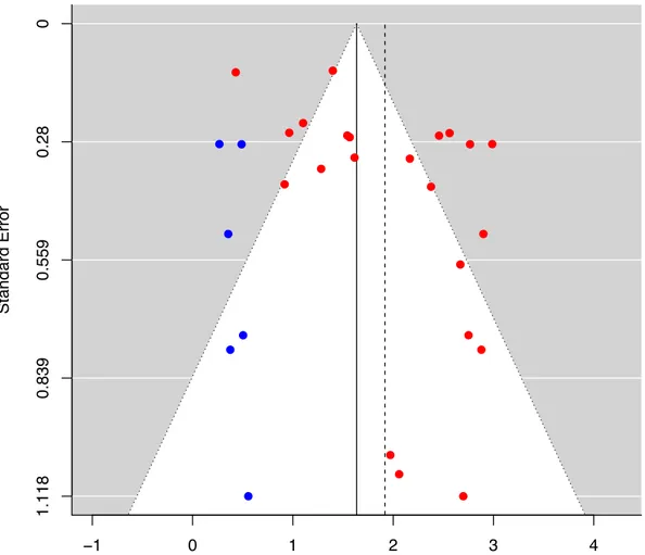
asymmetric due to the absence studies in the lower left corner, raising the possibility of publication bias (Egger’s regression asymmetry test p = 0.048). Addition of
hypothetical “missing” studies (N = 6) by the trim-and-fill method reduced the pooled CV mortality rate to 1.64 (95% CI: 1.29 – 1.98) per 100 person-years (Figure 5).
We observed a substantial between-study heterogeneity (I2 = 89.6%, p < 0.0001).
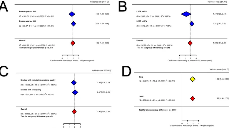
Therefore, we explored the clinical and statistical sources of heterogeneity by meta-regression and subgroup analyses. The meta-meta-regression analyses investigating the mediating effects of age, proportion of men, proportion of patients with NYHA > 2, LVEDD and LV NC:C did not identify any significant association. In subgroup analyses, studies enrolling patients with moderate-to-severe LV impairment (LVEF < 45%) appeared to have a higher incidence of cardiovascular mortality, compared to studies including patients with mildly impaired or normal LV systolic function (LVEF ³
45%) (2.21, 95% CI: 1.82 – 2.61 CV deaths per 100 person-years, I2 = 76.5% vs 1.19,
95% CI: 0.26–2.13 CV deaths per 100 person-years, I2 = 93.2%, p for subgroup
difference = 0.048). There was no significant difference in event rates when stratified by person-time at risk (an amalgamation of sample size and follow-up duration) of more than 3 years and high/intermediate vs low quality studies (Figure 6).
The overall estimate of CV mortality did not change significantly in the leave-one-out sensitivity analysis indicating that no single study had an overwhelming impact on the
combined meta-analysis estimate (SupplementalFigure 1).
Secondary outcomes
Twenty-four studies documented the incidence of all-cause mortality in 2122 patients over a median (IQR) follow-up of 2.6 (2.1 – 4.0) years. The pooled incidence rate of all-cause mortality was 2.16 (95% CI: 1.90 – 2.42) per 100 person-years (Supplemental
Figure 2). The funnel plot and Egger’s regression asymmetry test suggest possible
publication bias (Egger’s test p = 0.006). After addition of six hypothetical studies in the trim-and-fill sensitivity analysis, the pooled incidence rate decreased to 1.88 (95% CI: 1.60 – 2.16) per 100 person-years.
There was a substantial statistical heterogeneity among studies (I2 = 78.1%, p < 0.0001).
In meta-regression analyses, the proportion of male sex and the percentage of individuals with NYHA > 2 were positively associated with all-cause mortality. In subgroups stratified by LVEF, studies including patients with moderate-to-severe LV impairment (LVEF < 45%) appeared to have a higher incidence of all-cause deaths (p for subgroup difference = 0.011). The leave-one-out analysis was consistent with the overall result.
Stroke and systemic emboli
The event rates of stroke and systemic emboli was reported in 15 studies accounting for 1332 patients with a median (IQR) follow-up of 2.7 (2.4 – 3.8) years. The pooled incidence rate was 1.54 (95% CI: 1.22 – 1.86) per 100 person-years (Supplemental
Figure 3). We did not observe asymmetry in the funnel plot. Similar to the primary
outcome, we identified a substantial heterogeneity among studies (I2 = 73.4%, p <
0.0001). Meta-regression analyses did not reveal any mediating influence of age, sex, NYHA classification, LVEDD or prevalent AF. Stratification by the study quality, LVEF or person-time at risk did not show significant differences in the events rates
between subgroups. The leave-one-out sensitivity analysis showed no evidence of bias introduced by any one study.
Heart failure hospitalization
Twelve studies (1028 patients, median [IQR] follow-up: 2.5 [2.1 – 2.9] years) reported the incidence of heart failure hospitalization. The pooled event rate of heart failure hospitalization was 3.53 (95% CI: 2.95 – 4.11) per 100 person-years (Supplemental
Figure 4). The funnel plot did not appear asymmetric. There was a considerable
between-study heterogeneity (I2 = 87.7%, p<0.0001). Meta-regression analyses
identified a positive association between the proportion of symptomatic heart failure (NYHA > 2) at baseline and the incidence of heart failure admission at follow-up (regression coefficient = 0.04 per 1% increase in proportion of cohort with NYHA > 2, p = 0.049). In subgroup analyses, studies with an aggregate person-time at risk > 300
years appeared to have a lower incidence rate (2.77, 95% CI: 1.89 – 3.66, I2 = 92.5% vs
3.97, 95% CI: 3.34 – 4.60 per 100 person-years, I2 = 71.6%, p for subgroup difference =
0.031). The leave-one-out analysis was consistent with the overall pooled estimate.
Heart Transplantation
Data on cardiac transplantation rate was available in 14 studies (1576 patients, median [IQR] follow-up: 2.8 [2.4 – 4.9] years). The overall pooled event rate of heart
transplantation was 1.24 (95% CI: 0.98 – 1.50) per 100 person-years (Supplemental
Figure 5). The funnel plot showed sparsely distributed studies with evidence of
trim-CI: 0.96 – 1.48) per 100 person-years. The statistical heterogeneity of studies reporting
heart transplantation outcome was substantial (I2 = 71.6%, p < 0.0001). Meta-regression
analyses did not reveal any mediating effect of the selected covariates. We again found a lower incidence of heart transplantation in the subgroup with an aggregate person-time
at risk > 300 years (1.04, 95% CI: 0.81 – 1.26, I2 = 60.3% vs 1.79, 95% CI: 1.31 – 2.27
per 100 person-years, I2 = 35.1%, p for subgroup difference = 0.005). No undue
influence from any single study was detected in the leave-one-out analysis.
Ventricular arrhythmias
Nineteen studies with a total sample size of 1445 (median [IQR] follow-up of 2.8 [2.4 – 3.8] years) documented the incidence of ventricular arrhythmias. The calculated pooled
event rate was 2.17 (95% CI: 1.78 – 2.56) per 100 person-years (SupplementalFigure
6). There was no convincing evidence of publication bias in the funnel plot (Egger’s p =
0.05). We identified a substantial heterogeneity among studies (I2 = 84.4%, p < 0.0001).
Meta-regression analyses did not find any significant association with covariates but the subgroup with moderate-severe LV impairment (LVEF < 45%) appeared to have a
higher incidence of ventricular arrhythmias (2.30, 95% CI: 1.72 – 2.88, I2 = 88.4% vs
1.60, 95% CI: 1.23 – 1.97 per 100 person-years, I2 = 0%, p for subgroup difference =
0.047). The leave-one-out analysis did not show any significant deviation from the overall pooled result.
Cardiac device implantation
The incidence of cardiac device implantation was recorded in 15 studies (1278 patients, median [IQR] follow-up of 2.9 [2.0 – 3.8] years). The pooled incidence rate was 2.66
(95% CI: 1.93 – 3.39) per 100 person-years (SupplementalFigure 7). The funnel plot
appeared sparse but symmetric. A considerable between-study heterogeneity was
present (I2 = 95.3%, p < 0.0001). The meta-regression analyses identified a negative
association between the proportion of male sex and the incidence of cardiac device implantation (regression coefficient = -0.06 per 1% increase in male sex proportion, p = 0.04). Stratified analyses did not find any significant subgroup difference although the
inconsistency index (I2) appeared much lower in some subgroups. We did not find any
indication of bias in the leave-one-out analysis.
Comparison with DCM
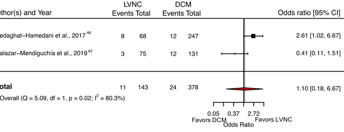
Two studies out of 22 reported the incidence of CV death in a comparable group of DCM patients. Overall, the LVNC patients did not have significantly higher CV
mortality than the DCM group (pooled OR: 1.10, 95% CI: 0.18 – 6.67) (Figure 7 43,44).
The pooled event rate of CV death in a previously published meta-analysis of DCM
patients 21 (19 studies enrolling 2466 individuals) was comparable to the pooled event
rate observed in our study (DCM: 1.92, 95% CI: 1.44 – 2.39 CV deaths per 100 person-years vs. LVNC: 1.92, 95% CI: 1.54 – 2.30 CV deaths per 100 person-person-years) (Figure 6,
panel D). Two studies out of 24 provided all-cause mortality data in a DCM control
group. In comparison with the DCM group, patients with LVNC did not have
significantly higher mortality (pooled OR: 0.67, 95% CI: 0.28 – 1.59). When compared
with an external previously published DCM meta-analysis 21, the heart failure
hospitalization rate in our study was significantly higher (3.53 vs 2.37 per 100 person-years, p = 0.003). There was no significant difference in the incidence rate of ventricular
DISCUSSION
In this meta-analysis investigating the prognosis of a large population of adult LVNC patients classified according to contemporary imaging criteria, we identified the following key findings: (i) the overall incidence rates of cardiovascular mortality, all-cause mortality, stroke and systemic emboli, heart failure admission, cardiac
transplantation, ventricular arrhythmias and cardiac device implantation were 1.92, 2.16, 1.54, 3.53, 1.24, 2.17, and 2.66, respectively, per 100 person-years, at an
intermediate-term follow-up (ii) the incidence of cardiovascular or all-cause mortality in LVNC patients were similar to DCM controls, (iii) the high level of statistical
heterogeneity was partly explained by the variability in clinical characteristics (LVEF in particular) and study characteristics such as sample size/study duration, (iv) the
incidence rate of ventricular arrhythmias was comparable to DCM patients but heart failure admission rate was higher in LVNC patients.
By investigating the prognosis of real-world patients with excessive trabeculations meeting the imaging diagnostic criteria for LVNC, we aimed to provide much needed information on the natural course of this controversial disease entity. The findings from our study can be regarded as a foundation for further discussion regarding the medical implications of an increasingly-recognized imaging finding, and also highlights important heterogeneity among available published studies.
The incidence rates of cardiovascular and all-cause mortality – arguably, two more reliable and objective outcomes – estimated to be 1.92 and 2.14 per 100 person-years,
respectively, in our meta-analysis, are 25- and 5-fold higher than the event rates in a general population (0.08 and 0.41 per 100 person-years for cardiovascular and all-cause
mortality, respectively, in 45-54 years age group in a North American population) 52.
Therefore, a diagnosis of LVNC by current clinical and imaging criteria appears to portend a heightened mortality risk despite a significant diversity of patient population in the individual studies. Nonetheless, when compared to non-ischemic DCM patients, LVNC patients carry a very similar risk of death from cardiovascular causes. We also observed elevated incidences of cardiovascular morbidities in LVNC patients with two most frequent complications being heart failure hospitalization and cardiac device implantation. The heart failure-related hospital admission rate in our meta-analysis was higher than the pooled incidence observed in a comparable DCM meta-analysis (3.52 vs 2.37 per 100 person-years). This finding should be interpreted with caution in view of variability in definition of heart failure decompensation and lack of data on the rigour of heart failure treatment.
There is a notion of an increased risk of systemic thromboembolism attributable to the sluggish blood flow in the deep inter-trabecular recesses in LVNC patients. However, no solid evidence is available to support this hypothesis. Indeed, in our study, the incidence rate of stroke and systemic emboli was 1.54 per 100 person-years which is either lower than or comparable to the event rates reported in: (i) V-HeFT trials in patients with systolic heart failure (2.1 – 2.7 per 100 person-years), (ii) patients with
ischemic cardiomyopathy (1.5 per 100 person-years) in SAVE trial 53, and (iii) DCM
It is important to consider the incidence rates reported in this meta-analysis in the context of cohort characteristics where 18 out of 28 included studies recruited individuals with significant LV systolic impairment. Subgroup analysis stratified by LVEF demonstrated a significant reduction in the number of CV deaths in the absence of moderate to severe LV systolic dysfunction. Therefore, the risk of achieving the endpoint may in part be contingent upon the development of LV dysfunction. Although the risk to individuals with excessive trabeculations in an unselected and otherwise healthy population is beyond the scope of this study, a previous population study of approximately 3000 asymptomatic individuals did not find any association between the degree of trabeculation and the decline in LV function or incident CV events over a course of nearly 10 years 56.
As anticipated, we observed a high degree of statistical heterogeneity among included studies which can be partially explained by the differences in cohort characteristics and study quality. In our quality assessment by QUIPS criteria, the two most commonly affected bias domains were study participation (i.e. selection bias) and treatment of confounders, reflecting the challenges associated with the observational studies reporting a relatively rare condition. The subsequent meta-regression and subgroup analyses revealed that severity of LV impairment measured by LVEF had an important influence on the variability of incidence rates reported in individual studies.
Equivalently, smaller studies with short follow-up duration tended to report higher
incidence rates of secondary outcomes. The indicator of between-study variability (I2
index) was noticeably lower in the subgroup analysis which further supports the importance of well-defined inclusion and diagnostic criteria.
All imaging diagnostic criteria for LVNC consider presence of excessive trabeculation as a cardinal signature of disease. There is a degree of confusion and uncertainty in assigning the disease status due to not-so-infrequent finding of increased trabeculation in otherwise healthy individuals and those with primary DCM or hypertrophic
cardiomyopathy. Recent evidence suggests that the extent of trabeculation in asymptomatic low-risk population, LVNC and DCM patients does not determine
prognosis 24,25,56. In this respect, the lack of mediating influence by the LV NC:C on
clinical outcomes in our study is concordant with the existing evidence in literature. A recently published meta-analysis of four CMR studies enrolling LVNC patients reported that in the absence of late gadolinium enhancement and LV systolic dysfunction, no
hard cardiac event was observed 57. Therefore, our study, together with mounting
evidence from existing literature, underscores the important prognostic role of focal myocardial injury and functional impairment, rather than the morphological appearance of LVNC. Indeed, it is notable that the conventional diagnostic criteria for LVNC have relied principally on ratio measurement and have not included other structural,
functional, clinical or familial parameters.
In this study, we systematically reviewed and performed the meta-analysis of the incidence of important cardiovascular outcomes in a large population of real-world LVNC patients. We attempted to synthesize the results in a robust manner giving due consideration to address potential biases where possible. However, we acknowledge several limitations associated with our study. First, the pooled analysis relied on
observational, mostly single centre, cohort studies with variable methodological quality as highlighted in the bias assessment, inclusion criteria and definitions of LVNC. Second, our study only focused on the adult population mostly free from congenital
heart disease, thus, the insights obtained from this work cannot be extended to pediatric LVNC or patients with coexisting congenital heart disease. Third, comparison of the rates of incident CV events between LVNC and hypertrophic cardiomyopathy was not performed and should be investigated in a future study. Fourth, meta-regression
analyses were limited to the studies without missing covariate information, hence, may be underpowered. Fifth, only a few studies reported the incidence rates in a comparable DCM cohort. Thus, the precision of pooled odds ratio and the level of evidence are weaker. Finally, the incidence rates of adverse events observed in this study only hold true for an intermediate follow-up duration and the long-term consequences of LVNC remain to be elucidated.
An expert group consensus approach to harmonize the diagnostic criteria, risk factors and endpoints is urgently needed to develop a more standardized assessment of LVNC. Future studies including prospective registries should address long term prognosis and could also investigate additional prognostic information provided by fractal analysis, T1 mapping and genotype over current LV NC:C ratio, systolic function and tissue
characterization by LGE.
CONCLUSIONS
Patients with LVNC have similar risks of cardiovascular mortality, all-cause mortality, thromboembolic complications and ventricular arrhythmias in comparison with DCM patients. The finding of increased incidence of heart failure hospitalization in isolation should be interpreted with caution and investigated in future well-designed studies.
Traditional indicators of cardiac disease severity such as low LVEF, not the burden of trabeculation, appear to be associated with worse outcomes.
FUNDING
NA is supported by a Wellcome Trust Research Training Fellowship (203553/Z/Z). SEP and NA acknowledge the support through the Barts Biomedical Research Centre which is funded by the National Institute for Health Research.
ACKNOWLEDGMENTS
This work was part of the portfolio of translational research of the NIHR Biomedical Research Centre at Barts and The London School of Medicine and Dentistry.
DISCLOSURES
SEP provides consultancy to Circle Cardiovascular Imaging Inc, Calgary, Canada. Other authors have no conflicts of interest to declare.
REFERENCES
1. Maron BJ, Towbin JA, Thiene G, Antzelevitch C, Corrado D, Arnett D, Moss AJ, Seidman CE, Young JB, American Heart Association, et al. Contemporary definitions and
classification of the cardiomyopathies: an American Heart Association Scientific Statement from the Council on Clinical Cardiology, Heart Failure and Transplantation Committee; Quality of Care and Outcomes Research and Functional Genomics and Translational Biology Interdisciplinary Working Groups; and Council on Epidemiology and Prevention. Circulation. 2006;113:1807–1816.
2. Elliott P, Andersson B, Arbustini E, Bilinska Z, Cecchi F, Charron P, Dubourg O, Kühl U, Maisch B, McKenna WJ, et al. Classification of the cardiomyopathies: a position
statement from the European Society Of Cardiology Working Group on Myocardial and Pericardial Diseases. Eur Heart J. 2008;29:270–276.
3. Arbustini E, Weidemann F, Hall JL. Left Ventricular Noncompaction: A Distinct Cardiomyopathy or a Trait Shared by Different Cardiac Diseases? J Am Coll Cardiol. 2014;64:1840–1850.
4. Gati S, Chandra N, Bennett RL, Reed M, Kervio G, Panoulas VF, Ghani S, Sheikh N, Zaidi A, Wilson M, et al. Increased left ventricular trabeculation in highly trained athletes: do we need more stringent criteria for the diagnosis of left ventricular non-compaction in athletes? Heart Br Card Soc. 2013;99:401–408.
5. Gati S, Papadakis M, Van Niekerk N, Reed M, Yeghen T, Sharma S. Increased left
ventricular trabeculation in individuals with sickle cell anaemia: physiology or pathology?
Int J Cardiol. 2013;168:1658–1660.
6. Gati S, Papadakis M, Papamichael ND, Zaidi A, Sheikh N, Reed M, Sharma R,
Thilaganathan B, Sharma S. Reversible de novo left ventricular trabeculations in pregnant women: implications for the diagnosis of left ventricular noncompaction in low-risk populations. Circulation. 2014;130:475–483.
7. Teekakirikul P, Kelly MA, Rehm HL, Lakdawala NK, Funke BH. Inherited
Cardiomyopathies: Molecular Genetics and Clinical Genetic Testing in the Postgenomic Era. J Mol Diagn. 2013;15:158–170.
8. Ross SB, Jones K, Blanch B, Puranik R, McGeechan K, Barratt A, Semsarian C. A
systematic review and meta-analysis of the prevalence of left ventricular non-compaction in adults. Eur Heart J. 2019;[ePub ahead of print] doi: 10.1093/eurheartj/ehz317
9. Stroup DF, Berlin JA, Morton SC, Olkin I, Williamson GD, Rennie D, Moher D, Becker BJ, Sipe TA, Thacker SB, et al. Meta-analysis of Observational Studies in Epidemiology: A Proposal for Reporting. JAMA. 2000;283:2008–2012.
10. Moher D, Liberati A, Tetzlaff J, Altman DG, Group TP. Preferred Reporting Items for Systematic Reviews and Meta-Analyses: The PRISMA Statement. PLOS Med. 2009;6:e1000097.
11. Petersen SE, Selvanayagam JB, Wiesmann F, Robson MD, Francis JM, Anderson RH, Watkins H, Neubauer S. Left Ventricular Non-Compaction: Insights From Cardiovascular Magnetic Resonance Imaging. J Am Coll Cardiol. 2005;46:101–105.
12. Chin TK, Perloff JK, Williams RG, Jue K, Mohrmann R. Isolated noncompaction of left ventricular myocardium. A study of eight cases. Circulation. 1990;82:507–513. 13. Jenni R, Oechslin E, Schneider J, Jost C, Kaufmann P. Echocardiographic and
pathoanatomical characteristics of isolated left ventricular non-compaction: a step towards classification as a distinct cardiomyopathy. Heart. 2001;86:666–671.
14. Jacquier A, Thuny F, Jop B, Giorgi R, Cohen F, Gaubert J-Y, Vidal V, Bartoli JM, Habib G, Moulin G. Measurement of trabeculated left ventricular mass using cardiac magnetic resonance imaging in the diagnosis of left ventricular non-compaction. Eur Heart J. 2010;31:1098–1104.
15. Grothoff M, Pachowsky M, Hoffmann J, Posch M, Klaassen S, Lehmkuhl L, Gutberlet M. Value of cardiovascular MR in diagnosing left ventricular non-compaction
cardiomyopathy and in discriminating between other cardiomyopathies. Eur Radiol. 2012;22:2699–2709.
16. Stacey RB, Andersen MM, St Clair M, Hundley WG, Thohan V. Comparison of systolic and diastolic criteria for isolated LV noncompaction in CMR. JACC Cardiovasc Imaging. 2013;6:931–940.
17. Stöllberger C, Gerecke B, Finsterer J, Engberding R. Refinement of echocardiographic criteria for left ventricular noncompaction. Int J Cardiol. 2013;165:463–467.
18. Captur G, Muthurangu V, Cook C, Flett AS, Wilson R, Barison A, Sado DM, Anderson S, McKenna WJ, Mohun TJ, et al. Quantification of left ventricular trabeculae using fractal analysis. J Cardiovasc Magn Reson. 2013;15:36.
19. Hayden JA, van der Windt DA, Cartwright JL, Côté P, Bombardier C. Assessing bias in studies of prognostic factors. Ann Intern Med. 2013;158:280–286.
20. Freeman MF, Tukey JW. Transformations Related to the Angular and the Square Root. Ann
Math Stat. 1950;21:607–611.
21. Becker MAJ, Cornel JH, van de Ven PM, van Rossum AC, Allaart CP, Germans T. The Prognostic Value of Late Gadolinium-Enhanced Cardiac Magnetic Resonance Imaging in Nonischemic Dilated Cardiomyopathy: A Review and Meta-Analysis. JACC
Cardiovasc Imaging. 2018;11:1274–1284.
22. Viechtbauer W. Conducting meta-analyses in R with the metafor package. J Stat Softw. 2010;36:1–48.
23. R Core Team. R: A Language and Environment for Statistical Computing [Internet]. Vienna, Austria: R Foundation for Statistical Computing; 2016 [accessed 2016 Jun 19]. Available from: https://www.R-project.org/
24. Heron M. Deaths: Leading Causes for 2016 [Internet]. [accessed 2018 Sep 24];Available from: https://www.cdc.gov/nchs/data/nvsr/nvsr67/nvsr67_06.pdf
25. Loh E, Sutton MStJ, Wun C-CC, Rouleau JL, Flaker GC, Gottlieb SS, Lamas GA, Moyé LA, Goldhaber SZ, Pfeffer MA. Ventricular Dysfunction and the Risk of Stroke after Myocardial Infarction. N Engl J Med. 1997;336:251–257.
26. Fuster V, Gersh BJ, Giuliani ER, Tajik AJ, Brandenburg RO, Frye RL. The natural history of idiopathic dilated cardiomyopathy. Am J Cardiol. 1981;47:525–531.
27. Koniaris LS, Goldhaber SZ. Anticoagulation in Dilated Cardiomyopathy. J Am Coll
Cardiol. 1998;31:745–748.
28. Zemrak F, Ahlman MA, Captur G, Mohiddin SA, Kawel-Boehm N, Prince MR, Moon JC, Hundley WG, Lima JAC, Bluemke DA, et al. The relationship of left ventricular
trabeculation to ventricular function and structure over a 9.5-year follow-up: the MESA study. J Am Coll Cardiol. 2014;64:1971–1980.
29. Andreini D, Pontone G, Bogaert J, Roghi A, Barison A, Schwitter J, Mushtaq S, Vovas G, Sormani P, Aquaro GD, et al. Long-Term Prognostic Value of Cardiac Magnetic
30. Amzulescu M-S, Rousseau MF, Ahn SA, Boileau L, de Meester de Ravenstein C, Vancraeynest D, Pasquet A, Vanoverschelde JL, Pouleur A-C, Gerber BL. Prognostic Impact of Hypertrabeculation and Noncompaction Phenotype in Dilated Cardiomyopathy: A CMR Study. JACC Cardiovasc Imaging. 2015;8:934–946.
31. Grigoratos C, Barison A, Ivanov A, Andreini D, Amzulescu M-S, Mazurkiewicz L, De Luca A, Grzybowski J, Masci PG, Marczak M, et al. Meta-Analysis of the Prognostic Role of Late Gadolinium Enhancement and Global Systolic Impairment in
Left Ventricular Noncompaction. JACC Cardiovasc Imaging. 2019;12:2141–2151. 32. Aras D, Tufekcioglu O, Ergun K, Ozeke O, Yildiz A, Topaloglu S, Deveci B, Sahin O,
Kisacik HL, Korkmaz S. Clinical features of isolated ventricular noncompaction in adults long-term clinical course, echocardiographic properties, and predictors of left ventricular failure. J Card Fail. 2006;12:726–733.
33. Asfalou I, Boulaamayl S, Raissouni M, Mouine N, Sabry M, Kheyi J, Doghmi N, Benyass A. Left ventricular noncompaction-A rare form of cardiomyopathy: Revelation modes and predictors of mortality in adults through 23 cases. J Saudi Heart Assoc. 2017;29:102–109. 34. Caliskan K, Szili-Torok T, Theuns DAMJ, Kardos A, Geleijnse ML, Balk AHMM, van
Domburg RT, Jordaens L, Simoons ML. Indications and outcome of implantable cardioverter-defibrillators for primary and secondary prophylaxis in patients with noncompaction cardiomyopathy. J Cardiovasc Electrophysiol. 2011;22:898–904. 35. Cetin MS, Ozcan Cetin EH, Canpolat U, Cay S, Topaloglu S, Temizhan A, Aydogdu S.
Usefulness of Fragmented QRS Complex to Predict Arrhythmic Events and Cardiovascular Mortality in Patients With Noncompaction Cardiomyopathy. Am J
Cardiol. 2016;117:1516–1523.
36. Correia E, Rodrigues B, Santos L, Faria R, Ferreira P, Gama P, Nascimento C, Dionisio O, Cabral C, Santos O. Noncompaction of the ventricular myocardium: characterization and follow-up of an affected population. Rev Port Cardiol Orgao Of Soc Port Cardiol Port J
Cardiol Off J Port Soc Cardiol. 2011;30:323–331.
37. Enríquez R A, Baeza V R, Gabrielli N L, Córdova A S, Castro G P. Non compaction cardiomyopathy: a series of 15 cases. Rev Médica Chile. 2011;139:864–871.
38. Gaye ND, Ngaïdé AA, Bah MB, Babaka K, Mbaye A, Abdoul K. Non-compaction of left ventricular myocardium in sub-Saharan African adults. Heart Asia. 2017;9:e010884. 39. Greutmann M, Mah ML, Silversides CK, Klaassen S, Attenhofer Jost CH, Jenni R,
Oechslin EN. Predictors of adverse outcome in adolescents and adults with isolated left ventricular noncompaction. Am J Cardiol. 2012;109:276–281.
40. Habib G, Charron P, Eicher J-C, Giorgi R, Donal E, Laperche T, Boulmier D, Pascal C, Logeart D, Jondeau G, et al. Isolated left ventricular non-compaction in adults: clinical and echocardiographic features in 105 patients. Results from a French registry. Eur J
Heart Fail. 2011;13:177–185.
41. Ivanov Alexander, Dabiesingh Devindra S., Bhumireddy Geetha P., Mohamed Ambreen, Asfour Ahmed, Briggs William M., Ho Jean, Khan Saadat A., Grossman Alexandra, Klem Igor, et al. Prevalence and Prognostic Significance of Left Ventricular Noncompaction in Patients Referred for Cardiac Magnetic Resonance Imaging. Circ Cardiovasc Imaging. 2017;10:e006174.
42. Kawasaki T, Azuma A, Taniguchi T, Asada S, Kamitani T, Kawasaki S, Matsubara H, Sugihara H. Heart rate variability in adult patients with isolated left ventricular noncompaction. Int J Cardiol. 2005;99:147–150.
43. Li Shijie, Zhang Ce, Liu Nana, Bai Hui, Hou Cuihong, Wang Jizheng, Song Lei, Pu Jielin. Genotype-Positive Status Is Associated With Poor Prognoses in Patients With Left Ventricular Noncompaction Cardiomyopathy. J Am Heart Assoc. 2018;7:e009910. 44. Lofiego C, Biagini E, Pasquale F, Ferlito M, Rocchi G, Perugini E, Bacchi-Reggiani L,
Boriani G, Leone O, Caliskan K, et al. Wide spectrum of presentation and variable
outcomes of isolated left ventricular non-compaction. Heart Br Card Soc. 2007;93:65–71. 45. Mazurkiewicz Ł, Petryka J, Śpiewak M, Miłosz-Wieczorek B, Małek ŁA, Jasińska A,
Jarmus E, Marczak M, Misko J, Grzybowski J. Clinical and prognostic relevancy of left ventricular trabeculation assessed by cardiac magnetic resonance in patients with dilated cardiomyopathy. Kardiologia Pol Pol Heart J. 2017;75:794–803.
46. Murphy RT, Thaman R, Blanes JG, Ward D, Sevdalis E, Papra E, Kiotsekoglou A, Kiotsekolglou A, Tome MT, Pellerin D, et al. Natural history and familial characteristics of isolated left ventricular non-compaction. Eur Heart J. 2005;26:187–192.
47. Peters F, Khandheria BK, Botha F, Libhaber E, Matioda H, Dos Santos C, Govender S, Meel R, Essop MR. Clinical outcomes in patients with isolated left ventricular
noncompaction and heart failure. J Card Fail. 2014;20:709–715.
48. Ritter M, Oechslin E, Sütsch G, Attenhofer C, Schneider J, Jenni R. Isolated noncompaction of the myocardium in adults. Mayo Clin Proc. 1997;72:26–31.
49. Salazar-Mendiguchía J, González-Costello J, Oliveras T, Gual F, Lupón J, Manito N. Long-term Follow-up of Symptomatic Adult Patients With Noncompaction Cardiomyopathy.
Rev Esp Cardiol Engl Ed. 2019;72:169–171.
50. Sedaghat-Hamedani F, Haas J, Zhu F, Geier C, Kayvanpour E, Liss M, Lai A, Frese K, Pribe-Wolferts R, Amr A, et al. Clinical genetics and outcome of left ventricular non-compaction cardiomyopathy. Eur Heart J. 2017;38:3449–3460.
51. Stämpfli SF, Erhart L, Hagenbuch N, Stähli BE, Gruner C, Greutmann M, Niemann M, Kaufmann BA, Jenni R, Held L, et al. Prognostic power of NT-proBNP in left ventricular non-compaction cardiomyopathy. Int J Cardiol. 2017;236:321–327.
52. Stanton C, Bruce C, Connolly H, Brady P, Syed I, Hodge D, Asirvatham S, Friedman P. Isolated left ventricular noncompaction syndrome. Am J Cardiol. 2009;104:1135–1138. 53. Steffel J, Hürlimann D, Namdar M, Despotovic D, Kobza R, Wolber T, Holzmeister J,
Haegeli L, Brunckhorst C, Lüscher TF, et al. Long-term follow-up of patients with isolated left ventricular noncompaction: role of electrocardiography in predicting poor outcome. Circ J Off J Jpn Circ Soc. 2011;75:1728–1734.
54. Stöllberger C, Wegner C, Finsterer J. Left ventricular hypertrabeculation/noncompaction, cardiac phenotype, and neuromuscular disorders. Herz. 2019;44:659–665.
55. Tian T, Liu Y, Gao L, Wang J, Sun K, Zou Y, Wang L, Zhang L, Li Y, Xiao Y, et al. Isolated left ventricular noncompaction: clinical profile and prognosis in 106 adult patients. Heart Vessels. 2014;29:645–652.
56. Tian T, Yang K-Q, Mao Y, Zhou L-L, Wang L-P, Xiao Y, Yang Y-K, Zhang Y, Meng X, Zhou X-L. Left Ventricular Noncompaction in Older Patients. Am J Med Sci.
2017;354:140–144.
57. van Waning JI, Caliskan K, Hoedemaekers YM, van Spaendonck-Zwarts KY, Baas AF, Boekholdt SM, van Melle JP, Teske AJ, Asselbergs FW, Backx APCM, et al. Genetics, Clinical Features, and Long-Term Outcome of Noncompaction Cardiomyopathy. J Am
Coll Cardiol. 2018;71:711–722.
FIGURE TITLES AND LEGENDS
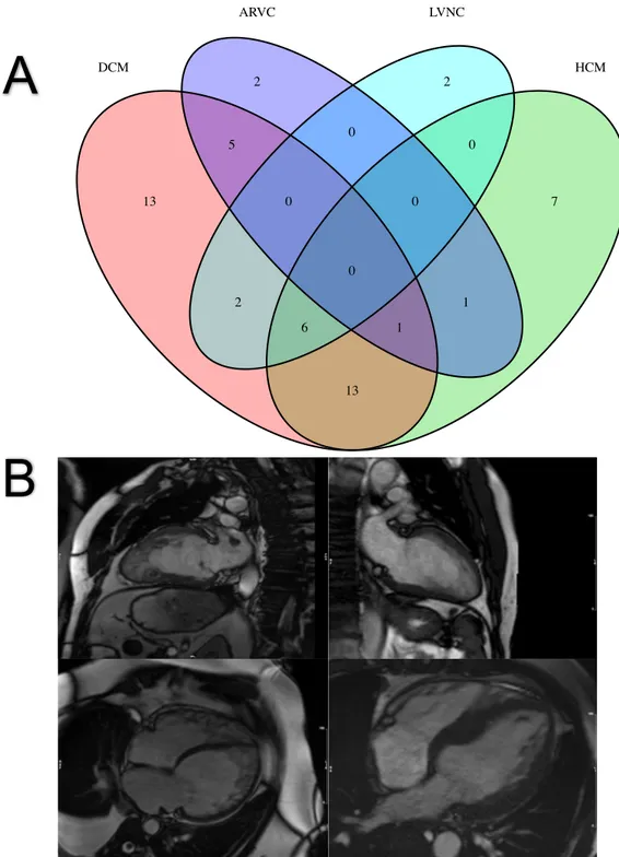
Figure 1. (A) Venn diagram of the number of genes associated with inherited
cardiomyopathy; (B) CMR images demonstrating a classic LVNC with a two-layer appearance of thin compact myocardium and excessive trabeculation (top left), isolated LVNC with normal chamber size and function (top right), mixed DCM and LVNC with biventricular involvement (bottom left) and HCM with features of LVNC (bottom right).
(DCM, dilated cardiomyopathy; ARVC, arrhythmogenic right ventricular cardiomyopathy; LVNC, left-ventricular non-compaction; HCM, hypertrophic cardiomyopathy; CMR, cardiovascular magnetic resonance)
Figure 2. Flow diagram demonstrating the process of study selection
Figure 3. Distribution of study quality according to QUIPS tool
Figure 4. Forest plot demonstrating the individual and overall incidences of
cardiovascular deaths per 100 person-years. The vertical dotted line indicates the pooled average incidence rate.
Figure 5. Funnel plot for cardiovascular mortality. The red dots represent the original
studies included in the meta-analysis while the blue dots represent the “missing” studies imputed by the trim-and-fill method. The vertical dashed line indicates the original pooled incidence rates and the vertical solid line indicates the revised pool incidence rates after inclusion of the imputed “missing” studies to counter publication bias. (CV, cardiovascular)
Figure 6. Subgroup analyses for cardiovascular mortality: (A) Incidence of
cardiovascular mortality in subgroups stratified by person-years > 300, (B) Incidence of cardiovascular mortality in subgroups stratified by LVEF < 45%, (C) Incidence of cardiovascular mortality in subgroups stratified by high vs low-moderate risk of bias, (D) Incidence of cardiovascular mortality in LVNC meta-analysis vs external DCM meta-analysis. The vertical dotted line indicates the pooled average incidence rate. (LVEF, left ventricular ejection fraction; LVNC, left ventricular non-compaction; DCM, dilated cardiomyopathy)
Figure 7. Forest plot of cardiovascular mortality in LVNC patients compared to DCM
controls. The vertical dotted line represents the pooled odds ratio.
TABLES
Table 1. Characteristics of included studies
Author Year characteristics Cohort Control group N (years) Age
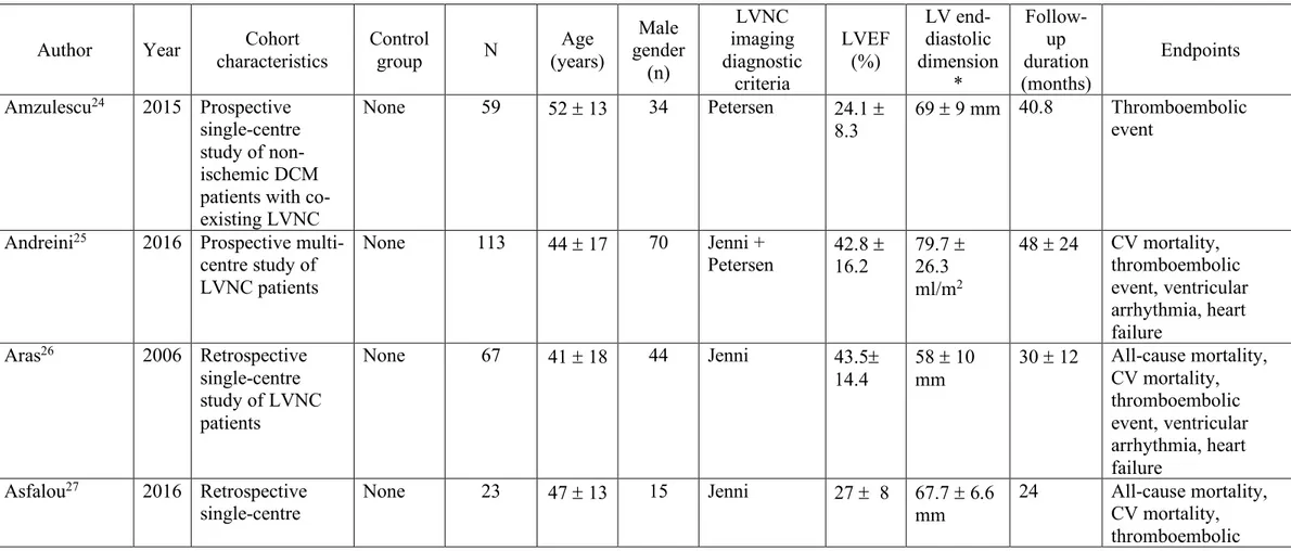
Male gender (n) LVNC imaging diagnostic criteria LVEF (%) LV end-diastolic dimension * Follow-up duration (months) Endpoints Amzulescu24 2015 Prospective single-centre study of non-ischemic DCM patients with co-existing LVNC
None 59 52 ± 13 34 Petersen 24.1 ±
8.3 69 ± 9 mm 40.8
Thromboembolic event
Andreini25 2016 Prospective
multi-centre study of LVNC patients None 113 44 ± 17 70 Jenni + Petersen 42.8 ± 16.2 79.7 ± 26.3 ml/m2 48 ± 24 CV mortality, thromboembolic event, ventricular arrhythmia, heart failure Aras26 2006 Retrospective single-centre study of LVNC patients None 67 41 ± 18 44 Jenni 43.5± 14.4 58 ± 10 mm 30 ± 12 All-cause mortality, CV mortality, thromboembolic event, ventricular arrhythmia, heart failure Asfalou27 2016 Retrospective
single-centre None 23 47 ± 13 15 Jenni 27 ± 8 67.7 ± 6.6 mm
24 All-cause mortality,
CV mortality, thromboembolic
study of LVNC patients event, ventricular arrhythmia, ICD implantation, heart failure Caliskan28 2011 Prospective single-centre study of LVNC patients None 77 40 ± 14 37 Jenni NR 60.4 ± 9.6 mm 33 ± 24 All-cause mortality, CV mortality, ventricular arrhythmia, cardiac transplantation, heart failure Cetin29 2016 Retrospective single-centre study of LVNC patients None 88 39 ± 18 57 Jenni 32.0 ± 12.5 59.3 ± 9.1 mm 42.4 All-cause mortality, CV mortality, ventricular arrhythmia, ICD implantation Correia30 2011 Retrospective single-centre study of LVNC patients None 20 53 ± 20 13 Jenni 45 ± 19 58 ± 11 mm 12 ± 6 All-cause mortality, CV mortality, cardiac transplantation, ICD implantation Enriquez31 2011 Retrospective single-centre study of LVNC patients None 15 52 ± 17 6 Jenni 27 ± 10 66 ± 11 mm 19 Ventricular arrhythmia, ICD implantation Gaye32 2017 Retrospective multi-centre study of LVNC patients None 35 47 ± 18 NR Jenni 32.5 ± 13.8 66.4 ± 9.6 mm 17.2 ± 14.5 All-cause mortality, ventricular arrhythmia, heart failure Greutmann33 2012 Retrospective single-centre None 132 41 ± 17 46 Jenni 41 ± 18 34 ± 7 mm/m2 32.4 CV mortality, thromboembolic event, ventricular
study of LVNC patients
arrhythmia, cardiac transplantation, heart failure
Habib34 2011 Prospective
multi-centre study of LVNC patients None 105 45 ± 17 69 Jenni 46 ± 18 63 ± 11 mm 30 ± 18 All-cause mortality, CV mortality, thromboembolic event, ventricular arrhythmia, ICD implantation, cardiac transplantation, heart failure Ivanov35 2017 Prospective single-centre study of LVNC patients Patients not fulfilling Petersen criteria and with no evidence of congenit al heart disease or valve disease 276 (LVNC) / 424 (non-LVNC with compara-ble age and LVEF) 57 147 Petersen 49 ± 17 77 ± 29 ml/m2 82 All-cause mortality, CV mortality Kawasaki36 2005 Retrospective single-centre study LVNC patients Age- and sex-matched with individu 10 (LVNC) / 80 (non-LVNC:
50 ± 13 8 Jenni NR NR 26 ± 14 All-cause mortality,
als with myocard ial infarctio n, hypertro phic cardiom yopathy and no CV disease 40 MI and 40 HCM)† Li37 2018 Prospective single-centre study of Chinese LVNC patients None 83 45 58 Jenni + Petersen 37 62 54 All-cause mortality, CV mortality, cardiac transplantation
Lofiego38 2007 Prospective
multi-centre study of LVNC patients None 65 45 ± 16 NR Jenni 31 ± 11 67 ± 11 mm 46 ± 44 All-cause mortality, CV mortality, thromboembolic event, ventricular arrhythmia, ICD implantation, cardiac transplantation, heart failure
Mazurkie-wicz39 2017 Prospective single centre study of
DCM patients with co-existing LVNC DCM patients not fulfilling LVNC criteria 127 (LVNC) / 149 (DCM) 33 ± 9 78 Grothoff 27.7 ± 7.5 172.9 ± 29.8 ml/m2 28.8 All-cause mortality, cardiac transplantation, thromboembolic event
Murphy40 2005 Prospective study of unrelated LVNC patients None 45 37 ± 17 28 Chin+Jenni NR 58 ± 11 mm 46 All-cause mortality, CV mortality, ventricular arrhythmia, ICD implantation Peters41 2014 Prospective single-centre study of idiopathic LVNC patients None 55 42 ± 12 21 Jenni 29.6 ± 11.8 59.1 ± 9.8 mm 16.7 ± 5.9 All-cause mortality, CV mortality, thromboembolic event, heart failure, ICD implantation
Ritters42 1997 Retrospective
single-centre study of LVNC patients
None 17 42 ± 17 14 Jenni NR NR 30 ± 28 All-cause mortality,
CV mortality, thromboembolic event, cardiac transplantation, heart failure, ventricular arrhythmia Salazar-Mendiguchía 43 2019 Retrospective multi-centre study of LVNC patients Sympto-matic DCM patients 75 50 ± 15 51 Jenni 32 63.8 60 CV mortality, thromboembolic event, cardiac transplantation, ventricular arrhythmia, ICD implantation
Sedaghat-Hamedani44 2017 Prospective multi-centre registry of
symptomatic LVNC patients Age-matched non-ischemic DCM patients 68 (LVNC) / 247 (DCM) 41 ± 14 48 Jenni+ Stöllberger+ Petersen 38 ± 15.3 62 ± 12.3 mm 61 All-cause mortality, CV mortality, thromboembolic event, cardiac transplantation, ventricular
arrhythmia, ICD implantation
Stampfli45 2017 Retrospective
multi-centre study of LVNC patients
None 153 43 ± 19 91 Jenni 45 NR 72 All-cause mortality,
cardiac transplantation Stanton46 2009 Retrospective single-centre study of LVNC patients Age-, sex- and LVEF-matched DCM patients 30 (LVNC) / 27 (DCM)
39 ± 20 18 Jenni 41 NR 30 ± 14 All-cause mortality,
ventricular arrhythmia, ICD implantation Steffel47 2011 Retrospective single-centre study of LVNC patients None 74 43 ± 16 53 Jenni 40 ± 19 32.7 ± 10 mm/m2 57.9 ± 41.5 All-cause mortality, CV mortality, ventricular arrhythmia Stollberger48 2018 Prospective single-centre study of LVNC patients; Prevalence of neuromuscular disease associated with LVNC was also assessed. None 273 53 ± 17 193 Stöllberger NR 60 ± 13 88.8 ± 68.4 All-cause mortality, CV mortality, ICD implantation, cardiac transplantation Tian49 2014 Retrospective single-centre study of LVNC patients None 106 46 ± 17 83 Jenni 39 ± 14 61 ± 10 mm 35 ± 25 All-cause mortality, CV mortality, thromboembolic event, ventricular arrhythmia, ICD
transplantation, heart failure Tian50 2017 Prospective single-centre study of older LVNC patients (age ³ 60 years)
None 35 65 ± 5 28 Petersen 30 ± 11 67 ± 8 mm 35 ± 28 All-cause mortality,
CV mortality, heart failure, ventricular arrhythmia, ICD implantation Waning51 2018 Retrospective multi-centre study of LVNC patients None 275 45 148 Jenni+
Petersen NR NR 60 All-cause mortality, thromboembolic
event, cardiac transplantation, ventricular arrhythmia, heart failure, ICD implantation LVNC, left ventricular non-compaction; MI, myocardial infarction; HCM, hypertrophic cardiomyopathy; DCM, dilated cardiomyopathy; LVEF, left ventricular ejection fraction; ICD, implantable cardioverter defibrillator; NR, not reported
* Unadjusted or indexed left ventricular end-diastolic diameter or volume; † The original study by Kawasaki et al. also reported the event rates in 40 healthy volunteers but these individuals were not included in the control group for this study.
Figure 1. (A) Venn diagram of the number of genes associated with inherited
cardiomyopathy; (B) CMR images demonstrating a classic LVNC with a two-layer
2 0 2 5 0 0 0 0 13 2 6 1 1 7 13 DCM HCM ARVC LVNC
A
B
LVNC with normal chamber size and function (top right), mixed DCM and LVNC with biventricular involvement (bottom left) and HCM with features of LVNC (bottom right).
(DCM, dilated cardiomyopathy; ARVC, arrhythmogenic right ventricular cardiomyopathy; LVNC, left-ventricular non-compaction; HCM, hypertrophic cardiomyopathy; CMR, cardiovascular magnetic resonance)
Figure 2. Flow diagram demonstrating the process of study selection. Citations identified and
retrieved (n= 2879)
Articles discarded after full-text review (n=66) Inclusion criteria not achieved (n=48) Non-outcome studies (n=8)
Review articles (n=6) Duplicated cohorts (n=4) Potentially relevant articles
(n= 94)
Studies included (n = 28)
Articles discarded after title and abstract (n= 2785) Abstract and review articles (n=652) Duplicates (n=879) Non-relevant (n=1254) Id e n ti fi c a ti o n Sc re e n in g El ig ib ili ty Inc lude d Search terms
(“Trabeculation” OR “left ventricular trabeculation” OR” excessive trabeculation” OR “hypertrabeculation” OR ”noncompaction” OR “non-compaction cardiomyopathy” OR ”noncompaction cardiomyopathy” OR ”left ventricular non-compaction” OR ”left ventricular noncompaction” OR “LVNC” OR “spongy myocardium”) AND (“major adverse cardiovascular events” OR “MACE” OR “death” OR “mortality” OR “myocardial infarction” OR “stroke” OR “thromboembolism” OR “left ventricular failure” OR “heart failure” OR “ventricular arrhythmia” OR “ventricular tachycardia” OR “ventricular fibrillation” OR “endpoint” OR “event” OR “outcome” OR ”prognosis”)
Figure 3. Distribution of study quality according to QUIPS tool.
(QUIPS, Quality In Prognosis Studies)
Overall Statistics Confounders Outcome Prognostic factor Attrition Participation 0% 25% 50% 75% 100%
Figure 4. Forest plot demonstrating the individual and overall incidences of
cardiovascular deaths per 100 person-years. The vertical dotted line indicates the pooled average incidence rate.
0 2 4 6 8
Incidence of cardiovascular mortality (n. events / 100 person−years) Tian et al., 2017 Tian et al., 2014 Stollberger et al., 2018 Steffel et al., 2011 Sedaghat−Hamedani et al., 2017 Salazar−Mendiguchía et al., 2019 Ritter et al., 1997 Peters et al., 2014 Murphy et al., 2005 Lofiego et al., 2007 Li et al., 2018 Kawasaki et al., 2005 Ivanov et al., 2017 Habib et al., 2010 Greutmann et al., 2012 Enriquez et al., 2011 Correira et al., 2011 Cetin et al., 2016 Caliskan et al., 2011 Asfalou et al., 2016 Aras et al., 2006 Andreini et al., 2016 8 23 39 8 8 3 3 5 1 6 24 0 3 11 21 0 1 27 3 3 9 5 1.01 3.07 20.2 3.57 3.46 3.75 0.42 0.77 1.73 2.49 3.73 0.22 18.86 2.45 3.56 0.24 0.2 3.08 2.12 0.46 1.68 4.52 2.90 [ 1.92, 3.87] 2.77 [ 2.21, 3.33] 1.40 [ 1.18, 1.62] 1.54 [ 1.02, 2.06] 1.57 [ 1.04, 2.09] 0.96 [ 0.46, 1.47] 2.88 [ 1.37, 4.39] 2.67 [ 1.55, 3.79] 0.92 [ 0.17, 1.66] 1.61 [ 0.99, 2.24] 2.56 [ 2.06, 3.07] 2.06 [−0.03, 4.15] 0.43 [ 0.20, 0.66] 2.17 [ 1.54, 2.79] 2.46 [ 1.94, 2.98] 1.97 [−0.03, 3.97] 2.70 [ 0.51, 4.89] 2.99 [ 2.43, 3.55] 1.28 [ 0.61, 1.95] 2.75 [ 1.31, 4.20] 2.38 [ 1.62, 3.13] 1.10 [ 0.64, 1.56] 1.92 [ 1.54, 2.30]
Author(s) and Year n. events Follow−up duration in 100 person−years Incidence rate [95% CI]
Total 211 81.59 Overall (Q = 202.88, df = 21, p < 0.0001; I2 = 89.6%) 25 26 27 28 29 30 31 33 34 35 36 38 40 41 42 44 47 48 49 50 37 43
Figure 5. Funnel plot for cardiovascular mortality
The red dots represent the original studies included in the meta-analysis while the blue dots represent the “missing” studies imputed by the trim-and-fill method. The vertical dashed line indicates the original pooled incidence rates and the vertical solid line indicates the revised pool incidence rates after inclusion of the imputed “missing” studies to counter publication bias.
(CV, cardiovascular)
Effect estimate (Number of CV deaths per 100 person−years)
Standard Error 1.118 0.839 0.559 0.28 0 ● ● ● ● ● ● ● ● ● ● ● ● ● ● ● ● ● ●● ● ● ● ● ● ● ● ● ● −1 0 1 2 3 4
Figure 6. Subgroup analyses for cardiovascular mortality: (A) Incidence of
cardiovascular mortality in subgroups stratified by person-years > 300, (B) Incidence of cardiovascular mortality in subgroups stratified by LVEF < 45%, (C) Incidence of cardiovascular mortality in subgroups stratified by high vs low-moderate risk of bias, (D) Incidence of cardiovascular mortality in LVNC meta-analysis vs external DCM meta-analysis. The vertical dotted line indicates the pooled average incidence rate. (LVEF, left ventricular ejection fraction; LVNC, left ventricular non-compaction; DCM, dilated cardiomyopathy) 1.92 [1.44, 2.39] 1.92 [1.54, 2.30] 0 1 2 3 DCM (Q = 223.95, df = 18, p < 0.0001; I2 = 92.0%) LVNC (Q = 202.88, df = 21, p < 0.0001; I2 = 89.6%)
Test for disease group difference: p = 0.997
Incidence rate [95% CI]
Cardiovascular mortality (n. events / 100 person-years) 1.83 [1.36, 2.29]
2.07 [1.53, 2.60]
1.92 [1.54, 2.30]
0 1 2 3
Studies with high to intermediate quality (Q = 185.05, df = 13, p < 0.0001; I2 = 93.0%)
Studies with low quality (Q = 12.21, df = 7, p = 0.094; I2 = 42.7%)
Overall
(Q = 202.88, df = 21, p < 0.0001; I2
= 89.6%) Test for subgroup difference: p = 0.51
Incidence rate [95% CI]
Cardiovascular mortality (n. events / 100 person-years)
1.19 [0.26, 2.13] 2.21 [1.82, 2.61] 1.92 [1.54, 2.30] 0 1 2 3 LVEF ≥ 45% (Q = 29.48, df = 2, p < 0.0001; I2 = 93.2%) LVEF < 45% (Q = 55.42, df = 13, p < 0.0001; I2 = 76.5%) Overall (Q = 202.88, df = 21, p < 0.0001; I2 = 89.6%)
Test for subgroup difference: p = 0.048
Incidence rate [95% CI]
Cardiovascular mortality (n. events / 100 person-years) 1.76 [1.22, 2.30] 2.04 [1.63, 2.46] 1.92 [1.54, 2.30] 0 1 2 3 Person-years > 300 (Q = 165.71, df = 9, p < 0.0001; I2 = 94.6%) Person-years ≤ 300 (Q = 22.27, df = 11, p = 0.0224; I2 = 50.6%) Overall (Q = 202.88, df = 21, p < 0.0001; I2 = 89.6%)
Test for subgroup difference: p = 0.413
Incidence rate [95% CI]
Cardiovascular mortality (n. events / 100 person-years)
A
C
B
Figure 7. Forest plot of cardiovascular mortality in LVNC patients compared to DCM
controls. The vertical dotted line represents the pooled odds ratio.
(LVNC, left-ventricular non-compaction; DCM, dilated cardiomyopathy)
Favors DCM0.05 0.37 2.72 Odds Ratio Salazar−Mendiguchía et al., 2019 Sedaghat−Hamedani et al., 2017 3 8 75 68 12 12 131 247 0.41 [0.11, 1.51] 2.61 [1.02, 6.67] 1.10 [0.18, 6.67]
Author(s) and Year Events Total Events Total LVNC DCM
Odds ratio [95% CI]
Total 11 143 24 378 Overall (Q = 5.09, df = 1, p = 0.02; I2 = 80.3%)
Favors LVNC
44