Dipartimento di Scienze del Farmaco
Dottorato di Ricerca in Biotecnologie Farmaceutiche ed Alimentari
XXVII ciclo a.a. 2011-2014
DESIGN AND CHARACTERIZATION OF
Ln(III)-CHELATES AS CONTRAST AGENTS
FOR CLINICAL DIAGNOSTIC TECHNIQUES
Dipartimento di Scienze del Farmaco
Dottorato di Ricerca in Biotecnologie Farmaceutiche ed Alimentari
XXVII ciclo a.a. 2011-2014
DESIGN AND CHARACTERIZATION OF
Ln(III)-CHELATES AS CONTRAST AGENTS
FOR CLINICAL DIAGNOSTIC TECHNIQUES
Claudia Guanci
Supervised by Prof. Giovanni B. Giovenzana
Dr. Luciano Lattuada – Bracco Imaging s.p.a.-
Claudia Guanci was the recipient of a Ph. D. fellowship by
Bracco Imaging S.p.A., Colleretto Giacosa (To), Italy,
To Giacomo, my force, my breath, my life
Contents
Chapter 1
1Introduction
1. Overview of Classical Imaging Modalities 3
2. Gd-based-MRI Contrast Agents: State of Art 16 3. Luminescent Probes for Optical Imaging 28
Chapter 2
41Outline of the thesis
Chapter 3
45 Synthesis of bifunctional chelating agents based on mono and diphosphonicderivatives of diethylenetriaminepentaacetic acid
Chapter 4
63Synthesis of phosphonic analogues of AAZTA and relaxometric evaluation of the corresponding Gd(III) complexes as potential MRI contrast agents
Chapter 5
79AMPED: a new platform for picolinate based luminescent lanthanide chelates
Chapter 6
99Conclusion
Introduction
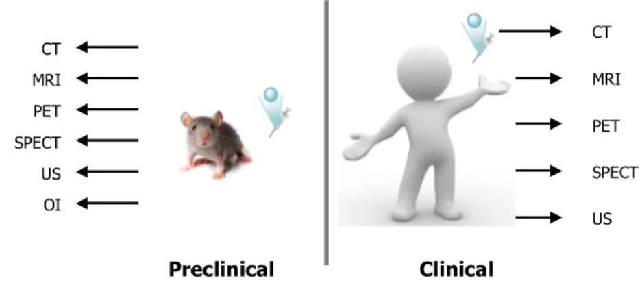
Clinical images are produced through a variety of processes relying on physical phenomena applied to human body, detecting tissues damages in strikingly different ways: submission of the body to photons of all energies (X-rays, gamma-rays, ultraviolet, visible, infrared, microwaves, radiofrequencies), weak electric and magnetic fields or ultrasound waves provides important anatomical and physiological information, differing substantially in their ability to penetrate in and interact with the body. Indeed, in the past three decades the use of non-invasive imaging techniques for disease diagnosis has been a tremendous growth, becoming an indispensable component in the clinical practice. Afterwards, imaging systems are in widespread clinical use: Computed Tomography (CT), Magnetic Resonance Imaging (MRI), Positron Emission Tomography (PET), Single Photon Emission Tomography (SPECT) and Ultrasound (US) are the most common imaging modalities used today. Only recently Optical Imaging (OI), witnessed significant advancements, showing an emerging clinical utility (Figure 1)1.
Figure 1: summary of modalities used for molecular imaging in preclinical/
clinical diagnosis. Clinical US CT MRI PET SPECT CT MRI PET SPECT US OI Preclinical
A crucial role in the technology for imaging was the advent of contrast agents (CAs): a contrast agent is a molecular media used to improve the spatial and temporal resolution of the common diagnostic technique, affording to higher-contrast images, with consequently advantages for the diagnosis of tissue diseases. For their relevant repercussion on diagnostic improvements, the research in the field of contrast agents (CAs) has received a marked impulse due to the increasing request of better agents to use for the detection of different diseases. In addition to this, recently CAs found a new alternative and well-promising application to the Molecular Imaging: to their initial role as emphasizer of the diagnostic, new potential uses were added by the introduction of targeting vectors, leading to therapy monitoring, drug discovery and development, with the aim to better-understand biochemical events at cellular and sub-cellular level.
Starting from this observations, a brief introduction to the classical imaging modalities, that are CT, MRI, PET, SPECT, US and OI, will be discussed in the next pages, referring to their relative applications, advantages, limitations and emerging technologies. After that, a well-detailed description of different classes of contrast agents for MRI and OI will be described as preface for the discussion of the results obtained during the PhD program, object of this defence.2
1. Overview of Classical Imaging Modalities
Imaging modalities can be broadly divided into primarily morphological/anatomical and primarily molecular imaging techniques.They are usually compared each other evaluating their use, but also the spatial and temporal resolution, the depth of penetration, the sensitivity, the safety profile and the costs of the technique (Table 1).
Figure 2: Pros and cons of the techniques used for molecular imaging
Each modality has particular characteristics, advantages and limitations, which are highlighted here. Most imaging modalities are used clinically and can be translated from animals to humans in the drug development process. The different imaging modalities should be generally considered complementary rather than competitive.
Primarily morphological and anatomical imaging technologies include CT, MRI and US: they are characterized by high spatial resolution but share the same limitation of detection diseases until tissue structural changes (for example, growth of a tumour). On the other hand, primary molecular imaging modalities, such as OI, PET, and SPECT (with fluorescent or radiotracer injected at nanomolar blood
SPECT Imaging Magnetic Resonance Imaging Ultrasound Imaging Advantages: Limitless depth of penetration Excellent sensitivity Quantitative data Clinical utility Advantages: Limitless depth of penetration
High spatial and temporal resolution Good quality Advantages: Limitless depth of penetration Excellent sensitivity Multiplexing capabilities Clinical utility Disadvantages: Poor sensitivity Primarly anatomical information Limited soft-tissue resolution Ionizing radiation Advantages: Relatively inexpensive Good temporal resolution Quantitative data No ionizing radiation Clinical utility Excellent sensitiviity Disadvantages: Relatively expensive Requires cyclotron/generator Ionizing radiation Limited spatial resolution No multiplexing Disadvantages: Relatively expensive Ionizing radiation Limited spatial resolution Lack of attenuation
correction Disadvantages:
Limited clinical translation Low depth penetration Disadvantages: Poor sensitivity (requires
large amount of imaging agent)
Relatively expensive Poor temporal resolution Disadvantages: Limitless depth of
penetration Primarily anatomical inf. Limited to imaging
soft-tissues only Advantages:
High-throughput screennig for target confirmation and compound optim. High sensitivity Advantages: Limitless depth of penetration High spatial resolution Quantitative data No ionizing radiation Optical Imaging
Im
agi
ng
PET Imaging CT Imagingdiseases (for example, before the tumour is large enough to cause structural changes). However, these modalities suffer from their poor spatial resolution with currently available technology (nuclear techniques) or from a low penetration depth (OI) (Figure 2).
A detailed description of the various imaging modalities is introduced in the next pages, taking account for key strengths and limitations of the single technique, introduced in the next table.
Combining the strengths of morphological/anatomical and molecular imaging modalities allows the detection of pathophysiological changes in an early disease phase at higher structural resolution (for example, PET-CT and PET-MRI technology). These technologies may change the current primary approach of diagnostic imaging into a more disease-oriented approach for both basic research and clinical applications3.
Table 1.Features of available and emerging imaging modalities
Modality Temporal
Resolution resolution Spatial Penetration Depth of Sensitivity Safety Profile Cost
CT Minutes
50-200 µm (preclinical) 0.5-1 mm
(clinical)
Limitless ND Ionizing radiation $$
MRI Minutes-hours 25-100 µm (preclinical) 0.5-1 mm (clinical) Limitless 10-3 to 10-5 M No ionizing radiation $$$ PET Second-minutes (preclinical) 1-2 mm
5-7 mm (clinical) Limitless 10 -11 to 10-12 M Ionizing radiation $$$ SPECT Minutes 1-2 mm (preclinical) 8-10 mm (clinical) Limitless 10 -10 to 10-11 M Ionizing radiation $$ US Second-minutes 0.01-0.1 mm for superficial applications 1-2 mm for deeper
mm-cm Excellent with microbubbles (10-12 M) Good safety profile $ OI Second-minutes 2-3 mm < 1 cm 10-9 to 10-12 M Good safety profile, but depends on fluorophore used and mass needed
CT is a technique that relies on differential level of X-ray attenuation by body tissues to produce images reflecting anatomy.
Unlike traditional X-ray examination, CT employs imaging by sections (tomography) resulting in a three-dimensional anatomic image of the subject being scanned. The X-ray source, linked to rotating detector arrays produces a “fanbeam” spanning the whole subject width. Usually, a large number of detectors are needed to obtain an adequate number of measurements and information: tissues that strongly absorb X-ray (e.g., bones) appear bright while other that absorb poorly (e.g., air) appear dark, creating a high contrast-image displaying detailed morphological information. Often, an iodinated-contrast agent will be used during CT imaging to improve spatial resolution and soft tissue contrast (Figure 3)4. In clinical, CT has obtained invaluable success in the identification and assignment of tumours, ischemia, brain injury, pulmonary embolism and a vast array of other conditions. Some advantages of CT include fast acquisition time, high spatial resolution (preclinical = 0.05 – 0.2 mm, clinical = 0.5-1.0 mm), cost effectiveness, availability, clinical utility and relative simplicity. One of the main limitations of CT is the high exposure to radiation, which often limits the number of scans that can be performed in the same patient in a given time frame. Addition to this, the low-quality of soft tissue contrast-image entails the use of non-specific iodinated-contrast agents that should be relatively toxic for patient.
A) Schematic illustration of CT: a suitable contrast agent is administered to the subject. The X-ray source produces a fanbeam, shining the entire subject. X-rays will be differentially attenuated depending on the type of tissues, obtaining a high-contrast image displaying detailed morphological information. B) Small animal CT axial images and 3D representation.
However, advances in X-ray detector sensitivity have been made in the last ten years affording to a significant dose reduction.
1.1 Magnetic Resonance Imaging (MRI)
MRI is a highly versatile imaging technique that uses a powerful magnet and a radiofrequency energy to visualize the internal structure and morphology of soft tissue in the body.
The tecnique is based on the ability of some nuclei to produce a small magnetic field able to interfer with an external magnetic field: this interference originates a detectable signal. In detail, nuclear particles (protons and neutrons) are in constant motion and spin about their axes, giving rise to angular momentum. Atoms with an equal number of protons and neutrons have a net angular momentum of zero, whereas atoms with an unequal number possess a specific spin angular momentum. Moreover, due to their mass, spin, and charge of protons , certain nuclei produce a small vectorial magnetic field, named magnetic moment. The ratio of angular momentum to magnetic moment is known as gyromagnetic ratio and it is unique for each magnetically active nucleus. During an MRI scan, a living subject is placed inside a magnet; when anatomic nuclei are placed in an external magnetic field, they behave like magnetic dipoles which assume either a parallel or anti-parallel alignment to the magnetic field. The MR signal is generated from the small net difference in the number of magnetic dipoles that align parallel versus those that align anti-parallel (named polarization). External magnetic field with 1.5- 3 T are usually employed. The MRI signal is proportional to
1) concentration of nuclei, 2) gyromagnetic ratio, 3) polarization.
magnetically active isotope of the element: all 1H nuclei are magnetically active and for this properties 1H is the most common nucleus used in clinical.
Anatomical images are usually performed using the hydrogen nuclei in tissue water, found at a concentration of ~80M in the body. Other nuclei such as 31P, 13C,
23Na, 19F and 17O can be used for imaging, usually achieved through magnetic
resonance spectroscopy (MRS).
A MRI scanner is composed of a set of embedded coils: one coil generating the main relatively homogeneous magnetic field, gradient coils that produce variations in the magnetic field in the X, Y, and Z directions that are used to localize the source of the MR signal, and a RF coil generating RF pulses at the Larmor frequency responsible for altering the alignment of the magnetic dipoles by the absorption process. After every RF pulse, the magnetic dipoles are “tipped” from equilibrium and they subsequently undergo into two forms of relaxation back towards equilibrium, spin-lattice (or longitudinal, T1) relaxation and spin-spin (or
trasverse, T2) relaxation. The contrast between different tissues in MR images is
generated from the different relaxation times of each tissue. MRI is of diagnostic value because it can be used to generate two or three-dimensional maps reflecting the spatial distribution of the values of T1 and T2 (Figure 4).
Proton relaxation times mainly depend on the degree of binding of the water molecules to nearby biomolecules and the water-biomolecule interactions are sensitive to the histologic characteristic of a tissue and its physiologic status. So a
T1 or T2-weighted MR image reveals information on both the anatomy and the state
of health of tissues: for instance, proton nuclei in fat and hydrocarbon-rich environments have relatively short relaxation times compared with those in aqueous environments.
iron oxide measuring 3-5 nm, coated with dextran, starch, polymer or citrate. On the other hand, T1 contrast agents usually consist of chelates of paramagnetic metal
ions, most commonly Gd(III), which exhibit a high magnetic moment due to its
seven unpaired electrons. The detection threshold of gadolinium chelates locates in the micromolar range, lower than iodinated contrast agents commonly used in CT, which is estimated to be in the range of hundreds of millimolar to molar concentrations. However, many molecular targets of interest are expressed in the low nanomolar range, and therefore the detection sensitivity of routinely used gadolinium chelates is inadequate for molecular MRI; to address these limitations, several new gadolinium constructs have been developed: examples include gadolinium-containing dendrimers5, micelles6, liposomes and high-density lipoproteins. These nanoparticles possess both longer intravascular half-lives and higher longitudinal relaxivities (r1)7.
MRI has a number of important advantages compared with other imaging modalities, including
1) the use of non-ionizing radiation, 2) unlimited depth of penetration,
3) high spatial resolution (clinical: ∼1 mm compared with 5-7 mm for PET, preclinical: micrometer, as opposed to millimeter resolution achievable via optical and radionuclide imaging),
4) unparalleled soft tissue contrast, superior to that attainable with CT, 5) excellent diagnostic potential.
One of the most important limitations of MRI is its extremely poor sensitivity that can lead to relatively long acquisition times and large amounts of imaging agents required to obtain an adequate signal.
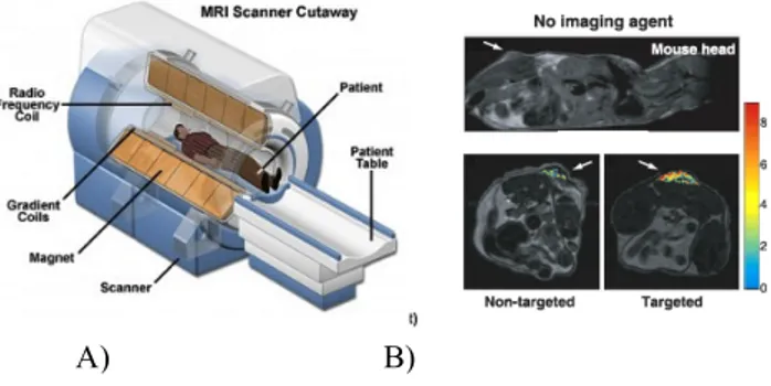
Figure 4: Magnetic resonance imaging (MRI)
A) Schematic illustration of MRI: a living subject is placed inside a magnet; unpaired nuclear spin within the body either align parallel or anti-parallel with the direction of the magnetic field. The MR signal is generated from the very small net difference in the number of parallel versus anti-parallel spins. B) MRI images, evaluating the presence and absence of contrast agents.
1.3 Positron Emission Tomography (PET)
PET is one of the first molecular imaging technologies, that permits the evaluation of biochemical changes and levels of molecular targets within a living subject. To image a certain molecular target using PET, identification and synthesis of a specific and selective radiolabeled imaging agent is needed in the beginning. The generation of short-lived radionuclides through a cyclotron , such as 18F (β+,
t1/2 = 109.8 min), 64Cu (β+, t1/2 = 12.7 h), 76Br (β+, t1/2 = 12.6 h), allows to take
advantage of the unique properties of radioactive isotopes to decay via positron emission, otherwise known as β+ decay. Indeed, nuclei that decay in this manner have an excess of protons, making them unstable: That instability is rectified by transforming a proton into a neutron, a positron and a neutrino.
A nanomolar amount of the chosen radiolabeled agent containing a positron emitting radioisotope is administered to the patient. One positron is emitted from each imaging agent, then they travel short distances and collide soon with
the production of two γ-rays, each with energy of 511 keV, travelling at opposite directions. The radioactivity is traced through the body and its distribution determined from scans obtained with a PET camera. PET detectors take the form of a closed ring, or set of rings, surrounding the subject to be imaged, designed to detect the annihilation events. The resulting electrical signals are converted into sinograms that reconstructes the tomographic image reflecting the distribution of the imaging agent in the subject and providing information on biochemical events
(Figure 5).
Isotopes of Zr, Y, In, Ga, and Cu have been investigated as radionuclide labels for biomolecules since they have the potential to combine their favourable decay characteristics with biological one of the targeting molecule to become useful radiopharmaceutical: 60Cu (β+, t1/2 = 0.4 h), 61Cu (β+, t1/2 = 3.3 h), 62Cu (β+, t1/2 =
0.16 h), 64Cu (β+, t1/2 = 12.7 h), 66Ga (β+, t1/2 = 9.5 h), 67Ga (β+, t1/2 = 78.3 h), and 68Ga (β+, t
1/2 = 67.71 min)find application in PET8.
Recently radioisotopes of gallium(III) find large interest in PET and SPECT technology: due to this potential application, the coordination chemistry of Ga(III) to several different polyaminocarboxylic ligands has been investigated leading to the development of new and improved chelating agents9.
Clinical PET is mainly used to image cancer through the use of 18F-labeled imaging agent10. In addition to its clinical utility, PET has a wide range of application in the basic research and preclinical studies. For example, PET can be used to investigate basic physiological and molecular mechanisms of human diseases through the use of appropriate radiolabeled imaging agents11.
Since biochemical changes generally occur before anatomical ones in disease, PET have a clear diagnostic advantage over classical CT and MRI. The key strengths of PET include excellent sensitivity (10-11-10-12M), requiring very small amounts of imaging agents (nanogram to milligram range) and limitless depth of penetration. The limited spatial resolution, the high-cost of the technique, the use of ionizing radiation and the need for a cyclotron are the key limitations of PET.
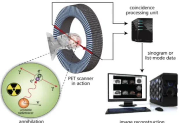
Figure 5: The basic principles of PET
Imaging agents containing a positron emitting radioisotope are administered to the patient. Positron are emitted from each imaging agent only once; these positrons travel short distances and collide with electrons in the surrounding tissue (annihilation) resulting in the production of two gamma rays, each with energy of 511 keV, travelling at the opposite directions to one another. Following the detection, the resulting electrical signal are converted into sinograms to reconstruct the tomographic image.
1.4 Single Photon Emission Computed Tomography
(SPECT)
SPECT is similar to PET, but the radioisotopes used for SPECT emit only a single high-energy (γ) photon: for this property, it requires the use of a contrast agent labelled with a γ-emitting radionuclide that is detected by a SPECT detector and elaborated giving an image of the radiotracer localization.
Generally, a SPECT contrast agent is administered to the subject and the gamma rays are detected via a rotating gamma camera; the detected γ−rays are then reconstructed into a tomographic image providing information on the location of the imaging agent in the subject (Figure 6).
67
Ga (γ, t1/2 = 78.3 h) and 111In (γ, t1/2 = 67.9 h) complexes find application as
According to the diversity of nuclides used in SPECT, a completely unique set-up is required to collect and reconstruct the data. Comparing to PET, the sensitivity of SPECT is several orders of magnitude lower than PET due to a different collimator used to detect the γ−rays, is less expensive and more widely available than PET, and it show a lower spatial resolution (8-10 mm, compared with 5-7 mm for PET). Like PET, SPECT has a clear diagnostic advantage over anatomical techniques as CT and MRI and it remains the most commonly used nuclear medicine modality in the clinic. Key disadvantage of SPECT are the lack of anatomical reference frame, and the safety profile: due to the use of ionizing radiation, each subject can be submitted to a limited number of scans per year. Unlike MRI and OI, both PET and SPECT only require small mass amounts of imaging agent (nanogram to milligram range).
Currently both PET and SPECT are being used either clinically or as research tool, to image a vast range of biological processes and disease states.
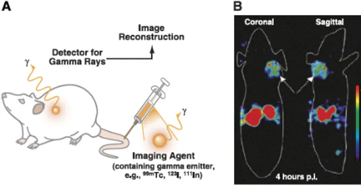
Figure 6: Single photon emission computed tomography (SPECT) principles
A) Schematic illustration of SPECT: an imaging agent contaning a
γ−emitting radioisotope is administered to the subject and gamma rays are detected via a gamma camera rotating around the subject. The detected signals re then reconstructed into a tomographic image. B) SPECT images, highlight detailed morphological/anatomical information.
Medical US is an imaging tool that exploits the properties and behavior of high-frequency sound waves as they travel through biological tissues, finding application in diagnostic imaging and therapeutic tool13.
During an US scan, a transducer sends sound waves and receives it back: in details, the transducer convert an electrical signal to US-waves, the US-waves enter the body, and some of them are reflected back to the transducer where they are detected and converted into electrical signals, subsequently processed by a computer and displayed as an image. Contrast agents, in the form of gas-filled microbubbles typically coated with lipids or biopolymers are used for enhancing the reflection signal-to-noise ratio for blood14 (Figure 7).
Resolution of US improves significantly with higher frequencies, but limiting the depth of penetration: this effect is due to the fact that high-frequencies sound waves possess shorter wavelengths, meaning they are more likely to interact with matter and slow down, thus reducing their travelling distance15.
The technique is cheap, it does not required the use of ionizing radiation and achieves excellent sensitivity with microbubbles and good temporal resolution. On the other hand, it shows limited depth of penetration, limited imaging applications (only for soft tissues) and it provides primarily anatomical information.
A) Schematic illustration of US: after administration of a targeted US imaging agent (microbubbles), high-frequency sound waves are transmitted into the subject. The sound wave reflections produced due to travelling through the subject are recorded, converted into electrical signals, analyzed affording to an image representative of the subject’s internal structure. B) US images demonstrating the significant advantage of using microbubbles contrast agents.
1.6 Optical Imaging (OI)
The visualization of cells and tissues using light has long been one of the most informative approach in basic research and medical diagnostic imaging. Optical techniques, unlike other imaging modalities, include multiple methods of imaging. The acquired images depends on the wavelength applied, the instrumental mode of detection, and whether or not intrinsic optical signatures or exogenously imaging agents are being explored as the constrast source. Generally, light interaction with tissues may involve the processes of absorption, photon scattering and the generation of fluorescence emission. All these phenomena have been exploited to differentiate tissue components. Depending on the light wavelength used, different penetration can be achieved.
In recent years, fluorescence microscopy and imaging have received particular attention. This is due to the increasing availability of fluorescent proteins, dyes and probes16 that enable the non-invasive study of a large number of cellular processes. On the other hand, there was an increasing list of fluorescent imaging techniques that offer microscopic resolutions, or methods that operate at resolution beyond the diffraction limit and offer single-molecule sensitivity, yielding unprecedented insights into biology.
Along the developments in live cells fluorescent microscopy, a number of non-invasive macroscopic optical imaging modalities have emerged: two examples are fluorescence and bioluminescence imaging.
Optical Fluorescence Imaging is associated with the administration of an engineered fluorescent imaging agent such as fluorophores, fluorescent proteins, and QDots17; after the administration of the fluorescent imaging agent, an appropriate excitation wavelength is used to illuminate the subject; this leads to excitation of the fluorophore and subsequent emission of light.
The light emitted is detected via a CCD camera, collected, analyzed and finally converted into a tomographic image detailing the location of emitted light from the subject.
important tools for mapping specific molecular events in mice and for tracking cells, e.g.: metastatic cells. These techniques are cheap, fast and require no radioisotopes. Structural and functional imaging with high resolution has become more and more important in the drug development process.
2. Gd-based-MRI Contrast Agents: State of
Art
Since the introduction of Magnetic Resonance Imaging (MRI) as diagnostic technique for the determination of diseases, the development of new contrast agents (CAs) has become of wide interest. Currently, about 35% of MRI scans make use of CAs and this percentage should further increase with the avaibility of more specific and sensitive agents. MRI-CAs are complexes of paramagnetic metal ions with cyclic and acyclic polyaminocarboxylic ligands, as the commercially available Gd-DTPA and Gd-DOTA (Table 2). The paramagnetic metal ions most extensively used in MRI are either transition metals or lanthanides: indeed, Gd(III) is preferred due to its high paramagnetism (7 unpaired electrons) and relaxation properties and it is the most investigated.18
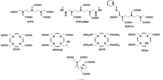
Figure 8: Common chelating agents for lanthanide(III) ions
Acyclic and cyclic chelating agents are reported: most of them are octadentate ligands, able to host only one water molecule in the inner-sphere. For this reason they generally show discrete relaxivity (4-5 mM-1s-1). On the other hand, DO3A and AAZTA
are heptadentate ligands with two water molecules in the inner sphere coordination. Some of Gd-L represented are clinically available (Gd-DTPA, Gd-DTPA-BMA, Gd-DOTA, Gd-HP-DO3A, Gd-BOPTA). N N N COOH COOH HOOC COOH COOH O BOPTA N N N COOH COOH HOOC COOH COOH DTPA N N N COOH COOH DTPA-BMA O HN NH O COOH N N N N HOOC HOOC COOH OH HPDO3A N N N N HOOC HOOC COOH COOH DOTA N N N N (HO)2OP (HO)2OP PO(OH)2 PO(OH)2 DOTP N N HN N HOOC HOOC COOH DO3A N N N COOH COOH COOH HOOC AAZTA
and cyclic polyaminocarboxylic chelating agents are used to wrap around the metal centre affording to very stable and inert complexes which should coordinate at least one molecule of water. In Figure 8 common cyclic and acyclic chelating agents are reported as example.
2.1. Properties of a good MRI-CAs
An efficient CAs for MRI is characterized by:
- suitable relaxometric properties (high relaxivity, r1);
- thermodynamic stability and kinetic inertness; - high water solubility;
- low toxicity.
2.1.1 Relaxometric Properties19. Signal intensity in Gd-based MRI is largely
related to the local value of the longitudinal relaxation rate of water protons, 1/T : indeed, signal tends to increase with increasing 1/T and
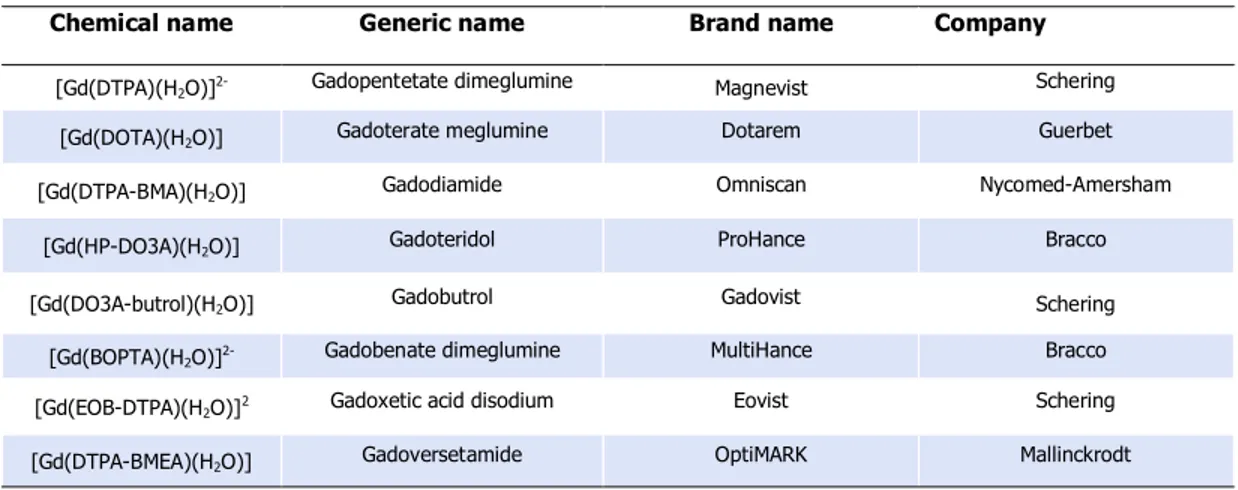
Table 2. Clinically relevant Gd-based-MRI-CAs
Chemical name Generic name Brand name Company
[Gd(DTPA)(H2O)]2- Gadopentetate dimeglumine Magnevist Schering [Gd(DOTA)(H2O)] Gadoterate meglumine Dotarem Guerbet [Gd(DTPA-BMA)(H2O)] Gadodiamide Omniscan Nycomed-Amersham
[Gd(HP-DO3A)(H2O)] Gadoteridol ProHance Bracco
[Gd(DO3A-butrol)(H2O)] Gadobutrol Gadovist Schering
[Gd(BOPTA)(H2O)]2- Gadobenate dimeglumine MultiHance Bracco [Gd(EOB-DTPA)(H2O)]2 Gadoxetic acid disodium Eovist Schering [Gd(DTPA-BMEA)(H2O)] Gadoversetamide OptiMARK Mallinckrodt
consequently, a pulse sequence that emphasize this parameter is referred as T1-weighted scan.
The longitudinal relaxation rate is matematically defined as the sum of diamagnetic and paramagnetic relaxation rate: the linear dependece of paramagnetic relaxation rate on the concentration of paramagnetic species permit to determine the relaxivity (r1) as mM-1s-1 from the
following equations:
(1/T1)obs = (1/T1)D + (1/T1)P (1)
(1/T1)P = r1*[Gd(III)] (2)
(1/T1)obs = (1/T1)D + r1*[Gd(III)] (3)
r1 = [(1/T1)obs - (1/T1)D]/ [Gd(III)] (4)
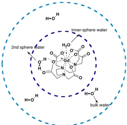
Relaxivity (r1) is defined as the enhancement in relaxation rate of water
protons of inner and outer sphere per mM of concentration at the field of 0.5 T and 298 K: the inner-sphere contribution is mainly related to the number of water molecule direcly coordinate to the metal centre (q) (Figure 9), the distance between the water proton and the unpaired electron spin (r), the lifetime of the solvent molecule in the complex (τm), the electronic relaxation time (τS), the molecular reorientational
time (τR) and the correlation time (τv).
Figure 9: water molecules of inner and outer sphere coordinated to a
Gd-complex N N N Gd O -O O O -O O -O O -H2O O O- 3+ inner-sphere water H OH 2nd sphere water H O H bulk water H OH H OH
(1/T1)P = (1/T1)inner-sphere + (1/T1)outer-sphere (5)
The second hydration sphere contribution, originating from water molecules interactions with the complex not directly bound to the metal centre, significantly partecipates to the relaxivity (5-15%) in complexes containing phosphonic or phosphinic ligands20.
The number of water molecule directly coordinated to the metal centre is closely related to the denticity of the chelating agents: octadentate ligands as DTPA and DOTA, employed as chelating agents for Gd, permit the inclusion of no more than one water molecule in the first coordination sphere (q = 1) affording to complexes with relatively low relaxivity; the decrease of denticity of the ligand corresponds to an increase of q and, consequently, marked relaxivity properties. On the other hand, the simple shift from octadentate to heptadentate ligand affects the thermodynamic stability, leading to toxic effects related to the release of free Gd(III) in the blood.
The distance between the water proton and the umpaired electron spin,
r, is a difficult parameter to measure and to control, but of relevant
importance: For example, because of the 1/r6 dependence, a decrease of about 0.2 Å would result in a 50% increase in relaxivity.
Rotation of the paramagnetic molecule is another critical variable, even if tunable: indeed, enancing the rigidity of the complex, relaxivity can be optimized; starting from these observations, supramolecular adducts of MRI-CAs with human serum albumin (HSA)21, low-density
lipoproteins and high-density lipoproteins (HDL), characterized by (1/T1) inner-sphere ∝q, 1/τm, 1/r6, 1/τr, 1/τv, and 1/τs
reduced mobility, were prepared, showing surprising and well-promising relaxivity values well higher than 20 mM-1s-1.22
b Relaxivity value determined at 40°C; c q = q’ correspond to the water molecules in the second-sphere; indeed for
DOTP q = 0.
2.1.2 Thermodynamic and kinetic properties. Thermodynamic stability and
kinetic inertness are important properties for a Gd-CAs because they are related to the release of free Gd(III) ion in in vivo applications through dissociation or transmetallation mechanism with Ca2+, Mg2+, Zn2+, Cu2+ presented in human blood causing toxicity at low doses (10-20 µmol/Kg). Indeed, recent reports outlined the development of pathologies related to the slow dissociation of Gd(III) complex from the complexes with acyclic ligands (nephrogenic systemic fibrosis / nephrogenic fibrosing dermopathy or NFS/NFD) in patients with renal failure23.
Stability constants, defined by Equations (6) and (7), are reported for different Gd-MRI-CAs in Table 4.
Gd + L = Gd-L (6) KGdL = [GdL]/[Gd][L] (7)
Table 3.Correlation time τv, molecular reorientational time τr, electronic relaxation τs, distance Gd-H
hydration number and relaxivity for a selection of Gd(III) complexes at 25°C
Ligand τv (ps) τr (ps) τs (ps) R (Å) q r1 DTPA 25 58 72 3.13 1 3.8 DTPA-BMA 25 66 81 3.13 1 4.39 DOTA 11 77 473 3.13 1 4.2 DO3A 14 66 129 3.15 2 4.8b HP-DO3A - - - - 1 3.7b DOTP 12.7 73 90.7 - 3c (ss) 3.5 AAZTA 31 74 - - 2 7.1 BOPTA 26 88 76 2.96 1 5.2
Stability constants for heptadentate ligands are generally some orders of magnitude lower than octadentate complexes due to the reduced denticity. Reduction of the ligand denticity in order to enhance q must take care of the possible detrimental effect on the stability.
2.1.3 Solubility and Toxicity. High solubility and low toxicity are not less
important parameters to be considered for a Gd-based MRI-CAs; high water solubility permits to prepare highly concentrated solutions of contrast agents to inject, while low toxicity is generally related to its thermodynamic stability and the kinetic inertness. Low molecular weight chelates are widely known to be among the safest drugs ever introduced due to their efficient excretion from the body. This minimizes the exposure to the drug and reduces the chance that slow uptake processes might internalize the agents in cells. As opposed to the free excreted small molecules, macromolecular conjugates tend to spend more times in the body and which, associated with the less complete
Table 4.Stability Constants for GdL
GdL Denticity LogKGdL () DTPA 8 22.46 DTPA-BMA 8 16.85 DOTA 8 25.3 DO3A 7 21.1 HP-DO3A 8 23.8 DOTP 8 28.8 AAZTA 7 20.24 BOPTA 8 22.59
elimination, increase the odds of cellular uptake, leading to the release of potentially toxic byproducts, including free gadolinium ions itself.
2.2 Classification of Chelating Agents
Chelating agents for Gd(III) ions could be classified into different classes: ionic/ non-ionic and acyclic/cyclic contrast agents; the second classification is of greater interest because it is closely related to the thermodynamic and kinetic properties (Figure 10).
MRI Chelating Agents
Acyclic N N N COOH COOH HOOC COOH COOH
Gd-DTPA (Magnevist) Gd-BOPTA
N N N COOtBu COOtBu BuOtOC COOtBu COOtBu HOOC n Bifunctional n = 0,1 Gd3+ N N N Gd O -O O O -O O -O O -H2O O O-3+ 2 Na+ O N N N COOH COOH C COOH COOH DTPA-DMPE O H N O P O O O O -H O O O Lipophilic Commercially available Cyclic N N N COOH COOH COOH HOOC N N N N HOOC HOOC COOH COOH DOTA N N N N HOOC HOOC COOH OH HPDO3A AAZTA Bifunctional N N N COOH COOH COOH HOOC R
R = OH, -OCO(CH2)2COOH
R = -(CH2)4COOH N N N HOOC HOOC COOH NH 2 Lipophilic N N N COOH COOH COOH HOOC C17H35 N N N N HOOC HOOC COOH DO3A-diph O N Lipo-AAZTA NOTA-BFCA Isoster Isoster N N N COOH COOH HOOC COOH PO3H2
Mixed Carboxylic - Phosphonic DTPA
N N N P P HOOC O OH O OH COOH
Mixed Carboxylic - Phosphinic AAZTA
Figure 10: Schematic classification of chelating agents for Gd(III) ions.
Common examples of each class of chelating agents are reported: they are divided into cyclic/acyclic; their ability to enhance the contrast effect can be modulate reducing the denticity, adding water molecule of inner or outer sphere (q), limitating the mobility, or substituting the coordinating group.
employed in clinical diagnostics with Gd-DTPA, also known as Magnevist. It is defined as linear or acyclic octadentate ligand which coordinates Gd(III) permitting the coordination of only one molecule of water in the inner coordination sphere. The linear structure entails low values of stability constant, while the high denticity is responsible of the relatively low values of relaxivity. Several octadentate ligands based to the DTPA scaffold were synthesized, substituting acetate moiety with different amides (DTPA-BMA) obtaining Gd-DTPA-bisamides complexes with relaxivity properties similar to DTPA.
2.2.2 Cyclic chelating agents: macrocyclic ligands as DOTA are preferable to
the DTPA-like open-chain ones, as the latter show lower thermodynamic stability and kinetic inertness. During the last decades, the research in the field of cyclic chelating agents for lanthanides were widely explored: in this framework DO3A and its derivates as HP-DO3A were investigated leading to interesting results and clinically employed derivatives. Recently a new cyclic heptadentate ligand with efficient chelating properties was synthesized, i.e.: AAZTA: comparing with DOTA and DTPA, an efficient size-match with lanthanide ions, the satisfactory thermodynamic and kinetic inertness and relaxometric properties of its Gd(III) complex make it a suitable choice as a cyclic chelating agent for Gd-based MRI-CAs24.
2.2.3 BFCAs: bifunctional chelating agents were introduced with the aim to
link the functionality of contrast agent to a targeting vector to accurately orient the diagnosis of tissue disease and therapy25: The most common
reactive functional group is added, better without altering the chelating properties of the coordination cage. Due to its easy formation and chemical stability, amide bond is one of the preferred choices for the preparation of coniugates containing metal complexes. Bifuctional DTPA-like ligands containing carboxylic, amino or phosphonic groups as functional groups were prepared and they have found several applications in the different diagnostic imaging techniques relying on contrast agents based on chelated metal ions.
2.2.3 Lipophilic Chelating Agents: the introduction of a lipophilic alkyl chain
on the skeleton of the common contrast agents supports the self-assembly into micelles that exhibit enhanced values of r1. In addition to
this, lipophilic CAs can further form supramolecular adducts with HSA, LDL and HDL with significative increments in the relaxivity due to the reduced and lower mobility of the systems: example of lipophylic MRI-CAs are Gd-DTPA-DMPE and Gd-AAZTA-C17 with potential applications for in vivo imaging of tumours and cardiovascular diseases. The importance of this aspect is highlighted if related to the current safety requirenments: indeed, by increasing the efficacy of Gd-chelated it may be possible to reduce the dose and minimize the potential toxicity associated with Gd probes exhibiting prolonged circulation times.
2.2.4 Phosphonic and Phosphinic Chelating Agents: as mentioned above, the
second hydration sphere, originating from interactions of the complex with water molecules not directly bound to the metal centre, can significantly contribute to the relaxivity particularly in complexes containing phosphonic or phosphinic ligands as P-DTPA, DOTP26:
indeed, the bulkier phosphonic group may hinder the access of water molecules in the inner sphere, while the ability to form strong H-bonds
with consequently increase in relaxation efficiency. Starting from that observations, substitution of carboxylic groups with phosphonic ones was extensively investigated: indeed, the tetrahedral deprotonated form –PO32- ion occurring at physiological pH values offers multiple
coordination modes, while the residual anionic charge after complexation provide a better solubility in water. Recently it was demonstrated that phosphonate-containing analogues of the commercially used Gd-DOTA and Gd-DTPA show faster water exchange than the original systems. Even though complete substitution of carboxylic with phosphonate groups affords to too much hindrance which leads to chelate systems unable to coordinate water molecules in the inner-sphere (q =0), partial substitution seem to guarantee better results: indeed, the hindrance of only one phosphonate group on the coordination cage usually does not cause a reduction of the number of molecules water bond with the metal centre. Alternative monophosphinic and monophosphonic complexes as Gd-DO3APBP and Gd-PC2AP with q≥ 1 were developed and they show higher relaxivity values27,28, respectively 7.4 mM-1s-1 and 8.3 mM-1s-1 (Figure 11).
Figure 11: examples of mono phosphonic/phosphinic chelating agents.
In the same context a mixed carboxylate-phosphinate AAZTA-like N N N COOH PO3H2 HOOC PCP2A N N N N COOH HOOC P HOOC O OH PO3H2 PO3H2 DO3APBP
6.1 mM-1s-1 (20 MHz, 298K), slightly lower than Gd-AAZTA (q = 2, r1
= 7.2 mM-1s-1), suggesting the presence of only one water molecule coordinated to the paramagnetic metal centre (q=1)29.
Conclusions
The relaxivity of a contrast agent is the measure of an MRI-CA efficacy to produce image-contrast. There is continuosly the desire to increase the longitudinal relaxivity of Gd-based contrast agents since the r1 values of currently available
commercial agents fall too short of what is theoretically possible. The octadentate coordination guarantees the formation of very stable complex but limited to the coordination of only one molecule of water in the inner sphere. Better results are obtained for octadentate cyclic chelating agents as Gd-DOTA and Gd-DO3A: indeed, they show r1 values of 4.3 and 4.6 s-1mM-1 respectively, while relaxivity of
Gd-DTPA is 3.8 s-1mM-1.
Reducing the denticity of the chelating agent interesting results were achieved with AAZTA, an heptadentate cyclic ligands which the reduced denticity is the explanation of relaxivity like 7.1 s-1mM-1, tipical value for MRI-CAs with q = 2. An alternative strategies to obtain this effect is the delivery of a high number of imaging reporters at the site of interest by conjugating several Gd-chelates to macromolecules or nanoparticles linked to a targenting vector, obtaining multimeric/dendrimeric MRI-CAs.
Finally, another parameter that could be modified to improve the longitudinal relaxivity of a contrast agent is the molecular reorientational time (τR) that is
related to mobility of the Gd-complex: indeed, the introduction of an alkyl chain limits the unrestrained movement of the contrast agent affording to surprising results, especially when they could form supramolecular adducts with HSA, LDL, and HDL.
during the last decade, affording to important results for clinical and also therapeutical applications; in spite of that, improvements are still needed to optimize the relaxometric properties and to reduce the toxicity risks.
3. Luminescent probes for Optical Imaging
The unique spectroscopic and magnetic properties of lanthanide ions allow the use of their complexes in a wide range of medical and biochemical applications. If the high magnetic moment and the slow electronic relaxation of gadolinium make it ideal for the design of magnetic resonance imaging relaxation agents, the long-lived luminescence of Eu(III) and Tb(III) complexes has been exploited in the development of sensor and time-resolved fluorescence imaging. The ligand design is crucial for the effective use of the lanthanide in such biochemical application and it has incited a large number of coordination chemistry studies.Optical Imaging is a high–resolution, sensitive and semi-quantitative technique of diagnosis developed in the last 30 years, contemporary with MRI. As MRI, Optical Imaging uses non-ionizing radiation and the signal or contrast could be modulated in response to biological events. The technique requires low concentration of contrast agent to produce high contrast images and cellular/subcellular resolution30. Lanthanide-based luminescent probes are particularly attractive for OI due to their long luminescent lifetime that offers a several advantage for the time-gated detection in biological samples.
3.1 Architecture of luminescent probes for OI
Lanthanides show intrinsic luminescence originating from f-f electron transitions in the 4f shell with lifetime in the milli to microsecond range. The 4f orbitals do not participate in chemical bonding, so the emission wavelengths are minimally perturbed by the surronding matrix and ligand field showing an emission spectra that preserves the same fingerprint of the corresponding free Ln(III) salts.
Despite that, lanthanides have low molar absorbivity requiring intense light sources to populate the excited states by direct excitation. Complexation with an appropriate chelate able to transfer energy to the metal centre would be a valid alternative to increase the population of excited state (Figure 12).
Figure 12: Fluorescence mechanism through energy trasfer activation.
Ln(III)-complex is initially at the groud state S0; excitation of
picolinic group with λexc = 273 nm affords to excited state S1;
Picolinic groups transfer energy to the metal centre that relax emitting fluorescence.
The architecture31 of emissive lanthanide complexes consists of a metal centre
surrounded by a chelate equipped with a sentitizer, named antenna. The chelate prevents the release of free Ln(III) into the biological system and protects the metal centre from the solvent coordination, avoiding the quenching and maximizing the quantum yield: chelates with high coordination numbers are requested for luminescent probes (n >8). In addition to this, also rigidity of the coordination cage can minimize the energy loss through X-H vibrations.
Europium and terbium complexes are the most favourable probes for Optical Imaging because they have high emission intensity in the visible region spectra, longer luminescent lifetime than most of the common organic fluorophores and low sensitivity to quenching by singlet oxygen and by vibrational transfer to X-H (X = C, N,O).
The antenna effect consists in the harvest of energy through high molar absorption to the singlet excited state. After undergoing ISC to the triplet state, the antenna transfers energy to the excited state of Ln(III) (5D
j) that relax to the ground state
(7Fj) emitting luminescence.
For a satisfactory antenna effect the following photophysical properties are required32 (Figure 13): N N N N N N COO -COO -COO -Ln3+ 1. hv, Excitation at 273nm 2. Energy transfer ligand-to-metal 3. Fluorescence
- excitation wavelength > 340 nm (but generally they are limited to excitations < 420nm);
- the ISC for S1 → T1 transition must be favoured over antenna fluorescence
and non radioactive relaxation S1 → S0;
Figure 13: Fluorescence mechanism in detail
The antenna harvest energy through high molar absorption to the singlet excited state. After undergoing intersystem crossing to the triplet state, the antenna transfers energy to the excite 5D
j state of
the lanthanide. The radiative transition of electrons from excited
5D
j to the 7Fj states results in luminescent emission from the
lanthanide ion.
Picolinic moyety was routinely introduced as antenna in the basic scaffold of typical chelating agents as DOTA and NOTA, promoting the energy transfer to the metal centre, affording to interesting luminescent probes for OI33,34.
3.2 Photophysical properties
The photophysical properties of lanthanides (sharp emission, long luminescence lifetime allowing to discriminate between the background fluorescence of
Linker Antenna Ln(III) hv' hv 2. Energy trasfer 1. Excitation 3. Emission S0 S1 T1 Excitation ISC Antenna Ln(III) 5D j 7F j Energy Transfer Linker Antenna Eu(III) 5D 1 7F Eu(III) 6 5 4 3 2 1 0 5 80 n m 59 0 n m 61 5 nm 65 0 nm 6 9 0 n m 7 10 n m Linker Antenna Tb(III) 5D 4 7F Tb(III) 0 1 2 3 4 5 6 49 0 n m 5 45 n m 5 90 n m 6 2 0 nm 6 50 n m
the application as luminescent labels in time-resolved imaging and in biomedical assays. The preparation of lanthanide complexes, that are stable and highly emissive in water environment, requires the design of polydentate ligands that contain suitable sensitizer of the lanthanide emission, capable to prevent nonradiative deactivation.
3.2.1 Absorption and emission spectra of Eu and Tb-complexes. The absorption
spectra of typical Eu- and Tb-chelates recorded in water generally show band with maxima in the UV region, assigned to π→π* and n→π* transitions centred on the (het)arene antenna moiety. The excitation spectra recorded upon metal-centred emission are very similar to the corresponding absorption spectra, indicating that the coordinated picolinate moiety provides an efficient energy transfer to the Eu(III) and Tb(III) ions. The corresponding emission spectra of ∼10-5M solutions of Eu- and Tb-complexes, obtained under excitation through the ligand bands usually display the 5D0→7Fj (Eu(III), j = 0-4) and 5D4→7Fj
(Tb(III), j = 6-0) transitions typical of Ln(III) ions. Specifically, Eu-complexes are characterized by five bands in the range of 570-730 nm with the main band attributed to the 5D0→7F2 transition appearing in the
600-630 nm range. Tb-complexes are characterized by seven bands in the range of 450-700 nm with the maximum around 540-550 nm attributed to the 5D4→7F5 transition (Figure 14).
Figure 14: Emission spectra of luminescent Eu- and Tb-complexes
The emission spectra of ∼10-5M solutions of Eu- (red) and Tb-complexes (green), embodying picolinate groups as coordinating/antenna residues. Excitation through the ligand band at 273 nm are reported: Eu-L is characterized by five bands in the range of 570-730 nm with the main band attributed to the 5D07F2
transition appearing in the 600-630 nm range with a maximum at 615 nm. Tb-L is characterized by seven bands in the range of 450-700 nm with the maximum at 545 nm attributed to the 5D47F5
transition.
3.2.2 Luminescent lifetime. Determination of luminescence lifetimes (τ) in
H2O and D2O allows an accurate estimation of the number of
coordinated water molecules present in solution (q) by using the equation of Beeby and co-workers35
q = A*[(1/τH2O)-(1/τD2O)-B]
For europium complex A= 1.2 ms and B = 0.25 ms-1, while for terbium complex A = 5 ms and B =0.06 ms-1 ;
For a luminescent probe q = 0 is desiderable because it strongly reduces the quenching effect due to the O-H vibrational oscillators.
3.2.3 Quantum yield. An efficient ligand-to-metal energy transfer is suggested
by the close matching of the excitation and emission spectra: indeed, quantum yield (φLn-L) is an indicative index related to the efficient
energy transfer and emission fluorescence in spite of radiationless deactivation mechanisms.
3.3 Chelating agents for OI
For biological imaging, lanthanides are typically chelated with multidentate ligands to attenuate the toxicity of free lanthanide ions. In luminescent applications, chelation has an additional role of protecting the metal center from solvent coordination. Indeed, coordination of water molecules shorten the excited-state lifetimes of the Ln(III) ions through nonradiative vibrational energy transfer to the high-frequency O-H oscillator. Thus, the number of water molecule directly coordinated to the matal centre, q, highly influences lanthanide emission. Chelate minimize q via coordinative saturation or steric hindrance, thereby attenuating solvent quenching and maximizing the φLn term.
For use as cellular probes, the chelate must form a complex that firmly saturates the Ln(III) coordination sphere over a wide pH range and resists hydrolysis. Given the hard-acid nature of lanthanide ions, hard bases, such as carboxylate ligands, amides and pyridines, are typically implemented as ligands. Macrocycles, such as NOTA and DOTA, and open nona-or octadentate chelates equipped with sterically bulky arms are commonly used for luminescent applications.
Increasing the rigidity of the chelate itself can further minimize energy loss through other X-H (X = C, N, O) vibrations of the chelate.
The majority of luminescent lanthanide probes exhibit the antenna covalently bound to the chelate through a linker or spacer, termed “pendant” antenna. It is
often derived from existing organic chromophores with well-defined fluorescence profiles that are appropriate for sensitization energy requirements. The photophysical properties of Eu(III) and Tb(III) sensitizers, such as azaxanthone, phenanthridine and tetracycline, have been extensively studied in the context of lanthanide probes for time-resolved luminescent bioassays and are being applied to cellular imaging applications (Figure 15).
A) B)
Figure 15: Pendant antennae used to sensitize lanthanide complexes.
A) Schematic of lanthanide chelates that utilize pendant antennae; the pendant antenna is attached to the chelate, as DOTA or DTPA, with a linker, and multiple pendant antennae can be incorporated. B) Examples of common sensitizers used as pendant antennae, deriving from organic fluorophores on the basis of favorable photophysiscal properties for sensitization.
Improving sensitization efficiency of pendant antennae has focused on increasing the energy gap T1(antenna) 5Dj(Ln) either decreasing linker length or using antennae
that can directly coordinate to the metal center. An alternative way consists in the increasing the number of pendant antennae attached to a single chelate: regarding that, high luminescent Eu(III) and Tb(III) probes incorporating pendant picolinic antennae into 1,4,7-triazacyclononane and cyclen derivate were synthesized: generally, the fully coordination of europium and terbium ions with nona-coordinating ligands as TPATCN and TPMPTCN affords to stable complex with q = 0, that means no molecules of water coordinated in the first coordination sphere of the metal in solutions: the lanthanide ion results fully saturated with the three endocyclic N-atoms, the three N-atoms of the pyridine and the three O-atoms of
N O O R N S O R H N O
azaxanthone azathiaxanthone acridone
N N N N N R R R OH O OH H OH OH CONH2 OH H OH NMe2
promoting these as well-promising luminescent probes.
The reduction of the ligand denticity affords to complexes with drastically reduced luminescent properties.
Figure 16: Picolinate ligands based on 1,4,7-triazacyclononane and cyclen with
interesting luminescent properties.
Table 5. Photophysical properties of such Ln(III)-L complex
Ligand λabs (nm)/ ε ( cm-1M-1) τH2O (ms) τD2O (ms) q φ (%) Eu(MPATCN) 273 nm, - 0.394 1.58 2 - Eu(BPATCN) 273 nm, 9050 cm-1M-1 0.542 1.67 1 5 Eu(TPATCN) 274 nm, 14400 cm-1M-1 1.08 1.47 0 9 Eu(TPMPTCN) 272 nm, 8400 cm-1M-1 1.56 1.60 0 7 Eu(BPDPA)+1 264 nm, - 0.66 1.70 0.7 1.3 Eu(DODPA)+1 272 nm, - 0.26 0.39 1.1 - Eu(Me-DODPA)+1 272 nm, - 0.91 1.27 0.0 - Tb(MPATCN) 273 nm, - 1.23 2.54 2 - Tb(BPATCN) 273 nm, 9100 cm-1M-1 1.49 2.46 1 43 Tb(TPATCN) 274 nm, 15800 cm-1M-1 2.00 2.14 0 60 Tb(TPMPTCN) 272 nm, 7500 cm-1M-1 2.59 2.98 0 60 Tb(BPDPA)+1 263 nm, - 1.16 1.53 0.7 16 Tb(DODPA)+1 272 nm 1.18 1.51 0.6 - Tb(Me-DODPA)+1 272 nm 2.39 2.68 0 - N N N COOH N COOH N N N N N N COOH COOH COOH N N N N N COOH COOH COOH N N N N N N P P OH O HO O P OH O N O N O N N COOH HOOC N N N N N N COOH HOOC COOH H3TPATCN N HN N NH N N COOH HOOC H2BP12C4 H2DODPA H2Me-DODPA N N N N N N HOOC COOH H2BPDPA H3BPATCN H3MPATCN H 3TPMPTCN
N N HN O NH O O O O HN HN O O NH O O HN N H O O O NH Tb3+ Tb(III)-IAM N N HN O NH O N N O O O O HN O N O O N H O N O O Eu3+ Et Et Et Et Eu(III)-HOPO
Comparing highly-coordinated chelates in Table 5, the lifetimes are longer than less coordinated complexes due to the absence of O-H oscillators in their coordination sphere.
Higher absorbing antennae with efficient ISC should be achieved with d-f bimetallic complexes where d-metal complexes can serve as sensitizing pendant antennae.
Finally, the use of chromophores that simultaneously serve as protective multidentate chelates, termed has chelating antennae, can sensitize Ln(III) luminescence: example of chelating agents are simple aromatic compounds such as pyridines and isophtalamides; Recently a chelating antennae based on 2-hydroxyisophtalamide (IAM) was used to sensitize Tb(III); initial evaluations of the sensitization efficiency of IAM binding units observed remarkably quantum yield leading to the development of octadentate ligands comprising a series of connected IAM units: while favourable properties of the free IAM unit, such as high molar absorptivity and quantum yield (ε = 26800 M-1cm-1; φ = 59%) were retained, the first generation of octadentate IAM derivatives was not water-soluble.
A) B)
Figure 17: Chelating antennae used to sesitize lanthanide complexes.
a) Schematic of a chelating antennae that serves a dual function as both the protective lanthanide and sensitizing antenna. B) Examples of recently reported lanthanide complexes with chelating antennae as IAM and 1,2-HOPO.
For this reasons, substitution of the methylamide with methoxyethyl was attempted to improve the solubility, retaining excellent photophysical properties. Addition to this, 1-hydroxypyridine-2-one (1,2-HOPO) derived ligands were tested for Eu(III)
sensitizing Tb(III) due to their energies being too close to the excited state of Tb(III), displaying lower quantum yield as consequence (Figure 17).
Conclusions
While stringent requirements on stability, solubility, and photophysical properties have limited advances of chelating agents/antenna systems in intracellular applications The examples highlighted above can be useful for the future design. Such properties are highly attractive for the remarkable sesitization efficiencies.
References
1. J.K. Willmann, N. Bruggen, L. M. Dinkelborg, A. A. Gambhir (2008) Nat.
Rev. Drug Disc. 7, 591-607.
2. M.L.James, S.S. Gambhir (2012) Physiol. Rev. 92, 897-965. 3. S. Lee, X. Chen (2009) Mol. Imaging 8(2), 87-100.
4. T.F. Massoud, S.S. Gambhir (2003), Genes & Dev. 17, 545-580. 5. G.Gugliotta, M.Botta, L.Tei (2010) Org. Biomol. Chem, 8, 4569-4574. 6. E. Gianolio, G.B. Giovenzana, A. Ciampa, D. Imperio, S. Aime (2008),
ChemMedChem 3, 60-62.
7. S. Crich, S. Lanzardo, D. Alberti, S. Belfiore, D. Ciampa, G.B. Giovenzana, C. Lovazzano, R. Pagliarin S. Aime (2007) Neoplasia 9(12), 1046-1056 8. T.J. Wadas, E.H. Wong, G.R. Weisman, C.J. Anderson (2010) Chem. Rev
110, 2858-2902.
9. B.P. Waldron, D. Parker, C. Burchardt, D. S. Yufit, M. Zimny, F. Roesch (2013) Chem. Commun. 49, 579-581.
10. W.Krause -Contrast Agent II: Optical, Ultrasound and Radiopharmaceutilca Imaging (2002)- Topics in Current Chemistry 222, p.291
11. M. F. Phelps (2000) J. Nucl. Med, 41, 661-681.
12. Z. Baranyai, F. Uggeri, A. Maiocchi, G.B. Giovenzana, C. Cavallotti, A. Takacs, I. Toth, J. Bànyai, A. Bényei, E. Brücher, S. Aime (2013) Eur. J.
Inorg. Chem, 147-162
13. R. Gessner, P.A. Dayton (2010) Mol. Imaging 9, 117-127. 14. M.F. Kircher, S.R. Willmann (2012) Radiology 263, 633-643. 15. A.B. Wolbarst, W.R. Hendee (2006) Radiology 238, 16-39.
16. D. Santra, D. Dutta, G.A. Walter, B.M. Moudyil (2005) Technology in
Sundaresan, A.M. Wu, S.S. Gambhir, S. Weiss (2005) Science 307(5079), 538-544K.Licha, C. Olbrich (2005) Adv. Drug Disc. Rev. 57, 1087-1108. 18. P. Caravan, J. J. Ellison, T.J. McMurry, R.B. Lauffer (1999) Chem. Rev. 99,
2293-2352.
19. L. M. Manus, R.C.. Strauch, A. H. Hung, A. L. Eckermann, T. J. Meade (2012), Anal. Chem. 84, 6278-6287.
20. G.A. Pereira, L. Balli, A. D. Sherry, J. A. Peters, C.F.G.C. Geraldes (2009) Helv. Chim Acta 92, 2532-2551.
21. S. Aime, D. Longo, I. Longo, I. Menegotto, E. Gianolio, G.B. Giovenzana (2007), Chem. Eur J. 13, 5785-5797.
22. S. Aime, K.C.Briley-Saebo, S. Geninatti-Crich, d. O. Cormode, A. Barazza, W. J. M. Mulder, W. Chen, E. A. Fisher, Z. A. Fayad, G.B. Giovenzana (2009), J.Phys. Chem. B 113, 6283-6289.
23. Z. Baranyai, F. Uggeri, G.B. Giovenzana, A. Bényei, E. Brucher, S. Aime (2009) Chem. Eur. J. 15, 1696-1705.
24. S. Aime, L. Calabi, C. Cavallotti, E. Gianolio, G.B. Giovenzana, P.Losi, A. Maiocchi, G. Palmisano, M. Sisti (2004), Inorg. Chem 43, 7588-7590. 25. Lattuada, L.; Barge, A.; Cravotto, G.; Giovenzana, G.B.; Tei (2010) L.
Chem. Soc. Rev. 40, 3019-3049.
26. Z. Kotkovà, G.A. Pereira, K. Djanasvli, J. Kotek, J. Rudorsky, P. Hermann, L. Vander Elst, R. N. Muller, C.F.G.C. Geraldes, I. Lukes, J.A. Peters (2009) Eur. J. Inorg. Chem, 119-136.
27. S. Aime, M. Botta, L.Frullano, S. Geninatti, G.B. Giovenzana, R. Pagliarin, G. Palmisano, F. Riccardi Sirtori, M. Sisti (2000) J. Med. Chem. 43, 4017-4024.
28. T. Vitha, V. Kubicek, J. Kotek, P. Hermann, L. Vander Elst, R.N. Muller, I. Lukes, J. Peters (2009) Dalton Trans, 3204-3214.
29. A. Ermelindo, G. Gambino, L. Tei (2013), Tetrahedron Letters 54, 6378-6380.