Università degli Studi del Piemonte Orientale
“Amedeo Avogadro”
Dipartimento di Scienze del Farmaco
Dottorato di Ricerca in Biotecnologie Farmaceutiche e Alimentari
XXVIII ciclo a.a. 2012-2015
BIOCHEMICAL AND STRUCTURAL
INVESTIGATION
ON HUMAN ALDH1A3
Andrea Moretti
Supervised by
Dott.ssa Silvia Garavaglia
Andrea Moretti was the recipient of a Ph.D. fellowship by the
Contents
Chapter 1
Introduction
Chapter 2
Outline of the thesis
Chapter 3
“Crystal structure and biochemical characterization of human
ALDH1A3 isoenzyme in complex with NAD
+and retinoic acid”
Chapter 4
“In silico virtual screening and development of high throughput
screening for identification of potential inhibitors of human
ALDH1A3”
Chapter 5
Conclusions
Introduction
Aldehyde dehydrogenases superfamily
Aldehydes dehydrogenases (ALDHs) are known to be NAD(P)+ dependent enzymes responsible for the oxidation of a wide spectrum of aldehydes into their corresponding carboxylic acids[1]. Phylogenetic studies suggested that ALDHs
derived from four ancestral genes that exist prior to the division between
Eubacteria and Eukaryotes about 2 billion years ago[2]. The evolutionary history of
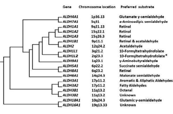
ALDH genes started with diversifications and duplications occurred from 890 million years ago to 70 million years ago[2]. The ALDH superfamily is represented across Archea, Eubacteria and Eukarya taxa with multiple genes, implying a critical role for these enzymes in the evolutionary history of many species[3]. The actual nomenclature of human ALDH superfamily was established during the Ninth International Symposium on Enzymology and Molecular Biology of Carbonyl Metabolism, held in 1998, in Varallo Sesia, Italy[1]. The ALDH superfamily comprises 19 functional protein-coding genes located on different chromosomes. ALDH isoforms belong to families from ALDH1 to ALDH9, ALDH16 and ALDH18 (Figure 1)[4], [5]. Human ALDHs are grouped in the same family by sharing more than 40% sequence identity, meanwhile they belong to the same subfamily when they exhibit more than 60% of amino acids identity[5]. The main biological role of ALDHs is regarded as being detoxifying enzymes, due to the ability to inactivate endogenous and exogenous toxic aldehydes. Nevertheless ALDHs are essential enzymes for the biosynthesis of key molecules in different metabolic pathways.
Figure1.Evolutionary tree for human aldehyde dehydrogenases with indication of chromosomal location and preferred substrate from Koppaka et al.[6].
ALDHs catalyze the conversion of exogenous and endogenous
aldehydes
Aldehydes are reactive electrophilic molecules with at least one hydrogen substituent on the carbonyl carbon at the end of the carbon chain. Aldehydes can be grouped in four large subclasses: short chain aldehydes, long chain alkanals, aromatic aldehydes and α,β-unsaturated aldehydes[7]. The anthropic activities such as combustion of hydrocarbon fuels, solvent utilization, air pollution, cigarette
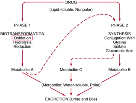
neurotransmitters catabolism, vitamins and steroids metabolism[8]. Cytotoxic endogenous and exogenous aldehydes are detoxified mainly through the oxidation catalyzed by ALDHs that prevents mutagenesis and cell death caused by DNA-adducts and cross-linking of proteins, consequence of aldehydes reactivity[9], [10]. Besides their key role as detoxifying enzymes, ALDHs are important catalysts in the xenobiotic metabolism (Figure 2). The phase 1 of drug metabolism consists in the biotransformation of the pharmaceutical compound that involves the oxidation of the drug functional group catalyzed by ALDHs.
Figure 2. Schematic representation of the xenobiotic metabolism of drugs in humans.
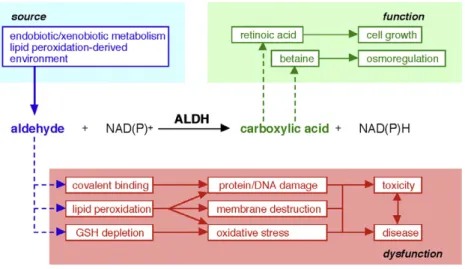
ALDHs are implicated both in the detoxification of cells and in important metabolic and signaling processes (Figure 3). Different ALDH isoforms catalyze the biosynthesis of physiologically relevant molecules involved in embryonic development (retinoic acid), neurotransmission (γ-aminobutyric acid) and osmosis (betaine)[4].
4
Figure 3. Schematic representation of ALDHs catalytic activity and their role in cell detoxification and metabolism, from Vasiliou et al.[5].
Representative isoforms of human ALDH superfamily and their
biological roles
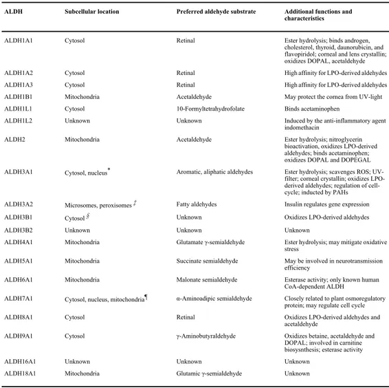
The 19 ALDH isoforms composing the ALDH superfamily are listed in Table 1 with a brief description of the main characteristics for each isoforms.
ALDH1A1, ALDH1A2 and ALDH1A3 are the three known isoforms of the 1A subfamily participating in the biosynthesis of retinoic acid (REA), a morphogen molecule that regulates embryonic development and cellular differentiation. By oxidizing retinaldehyde to retinoic acid the three ALDH1A isoforms are essential for the REA signaling pathway.
amino acid identity[4]. A database and website (www.aldh.org) maintained by the laboratory of Dr. Vasilis Vasiliou at the University of Colorado Denver is dedicated to providing a detailed, up-to-date resource for the ALDH gene superfamily[6].
The eukaryotic ALDH gene superfamily currently consists of 24 families that contain putatively functional genes with distinct chromosomal locations. It is anticipated that the number of ALDH families will expand after the inclusion of the bacterial ALDHs (Vasiliou et al., unpublished). Human genes are found in ALDH1– 9, ALDH16 and ALDH18 families, which also contain genes from other species including plant and fungi[7].
1.3. ALDH families
The ALDH1A subfamily comprises ALDH1A1, ALDH1A2 and ALDH1A3, all of which synthesize RA from retinaldehyde and, as such, are crucial in regulating RA signaling [4]. These isozymes have a high affinity for the oxidation of both all-trans- and 9-cis-retinal and exhibit a Kmfor these molecules in the low
micro-molar range[8]. Although ALDH2 officially qualifies as a member of the ALDH1 family, its longstanding name of ‘‘ALDH2’’ (associated with ethanol metabolism) has been grandfathered into the ALDH nomenclature system based on evolutionary divergence. ALDH2 is a mitochondrial enzyme that is primarily involved in the metab-olism of acetaldehyde generated during alcohol metabmetab-olism [1]. ALDH1B1, a mitochondrial enzyme very similar to ALDH2 (75% identical to ALDH2), also contributes to acetaldehyde oxidation, albeit with lower affinity than ALDH2[9]. ALDH1B1 is a potential colon cancer biomarker[10]. The ALDH1L1 gene codes for the cyto-solic 10-FTHF dehydrogenase (FDH), which converts 10-formyltet-rahydrofolate (10-FTHF) to tet10-formyltet-rahydrofolate [11]. The ALDH1L2 gene is very similar to ALDH1L1 and encodes the mitochondrial FDH that has enzymatic properties similar to its cytosolic counter-part. Unlike ALDH1L1, ALDH1L2 does not metabolize short-chain aldehyde substrates[12]. The ALDH3A subfamily contains the di-oxin-inducible ALDH3A1 and ALDH3A2 enzymes, both of which are involved in the oxidation of medium- and long-chain aliphatic and aromatic aldehydes[13,14]. The ALDH3B subfamily consists of
isozyme, also known as succinic semialdehyde (SSA) dehydroge-nase, catalyzes the NAD+-dependent conversion of SSA to succinate
in the last step GABA catabolism[17]. ALDH6A1 is a mitochondrial enzyme also known as acetyl CoA-dependent methylmalonate semialdehyde (MMS) dehydrogenase. ALDH6A1 is involved in valine and pyrimidine catabolism and catalyzes the oxidative decarboxylation of malonate semialdehyde and MMS to acetyl-CoA and propionyl-acetyl-CoA, respectively[18]. Human ALDH7A1 has a primary role in the pipecolic acid pathway of lysine catabolism, catalyzing the oxidation of alpha-aminoadipic semialdehyde (AASA) to alpha-aminoadipate[3,19]. ALDH8A1 is a cytosolic en-zyme that appears to metabolize retinaldehyde to retinoic acid
[20,21]. ALDH9A1 codes for an enzyme that participates in the
metabolism of c-aminobutyraldehyde and aminoaldehydes de-rived from polyamines [22]. The ALDH16A1 gene encodes an 802-amino acid protein with as-yet unknown function that most likely does not possess catalytic activity (Vasiliou et al., in this vol-ume). ALDH16A1 is a novel and rather unique member of the ALDH superfamily in that it contains two ALDH active site domains (as opposed to one in the other members of the superfamily), four transmembrane domains and a coiled-coil domain. Interestingly, the ALDH16 enzyme active site in frog and many invertebrates (e.g. sea squirt, sea anemone, sea urchin, lancelet, and Trichoplax adhaerens), as well as in bacteria, contains the catalytically-important cysteine residue (Cys-302); in contrast, this residue is absent from the mammalian and fish orthologous protein. The Vasiliou et al. chapter in this issue describes this ALDH as a protein interacting molecule[23].
The ALDH10 family contains plant genes encoding enzymes known as aminoaldehyde dehydrogenases (AMADHs), 4-aminobu-tyraldehyde dehydrogenases, 4-guanidinobu4-aminobu-tyraldehyde dehydro-genases, and betaine aldehyde dehydrogenases (BADHs). The ALDH11 family of genes codes for the cytosolic non-phosphorylat-ing glyceraldehyde 3-phosphate dehydrogenases (GAPNs). These enzymes catalyze the irreversible NADP+-dependent oxidation of
glyceraldehyde 3-phosphate (GAP) to 3-phosphoglycerate and NADPH [24]. The ALDH12 family contains mostly plant genes encoding D-1-pyrroline-5-carboxylate dehydrogenases (P5CDH),
Fig. 1. Illustration of ALDH catalytic activity.
Table 1. List of ALDH isoforms with brief description of localization, substrate preference and other functions,
from Marchitti et al.[4].
ALDH1A1 is a homotetramer cytosolic enzyme distributed in retina, eye lens, lung, kidney, liver and brain. ALDH1A1 represent 2-3% of cytosolic proteins in retina and it is important to protect the eye from UV - damage generated aldehydes[11]. ALDH1A1 is mainly involved in embryonic development and in the last years has been investigated for its role in cancer stem cell biology and chemotherapy resistance[11]. Hematopoietic progenitor cells expressing high levels of ALDH1A1 showed an increased tolerance to active metabolites of cyclophosphamide anticancer drug[12]. Moreover, breast cancer cells that
NIH-PA Author Manuscript
NIH-PA Author Manuscript
NIH-PA Author Manuscript
Marchitti et al. Page 36
Table 1
Human ALDH proteins.
ALDH Subcellular location Preferred aldehyde substrate Additional functions and
characteristics
ALDH1A1 Cytosol Retinal Ester hydrolysis; binds androgen,
cholesterol, thyroid, daunorubicin, and flavopiridol; corneal and lens crystallin; oxidizes DOPAL, acetaldehyde
ALDH1A2 Cytosol Retinal High affinity for LPO-derived aldehydes
ALDH1A3 Cytosol Retinal High affinity for LPO-derived aldehydes
ALDH1B1 Mitochondria Acetaldehyde May protect the cornea from UV-light
ALDH1L1 Cytosol 10-Formyltetrahydrofolate Binds acetaminophen
ALDH1L2 Unknown Unknown Induced by the anti-inflammatory agent
indomethacin
ALDH2 Mitochondria Acetaldehyde Ester hydrolysis; nitroglycerin
bioactivation, oxidizes LPO-derived aldehydes; binds acetaminophen; oxidizes DOPAL and DOPEGAL ALDH3A1 Cytosol, nucleus* Aromatic, aliphatic aldehydes Ester hydrolysis; scavenges ROS;
UV-filter; corneal crystallin; oxidizes LPO-derived aldehydes; regulation of cell-cycle; inducted by PAHs ALDH3A2 Microsomes, peroxisomes ‡ Fatty aldehydes Insulin regulates gene expression
ALDH3B1 Cytosol § Unknown Oxidizes LPO-derived aldehydes
ALDH3B2 Unknown Unknown Unknown
ALDH4A1 Mitochondria Glutamate γ-semialdehyde Ester hydrolysis; may mitigate oxidative stress
ALDH5A1 Mitochondria Succinate semialdehyde May be involved in neurotransmission
efficiency
ALDH6A1 Mitochondria Malonate semialdehyde Esterase activity; only known human
CoA-dependent ALDH
ALDH7A1 Cytosol, nucleus, mitochondria¶ α-Aminoadipic semialdehyde Closely related to plant osmoregulatory protein; may regulate cell cycle
ALDH8A1 Cytosol Retinal Oxidizes LPO-derived aldehydes and
acetaldehyde
ALDH9A1 Cytosol γ-Aminobutyraldehyde Oxidizes betaine, acetaldehyde and
DOPAL; involved in carnitine biosysnthesis; esterase activity
ALDH16A1 Unknown Unknown Unknown
ALDH18A1 Mitochondria Glutamic γ-semialdehyde Unknown
*
While predominantly cytosolic, ALDH3A1 is also found in the nucleus. ‡
A variant of ALDH3A2, FALDHv, is believed to be localized to the peroxisomes.
§
Marchitti SA, Vasiliou V. unpublished data. ¶
Brocker C, Cantore M, Pappa A, et al. unpublished data.
overexpress ALDH1A1 demonstrated an enhanced resistance to doxorubicin and paclitaxel[13]. ALDH1A2 is the second isoform involved in REA biosynthesis. It is expressed in various embryonic tissues as a cytosolic homotetramer and it regulates early heart development in mice[4]. Furthermore human spina bifida disease is significantly associated with ALDH1A2 single nucleotide polymorphisms[14]. ALDH1A3 is the third isoform implicated in REA signaling pathway and its role has been demonstrated crucial in nasal development and more general in embryonic development[15]. ALDH1A3 is expressed in fetal nasal mucosa, kidney, breast, stomach and salivary gland and it is involved in the development of eye, olfactory bulbs, hair follicles, forebrain and cerebral cortex[4]. Mutation in human
ALDH1A3 gene has been identified as the major cause of
microphtalmia/anophtalmia[16]. Recently the isoenzyme is assuming a central role
in the biology of different cancers in relation to its overexpression in ALDH- positive cancer stem cells.
ALDH1L1 is a cytosolic multi domain homotetramer important for the conversion of 10-formyltetrahydrofolate to tetrahydrofolate (THF). THF is an important metabolite in the dietary folate, in one-carbon metabolism and in the purine biosynthesis[17].
ALDH2 is a mitochondrial homotetramer responsible for the conversion of acetaldehyde to acetic acid. ALDH2 is the main enzyme of the ethanol metabolism and its well-known dominant allelic variant ALDH2*2 is the main cause of alcoholic liver disease and cirrhosis in Asian population [18], [19].
ALDH3A1 detoxifies aromatic and medium-chain aldehydes derived from lipid peroxidation[20]. ALDH3A2 is a microsomal homodimer that catalyzes the oxidation of fatty aldheydes and it is part of the fatty alcohol:NADoxidoreductase enzyme complex[21]. Deletions at the first five exon of the enzyme are correlated
ALDH4A1 participates in glutamate biosynthesis and proline degradation by promoting the conversion of pyrroline 5-carboxylate to glutamate[23]. Mutations in its coding gene are responsible of type II hyperprolinemia[24].
ALDH5A1 is a mitochondrial homotetramer, also known as succinic semialdehyde dehydrogenase (SSADH) that is implicated in the catabolism of γ-aminobutyric acid (GABA) by converting succinic semialdehyde to succinate[25]. The 4-hydroxybutyric aciduria, a neurological autosomal disorder, is a disease related to ALDH5A1 malfunction[26].
ALDH6A1 is involved in the valine and pyrimidine catabolism. The enzyme catalyzes the irreversible oxidative decarboxylation of malonate and methylmalonate semialdehydes to acetyl- and propionyl-CoA. Mutations to ALDH6A1 are responsible of dysmielination and methylmalonate aciduria[27].
ALDH7A1 is a homotetramer found both in cytosol and mitochondria. The enzyme participates to the lysine metabolism and mutations on ALDH7A1 gene cause pyridoxine dependent epilepsy[28], [29].
ALDH8A1 is the only isoform outside the ALDH1A subfamily involved in the retinoic acid metabolism. ALDH8A1 has the characteristic to prefer 9 cis-retinal substrate compared to all trans-retinal, showing a 40 times higher affinity[30].
ALDH9A1 is a key enzyme in the alternate metabolism of GABA. The cytosolic tetramer of ALDH9A1 oxidizes γ-aminobutyraldehyde to GABA[31]. ALDH16A1 is a recently discovered non-catalytic ALDH isoform. It has a preference for the NADP+ cofactor and seems to interact with maspardin, a protein responsible of spastic paraplegia[32]. ALDH18A1 is the second mitochondrial ALDH involved in the proline synthesis and it is a bifunctional enzyme that utilizes ATP and NADP for γ-glutamyl kinase and γ-glutamyl phosphate reductase activities, respectively[33].
Structural and functional aspects of ALDH
ALDHs are built on three functional domains: the NAD+ binding domain at the N-terminal, the catalytic domain and the oligomerization domain at the C-terminal (Figure 4)[34]. The global tertiary structure of ALDHs is conserved among isoforms across all families, and both the N-terminal and the C-terminal domains involve well-known and conserved amino acids in the binding of the cofactors and in the catalysis of the substrates, respectively[35].
Figure 4. Structure of sheep liver cytosolic ALDH1A1 in complex with NAD+[34]. The α-helix, the β-sheets and the connecting loop composing the N-domain, the C-domain and the oligomerization domain are respectively showed in red, blue and yellow.
ALDHs function as catalysts for the oxidation of aldehydes, although these enzymes are known to possess also an esterase activity. Beside the large differences in substrate selectivity, the ALDH superfamily shares the same catalytic mechanism. The only known exception is ALDH6A1 that uses CoA
ordered reaction in which the enzyme first binds the cofactor than allows aldehyde or ester to enter the catalytic site[37]. The nucleophilic attack on the substrate is operated by the active nucleophile C302 that is conserved among all the ALDHs (the numbering of amino acids is conventionally based on the mature human ALDH2). The thiol of the C302 is activated by a water-mediated proton abstraction operated by the highly conserved E268 that act as a general base[6]. The nucleophile C302 attacks the carbonyl carbon of the substrate driving through the formation of the thioemiacetal intermediate. The hydride ion transfer from the intermediate to the cofactor occurs on the C4 of the nicotinamide ring and it is stereospecific for the pro-R side[38]. After the hydride transfer, the E268 activates a second water molecule that is responsible of the hydrolysis of the thioester intermediate and the consequent release of the product. Simultaneously the reduced cofactor leaves the enzyme that is finally regenerated by the binding of a new NAD(P)+ molecule through the conserved residues E399 and K192[6]. Studies on ALDH three-dimensional structure and its catalytic mechanism has been reported in three pivotal works on ALDHs[34], [35], [39]. Liu and collaborators focused on the different way ALDH binds NAD(P) respect to the typical binding of other enzymes sharing the Rossmann fold. Usually the pyrophosphate moiety of NAD closely interacts with the fingerprint sequence GlyXGlyXXGly that is located in the loop between the β1-αA motifs of the βαβ-conserved Rossmann fold domain. Instead, in ALDH
the nucleotide cofactor binds between the β4-αD loop rather than in the canonical
β1-αA and the pyrophosphates does not interact strongly with any residue (Figure
6)[39]. In all the ALDH structures solved in complex with NAD, the cofactor lies in an hydrophobic pocket and it is more likely stabilized through hydrogen bonds between K192, E195, the main chain carbonyl oxygen of I166 and the adenine ring
Figure 5. Description of ALDH catalytic mechanism by Koppaka et al.[6] 1: The enzyme binds the NAD(P) cofactor and E268 activate the water molecule responsible of the thiol deprotonation. 2: Nucleophilic attack of C302 on the carbonyl carbon of the aldehyde 3: Thioemiacetlal intermediate formation and concomitant hydride transfer to the NAD(P)+ cofactor 4: Activation of a water molecule through E268 and hydrolysis of the thioester 5: Release of the carboxylic acid and reduced cofactor.
In the sheep liver ALDH1A1 structure, for the first time, was described the flexibility of the nicotinamide ring in complex with ALDH. The cofactor was found in two major conformations belonging to two different catalytic steps: the hydride transfer conformation, in which NAD+ is closer to the active site, ready to accept the hydride, and the release conformation where the reduced cofactor moved away from the catalytic site to make room for the product hydrolysis[34].
Figure 6.Comparison of NAD binding mode between ALDH3 and alcool dehydrogenase (ADH). The ALDH3 structure is reported as cartoon on the left side of the figure and it evidences how NAD binds between αD β4 and αC of the Rossmann fold. In the ADH structure, reported as cartoon on the right side of the figure, NAD exhibits the classical binding between αA and β1 of the Rossmann fold. From Liu et al., 1997[39].
ALDHs are known to process a wide spectrum of substrates, albeit each isoform displays a major affinity for its “preferred” or natural substrate. Moore et al. compared ALDH1 and ALDH2 structures to identify mechanisms involved in substrate selectivity and affinity[34]. The authors highlight the determinant role of the substrate access tunnel in the selectivity of ALDH1 towards bulky substrates such as retinal, whereas the bovine mitochondrial ALDH2 reveals a smaller and longer tunnel accessible to small substrates such as acetaldehyde[35]. The geometry
of the catalytic tunnel is not the only mechanism exploited by ALDHs for substrates selectivity. Structural and biochemical studies on ALDH1A1 and ALDH1A2 suggested that the interaction of bulky substrates with a disorder loop (~ 457 - 477) located at the entrance of the substrate tunnel promotes a disorder to order transition[40], [41]. Activity assays, direct mutagenesis, Fluorescence Resonance Energy Transfer (FRET) analysis and structural studies on both hALDH1A1 and rat ALDH1A2 confirmed that the conformational adaptation of the disordered loop is a key step for the substrate recognition and catalysis[40], [41]. The methods for substrate recognition are not universal across all isoforms and
many aspects about substrate selectivity in ALDHs still need to been elucidated. Recently, MA Keller et al. (2014) published the structure of the fatty aldehyde dehydrogenase (FALDH). Their work identified a new mechanism in which FALDH uses a unique gatekeeper α-helix, at the entrance of the substrate tunnel, to regulate the access of the substrates[42].
Retinoid metabolism and retinoic acid signaling
The etymology of retinol, retinal and retinoic acid derives from the term retina that refers to the organ where they were first identified[43]. Retinoids accomplish different roles in human metabolism. They are at the basis of the visual cycle, but at the meantime, they are crucial for embryonic development and tissues differentiation. The fat-soluble retinol, also known as vitamin-A, is up taken by dietary intake and it is the main source for retinoic acid (REA) biosynthesis. The conversion of retinol to all-trans retinoic acid goes through two reactions: the reversible conversion of retinol to all-trans retinal by alcohol dehydrogenase (ADH) or short-chain dehydrogenase (SDR), and the irreversible oxidation of
all-trans retinaldehyde to all-all-trans retinoic acid catalyzed by one of the three
ALDH1A isoforms (Figure 7)[44]. The inactivation of retinoic acid is the last important step of the metabolism catalyzed by Cyp26 (also referred as P450RAI) that hydroxylate REA causing its inactivation[45]. Retinoids play an essential role in visual cycle that occurs in cone photoreceptor cell outer segments (OS) and in the retinal pigment epithelium (RPE). Photo isomerization of the 11-cis-retinal chromophore of rhodopsin triggers a complex set of metabolic transformations collectively termed phototransduction that ultimately lead to light perception[46].
Chapter 1
Figure 7.Overview of metabolism and catabolism of retinoids with main enzymes involved in synthesis of retinoic acid and its inactivation[44].
The signaling role of retinoic acid have to be accomplished inside target cells, since that retinol is transported from plasma into target cells through the interaction between the retinol binding protein (RBP) and its receptor stimulated by retinoic acid 6 (STRA6)[47]. Inside the cells retinol can be segregated by cellular retinol
binding protein (CRBP) or can be available as substrate for ADH and SDR and enter the retinoid metabolism (Figure 7)[48]. The final product of the retinoid metabolism is retinoic acid, a well-known signaling molecule with pleiotropic functions, fundamental for the spatial-temporal development of the embryo and the tissue development in adults[49]. The morphogen REA is essential in somitogenesis and axial elongation, cardiogenesis and neurogenesis. Therefore, unbalance in REA biosynthesis is cause of severe diseases at embryonic stage and in adult.
nature genetics • volume 31 • may 2002 7
Using mice as flies is a mouse geneticist’s dream. With an ever-increasing number of mouse knockout strains available, the dream is fast becoming a reality. We can now carry out experiments to uncover genetic interactions that until recently were almost exclusively identified in inverte-brates such as Drosophila melanogaster and
Caenorhabditis elegans. An example of such
an experiment is presented in the accompa-nying paper1by Karen Niederreither and
colleagues. These authors have identified genetic interactions between two enzymes involved in retinoid metabolism by crossing mouse knockout strains carrying targeted mutations in each. Their results deepen our understanding of retinoid biology. Retinoid metabolism
Retinoids are derived from dietary vita-min A. Their main function is in cell sig-naling, in which they bind two classes of retinoid receptors, RARs and RXRs, that belong to the family of nuclear hormone receptors. These receptors are ligand-reg-ulated transcription factors with essential roles in embryonic development and adult physiology. The biological impor-tance of retinoids has long been known, as major developmental abnormalities fol-low retinoid deprivation or exposure to excess retinoids. Some of these abnormal-ities resemble congenital birth defects that are relatively common in humans, includ-ing spina bifida and cleft palate.
How are retinoids synthesized and catabolized? Vitamin A (retinol) is con-verted to retinaldehyde and then to
all-trans retinoic acid by way of two
oxidation steps (see figure). All-trans retinoic acid is present in embryonic and adult tissues at high levels, binds effi-ciently to RARs, and is a major biologi-cally active retinoid in vivo2. Some of the
remaining uncertainties stem from the existence of additional retinoid metabo-lites. For example, 4-hydroxy-retinoic acid and 4-oxo-retinoic acid (see figure) have been suggested to be inactive break-down products of all-trans retinoic acid.
Other results, however, have raised the possibility that these retinoids have important biological functions3,4.
Although the picture remains incom-plete, many of the critical enzymes involved in retinoid metabolism have been identi-fied. Oxidation of retinol to retinaldehyde requires the activities of several alcohol dehydrogenases (see figure)5. The second
step—oxidation of retinaldehyde to
all-trans retinoic acid—requires the action of
three related retinaldehyde dehydrogenases and is generally believed to be the rate-lim-iting step in the biosynthesis of all-trans retinoic acid. This conclusion is supported by several observations. For example, there is a striking correlation between embryonic expression of the aldehyde dehydrogenase
Retinoid metabolism: a balancing act
Thomas Perlmann
Ludwig Institute for Cancer Research, Karolinska Institute, S-171 77 Stockholm, Sweden. e-mail: [email protected]
Published online: 15 April 2002, DOI: 10.1038/ng877
The recent characterization of the aldehyde dehydrogenase Aldh1a2 and the cytochrome P450 Cyp26, two enzymes involved in retinoid metabolism, has helped to explain how bioactive retinoids are made and catabolized. By the elegant definition of an
Aldh1a2 null mutation as a dominant suppressor of a Cyp26 null mutation, it is now unequivocally demonstrated that the main
function of Cyp26 is to degrade endogenous all-trans retinoic acid rather than to synthesize bioactive hydroxylated retinoids.
BOB CRIMI
CH2OH
retinol (vitamin A)
all-trans retinaldehyde
all-trans retinoic acid
alcohol dehydrogenases (ADH) short-chain dehydrogenases (SDR) Aldh1a1 Aldh1a2 Aldh1a3 Cyp26 OH
4-hydroxy–retinoic acid 4-oxo–retinoic acid
O COOH
COOH CHO
COOH
Retinoid roundup. The biosynthetic pathway for generating all-trans retinoic acid involves two oxidation
steps, the first leading to the generation of all-trans retinaldehyde. The rate-limiting step in production of all-trans retinoic acid in the embryo is mediated by three related aldehyde dehydrogenases, Aldh1a1, Aldha2 and Aldha3. All-trans retinoic acid is converted to hydroxylated metabolites by Cyp26 (refs 7,8). These hydroxylated derivatives are metabolized to other polar retinoid metabolites by additional enzy-matic pathways. Niederreither et al.1show that the impaired retinoid balance resulting from a Cyp26 null
mutation is rescued on an Aldh1a2 heterozygous background. This finding indicates that the main, if not only, function of Cyp26 is to protect tissues from excess all-trans retinoic acid.
©2 0 0 2 N a tu re P u b li s h in g G ro u p h tt p :/ /g e n e ti c s .n a tu re .c o m
In late 1980s were identified for the first time the Retinoic acids Receptors (RARs) that are nuclear transcription factors inducible by retinoic acid binding[50], [51]. RARs are member of the nuclear hormone receptor superfamily and are composed by three subtypes RARα, RARβ and RARγ. RARs function as heterodimers with one member of the retinoid X receptors that are in turn composed by three subtypes RXRα, RXRβ and RXRγ. RAR-RXR heterodimers exert their pleiotropic effect of retinoic acid-dependent transcriptional regulators by binding to the specific Retinoic Acid Response Element (RARE) DNA sequences found in the promoter region of retinoid target genes[52]. The repression and activation of target response genes mechanism for RXR-RAR heterodimer is illustrated in Figure 8. The RXRs are able to bind 9-cis retinal and other ligands termed retinoxids, but 9-cis retinal cannot be detected endogenously in embryos or adult tissues. It is thus likely that RAR-RXR dimers act mainly, if not solely, by ligand binding of the RAR moiety, while RXR alone is not able to determine the activation of the target gene. This theory is supported by the fact that neither 9-cis retinal or other RXR ligands can rescue embryonic disease related to REA defeat[53]. Once identified the molecular basis of REA signaling, it is still tricky to elucidate all the mechanisms that are relevant to the REA mediated response. If on the one hand only about 20 genes have unambiguously RARE elements, on the other hand hundreds genes are involved in REA signaling or through direct interaction or through cross talk and signal cascades activation. Reflecting the complexity of REAresponse, the work of Liu et al. reported a total of 169 genes, including transcriptional factors, signal transduction modulators, protein modulators and cell cycle promoters and inhibitors, to be REA-modulated and form a harmonious network in the course of the REA-induced NB4 cell differentiation[54].
Figure 8. a) RXR-RAR heterodimers transcription factors bind to Retinoic Acid Response Element target
genes. b) In absence of retinoic acid (REA) the system is repressed by transcriptional co-repressor (SMRT or NcoR) and by the histone deacetylase (HDACs). The binding of REA induces the release of the HDCA and the recruitment of Histone Acetyltransferase (HAT) co-activator complex that acetylates the histones for the de-repression. Activation of the target gene is consequence of the transcription started by the RNA polymerase II holoenzyme, together with the TATA-binding protein (TBP), the TBP-associated factors (TAFs) and mediator complexes (MEDs). From Clarke et al.[55].
The embryonic development and cell fate determination of organisms are orchestrated by REA signaling, since that unbalance or absence of REA in embryos is cause of defects in the growth of central nervous system (CNS), craniofacial region, limb, urogenital system, lungs and heart. REA balance is determinant also in post-embryonic development and regeneration of lung, hair, ear and CNS[49]. Although is known that REA-activated pathway plays important, evolutionarily and conserved roles in neurogenesis, many of the detailed molecular interactions required for the neural progenitors proliferation and differentiation into neurons remain obscure[56].
Retinoic acid was tested as therapeutic compound on brain cancer cell lines such as neuroblastoma and glioma. The thinking behind the use of retinoic acid against brain tumors is based on the observation that an undifferentiated cancer cell population is cause of a more aggressive cancer and poor prognosis. Since differentiation of cancer cells population is a goal in cancer treatment, the signaling role of REA in tissue differentiation should help to threat cancer. Even thought the ability of REA to differentiate neuroblastoma cell lines have been demonstrated, the clinical response to REA treatment is too variable[56]. REA-induced alterations, targeting undifferentiated glioblastoma cancer cells, lead to a differentiation of the cancer cells population, rendering them sensitive to targeted therapy. However retinoid signaling is even more complex in clinical gliomas, so that resistance and side effects were common[56],[57]. Hypotheses on inefficiency of REA treatment
against brain tumors involves the possibility that REA is channeled to a pro-proliferative, oncogenic pathway depending on the relative abundance of the REA[56].
ALDHs in Cancer Stem Cells (CSCs)
First evidence of a cancer cell population with stem-like properties was reported by Bonnet and Dick in 1997 with their studies on acute myeloid leukemia[58]. In the early 2000s the Cancer Stem Cells (CSCs) theory was extended to breast cancer and glioma, two of the most studied tumors in relation to CSCs behavior[59], [60]. After almost 20 years of research, the scientific community agrees on the existence of a subpopulation of multipotent cancer cells with the ability to initiate the tumor. CSCs own the typical self-renewal/differentiation capacity of stem cells and it is quite likely that they are the engine of tumorigenisis in many cancers, not only in
17
which CSCs can be originated (Figure 9). The first scenario explains how the cancer stem cell that generates the primary tumor can derives from aberrant mutations of a normal stem cell or progenitor cell. The second scenario describes a situation in which refractory cancer stem cells, able to form a relapsed tumor, are consequence of chemotherapy treatment. The last scenario describes a tumor cell escape of a metastatic cancer stem cell that originates metastasis.
Figure 9. Three scenarios describing the origin of Cancer Stem Cells (CSCs) and their involvement in
tumorigenesis, drug resistance and tumor propagation A) CSC generates from mutations to normal stem cell or progenitor cell. The generated CSC gave rise to the primary tumor. B) Chemotherapy treatment of the primary tumor cause a refractory cancer stem cell able to generates a relapsed tumor. C) A tumor cell escape from the primary cancer generates a metastatic cancer stem cell that originates metastasis[61].
Although the existence of a CSCs subpopulation has been proof and accepted, their identification is still controversial. Over the recognition through associated markers, CSC detection needs to be confirmed through a “gold standard” test that consists in the validation of CSCs ability to start a new tumor ex vivo or after xenotransplantation in animal models[60]. Up to date the relation between specific
cancer markers and CSCs presence is still argument of debate. In Table 2 are listed the majority of the markers that have been somehow correlated to CSCs. Diverse
T h e ne w e ngl a nd jou r na l o f m e dicine
n engl j med 355;12 www.nejm.org september 21, 2006 1256
Unique molecular features of leukemia stem cells may provide opportunities for therapeutic intervention. For example, there is evidence of constitutive activation of both the nuclear factor-κB (NF-factor-κB) and phosphatidylinositol3′ (PI3) ki-nase signaling pathways in AML stem cells.28,44
Neither NF-κB nor PI3 kinase activity is detect-able in resting, normal hematopoietic stem cells, so both of these molecular factors could be tumor-specific targets. Two studies with different meth-ods of pharmacological inhibition of NF-κB have reported specific eradication of AML stem cells in vitro, without apparent harm to normal hema-topoietic stem cells.45,46 A separate study
demon-strated that inhibition of PI3 kinase reduced the growth of AML stem cells.44 Similarly, inhibition
of the downstream PI3-kinase mammalian target of rapamycin (mTOR) appears to enhance the activity of the chemotherapeutic agent etoposide
against AML stem cells.47 Inhibition of mTOR
also blocks the growth of leukemia-initiating cells in a mouse model of AML.48 Taken together,
these findings indicate that leukemia stem-cell– specific therapies may be attainable.
Cancer Stem Cells in the Central Nervous System
Isolation of cancer stem cells of the central ner-vous system (CNS) has been achieved by means of antigenic markers and by exploiting in vitro culture conditions developed for normal neural stem cells. As was first observed in 1992,49,50 CNS
cells grown on nonadherent surfaces give rise to balls of cells (neurospheres) that have the capac-ity for self-renewal and can generate all of the principal cell types of the brain (i.e., neurons, astrocytes, and oligodendrocytes). Neurospheres in which the stem-cell compartment is maintained
Normal stem cell or progenitor cell A New cancer stem cell Refractory cancer stem cell Metastatic cancer stem cell Primary tumor Primary tumor Primary tumor Primary tumor Primary tumor Primary tumor Primary tumor Primary tumor Primary tumor Metastases Metastases Metastases Relapsed tumor Relapsed tumor Relapsed tumor Mutations Chemotherapy Tumor cell escape B C
Figure 3. Scenarios Involving Cancer Stem Cells.
For tumors in which cancer stem cells play a role, at least three scenarios are possible. First, mutation of a normal stem cell or progenitor cell may create a cancer stem cell, which will then generate a primary tumor (Panel A). Sec-ond, during treatment with chemotherapy, the majority of cells in a primary tumor may be destroyed, but if the can-cer stem cells are not eradicated, the tumor may regrow and cause a relapse (Panel B). Third, cancan-cer stem cells aris-ing from a primary tumor may emigrate to distal sites and create metastatic lesions (Panel C).
markers were scrutinized only for cell lines and does not have evidence in other cancer cell lines or in vivo[62]. The discovery of more specific and reliable markers among with the development of standard protocol for the evaluation of CSCs presence is a desirable goal for cancer research.
Table 2. List of molecular markers for CSC adapted from[62]. This list is not exhaustive and includes markers not exhaustively tested.
Breast Colon Glioma Liver Lung
ALDH1 CD24 CD44 CD90 CD133 Hedgehog-Gli activity α6-integrin ABCB5 ALDH1 β-catenin CD24 CD26 CD29 CD44 CD133 CD166 LGR5 CD15 CD90 CD133 α6-integrin nestin CD13 CD24 CD44 CD90 CD133 ABCG2 ALDH1 CD20 CD133 CD271
Melanoma Ovarian Pancreatic Prostate
ABCG2 ALDH1 CD20 CD133 CD271 CD24 CD44 CD117 CD133 ABCG2 ALDH1 CD24 CD44 CD133 c-Met CXCR4 Nestin Nodal - Activin ALDH1 CD44 CD133 CD166 α2-β1-integrin α6-integrin
ALDHs as cancer stem cell markers
ALDHs are good candidates for the identification of CSCs and has been used as marker for many solid tumors including breast, brain, lung, liver, colon, pancreatic, ovarian, head and neck, prostate and melanoma[5]. ALDHs are a multipurpose tool in CSCs studies because they couple the detection of the CSC subpopulation with the selection of ALDH-positive cells through Fluorescence Activated Cell Sorting (FACS) with ALDEFLUOR™ assay. This technique is suitable for FACS that uses BODIPY aminoacetaldehyde (BAAA) as modified fluorescent substrate for ALDH. Cells with high ALDH activity convert BAAA in the cytoplasm into the negatively charged BODIPY aminoacetate (BAA-), which is retained into the cells that appear as a distinct cohort of cells exhibiting green fluorescence[63]. High
aldehyde dehydrogenase activity in breast, myeloid leukemia, prostate, rectal, oesophageal, lung, ovarian and gallbladder cancers is predictive of worst outcome, therefore ALDHs are being scrutiny as a potential prognostic markers[64]. The identification and correlation of specific ALDH isoforms to the presence of CSC subpopulation and a poor cancer prognosis has been reviewed from Marcato et al.
[65] and Pors et al.[66] (Table 3). Initially most of the studies reported ALDH1A1 as
the signature marker for CSCs in many cancers, but its correlation with CSCs was overestimated because ALDEFLUOR™ does not specifically detect only ALDH1A1 activity. The capability of ALDH to process different aldehydes doesn’t guarantee the isoform specificity for the ALDEFLUOR™, therefore detection of specific isoforms in CSC is under review and it need to be supported with different experiments to investigate the specific ALDH isoform biochemical profile.
Table 3. List of ALDH isoforms detected in various cancers and their correlation with cancer prognosis.
Adapted from Rodriguez-Torres et al.[64]. IHC is immunohistochemistry, HNSCC is Head and Neck Squamous cell carcinoma.
Tumor Type Method of ALDH detection
ALDH isoform Clinical observation
Breast Cancer ALDEFLUOR, IHC,
Immunoblotting, qPCR
ALDH1 ALDH1A1 ALDH1A3
Poor clinical outcome, tumor recurrence, poor response to chemoterapy
Ovarian Cancer ALDEFLUOR, IHC,
Immunoblotting, qPCR
ALDH1 ALDH1A1
Poor clinical outcome
Brain cancer ALDEFLUOR, IHC,
qPCR, DNA methylation
ALDH1 ALDH1A1 ALDH1A3
Increased metastases
Bone cancer ALDEFLUOR ALDH Poor clinical outcome
Prostate Cancer ALDEFLUOR, IHC ALDH7A1
ALDH3A1
Poor clinical outcome
HNSCC ALDEFLUOR, IHC ALDH1A1 Poor clinical outcome
Colorectal cancer ALDEFLUOR, IHC ALDH1
ALDH1A1
Loss of ALDH expression
Lung cancer ALDEFLUOR, IHC
immunoblotting
ALDH1A1 ALDH7A1
ALDH1A1 poor clinical
outcome, recurrence
Cervical cancer ALDEFLUOR ALDH Not assessed
Melanoma ALDEFLUOR,
Immunoblotting
ALDH1A1 ALDH1A3
Not assessed
Endometrial cancer ALDEFLUOR, IHC ALDH1
ALDH1A1
Not assessed
Renal cancer IHC, Immunoblotting ALDH1
ALDH1A1
Poor clinical outcome
Pancreatic cancer pPCR, Chromatin,
Immunoprecipitation
ALDH1A1 ALDH1A3
Not assessed
Heptobiliary cancer ALDEFLUOR, IHC ALDH1A3
ALDH3A1
Poor clinical outcome
Oesophageal cancer ALDEFLUOR, IHC ALDH1
ALDH1A1
Focusing on specific ALDH isoforms that concerned cancer research, ALDH1A3 is gaining more importance in CSC biology and detection. Marcato et al. in 2011 found that the overexpression of ALDH1A3, and not ALDH1A1 was predictive of metastasis in breast cancer stem cells[67]. In 2014 Shao et al. demonstrated the importance of ALDH1A3 for the maintenance of non-small lung cancer stem cells[68], whereas Mao et al. in 2013 found overexpression of ALDH1A3 in glioblastoma stem cells[69].
ALDH1A3 in glioma
The World Health Organization (WHO) classified gliomas in four histological grades by increasing degrees of undifferentiation, anaplasia, and aggressiveness[70].
Glioblastoma or High Grade Gliomas (HGGs) are classified as WHO grade III and IV and account 82% of gliomas[71]. Glioblastoma are highly invasive, infiltrative
and recurrent brain tumors with a 5 years survival < 5%[71], [72]. The presence of a
cancer stem cell population in glioma and in its more aggressive declinations has been proof, thereafter glioma stem cells (GSCs) are on the apex in a cellular tumorigenic hierarchy of HGGs[73], [74]. Microarray gene expression data, immunohistochemistry and biomarkers studies propose the existence of two glioma stem cell populations: the proneural glioma stem cells (PN GSCs) and the mesenchymal glioma stem cells (Mes GSCs)[69]. In the mesenchymal phenotype of HGG it has been demonstrated that the concurrent activation of two conflicting transcriptional regulators for neurogenesis and gliogenesis operates to permanently drive the aberrant mesenchymal phenotype in the context of the genetic and epigenetic changes that accompany high-grade gliomagenesis[75].
Mao et al. found high expression of ALDH1A3 isoform in GSCs. The expression of hALDH1A3 was detected by whole transcriptome microarray analysis and was found to be about 1000 times more in Mes GSCs compared to other cancer stem cells subpopulation (Figure 10). In vivo studies and xenograft experiments demonstrated an enhanced tumorigenicity of HGG derived from Mes GSCs.
Figure 10. ALDH1A3 is a functional Mes GSC marker. (A) qRT-PCR analysis of ALDH1A3 expression in PN
and Mes GSCs (**P < 0.01). (B) FACS analysis using Aldeflour™. ALDH activities in PN GSCs (n = 3), Mes GSCs (n = 3), and non-GSCs (n = 3) derived from Mes GSCs (**P < 0.01)[69].
The high activity (detected by ALDEFLUOR™) and the overexpression (detected by mRNA microarray) of ALDH1A3 in Mes GSCs are related with the high tumorigenicity, aggressiveness and worst prognosis of Mes GSC derived HGG. This research work lays the foundation for the validation of ALDH1A3 protein as interesting target in glioma.
ALDHs and cancer stem cell therapy resistance
Chemotherapy and radio resistance in tumors are the major difficulties that cancer research is facing and represent one of the most challenging barrier in cancer treatment. The resistance to treatments can be inherent or acquired by cancer cells, and mechanism such as DNA repair, drug efflux, drug inactivation and other adaptive responses are the known defenses of cancer cells against therapies[76] (Figure 11).
Figure 11. Overview on cancer drug resistance. Pharmacokinetics (PK) and Pharmodynamics (PD) are the two
critical mechanisms where the drug can be limited on its effect in cancer treatment by drug resistance mechanism of the cell or absorption, distribution, metabolism and elimination factors. From Holohan et al.[76].
The plethora of mechanisms that tumor cells use to contrast cancer treatments includes CSCs that are often mentioned as one of the main actors in therapy resistance. Radio and chemo resistance of CSCs are in turn a consequence of a multiplicity of defense mechanisms such as DNA damage response, ABC transporters, slow cycling rate and high ALDH expression[12]. First evidence of cytotoxic drug resistance, due to ALDH high activity, was observed for cyclophosphamide treatment[12], in which ALDH1A1 has been observed to oxidize pivotal aldehyde intermediates of pro-drug cyclophosphamide and its analogues[77]. Other drugs such as doxorubicin, paclitaxel and A459-Taxol used in breast and lung cancer chemotherapy might be associated with high ALDH expression. It is still to be understood case by case if ALDHs are the cause of drug resistance or biomarkers of cancer cells that gain drug resistance[12]. The different situations in which ALDHs are involved in CSC biology can lead to different strategies: the development of selective inhibitors or the use of ALDH isoforms as biomarker and prognosis predictors. The structure determination of the ALDH isoforms, notably
in complex with its ligand and the structured-based design of small molecules are indispensable tools to investigate the ALDH biology itself or in relation to CSCs.
Bibliography
1 Vasiliou, V., Bairoch, A., Tipton, K. F. and Nebert, D. W. (1999) Eukaryotic aldehyde dehydrogenase (ALDH) genes: human polymorphisms, and recommended nomenclature based on divergent evolution and chromosomal mapping. Pharmacogenetics 9, 421–434.
2 Yoshida, A., Rzhetsky, A., Hsu, L. C. and Chang, C. (1998) Human aldehyde dehydrogenase gene family. Eur. J. Biochem. FEBS 251, 549–557.
3 Jackson, B., Brocker, C., Thompson, D. C., Black, W., Vasiliou, K., Nebert, D. W. and Vasiliou, V. (2011) Update on the aldehyde dehydrogenase gene (ALDH) superfamily. Hum. Genomics 5, 283–303.
4 Marchitti, S. A., Brocker, C., Stagos, D. and Vasiliou, V. (2008) Non-P450 aldehyde oxidizing enzymes: the aldehyde dehydrogenase superfamily.
5 Vasiliou, V., Thompson, D. C., Smith, C., Fujita, M. and Chen, Y. (2013) Aldehyde dehydrogenases: From eye crystallins to metabolic disease and cancer stem cells. Chem. Biol. Interact. 202, 2–10.
6 Koppaka, V., Thompson, D. C., Chen, Y., Ellermann, M., Nicolaou, K. C., Juvonen, R. O., Petersen, D., Deitrich, R. A., Hurley, T. D. and Vasiliou, V. (2012) Aldehyde dehydrogenase inhibitors: a comprehensive review of the pharmacology, mechanism of action, substrate specificity, and clinical application. Pharmacol. Rev. 64, 520–539.
7 LoPachin, R. M. and Gavin, T. (2014) Molecular mechanisms of aldehyde toxicity: a chemical perspective. Chem. Res. Toxicol. 27, 1081–1091.
8 Vasiliou, V., Pappa, A. and Petersen, D. R. (2000) Role of aldehyde dehydrogenases in endogenous and xenobiotic metabolism. Chem. Biol. Interact. 129, 1–19.
9 Brooks, P. J. and Theruvathu, J. A. (2005) DNA adducts from acetaldehyde: implications for alcohol-related carcinogenesis. Alcohol Fayettev. N 35, 187– 193.
10 Burcham, P. C., Fontaine, F. R., Kaminskas, L. M., Petersen, D. R. and Pyke, S. M. (2004) Protein adduct-trapping by hydrazinophthalazine drugs: mechanisms of cytoprotection against acrolein-mediated toxicity. Mol.
12 Januchowski, R., Wojtowicz, K. and Zabel, M. (2013) The role of aldehyde dehydrogenase (ALDH) in cancer drug resistance. Biomed. Pharmacother. Bioméd. Pharmacothérapie 67, 669–680.
13 Croker, A. K. and Allan, A. L. (2012) Inhibition of aldehyde dehydrogenase (ALDH) activity reduces chemotherapy and radiation resistance of stem-like ALDHhiCD44+ human breast cancer cells. Breast Cancer Res. Treat. 133, 75–
87.
14 Deak, K. L., Dickerson, M. E., Linney, E., Enterline, D. S., George, T. M., Melvin, E. C., Graham, F. L., Siegel, D. G., Hammock, P., Mehltretter, L., et al. (2005) Analysis of ALDH1A2, CYP26A1, CYP26B1, CRABP1, and CRABP2 in human neural tube defects suggests a possible association with alleles in ALDH1A2. Birt. Defects Res. A. Clin. Mol. Teratol. 73, 868–875. 15 Zhang, X., Zhang, Q.-Y., Liu, D., Su, T., Weng, Y., Ling, G., Chen, Y., Gu, J.,
Schilling, B. and Ding, X. (2005) Expression of cytochrome p450 and other biotransformation genes in fetal and adult human nasal mucosa. Drug Metab. Dispos. Biol. Fate Chem. 33, 1423–1428.
16 Abouzeid, H., Favez, T., Schmid, A., Agosti, C., Youssef, M., Marzouk, I., El Shakankiry, N., Bayoumi, N., Munier, F. L. and Schorderet, D. F. (2014)
Mutations in ALDH1A3 Represent a Frequent Cause of
Microphthalmia/Anophthalmia in Consanguineous Families. Hum. Mutat. 35, 949–953.
17 Tsybovsky, Y., Donato, H., Krupenko, N. I., Davies, C. and Krupenko, S. A. (2007) Crystal structures of the carboxyl terminal domain of rat 10-formyltetrahydrofolate dehydrogenase: implications for the catalytic mechanism of aldehyde dehydrogenases. Biochemistry (Mosc.) 46, 2917–2929. 18 Xiao, Q., Weiner, H. and Crabb, D. W. (1996) The mutation in the mitochondrial aldehyde dehydrogenase (ALDH2) gene responsible for alcohol-induced flushing increases turnover of the enzyme tetramers in a dominant fashion. J. Clin. Invest. 98, 2027–2032.
19 Nagata, N., Hiyoshi, M., Shiozawa, H., Shiraishi, K., Watanabe, N., Tsuda, M. and Matsuzaki, S. (2002) Assessment of a difference in ALDH2 heterozygotes and alcoholic liver injury. Alcohol. Clin. Exp. Res. 26, 11S–14S.
20 Black, W., Chen, Y., Matsumoto, A., Thompson, D. C., Lassen, N., Pappa, A. and Vasiliou, V. (2012) Molecular mechanisms of ALDH3A1-mediated cellular protection against 4-hydroxy-2-nonenal. Free Radic. Biol. Med. 52, 1937–1944.
21 Rizzo, W. B. (2014) Fatty aldehyde and fatty alcohol metabolism: review and importance for epidermal structure and function. Biochim. Biophys. Acta 1841, 377–389.
22 Gaboon, N. E. A., Jelani, M., Almramhi, M. M., Mohamoud, H. S. A. and Al-Aama, J. Y. (2015) Case of Sjögren-Larsson syndrome with a large deletion in the ALDH3A2 gene confirmed by single nucleotide polymorphism array analysis. J. Dermatol. 42, 706–709.
23 Hu, C. A., Lin, W. W. and Valle, D. (1996) Cloning, characterization, and expression of cDNAs encoding human delta 1-pyrroline-5-carboxylate dehydrogenase. J. Biol. Chem. 271, 9795–9800.
24 Geraghty, M. T., Vaughn, D., Nicholson, A. J., Lin, W. W., Jimenez-Sanchez, G., Obie, C., Flynn, M. P., Valle, D. and Hu, C. A. (1998) Mutations in the Delta1-pyrroline 5-carboxylate dehydrogenase gene cause type II hyperprolinemia. Hum. Mol. Genet. 7, 1411–1415.
25 Tillakaratne, N. J., Medina-Kauwe, L. and Gibson, K. M. (1995) gamma-Aminobutyric acid (GABA) metabolism in mammalian neural and nonneural tissues. Comp. Biochem. Physiol. A Physiol. 112, 247–263.
26 Pearl, P. L., Parviz, M., Vogel, K., Schreiber, J., Theodore, W. H. and Gibson, K. M. (2014) Inherited disorders of gamma-aminobutyric acid metabolism and advances in ALDH5A1 mutation identification. Dev. Med. Child Neurol. 27 Marcadier, J. L., Smith, A. M., Pohl, D., Schwartzentruber, J., Al-Dirbashi, O.
Y., FORGE Canada Consortium, Majewski, J., Ferdinandusse, S., Wanders, R. J. A., Bulman, D. E., et al. (2013) Mutations in ALDH6A1 encoding
methylmalonate semialdehyde dehydrogenase are associated with
dysmyelination and transient methylmalonic aciduria. Orphanet J. Rare Dis. 8, 98.
28 Chang, Y. F., Ghosh, P. and Rao, V. V. (1990) L-pipecolic acid metabolism in human liver: L-alpha-aminoadipate delta-semialdehyde oxidoreductase. Biochim. Biophys. Acta 1038, 300–305.
29 Yang, Z., Yang, X., Wu, Y., Wang, J., Zhang, Y., Xiong, H., Jiang, Y. and Qin, J. (2014) Clinical diagnosis, treatment, and ALDH7A1 mutations in pyridoxine-dependent epilepsy in three Chinese infants. PloS One 9, e92803. 30 Lin, M. and Napoli, J. L. (2000) cDNA cloning and expression of a human
aldehyde dehydrogenase (ALDH) active with 9-cis-retinal and identification of a rat ortholog, ALDH12. J. Biol. Chem. 275, 40106–40112.
31 Kikonyogo, A. and Pietruszko, R. (1996) Aldehyde dehydrogenase from adult human brain that dehydrogenates gamma-aminobutyraldehyde: purification, characterization, cloning and distribution. Biochem. J. 316 ( Pt 1), 317–324. 32 Hanna, M. C. and Blackstone, C. (2009) Interaction of the SPG21 protein
ACP33/maspardin with the aldehyde dehydrogenase ALDH16A1. Neurogenetics 10, 217–228.
33 Hu, C. -a. A., Khalil, S., Zhaorigetu, S., Liu, Z., Tyler, M., Wan, G. and Valle, D. (2008) Human Delta1-pyrroline-5-carboxylate synthase: function and regulation. Amino Acids 35, 665–672.
34 Moore, S. A., Baker, H. M., Blythe, T. J., Kitson, K. E., Kitson, T. M. and Baker, E. N. (1998) Sheep liver cytosolic aldehyde dehydrogenase: the
35 Steinmetz, C. G., Xie, P., Weiner, H. and Hurley, T. D. (1997) Structure of mitochondrial aldehyde dehydrogenase: the genetic component of ethanol aversion. Struct. Lond. Engl. 1993 5, 701–711.
36 Kedishvili, N. Y., Popov, K. M., Rougraff, P. M., Zhao, Y., Crabb, D. W. and Harris, R. A. (1992) CoA-dependent methylmalonate-semialdehyde dehydrogenase, a unique member of the aldehyde dehydrogenase superfamily. cDNA cloning, evolutionary relationships, and tissue distribution. J. Biol. Chem. 267, 19724–19729.
37 Feldman, R. I. and Weiner, H. (1972) Horse liver aldehyde dehydrogenase. II. Kinetics and mechanistic implications of the dehydrogenase and esterase activity. J. Biol. Chem. 247, 267–272.
38 Jones, K. H., Lindahl, R., Baker, D. C. and Timkovich, R. (1987) Hydride transfer stereospecificity of rat liver aldehyde dehydrogenases. J. Biol. Chem. 262, 10911–10913.
39 Liu, Z. J., Sun, Y. J., Rose, J., Chung, Y. J., Hsiao, C. D., Chang, W. R., Kuo, I., Perozich, J., Lindahl, R., Hempel, J., et al. (1997) The first structure of an aldehyde dehydrogenase reveals novel interactions between NAD and the Rossmann fold. Nat. Struct. Biol. 4, 317–326.
40 Bordelon, T., Montegudo, S. K., Pakhomova, S., Oldham, M. L. and Newcomer, M. E. (2004) A disorder to order transition accompanies catalysis in retinaldehyde dehydrogenase type II. J. Biol. Chem. 279, 43085–43091. 41 Bchini, R., Vasiliou, V., Branlant, G., Talfournier, F. and Rahuel-Clermont, S.
(2013) Retinoic acid biosynthesis catalyzed by retinal dehydrogenases relies on a rate-limiting conformational transition associated with substrate recognition. Chem. Biol. Interact. 202, 78–84.
42 Keller, M. A., Zander, U., Fuchs, J. E., Kreutz, C., Watschinger, K., Mueller, T., Golderer, G., Liedl, K. R., Ralser, M., Kräutler, B., et al. (2014) A gatekeeper helix determines the substrate specificity of Sjögren-Larsson Syndrome enzyme fatty aldehyde dehydrogenase. Nat. Commun. 5, 4439. 43 Wald, G. (1968) The molecular basis of visual excitation. Nature 219, 800–
807.
44 Perlmann, T. (2002) Retinoid metabolism: a balancing act. Nat. Genet. 31, 7–8. 45 White, J. A., Ramshaw, H., Taimi, M., Stangle, W., Zhang, A., Everingham, S., Creighton, S., Tam, S. P., Jones, G. and Petkovich, M. (2000) Identification of the human cytochrome P450, P450RAI-2, which is predominantly expressed in the adult cerebellum and is responsible for all-trans-retinoic acid metabolism. Proc. Natl. Acad. Sci. U. S. A. 97, 6403–6408.
46 Kiser, P. D., Golczak, M., Maeda, A. and Palczewski, K. (2012) Key enzymes of the retinoid (visual) cycle in vertebrate retina. Biochim. Biophys. Acta 1821, 137–151.
47 Das, B. C., Thapa, P., Karki, R., Das, S., Mahapatra, S., Liu, T.-C., Torregroza, I., Wallace, D. P., Kambhampati, S., Van Veldhuizen, P., et al. (2014) Retinoic
acid signaling pathways in development and diseases. Bioorg. Med. Chem. 22, 673–683.
48 Napoli, J. L. (1996) Retinoic acid biosynthesis and metabolism. FASEB J. Off. Publ. Fed. Am. Soc. Exp. Biol. 10, 993–1001.
49 Maden, M. (2000) The role of retinoic acid in embryonic and post-embryonic development. Proc. Nutr. Soc. 59, 65–73.
50 Petkovich, M., Brand, N. J., Krust, A. and Chambon, P. (1987) A human retinoic acid receptor which belongs to the family of nuclear receptors. Nature 330, 444–450.
51 Giguere, V., Ong, E. S., Segui, P. and Evans, R. M. (1987) Identification of a receptor for the morphogen retinoic acid. Nature 330, 624–629.
52 Germain, P., Chambon, P., Eichele, G., Evans, R. M., Lazar, M. A., Leid, M., De Lera, A. R., Lotan, R., Mangelsdorf, D. J. and Gronemeyer, H. (2006) International Union of Pharmacology. LX. Retinoic acid receptors. Pharmacol. Rev. 58, 712–725.
53 Niederreither, K. and Dollé, P. (2008) Retinoic acid in development: towards an integrated view. Nat. Rev. Genet. 9, 541–553.
54 Liu, T. X., Zhang, J. W., Tao, J., Zhang, R. B., Zhang, Q. H., Zhao, C. J., Tong, J. H., Lanotte, M., Waxman, S., Chen, S. J., et al. (2000) Gene expression networks underlying retinoic acid-induced differentiation of acute promyelocytic leukemia cells. Blood 96, 1496–1504.
55 Clarke, N., Germain, P., Altucci, L. and Gronemeyer, H. (2004) Retinoids: potential in cancer prevention and therapy. Expert Rev. Mol. Med. 6.
56 Janesick, A., Wu, S. C. and Blumberg, B. (2015) Retinoic acid signaling and neuronal differentiation. Cell. Mol. Life Sci. CMLS 72, 1559–1576.
57 Karsy, M., Albert, L., Tobias, M. E., Murali, R. and Jhanwar-Uniyal, M. (2010) All-trans retinoic acid modulates cancer stem cells of glioblastoma multiforme in an MAPK-dependent manner. Anticancer Res. 30, 4915–4920.
58 Bonnet, D. and Dick, J. E. (1997) Human acute myeloid leukemia is organized as a hierarchy that originates from a primitive hematopoietic cell. Nat. Med. 3, 730–737.
59 Al-Hajj, M., Wicha, M. S., Benito-Hernandez, A., Morrison, S. J. and Clarke, M. F. (2003) Prospective identification of tumorigenic breast cancer cells. Proc. Natl. Acad. Sci. U. S. A. 100, 3983–3988.
60 Galli, R., Binda, E., Orfanelli, U., Cipelletti, B., Gritti, A., De Vitis, S., Fiocco, R., Foroni, C., Dimeco, F. and Vescovi, A. (2004) Isolation and characterization of tumorigenic, stem-like neural precursors from human glioblastoma. Cancer Res. 64, 7011–7021.
63 Alison, M. R., Guppy, N. J., Lim, S. M. L. and Nicholson, L. J. (2010) Finding cancer stem cells: are aldehyde dehydrogenases fit for purpose? J. Pathol. 222, 335–344.
64 Rodriguez-Torres, M. and Allan, A. L. (2015) Aldehyde dehydrogenase as a marker and functional mediator of metastasis in solid tumors. Clin. Exp. Metastasis.
65 Marcato, P., Dean, C. A., Giacomantonio, C. A. and Lee, P. W. K. (2011) Aldehyde dehydrogenase: its role as a cancer stem cell marker comes down to the specific isoform. Cell Cycle Georget. Tex 10, 1378–1384.
66 Pors, K. and Moreb, J. S. (2014) Aldehyde dehydrogenases in cancer: an opportunity for biomarker and drug development? Drug Discov. Today 19, 1953–1963.
67 Marcato, P., Dean, C. A., Pan, D., Araslanova, R., Gillis, M., Joshi, M., Helyer, L., Pan, L., Leidal, A., Gujar, S., et al. (2011) Aldehyde dehydrogenase activity of breast cancer stem cells is primarily due to isoform ALDH1A3 and its expression is predictive of metastasis. Stem Cells Dayt. Ohio 29, 32–45.
68 Shao, C., Sullivan, J. P., Girard, L., Augustyn, A., Yenerall, P., Rodriguez-Canales, J., Liu, H., Behrens, C., Shay, J. W., Wistuba, I. I., et al. (2014) Essential role of aldehyde dehydrogenase 1A3 for the maintenance of non-small cell lung cancer stem cells is associated with the STAT3 pathway. Clin. Cancer Res. Off. J. Am. Assoc. Cancer Res. 20, 4154–4166.
69 Mao, P., Joshi, K., Li, J., Kim, S.-H., Li, P., Santana-Santos, L., Luthra, S., Chandran, U. R., Benos, P. V., Smith, L., et al. (2013) Mesenchymal glioma stem cells are maintained by activated glycolytic metabolism involving aldehyde dehydrogenase 1A3. Proc. Natl. Acad. Sci. U. S. A. 110, 8644–8649. 70 Louis, D. N., Ohgaki, H., Wiestler, O. D., Cavenee, W. K., Burger, P. C.,
Jouvet, A., Scheithauer, B. W. and Kleihues, P. (2007) The 2007 WHO classification of tumours of the central nervous system. Acta Neuropathol. (Berl.) 114, 97–109.
71 Omuro, A. and DeAngelis, L. M. (2013) Glioblastoma and other malignant gliomas: a clinical review. JAMA 310, 1842–1850.
72 Ostrom, Q. T., Bauchet, L., Davis, F. G., Deltour, I., Fisher, J. L., Langer, C. E., Pekmezci, M., Schwartzbaum, J. A., Turner, M. C., Walsh, K. M., et al. (2014) The epidemiology of glioma in adults: a “state of the science” review. Neuro-Oncol. 16, 896–913.
73 Heywood, R. M., Marcus, H. J., Ryan, D. J., Piccirillo, S. G. M., Al-Mayhani, T. M. F. and Watts, C. (2012) A review of the role of stem cells in the development and treatment of glioma. Acta Neurochir. (Wien) 154, 951–969; discussion 969.
74 Cheng, L., Huang, Z., Zhou, W., Wu, Q., Donnola, S., Liu, J. K., Fang, X., Sloan, A. E., Mao, Y., Lathia, J. D., et al. (2013) Glioblastoma stem cells generate vascular pericytes to support vessel function and tumor growth. Cell 153, 139–152.
75 Carro, M. S., Lim, W. K., Alvarez, M. J., Bollo, R. J., Zhao, X., Snyder, E. Y., Sulman, E. P., Anne, S. L., Doetsch, F., Colman, H., et al. (2010) The transcriptional network for mesenchymal transformation of brain tumours. Nature 463, 318–325.
76 Holohan, C., Van Schaeybroeck, S., Longley, D. B. and Johnston, P. G. (2013) Cancer drug resistance: an evolving paradigm. Nat. Rev. Cancer 13, 714–726. 77 Sládek, N. E., Kollander, R., Sreerama, L. and Kiang, D. T. (2002) Cellular
levels of aldehyde dehydrogenases (ALDH1A1 and ALDH3A1) as predictors of therapeutic responses to cyclophosphamide-based chemotherapy of breast cancer: a retrospective study. Rational individualization of oxazaphosphorine-based cancer chemotherapeutic regimens. Cancer Chemother. Pharmacol. 49, 309–321.
Outline of the thesis
Aldehyde dehydrogenases (ALDHs) are detoxifying enzymes that uses NAD(P) as cofactor to oxidizes aldehydes into their corresponding carboxylic acids. The human ALDH superfamily consist of 19 isoforms classified in families and subfamilies by sequence identity. Human ALDHs comprise crucial enzymes involved in many metabolic and signaling processes such as the retinoid metabolism, the neurotransmitter (GABA) biosynthesis and osmosis (betaine) regulation.
ALDH1A1, ALDH1A2 and ALDH1A3 are the three cytosolic isoenzymes composing the ALDH1A subfamily. They are key regulators of the biosynthesis of retinoic acid (REA) that is an important signaling molecule at the basis of the embryonic development and tissue differentiation. ALDH1A1 and ALDH1A3 are the two isoforms that are gaining importance in cancer studies for their role in Cancer Stem Cells (CSCs) biology. CSCs are a reservoir of tumorigenic cells with self-renewal/differentiation capacity, high tolerance to radio and chemotherapy and with the ability to initiate and propagate tumors. The intrinsic CSCs high-ALDH activity is exploited for their identification and selection through ALDEFLUOR™. In summary, the high expression of ALDH isozymes in each cancer type is likely correlated to CSCs presence, worst outcome of the tumor and drug resistance. Focusing on ALDH1A3, its high expression and activity has been detected in mesenchymal Glioma Stem Cells (Mes GSCs) of High Grade Gliomas (HGGs) that are the most aggressive brain cancers, affecting people of all ages. The primary treatment for glioma is the surgical removal however, because it is an invasive and aggressive cancer, the neurosurgery can’t remove the entire tumor mass and doesn’t avoid the return of the glioma. The high invasive nature of HGG and its recurrence have been associated with the presence of Glioma Stem Cells (GSCs), in which ALDH1A3 seems to play a critical role.
![Figure 5. Description of ALDH catalytic mechanism by Koppaka et al. [6] 1: The enzyme binds the NAD(P) cofactor and E268 activate the water molecule responsible of the thiol deprotonation](https://thumb-eu.123doks.com/thumbv2/123dokorg/4814833.50065/16.774.101.679.108.566/description-catalytic-mechanism-koppaka-activate-molecule-responsible-deprotonation.webp)
![Figure 7. Overview of metabolism and catabolism of retinoids with main enzymes involved in synthesis of retinoic acid and its inactivation [44]](https://thumb-eu.123doks.com/thumbv2/123dokorg/4814833.50065/19.774.183.587.129.560/overview-metabolism-catabolism-retinoids-involved-synthesis-retinoic-inactivation.webp)
![Table 2. List of molecular markers for CSC adapted from [62] . This list is not exhaustive and includes markers not exhaustively tested](https://thumb-eu.123doks.com/thumbv2/123dokorg/4814833.50065/24.774.84.676.295.683/table-molecular-markers-adapted-exhaustive-includes-markers-exhaustively.webp)
