R E V I E W
Open Access
Recent advances on
Dirofilaria repens in
dogs and humans in Europe
Gioia Capelli
1*†, Claudio Genchi
2†, Gad Baneth
3, Patrick Bourdeau
4, Emanuele Brianti
5, Luís Cardoso
6,
Patrizia Danesi
1, Hans-Peter Fuehrer
7, Alessio Giannelli
8, Angela Monica Ionic
ă
9, Carla Maia
10, David Modrý
11,12,
Fabrizio Montarsi
1, Jürgen Krücken
13, Elias Papadopoulos
14, Du
šan Petrić
15, Martin Pfeffer
16, Sara Savi
ć
17,
Domenico Otranto
8, Sven Poppert
18,19and Cornelia Silaghi
20,21Abstract
Dirofilaria repens is a nematode affecting domestic and wild canids, transmitted by several species of mosquitoes. It usually causes a non-pathogenic subcutaneous infection in dogs and is the principal agent of human dirofilariosis in the Old World. In the last decades, D. repens has increased in prevalence in areas where it has already been reported and its distribution range has expanded into new areas of Europe, representing a paradigmatic example of an emergent pathogen. Despite its emergence and zoonotic impact, D. repens has received less attention by scientists compared to Dirofilaria immitis. In this review we report the recent advances of D. repens infection in dogs and humans, and transmission by vectors, and discuss possible factors that influence the spread and increase of this zoonotic parasite in Europe. There is evidence that D. repens has spread faster than D. immitis from the endemic areas of southern Europe to northern Europe. Climate change affecting mosquito vectors and the facilitation of pet travel seem to have contributed to this expansion; however, in the authors’ opinion, the major factor is likely the rate of undiagnosed dogs continuing to perpetuate the life-cycle of D. repens. Many infected dogs remain undetected due to the subclinical nature of the disease, the lack of rapid and reliable diagnostic tools and the poor knowledge and still low awareness of D. repens in non-endemic areas. Improved diagnostic tools are warranted to bring D. repens diagnosis to the state of D. immitis diagnosis, as well as improved screening of imported dogs and promotion of preventative measures among veterinarians and dog owners. For vector-borne diseases involving pets, veterinarians play a significant role in prevention and should be more aware of their responsibility in reducing the impact of the zoonotic agents. In addition, they should enhance multisectorial collaboration with medical entomologists and the public health experts, under the concept and the actions of One Health-One Medicine.
Keywords: Dirofilaria repens, Vector-borne infections, Mosquitoes, Zoonosis, Emergent parasite, One Health Background
Amongst mosquito-transmitted nematodes with a zoo-notic potential, Dirofilaria repens and Dirofilaria immi-tis(Spirurida: Onchocercidae) play significant roles from a public health perspective. Dirofilaria immitis causes a severe disease (heartworm disease) in dogs and other carnivores and occasionally infects humans, while D. repens usually causes a non-pathogenic subcutaneous
infection in dogs and it is the principal agent of human dirofilariosis in the Old World [1].
Dirofilaria repens Railliet & Henry, 1911 (subgenus Nochtiella) is endemic in many countries of the Old World [2] and affects domestic and wild canids [3]. In these hosts, the adult worms are usually beneath the skin, in the subcutaneous tissues, whereas microfilariae circulate in the blood stream and are ingested by several species of competent mosquito vectors during their blood-feeding.
Microfilaremic dogs are the most important reservoir of infection, with wild canids and domestic and wild fe-lids rarely positive for circulating microfilariae [3, 4]. In humans the parasite does not usually reach the adult
* Correspondence:[email protected]
†Gioia Capelli and Claudio Genchi contributed equally to this work. 1Laboratory of Parasitology, National reference centre/OIE collaborating
centre for diseases at the animal-human interface, Istituto Zooprofilattico Sperimentale delle Venezie, Legnaro, Italy
Full list of author information is available at the end of the article
© The Author(s). 2018 Open Access This article is distributed under the terms of the Creative Commons Attribution 4.0 International License (http://creativecommons.org/licenses/by/4.0/), which permits unrestricted use, distribution, and reproduction in any medium, provided you give appropriate credit to the original author(s) and the source, provide a link to the Creative Commons license, and indicate if changes were made. The Creative Commons Public Domain Dedication waiver (http://creativecommons.org/publicdomain/zero/1.0/) applies to the data made available in this article, unless otherwise stated.
stage and remains confined to an immature form. It may cause a larva migrans syndrome and form subcutaneous nodules. The worm often reaches the ocular region and occasionally other organs, such as the lungs [1,5–7].
In the last decades, D. repens has increased its preva-lence in areas where it has already been reported and its distribution range has expanded into new areas of Eur-ope, with new clinical cases in both dogs and humans increasingly reported [7–11]. Thus, D. repens can be considered as a paradigmatic example of an emergent pathogen.
Despite its emergence and zoonotic impact, D. repens has received less attention by scientists compared to D. immitis. A thematic search in PubMed (accessed 1st May 2018) of papers focused on D. repens only (repens and NOT immitis in title/abstract and vice versa), re-sulted in approximately one fifth of the number of publi-cations compared to D. immitis (i.e. 345 vs 1817). Consequently, many aspects of D. repens infection and epidemiology are still poorly known, for example its pathogenicity, geographical distribution, therapy and genomics.
In this paper we review the recent advances of D. repens infection in dogs, humans and transmission by vectors, and discuss possible factors that influence the spread and increase of the prevalence of this zoonotic parasite in Europe.
History ofDirofilaria repens in dogs and humans The first observation of D. repens was likely reported in a human being in 1566 by Amato Lusitano, a Portuguese medical doctor, who stated in his Curationum Medicina-lium Centuriae“puella trima … per oculi internam par-tem, quam angulum magnum appellamus, a jumbrici cuius dam caput appere coepis…” (in a 3-year-old girl, in the area we call big angle of the eye, suddenly it started to appear the tip of one worm which sometimes is sited in the eye making its opacity) [12]. Between 1864–1879,
three reports were published in Europe (Italy and Hungary) on subcutaneous and ocular human infections (reviewed in [13]), before Addario’s paper on Filaria
con-junctivae [14], later considered synonymous with D. repens [15]. Ercolani [16] demonstrated that when no worms are found in the heart of microfilaremic dogs, they are usually present in subcutaneous connective or in other sites of the body, suggesting that two species of Dirofilariawere involved in canine filarial infections. Fil-arial larvae of D. repens collected from dogs captured in the Roma area (Italy) as well as in mosquitos were most likely described by Fulleborn [17], although at that time there was a notable uncertainty in the classification of filarial worms obtained both from the subcutaneous tis-sues of dogs and from ocular localization in humans. For instance, “fully developed” filariae in subcutaneous
tissue of microfilaremic dogs were misdiagnosed as Fil-aria immitisin Pisa and in Milan [18]. In the first exper-iments to demonstrate the ability of mosquitoes to transmit parasites throughout their puncture, it is prob-able that D. repens larvae were used and not D. immitis as erroneously stated, as the adult worm was found in subcutaneous tissues [19]. Dirofilaria repens Railliet & Henry, 1911 was first described and named in 1911 on the basis of specimens sent by Bonvicini, a clinician pro-fessor of Bologna [20]. Some years later, the L1-L3 devel-opment of the parasite in the mosquito intermediate host was elucidated [21]. As far as the clinical presenta-tion of the infecpresenta-tion is concerned, dermatitis by D. repens was reported in dogs [22–24] although no clear etiological evidence was provided.
Geographical distribution ofDirofilaria repens in dogs, humans and mosquitoes
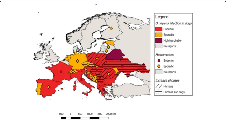
Autochthonous D. repens infections have been found in dogs in most European countries, from Portugal to Russia (Fig.1). Accordingly, human cases of dirofilariosis occur in the same areas where the infection is endemic in dogs [7] and their distribution has been previously reviewed [7,9,25–28]. The highest incidence of human cases has been recorded in the Mediterranean countries (Italy, southern France, Greece) and in the last two de-cades in some eastern European countries, namely Ukraine, Russian Federation and Belarus [7, 13, 29]. Nonetheless, many human cases are not published and the overall picture of the distribution of human dirofilar-iosis remains uncertain.
In the following chapters we briefly summarize and up-date the current distribution of D. repens in dogs, humans and mosquitoes in Europe, which has been divided into four zones following the Köppen-Geiger Climate Classifi-cation [30] (available at: http://koeppen-geiger.vu-wien.a-c.at/pdf/kottek_et_al_2006_A4.pdf), namely Mediterranean countries (Portugal, Spain, southern France, southern Italy, and Greece), west-central and Balkan countries (northern Italy, central and northern France, UK, Belgium, Denmark, Netherlands, Germany, Switzerland, Austria, Czech Repub-lic, Poland, Hungary, Bulgaria), eastern countries (Slovakia, Romania, Moldova, Ukraine, Belarus, Russian Federation, Lithuania, Estonia, Latvia), and Nordic countries (Norway, Sweden, Finland). Countries falling into different climatic zones have been placed in the one covering the majority of the area. Reports from other countries bordering Europe or the Mediterranean basin are also briefly mentioned.
Mediterranean countries
In Italy, the first extensive data of canine D. repens prevalence were obtained in the north of the country in the second half of the last century [31,32]. Interestingly, the results showed a higher prevalence of D. repens
compared to D. immitis (30 vs 5% respectively) [31,32], while 25 years later, surveys in the same areas showed a dramatic increase of D. immitis in dogs (20–40%) [33]. The most recent data indicate that D. repens is practic-ally endemic in all of the peninsula and the major islands (Sicily and Sardinia) with a prevalence ranging between 1.5–12% [34–37], and that dogs are often co-infected with other filarioids, such as Acanthocheilonema recon-ditum and D. immitis [38–40]. Dirofilaria repens was also found in the mosquito species Culex pipiens in the northeastern part of the country [41], with an infection rate ranging between 0.23–0.71%.
Accordingly, Italy is one of the countries with the most significant number of human cases [1, 8, 9, 42], and case series of up to 60 patients were published [8]. A spatial correlation has been observed, with human cases reported more frequently in areas where D. repens infection in dogs is highly endemic [43, 44]. For ex-ample, out of 14 cases of human ocular dirofilariosis re-ported in Sicily (southern Italy), eight (57.1%) occurred in the Province of Trapani where the infection rate in dogs was as high as 20.4% [45].
Canine filariosis, caused by D. repens, has been docu-mented in dogs from continental Spain and the Balearic Islands. In a study conducted in Salamanca Province (north-west Spain), blood samples from 293 dogs re-vealed D. repens in 0.3% of the animals [25]. A similar prevalence (0.2%) was obtained after examining 1683 dogs from three areas on the Mediterranean coastline of Spain and one in the Madrid Province (central Spain)
[46]. In southeastern Spain, the presence of D. repens in-fection was evaluated in 114 kenneled dogs with the highest prevalence of infection (84.6%) observed in the Alicante Province [47].
Although Spain is frequently the country of origin for human infections diagnosed in Norway, Slovenia, Netherlands and UK [48], few autochthonous human cases have been reported, namely on the island of Ibiza [49] and in the Province of Alicante [50].
In Portugal, canine or other animal cases of D. repens infection have not been reported until very recently, when the first case of canine infection was found in the Algarve, the southernmost part of the country [51]. Cur-rently there are no reports of human infection, apart from the description of an imported case [52].
Dirofilariosis is a common parasitic disease of dogs in Greece, with a higher prevalence of D. repens in north-ern Greece (30%) [53] compared to southern Greece (0.68%) [54]. The infection is also expanding in the west-ern province (Achaia), where a positive dog was recently recorded for the first time [55]. Therefore, it is not sur-prising that human infections in Greece have been re-ported since the year 2000 [56] both in residents and tourists [57].
West-central and Balkan countries
In France, D. repens has only recently received attention. Epidemiological studies conducted on military dogs in the southeast of France in 1986 and 1990 [58], showed a wider distribution of D. repens in comparison to D.
immitis.A national survey on Dirofilaria infections seen in veterinary clinics conducted in 2006 [59] pointed out that at least one case of canine cutaneous dirofilariosis was diagnosed in 8.5% of the clinics. In general, the case frequency was considered relatively stable in the ten-year period 1996–2006, with a national average an-nual clinical prevalence calculated to be 0.005%. The majority of cases (74.4%) were considered autochthon-ous in the sampling area. The parasite was mainly dis-tributed in the southern (Mediterranean), central and western (Atlantic) parts of the country [59].
A review of human cases reported in France during the period of 1923–1999 counted 75 descriptions, mainly from the southeastern part of the country [60]. Since then, another five cases have been described, in-cluding apparently new areas, resulting in a cumulative total of 80 cases until 2007. Interestingly, D. repens has been observed in 22 (23.5%) departments of France, most of them overlapping with those where canine filar-iosis was previously reported [58–67]. On the island of Corsica, human cases have been reported since 1994 [68] and DNA of D. repens has been recently found in 1.5% of Aedes albopictus mosquitoes [69].
The first empirical evidence of northern spreading of Dirofilaria infections over the Alps was in a dog from southern Switzerland at the end of the last century [70]. A few years later, another two positive dogs were found in Canton Ticino, the region bordering northern Italy [71]. Considering the close proximity of Switzerland to hotspots in Italy, it is not surprising to find some human infections in this area [72].
Other cases of possibly autochthonous D. repens in-fections in dogs of central Europe are reported from Germany [73–76]. However, the screening of 1023 blood samples collected in 2013 and 2014 in Branden-burg (north-eastern Germany) did not provide any evi-dence for autochthonous D. repens infections [77]. The finding of D. repens in the mosquito species Culiseta annulata, Anopheles maculipennis (sensu lato), Aedes vexans [78, 79] and Anopheles daciae [80], along with an analysis of weather data, suggests that active trans-mission within the area may occur [81]. Accordingly, in 2014 the first autochthonous human case was reported in Germany [82].
A single autochthonous case of D. repens infection in a dog was reported in the Netherlands in 2008 [83].
In Austria, a recent review of cases occurring from 1978 to 2014 found autochthonous D. repens infection in seven dogs [28]. The first autochthonous human case was described in 2008 [84]. The finding of the nematode in the mosquitoes An. maculipennis (s.l.) and Anopheles algeriensis[85] suggest the endemisation of the infection as well as the introduction of D. repens from eastern neighboring countries.
In Poland, the first foci of canine D. repens infection were signaled in 2009 with a high mean prevalence of 37.5% [86]. A survey conducted between 2011 and 2013 on 1588 dogs originating from all 16 provinces of Poland, revealed a nationwide distribution, with an over-all prevalence of 11.7% and local values ranging from 1.2 to 25.8% [87]. A high prevalence (38%) was recently con-firmed in dogs in central Poland [88]. The first human autochthonous case was published in 2008 [89], then a retrospective survey on affected human tissues since 2007 revealed a total of 18 cases of D. repens infections in Poland [90].
In the Czech Republic, D. repens occurs only in low-lands in the south-east of the country, in the triangle be-tween the rivers Dyje (= Thaya) and Morava [91,92], with indication of recent movement northwards along the River Morava (Modrý et al., unpublished). Recently, a re-port on emergence of autochthonous human infections in the Czech Republic was published, geographically overlap-ping with known distribution of D. repens in dogs [93].
In Hungary, the first dog with an autochthonous D. repens infection was diagnosed in 1995 [94]. An epi-demiological survey carried out during 2005–2006 re-vealed a prevalence of 14% in dogs [95]. In the following years the national prevalence of D. repens microfilaremic dogs was 18%, with significant local variations of preva-lence up to 30%. [96]. Accordingly, human cases are in-creasingly reported and D. repens infection is considered an emerging zoonosis in Hungary [97–101].
Cases of D. repens in dogs are reported in the whole Balkan region [27], with high variations of prevalence ac-cording to the area and the type of study, such as 14– 47.3% in Croatia, 11% in Albania and Kosovo, 1.9% in Bosnia and Herzegovina and 21% in Macedonia (FYROM) [27,102,103].
Although prevalence surveys are not available for Slovenia, the parasite was diagnosed in a dog as an imported case to Germany [104].
One of the most affected countries in the Balkan area is Serbia, where D. repens has been found in dogs, with prevalence ranging from 17 to 49% [105]. Infection was also found to be prevalent in wild canids [106]. Dirofi-laria repens has repeatedly been reported in humans [106–108] and a recent survey on canine and human cases revealed an endemic status of dirofilariosis in parts of Serbia [109].
Human cases are also reported in Croatia [110–112] and more rarely in Bosnia and Herzegovina [113], in Montenegro [107,114] and in Slovenia [13]. The infec-tion by D. repens in dogs of the Balkan countries is cur-rently considered in expansion and human cases are correspondingly reported [110].
Studies performed in dogs in Bulgaria reported two positive (1%) out of 192 stray dogs [115], while in Sofia
ten years later (2005–2007), 18 (4.8%) dogs out of 378 were found microfilaremic [116]. The analysis of data for a 39-year period found 47 cases of human dirofilariosis with various organ localizations [116].
Eastern countries
In Slovakia the first microfilaremic dogs for both Dirofi-laria species were identified in 2005 during routine blood testing [117]. The first systematic research de-tected microfilariae of D. repens in 99/287 (34.5%) dogs, confirming the country as a new endemic area of central Europe [118,119].
In 2007 the first human case was also detected in Slovakia [120], two years after the first case in dogs. Since then, a total of 12 human cases have been regis-tered at the Institute of Parasitology, Slovak Academy of Sciences [121–123]. The majority of cases came from the southern regions of the country, bordering Austria and Hungary [123]. Recently, D. repens was identified in Anopheles messeae and unidentified mosquitoes of the An. maculipennisand Cx. pipiens complexes [124].
In Romania D. repens was mentioned in dogs during expeditions that took place in1963–1964 [125]. In 2008, adult D. repens were found in a dog from the northeast-ern part of the country [126]. In the western counties, the prevalence of infection ranged between 2.2–7.2%, close to the Hungarian border [127, 128]. In a recent survey focused mainly on the southern parts of the country, the highest prevalence (18.8%) was recorded in the Danube Delta (southeast), while in the southwestern counties the prevalence values ranged between 2.2– 13.4%, near the Danube [129].
The first human case report in Romania was published in 2009 [130], followed by a few other reports [131– 133]. It may be assumed that D. repens is endemic in Romania and that a considerable number of human and canine cases remain undetected.
In the former USSR, first records of D. repens infec-tion in dogs originating from Ukraine and the Rostov re-gion of Russia were reported in the first half of the 20th century [134]. More recently (2002–2009), 20.25% of tested dogs were positive for Dirofilaria spp. microfilar-iae in the Rostov region, with D. repens single infection (44.7%) superseding mixed infections with D. immitis (25%) [135]. A large-scale survey conducted between 1995 and 2012 on 3258 canine blood samples revealed a prevalence of D. repens infection ranging between 10– 43% in southern Russia, and up to 12% and 36% in pet dogs and service dogs of northern regions, respectively [136]. Between 2000 and 2002, a similar prevalence was recorded in Kiev (Ukraine), with 30% and 22% of stray and owned dogs, respectively, being positive. More re-cently, similar rates (18%) were found in client-owned dogs in Kiev [137].
In southern Russia and Ukraine, D. repens in humans is endemic and well known by local physicians [136,
138–148]. Of 264 cases of human dirofilariosis recorded in Russia between 1915 and 2001, 43% occurred during the last three years of the period analyzed (1999–2001) [149]. According to a genetic analysis of strains isolated from patients who acquired infection in Ukraine, there are only negligible genetic differences as compared to strains from southern Europe [150]. A recent analysis of 266 cases detected in Rostov-on-Don, Russia from 2000 to 2016 reports a relatively high proportion (10%) of ma-ture females [151].
In various territories of Russia, infection prevalence within 6232 mosquitoes of the genera Anopheles, Aedes and Culex ranged between 1–14% [137]. Dirofilaria repens has also been found in 1% of mosquitoes col-lected in Tula region, in the species Ae. vexans, Aedes geniculatus, Aedes cantans and Cx. pipiens [152].
In Moldova, few human cases were reported, but the finding of DNA of D. repens in mosquitoes from 13 of 25 trapping sites and the suitability of temperature con-ditions for transmission of Dirofilaria spp. within the entire country suggest an endemic status [153]. Indeed from 2010 to 2015, the highest infection rate of D. repens (4.91%) was found in An. maculipennis (s.l.), whereas the most frequent mosquito species Cx. pipiens (s.l.)/Cx. torrentium had significantly lower infection rates (0.88%) [153].
Thus far, the northernmost European site where the parasite life-cycle has been confirmed is Estonia (Tartu 58°23'N, 26°43'E) where D. repens microfilariae were re-ported in three dogs in 2013–2014 [154], while no hu-man cases have been suspected or confirmed.
A human case was diagnosed after surgery in 2011 in Latvia [155].
Scandinavia
In 2016, a survey of 125 veterinarians in the Baltic (Estonia, Latvia, and Lithuania) and the Nordic countries (Denmark, Finland, Iceland, Norway and Sweden) inter-viewed by a questionnaire on the presence of canine babesiosis, D. immitis and D. repens, suggested that au-tochthonous cases of the three vector-borne parasitic in-fections occur in the region [156]. Accordingly, an autochthonous human case has been diagnosed in Finland in 2015 [157].
Other countries
Autochthonous D. repens infections have been reported in both dogs and humans in Egypt [158], Tunisia [159], Israel [160, 161], Iraq [162], Saudi Arabia [163], Dubai [164], Kuwait [165], Iran [166] and Turkey [167, 168]. While D. immitis is apparently absent from some Middle Eastern countries such as Israel where D. repens is
present, D. immitis seems to be more common in dogs than D. repens in other countries such as Iran and Turkey [169,170].
Imported human cases in central and northern Europe
Most cases reported in central and northern Europe have been seen in travelers to endemic areas or in mi-grants. Most infections are acquired in southern Europe (e.g. Italy, Spain, Greece) and to a considerable extent in southern regions of Russia and Ukraine. However, infec-tions are further imported from non-European coun-tries, especially India and Sri Lanka. Interestingly, molecular analysis of human cases imported from India repeatedly revealed these as caused by Dirofilaria sp. “hongkongensis”, which is closely related to D. repens [171,172]. Thus, cases from Asia, attributed to D. repens in the past, may indeed have been caused by Dirofilaria sp.“hongkongensis”.
Additionally, human cases of D. repens were repeatedly diagnosed from travelers returning from Africa, includ-ing cases from countries with no previous information on the presence of D. repens (e.g. Senegal and Namibia; unpublished experience of the authors).
Life-cycle
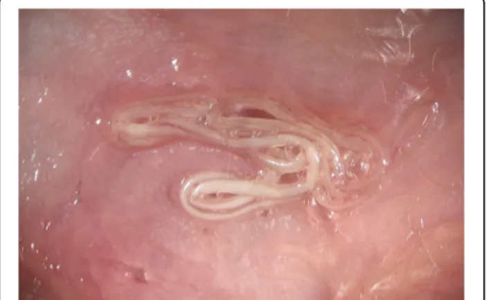
Dirofilaria repens worms are parasites of subcutaneous and intramuscular connective tissues of dogs and other carnivores (e.g. foxes, wolves and coyotes) (Fig. 2). The females of D. repens are viviparous and after mating, microfilariae are released in the peripheral blood and are picked up by a mosquito, the intermediate host, during the blood meal. Soon after ingestion, microfilariae mi-grate from the midgut to the Malpighian tubules through the haemocoel of the insect, where they molt into the second (L2) and third (L3) infective larval stages (Fig. 3). The L3 then actively leave the Malpighian tu-bules to migrate through the body cavity and the thorax
to the head and finally the proboscis where they wait until they are transmitted to the next host. The develop-mental process is temperature-dependent and takes about 8–13 days at 27–30 °C, 10–12 days at 24–26 °C and 16–20 days at 22 °C [173–175]. A delay of four days has been observed in the development at 22.5 °C and 29.4% relative humidity (RH) in comparison to 24.5 °C and 80.9% RH [174, 176]. At 18 °C, the development needs 28 days [173, 175, 177]. In the mammalian host, the L3 migrate to the subcutaneous tissue and undergo two additional molts (from L3 to L4 and to preadult worms), finally maturing into adults. In dogs, the prepa-tent period is 189–239 days [175], although in a recent study the first microfilariae were found in the blood-stream on day 164 post-infection (pi) [178]. Dirofilaria repens nematodes may live up to ten years (on average two to four years) and females potentially produce microfilariae throughout their lifespan [4].
Epidemiology
Vectors and transmission
In Europe, the known vectors of D. repens are mosquito species of the genera Anopheles, Aedes, Culex and Coquil-lettidia, with Culex pipiens pipiens [28,41,177,179,180] and Aedes albopictus implicated as the main vectors in southern Europe [177, 179, 181]. In central Europe, Ae. vexans and mosquitoes of the Cx. pipiens complex may readily act as potential vectors [41,182–184].
Other mosquito species indigenous to Europe are indi-cated as possible vectors in nature: An. algeriensis [185], An. daciae [186], An. maculipennis (s.l.) [79, 182, 185], Ae. caspius [179] and Cs. annulata [79]. Recent studies conducted in highly endemic areas in southern Hungary and northeastern Italy have shown that the molecular screening of blood-fed or host-seeking mosquitoes is an adequate tool to verify the presence of D. repens and other mosquito-transmitted filarioid helminths in a cer-tain area [41,182]. However, the simple detection of fil-arial DNA is not enough to confirm the occurrence of microfilariae development into infective L3 stages. Filar-ial DNA must be detected in separate body regions of the mosquito and the positivity of the head/thorax sam-ples can indicate that infective larval stages had devel-oped within the mosquito host [177,180,181].
Vector competence
Several factors define the vectorial capacity of a mos-quito species for a specific pathogen: vector competence (i.e. the percentage of vector individuals able to support the development to the infective stage), mosquito dens-ity and seasonaldens-ity, extrinsic incubation time, host pref-erence and daily biting rate, expected infective lifetime, the mosquito daily survival rate, as well as the availabil-ity and densavailabil-ity of infected vertebrate hosts [80,81,187].
Fig. 2 Adult specimen of Dirofilaria repens detected in the subcutaneous tissue of a dog during a necropsy (courtesy of Riccardo Paolo Lia)
For the successful transmission of D. repens L3 to a ca-nine (or other vertebrate) host, an infected mosquito must survive for at least the extrinsic incubation time until the highly motile L3 have reached the proboscis. Furthermore, the mosquito species needs to be endemic at localities where dogs are present to acquire and trans-mit the infection, and it needs to have a particular biting preference for canines. Therefore, this renders mosquito species with a mammalian host preference present in urban and suburban localities suitable for the support of an endemic D. repens cycle.
The vector competence of several mosquito species for D. repens has been shown in experimental laboratory studies by observation of the development to the infect-ive L3 stage: Ae. aegypti [15,174,176,188]; Ae. albopic-tus [189]; Ae. caspius, Aedes detritus [173]; Aedes mariae [174]; Ae. vexans, Anopheles stephensi [175]; Anopheles claviger; An. atroparvus [175]; Anopheles sinensis [174]; Culex pipiens molestus [188]; Aedes togoi [190]; Ae. geniculatus; and Aedes japonicus [191]. Differ-ent methods for the infection of the mosquitoes were applied in these studies such as the direct feeding on a
microfilaraemic animal [173, 176, 188] or the artificial membrane feeding with infected blood [192].
Furthermore, within a certain species of mosquitoes, susceptibility or refractoriness may vary considerably and may be dependent on certain genes, as has been shown for Ae. aegypti [193]. Controversial results exist also for Cx. pipiens, as it has been shown both suscep-tible and refractory in laboratory experiments [176]. This might be attributed to testing of different biotypes (pipiens, molestus and their hybrids) that possess differ-ent vectorial capacity. Culex pipiens fatigans, Anopheles gambiaecomplex, Aedes vittatus, Ae. aegypti and Man-sonia africana were also shown to be refractory to D. repens infection in laboratory investigations [176, 191]. All microfilariae in the latter mosquito species were trapped inside the midgut in the blood clot and were disintegrated and no longer observable after day 5 pi. This retention of microfilariae has been described as potentially beneficial to the vector-parasite interaction system. A reduced microfilarial burden can lead to in-creased mosquito longevity, potentially making it more efficient transmitting host [194]. Microfilaria burden can
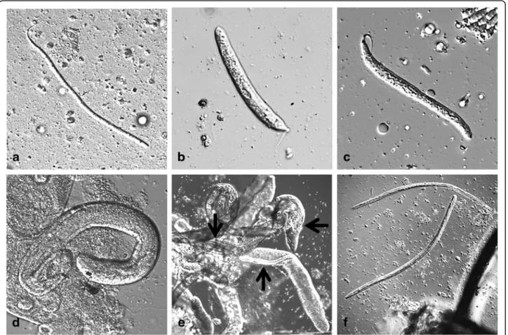
Fig. 3 Developmental stages of Dirofilaria repens inside a mosquito (Aedes vexans) (courtesy of Cornelia Silaghi). a L1 day 2 pi; 335 × 9μm, the stage still resembling a microfilaria. b L1 day 3 pI; 167 (214) × 18μm, so-called sausage stage. c L1 day 5 pi; 198 (220) × 16.8 μm, so-called sausage stage, but more elongated. d L2 day 7 pi; 425 × 35μm. e L2 late stage or L3 inside Malpighian tubules (black arrows), day 19 pi. f L3 day 16 pi, transition from thorax to head; 962 × 30 and 934 × 23μm
vary greatly in a canine host and consequently also the uptake of microfilariae by a mosquito vector. This variation may be due to the circadian rhythms of microfilariae in the peripheral blood and mosquito vector biting [6, 175].
Apart from the process of microfilaria degradation and melanisation as part of an innate immune response of the mosquito host [195], it was also assumed that the anatomical structures of the alimentary channel and the physiology of the respective mosquito species influence the development of microfilariae, for example the speed of blood clotting after blood intake (discussed in [188]). Some authors have highlighted the importance of mos-quito cibarial armature and peritrophic membrane in the transmission of D. repens. Indeed, cibarial armature and dome can mechanically damage a high proportion of microfilariae, which are ingested with the blood meal, and possibly serve to protect mosquitoes [188,189]. De-velopment and complexity of the cibarial armature differ between different species. In some it is absent (An. atro-parvus, An. claviger, Ae. aegypti and Ae. mariae), in others it has one (Anopheles albimanus and Anopheles farauti) or two (An. gambiae, Anopheles stephensi and Anopheles superpictus) rows of cibarial teeth, whereas in Cx. p. pipiensteeth of cibarial armature are spoon-shaped and the cibarial dome is strongly denticulated [196,197]. The number of damaged erythrocytes varied between 2–4% in the first, and 45–50% in the last group. The time needed for formation of peritrophic membranes in adult mosquito varies between 4 and 12 h in dif-ferent species [198].
Risk factors
No study has been published on risk factor analyses using a multivariate approach, which would be more suitable for highlighting confounding factors and biases. Therefore, some of the associations found and often re-ported as risk factors (Table 1) are likely the results of the interaction of different factors related to the host (sex, age, breed and lifestyle), the vector (presence, density, vectorial capacity and attraction to dogs), the environment (rural, urban, climate) and the human intervention (use of specific chemoprophylaxis and/or physical or chemical protection against mosquitoes).
The evaluation of the frequency of the factors associ-ated with D. repens prevalence in literature, in particular male and guard dogs, older age and outdoor lifestyle, suggests that the higher exposure to mosquito bites is the only risk factor clearly associated with D. repens prevalence.
Canine subcutaneous dirofilariosis
Although canine D. repens infections very often runs asymptomatically, a plethora of nonspecific dermal
alterations has been reported such as skin nodules, prur-itus, thinning, itching and asthenia [10, 59, 199, 200]. Usually, no inflammatory reaction or connective cap-sules are surrounding the living parasite (Fig.2a), which can be seen moving actively under the connective serous layers [4]. Non-inflammatory subcutaneous nodules, cold, not painful and mobile, can be seen on the skin surface of infected animals. Inflammatory and painful nodules may be associated with localizations such as the scrotum. Granulomatous capsules generally surround dying and degenerating worms. These clinical alter-ations, however, must be supported by histopathological data or D. repens microfilaria-positive blood examina-tions or molecular identification from biopsy. Lesions may also appear as circular alopecic areas with lichenifi-cation, hyperpigmentation and erythematous and scaling margins [201] and they can occur in the lumbosacral and perianal regions [164]. Skin affections may be prur-itic or not, suggesting that itching is not crucial for a presumptive diagnosis of D. repens-associated dermatitis. An unusual case of allergic non-pruritic diffused derma-titis caused by D. repens, confirmed by histological examination, has also been described [201].
Dirofilaria repensinfection was the aetiological cause of ocular lesions in a dog reporting conjunctivitis and later additional ocular and nasal mucopurulent dis-charge [202]. Worms were then found in a dorsonasal bulbar conjunctival mass and in the ventral palpebral conjunctival fornix and confirmed as D. repens by PCR. Rarely, D. repens may reach ectopic body parts. A case of adults in the pelvic cavity and mesentery
Table 1 Factors significantly associated with Dirofilaria repens prevalence in dogs of Europe
No. of dogs tested Country Potential risk factors Reference 114 Southern Spain Kenneled dogs (lack of
preventative measures) [47]
Geographical location 2406 Central Italy Older age [294]
Male sex Pure breed Traveling dogs
151 Eastern Slovakia Older age [119] Lifestyle (outdoors)
Geographical location 972 Central Italy Rural environment [34]
Geographical location 194 Northern Serbia Older age [295]
Geographical location 2512 Southern Italy Lifestyle (guard dogs) [296]
was reported in a dog with a diagnosis of kidney fail-ure and chronical cystitis [203].
The histological examination of lesions may reveal the presence of multifocal purulent dermatitis, panniculitis, hyper-pigmentation and hyperkeratosis [10]. Generalized cardio-hepato-renal insufficiency may also occur [87]. Pathological changes are most likely associated with the presence of adult nematodes or microfilariae [10]; how-ever, symbiotic Wolbachia bacteria, which live in the hy-podermal chords of Dirofilaria male and female adults, and in the female germline [204], have been shown to increase the level of pro-inflammatory cytokine (e.g. IL-8) and induce chemoattraction [205,206].
Human infections
Humans acquire the infection in the same manner as dogs, by the bite of a mosquito, but it is probable that most of the infective larvae die shortly after, with the in-fection resolving unrecognized and without causing any specific symptom [1, 8]. No predisposing factors are known to explain why in some cases larvae may develop further. After the bite of an infective mosquito, a stron-ger reaction with erythema, swelling and pruritus lasting 5–8 days is reported [1, 8]. In most of the cases a single worm develops, probably because the stimulation of the immune system prevents the development of others [1,
8]. In rare cases the worm may develop to a mature adult [1, 207, 208] and even fertilized worms releasing microfilariae have been described, especially in immuno-suppressed patients [1, 8, 42, 146, 209–212], which in very rare cases may even reach the bloodstream [213].
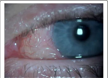
In infected patients, the developing stages of D. repens migrate subcutaneously [1,8,61] for weeks up to several months in several parts of the body, usually with mild and unrecognized symptoms [1, 8, 61] and only some-times causing larva migrans-like symptoms (i.e. irritation and itching) [1, 8, 42, 61, 131, 211, 214]. In one case, a patient, after scratching a pruritic lesion, removed a 6 cm long whitish worm from the wound [215]. During migration D. repens may reach the eyes [1, 8, 61, 211], becoming visible through the subconjunctiva [1, 5, 72,
110,113,168,214,216–219] (Fig.4). Larval stages local-ized in the eyes can be removed surgically without ser-ious damage [1, 214, 219]. However, in rare cases, serious sequelae (glaucoma, uveitis, episcleritis and retina-detachment) may develop and ultimately lead to significant loss of vision [1,8,100,147,220–222].
After weeks to several months from the infection, D. repensmay stop to migrate and form a nodule of about one centimeter [1,8]. In most cases, the nodules develop subcutaneously [1,8,48,63,93,108,111,116,138,158,
212, 223–228]. Nodules have been reported in various human body areas and tissues, mostly in the superficial tissues of the facial regions [1, 8], as perioral and
periorbital tissues [107, 167, 224, 226, 227, 229–234], forehead [235], skin of the lower leg [93], soft tissues of the hand [236] or finger [93], subcutaneous tissue of the hypogastrium [93] and of the neck [237]. Other predilec-tion sites are scrotum and testicles and, to a lesser ex-tent, the breasts of women [1,8,65,223,235,238–245]. Various reasons have been hypothesized for these prefer-ences, such as lower body temperature of these areas, higher awareness of patients for these body parts or a tropism of D. repens to higher concentrations of sexual hormones [1].
The nematodes may also reach deeper body areas, such as lymph nodes [93], the abdominal cavity [93,99], lungs [1,56,158,246], muscles [247] and even the dura [64].
If left untreated, D. repens may survive for up to one and a half years [1,8]. The symptoms caused by D. repens nodules depend on their localization, usually being limited to a local irritation, erythema and pruritus [1, 8, 93]. Rarely, a strong local immune reaction develops, and the nodules may appear like a suppurating abscess with local infection accompanied by a mild systemic reaction, in-cluding elevation of body temperature and mild eosino-philia [1, 8, 206]. In very rare cases, even more severe systemic immunoreactions may develop, manifesting as fever or lymphadenopathy. A case of meningoencephalitis has also been reported [211]. Comparatively severe symp-toms are seen in immunosuppressed patients and in the rare cases where microfilariae develop [1,8].
Diagnosis in dogs
Diagnosis of D. repens may be performed by detection and identification of circulating microfilariae, morpho-logical and molecular identification of adult parasites, cytological examination of fine-needle aspiration biop-sies and histopathological examination of excised nod-ules. In the case of localized skin lesions, the adult
Fig. 4 Dirofilaria repens visible in the subconjunctiva of a human eye (courtesy of Ramin Khoramnia and Aharon Wegner)
nematodes can be recovered from the nodules located in different anatomical sites of the animal (e.g. chest or lower limbs) [10] (Fig. 5), while in cases of localized or generalized dermatitis adults are almost impossible to find.
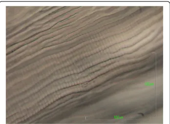
On gross examination, the cuticle of D. repens speci-mens is whitish, with distinct longitudinal ridges on the surface (Figs. 6 and7), and narrows at the ends. Males measure 48–70 mm in length and 3.7–4.5 mm in width, while the females are larger, reaching 100–170 mm in length and of 4.6–6.5 mm in width [248,249]. Upon ac-curate microscopic observations, the clarification of specimens with lactophenol or with glycerine for tem-porary mounts, allows the observation of distinct mor-phological features, such as the vagina in the female, which opens at 1.1–1.9 mm from the oral aperture, or the two spicules in the male, measuring 430–590 and 175–210 μm, respectively, as well as 4–6 precloacal pa-pillae (1–2 post-anal and 3 caudal). In the case of adults embedded in the nodule, D. repens specimens are identi-fied at the histology on the basis on their body diameter (220–600 μm), and by the presence of the longitudinal ridges, each separated from the others by a distance that is larger than the width of the actual ridge itself [250]. In transverse sections stained with haematoxylin-eosin, the occurrence of longitudinal muscles and the multilayered cuticle, expanding in the region of the two large lateral chords, is indicative for D. repens [10,250].
The subcutaneous nodules can be also examined by ultrasound and the parasite is visualized as double linear parallel hyperechoic structures [251].
More often the diagnosis of subcutaneous dirofilariosis is based on the visualization (see Additional file 1) and morphological identification of the blood-circulating microfilariae, by concentration methods (e.g. modified Knott’s test or filtration) (Fig. 8), histochemical staining (e.g. acid phosphatase activity) and fine needle sampling of nodules containing fertile adults. A blood sample
taken in the evening may maximize the chance to find circulating microfilariae, due to the circadian variation of microfilariae in naturally infected dogs [6,252].
Dirofilaria repens microfilariae are unsheathed, having an obtuse-rounded cephalic margin (Fig. 5), and a long sharp tail, often curved [253,254]. Their size may vary as a consequence of collection and fixation methods. The mean length is 300–370 μm and the mean width is 6–8 μm [253]. In a recent study [254], a mean length of 369.44 ± 10.76μm and a mean width of 8.87 ± 0.58 μm was re-ported using the Knott’s test on 171 microfilaraemic dog blood samples originating from eight European countries. The test was able to clearly distinguish between D. immi-tis, D. repens and Acanthocheilonema spp. [254].
On the contrary to D. immitis infection, for which sev-eral, easy and rapid in-clinic test kits, based on
Fig. 5 Adult Dirofilaria repens removed from the subcutaneous tissue of a dog during necropsy (courtesy of Riccardo Paolo Lia)
Fig. 6 Aspect of the ridges of the cuticle of Dirofilaria repens under scanning electron microscopy (courtesy of Sven Poppert). Scale-bars: 100μm
Fig. 7 Cuticle morphology of Dirofilaria repens under scanning electron microscopy (courtesy of Salvatore Giannetto). Scale-bar: 200μm
detections of circulating antigens mainly produced by fe-males, are commercially available for the serological diagnosis of the infection, no similar specific serological tests are available for D. repens.
The identification of D. repens may be carried out by molecular methods testing parts of adult specimens, microfilariae (in whole blood or on filter paper), or larval stages in the mosquito vectors. Various techniques have been developed for the specific detection of D. repens, such as multiplex PCRs targeting several filarioid spe-cies, but also for the entire superfamily Filarioidea. Amongst these are conventional and real-time PCRs, probe-based or high-resolution melting analysis tech-niques. The most common gene targets used are the cytochrome c oxidase subunit 1 (cox1) as a barcoding gene, the inter-genic spacer (ITS) regions, and 12S rRNA gene [41,184,185,255–259]. Other target genes used to identify the nematode are listed in Table 2. The high sensitivity of real-time PCR allows the detection of small amounts of genomic DNA either in dog blood or
mosquitoes (2.5 and 0.3 pg/μl for D. immitis and D. repens, respectively) being potentially useful for epi-demiological studies [41]. In addition, a multiplex PCR targeting a barcoding region within the cox1 gene was developed for the simultaneous detection of almost all the filarioids infecting dogs in Europe (i.e. D. immitis, D. repens, A. reconditum and Cercopithifilaria sp.) [260], therefore representing a new tool for the molecular de-tection and differentiation of canine filarioids in blood and skin samples. Nonetheless, positive PCR alone should not be considered sufficient to establish D. repens as a cause of subcutaneous nodular lesions in the ab-sence of clear cytological picture [261].
Diagnosis in humans
The diagnosis of a D. repens infection in humans is affected by the localization of the worm and the clinical symptoms. If the infection occurs as larva migrans, especially in the subconjunctiva, and the patient was not exposed to other potential causes of larva migrans, the clinical picture is highly suggestive of D. repens. The anamnesis should ex-clude the visit of the patient to endemic areas of other filar-ioids, such as Loa loa in Africa. In case of intraocular cysts or subcutaneous nodules, the diagnosis is more difficult, but a live moving worm can be seen using a pre-operative high-resolution ultrasound [231,245].
In most cases, the definitive diagnosis is obtained after the worm removal, using the same methods applied for animals. Microscopically, D. repens females do not usu-ally contain microfilariae. The most discriminative fea-tures of D. repens are the longitudinal ridges of the cuticle (Figs. 6 and 7), not present in any other filarial worm infecting humans except for Dirofilaria sp. “hon-gkongensis”, a recently proposed new species from Hong Kong [262] and Dirofilaria ursi present in North Amer-ica, North Europe and Japan in bears and rarely also in humans [171].
Since none of the described features are entirely specific, molecular tools should be applied in order to confirm the morphological diagnosis and avoid misdiagnosis, which may occur in some case with D. immitis [263]. In this re-spect, it should be suggested to surgeons to conserve the removed worm, one part in formalin for histology and an-other refrigerated or frozen for molecular identification. Most typical features are recognizable in histological slides, if a proper section is available and the worm not degraded. In these cases, it is still possible to perform mo-lecular investigations from paraffin sections. An extensive description of D. repens in human tissue is already avail-able [264].
Serological investigations are not helpful in human cases. In filarial infections, the immunological reaction is mainly triggered by microfilariae, which rarely develop in humans. Therefore, in most human D. repens cases, no
Fig. 8 The round head of the microfilaria of Dirofilaria repens (Knott’s test). Scale-bar: 20μm
Table 2 Target genes used to identify Dirofilaria repens in animals, humans and mosquitoes, available on GenBank (accessed 10th September 2018)
Gene Hosts Length (bp)
12S rDNA Human, dog, cat, mosquito 116–510 cox1 Human, dog, cat, mosquito,
beech marten
123–715
16S rDNA Human, mosquito 366–487
18S-5.8S-28S rDNA Human, dog, mosquito 153–2230 18S-small subunit
ribosomal RNA
Human, dog, jackal 613–839
hsp70 Dog 553
rbpI Dog 594
antibodies against filariae are detectable or very low titers can be found [47]. However, such low titers are also seen in other nematode infections due to cross-reactive anti-bodies. The investigation of blood samples by microscopy or PCR is not useful for the same reason.
Mitochondrial genotypes and potential cryptic species
A new species of Dirofilaria infecting dogs and humans has been first described from Hong Kong and designated as Dirofilaria sp. “hongkongensis” [262, 265]. This new species was proposed on the basis of relative short DNA sequences from the mitochondrial cytochrome c oxidase 1 and the nuclear ITS1 locus. Unfortunately, at that time all ITS1 sequences on GenBank were from D. repens samples collected from Thailand while all ITS2 sequences were of European origin which hampered comparisons with Euro-pean D. repens data. Complete sequencing of mitochon-drial genomes from four worms initially identified as D. repens using morphological features and short DNA se-quences, revealed that three sequences from European samples were very similar while a fourth one collected from a patient after traveling in India was very similar to Dirofilaria sp. “hongkongensis” [171]. An additional D. repens mitochondrial genome sequence available from GenBank (accession no. KR071802) is also highly similar to the other European samples but its geographical origin is not available from the database entry. The organization of these mitochondrial genomes is identical to those of other onchocercids and like all clade III nematode mito-chondrial genomes lacks the atp-8 gene that is present in most animal mitochondrial genomes. It is slightly smaller than any of the other mitochondrial genomes described for the Onchocercidae and has the most extreme AT skew with a very high T content on the coding strand.
Phylogenetic analysis using all coding regions from the whole genomes revealed that D. repens and Dirofilaria sp. “hongkongensis” are more closely related to each other than to D. immitis [171]. However, as long as no other mitochondrial genomes from species of the sub-genus Nochtiella are available, it remains speculation how closely related both species actually are. The overall similarity of mitochondrial genomes was lower than for the comparison between the human parasite Onchocerca volvulus and its sibling species Onchocerca ochengi in-fecting cattle. This suggests that both might represent valid species [171]. Sequencing of partial genomic frag-ments of approximately 2.55 kb, including the most vari-able long non-coding region of the mitochondrial genome, from 41 canine samples (29 from Europe and two from Thailand) and one human sample from Vietnam, revealed further heterogeneity. In the phylo-gram, all European and the Vietnamese sequences were located in the same statistically highly supported cluster
with the complete D. repens mitochondrial genome se-quences. With the exception of only two samples (one from Hungary and one from Poland), differences be-tween the remaining D. repens sequences were small although there were some subclusters containing prefer-entially samples from Poland and Hungary or from southwestern Europe and Hungary in addition to a Ger-man sample. The two samples from Thailand had very similar sequences and were more similar to Dirofilaria sp.“hongkongensis” than to the D. repens cluster. How-ever, the genetic distance between samples from India and Thailand was considerable and the latter might rep-resent a third species [171]. These data support the view that what is currently considered to be D. repens is in fact a species complex with different genotypes. However, data are not sufficient yet to decide whether different geno-types from various geographical origins represent valid species, subspecies with limited geographical range or only variants within a population. Multi-locus phylogenetic analyses using samples from diverse endemic regions combined with experimental crosses would be required to define valid genospecies within the D. repens complex. Treatment and prevention
Dogs
Due to the lack of specific clinical alterations, the treat-ment of D. repens infection in dogs often goes along with its prevention, which should be routinely per-formed in order to reduce the risk for the transmission to humans (Table 3). Most therapeutic protocols cur-rently available have been translated from the experience developed in the prevention of heartworm disease and are based on the administration of macrocyclic lactones. However, contrary to heartworm disease, very few ex-perimental studies have been carried out to assess the ef-ficacy of macrocyclic lactones against D. repens [4].
A complete clearance of D. repens microfilariae was achieved in a dog treated with an off-label protocol based on melarsomine injection followed by doramectin [160], but this fact needs further confirmation since no
Table 3 Macrocyclic lactones tested for the prevention of Dirofilaria repens infections in dogs
Active ingredient Formulation Dosage Ivermectin Tablet/Chewable 6μg/kg
Ivermectin + Praziquantel Chewable 6μg/kg + 5mg/kg Ivermectn + Doxycycline Chewable + Tablet 6μg/kg + 10mg/kg
Doramectin Injectable 0.4 mg/kg
Milbemycin oxime + Praziquantel
Chewable 0.5–5 mg/kg Moxidectin Injectable 0.17 mg/kg Moxidectin + Imidacloprid Spot-on 2.5–10 mg/kg
efficacy was found in previous clinical studies [4]. Differ-ent dosages of moxidectin in oral, injectable sustained-release and spot-on formulations showed long-term sup-pression of D. repens microfilaraemia, being highly efficacious for the treatment of dogs positive for sub-cutaneous dirofilariosis in both natural conditions and experimental studies [266–271]. Currently, the only protocol claiming adulticidal activity for this filarioid is represented by the use of a spot-on product containing imidacloprid/moxidectin for six consecutive months, a protocol which has also been used to prevent the onset of skin lesions and dermatitis caused by the parasite [178]. Interestingly, the microfilaricidal efficacy of monthly administration of ivermectin [272] may be im-proved by including doxycycline [273]. This therapeutic schedule represents a novel approach for the treatment of dirofilariosis, targeting the Wolbachia endosymbionts of the nematode [274] and allows the reduction of the recommended ivermectin dosage, along with a minor risk of drug resistance.
As in the case of the treatment, the prevention of D. repens infection is largely based on the regular use of macrocyclic lactones (Table 3). When designing a ra-tional approach for the control of dirofilariosis, the re-gional distribution patterns and the transmission period of the parasite should be taken into account, which de-rives from detailed epidemiological maps of the disease.
The prevention of D. repens transmission becomes in-creasingly important, considering that reducing the bur-den of canine dirofilariosis represents the only effective measure to decrease the risk for human infection, as dogs are the most important reservoir of the parasite.
Monthly applications of selamectin in a spot-on for-mulation were successfully used to reduce the pathogen transmission under natural field conditions for six months [275]. In addition, when infected animals were treated twice a month, the period of dog protection in-creased to nine months [276]. The use of moxidectin in a sustained-release formulation administered subcutane-ously was found to have a complete efficacy in the pre-vention of D. repens in an experimental trial [269] and the authors suggested that the excellent action of the formulation was most likely attributed to the high lipophily of this active ingredient, which is stored in the body fat. Furthermore, moxidectin may be of great value towards the prevention of this filarial parasite and against adult parasites, when given as a spot-on treat-ment in combination with imidacloprid (imidacloprid 10% and moxidectin 2.5%) [40,178].
Finally, milbemycin oxime, another macrocyclic lactone, given orally once per month also proved to be effective in protecting dogs from subcutaneous dirofilariosis in endemic areas and may offer further chemoprevention option [277].
Another important part of the prevention of infection is based on contact repellent insecticides. This can be obtained by the use of veterinary products that contain pyrethroids with a specific label on the prevention of Culexand/or Aedes bites. This prevention is particularly important in periods of activity of mosquitoes and in areas where the risk of transmission is high. The use of topic repellent may also decrease the transmission of Dirofilariafrom infected dogs to mosquitoes [278].
Humans
Theoretically, no special treatment is necessary in humans, because D. repens does not cause severe symp-toms and usually dies after some time [1,8]. The nema-tode can be removed by surgery, a practice that is also needed for the etiological diagnosis and to exclude other severe diseases, such as a carcinoma [1, 8]. As soon as D. repens has formed a stationary nodule, surgical re-moval can be conducted following standard procedures corresponding to the site of infection.
If a migrating D. repens is discovered in the conjunc-tiva, removal is comparably easy because the worm is visible through the conjunctiva [1, 8, 172, 214]. On the contrary, surgical removal of subcutaneous worms may be unsuccessful, due to the difficulties in precisely locat-ing the parasite.
Medical treatment with anthelminthic drugs, such as albendazole, coupled with doxycycline, was found to stop migration of the worm and promote the formation of a fixed nodule, which can then be removed [136]. The efficacy of such treatment suggests that doxycycline may have a role targeting the endosymbiont Wolbachia, as has been found in dogs [274]. In addition, the immune response of humans to Wolbachia may be used for fur-ther confirmation of exposure to the parasite [279].
As soon as D. repens is removed, no further medical treatment is required, unless the patient is immunosup-pressed or in the extremely rare case of a suspected sec-ond nematode [1, 8]. Because of the rareness of the disease in humans, there are no guidelines or treatment studies and the physician have to rely on their experi-ence. However, either with or without treatment, there is not a single report of a fatality or of permanent body damage.
Prevention of dirofilariosis in humans can be achieved by protecting people from the bites of mosquitoes through the use of repellents and by reducing the preva-lence of D. repens in dogs, the principal reservoir of the parasite [280].
Potential drivers for the emergence ofDirofilaria repens
The enhanced dissemination of D. repens in Europe has been primarily attributed to global warming and the
rapid geographical expansion of some invasive mosqui-toes (and/or increases in their density), but also to in-creased travel and movement of infected animals into non-endemic areas along with a change in human activ-ities [4,11].
The effects of climate change in Europe have been exten-sively debated [281], since warmer climates could favor mosquito breeding and shorten extrinsic incubation periods [282], thus enhancing the risk of Dirofilaria spp. transmis-sion. The projected increment in temperature will impact on insect vectors through broadening areas of colonization, invasion of new sites and, eventually, resulting in physio-logical changes and increased vectorial capacity. The most recent example is the finding of Uranotaenia unguiculata, a thermophilic mosquito species frequently occurring in the Mediterranean, in northern Germany, some 300 km north of previous collection sites [283].
An increase in mean temperatures has affected the mosquito abundance and their seasonal survival in many areas of Europe greatly impacting on the spread of filar-ial infestation and making most of the European coun-tries suitable for Dirofilaria spp. transmission [284,285]. A recent climatic model studied the impact of a re-gional warming (Russia, Ukraine, and other countries of the former USSR) on the spreading of D. repens and the risk of transmission to humans [26]. The model pre-dicted an increase of 18.5% in transmission area and 10.8% in population exposure by 2030.
In addition, several intrinsic factors linked to the spe-cific vector mosquito species also impact on the distri-bution of D. repens. The expansion of dirofilariosis somehow matched the second introduction of Ae. albo-pictusin Europe (Italy) [286]. Furthermore, over the past decades, Cx. p. pipiens has changed its endophagic and anthropophagic behavior in central Europe [287], where it also searches for human blood outdoors, close to the houses, as happens in southern parts of the continent.
The introduction of the Pet Travel Scheme in 2000, allowing an easier movement of companion animals throughout the European Union [288], has likely con-tributed to the diffusion of D. repens in Europe. The first case of D. repens in a dog resident in UK was recently reported in a dog originated from Romania and was not easily identified [202], thus reactivating the discussion on the implications for establishment and spread of D. repensin non-endemic countries.
Once D. repens has been introduced in a new area with an infected dog, the availability of suitable hosts for D. repens, the presence and density of competent mos-quito vectors and their feeding behavior are among the most important factors impacting on its further distribu-tion. Dogs are optimal reservoirs of D. repens also be-cause they attract competent mosquito vectors and are quite tolerant to mosquito bites [11]. Prevalence of
microfilaraemic dogs and presence and abundance of competent vectors also affect the rate of infestation within a given mosquito population, which in turn is dir-ectly related to the risk for a native dog to be infested.
The factors enhancing the exposure of the host to the vector (i.e. the dog’s size, the age and especially the outside exposure) may further increase the risk of D. repens infest-ation [2]. The role of cats and foxes as reservoirs is mar-ginal, because these hosts are rarely microfilaraemic [289]. However, the general factors discussed above should have affected the emergence of both D. repens and D. immitis. Although a few reports have been published until now on the spread of D. immitis towards northern Europe [118,290–292], there is no doubt that D. repens has spread faster than D. immitis from the endemic areas of southern European countries and currently it is more prevalent in northern Europe, as confirmed by the emergence of human infections (reviewed in [4,7,9,27,
136]. The reasons for this could be linked to the fact that while heartworm infection causes a severe clinical condi-tion in dogs, D. repens in most cases is difficult to diag-nose and the course of the infection can be completely asymptomatic. As a consequence, many canine infec-tions can run unnoticed and the infected dog continues to act as a reservoir for competent mosquitoes locally and if transported to non-endemic areas.
In addition, for heartworm infections, several rapid, easy, in clinic whole blood/serological kits are available that detect the circulating antigens of female worms. This allows veterinarians a prompt diagnosis while no serological diagnostic is commercially available for D. repens, hampering a rapid screening in dog populations. Blood examination for circulating microfilariae remains the most diffuse test for D. repens diagnosis. However, the Knott’s test, which allows the visualization and the identification of microfilariae, is not familiar to veteri-narians in areas of recent introduction of the parasite. Furthermore, an interaction between the two species of Dirofilariahas been suggested [33], which seems to slow the spread of D. immitis in areas where D. repens has firstly settled.
Another aspect which deserves attention is the higher prevalence of human infection by D. repens compared to D. immitisin Europe, even in countries where the latter is endemic [4]; this is in contrast to the prevalence in the New World, where the human cases of dirofilariosis by D. immitis are relatively frequent [293]. There is cur-rently no evidence of a higher virulence of D. repens re-spect to D. immitis and of a difference of virulence among strains of the same species, or of a difference in the mosquito vectors of the two parasites. It has been hypothesized that the localization in the subcutaneous tissues may help D. repens to escape the natural immune response of unusual hosts, such as humans.
Conclusions
There is evidence that D. repens has spread faster than D. immitis from the endemic areas of southern Europe to northern Europe. Climate change affecting mosquito vectors and the facilitation of pet travel seem to have contributed to this expansion; however, the major factor is likely the rate of undiagnosed dogs perpetuating the life-cycle of D. repens. Many infected dogs remain un-detected due to the subclinical nature of the disease, the lack of rapid and reliable diagnostic tools and the poor knowledge and still low awareness of D. repens in non-endemic areas. Research and education should fill this gap. Indeed, improved diagnostic tools are war-ranted to bring D. repens diagnosis to the state of D. immitis diagnosis, as well as improved screening of imported dogs and promotion of preventative measures among veterinarians and dog owners. In this respect, to transform the disease in a notifiable disease, at least in humans, would help Europe to have official and compar-able data on the presence and variations of prevalence among countries. Upcoming studies should also focus on (i) the vector competence and vectorial capacity of mosquito species; (ii) the presence of different genospe-cies or genotypes of D. repens and their specific interac-tions with hosts and vectors; and (iii) the possible selection of resistance to macrocyclic lactones if pre-ventative measures increase. For vector-borne diseases where an animal species serves as a reservoir, especially a pet, veterinarians play a significant role in prevention and should be more aware of their responsibility in re-ducing the impact of the zoonotic agents. In addition, they should enhance multisectorial collaboration with medical entomologists and the public health experts, under the concept (and the actions) of One Health-One Medicine.
Additional file
Additional file 1: Live microfilaria of Dirofilaria repens in the bloodstream of a dog. This movie shows the morphology and movement of the microfilaria of D. repens in a direct blood smear. (MOV 9179 kb)
Abbreviations
FYROM:Former Yugoslav Republic of Macedonia; L2: Second larval stage; L3: Third larval stage; PCR: Polymerase chain reaction
Acknowledgements
This work was done under the frame of COST action TD1303 (EURNEGVEC).
Funding
The study was partially funded by the Veneto region, Venice, Italy.
Availability of data and materials
All data generated and/or analysed during this study are included in this published article and its additional file.
Authors’ contributions
GC, CG, DO and CS assembled the first draft of the review. Authors were responsible for the search of data and references in their respective countries or areas: GB (other mediterranean countries), PB (France), EB, GC, DO, PD and FM (Italy), LC and CM (Portugal and Spain), HPF (Austria), AMI (Romania), DM (Czech Republic), EP (Greece), DP and SS (Balkan area), MP and CS (Germany). CG wrote the hystory of D. repens; LC, CM and GC wrote risk factors; JK wrote the sections on genetics; AG and EB wrote therapy and prevention; DP, FM and CS wrote the sections on vector transmission and competence; SP wrote the parts related to human infections. All authors critically reviewed the manuscript and read and approved the final manuscript.
Ethics approval and consent to participate Not applicable.
Consent for publication Not applicable. Competing interests
The authors declare that they have no competing interests.
Publisher’s Note
Springer Nature remains neutral with regard to jurisdictional claims in published maps and institutional affiliations.
Author details
1Laboratory of Parasitology, National reference centre/OIE collaborating
centre for diseases at the animal-human interface, Istituto Zooprofilattico Sperimentale delle Venezie, Legnaro, Italy.2Department of Veterinary
Medicine, Università degli Studi di Milano, Milan, Italy.3Koret School of Veterinary Medicine, The Hebrew University, Rehovot, Israel.4Veterinary
School of Nantes ONIRIS, University of Nantes, LUNAM, Nantes, France.
5Department of Veterinary Science, Università degli Studi di Messina,
Messina, Italy.6Department of Veterinary Sciences, School of Agrarian and Veterinary Sciences, University of Trás-os-Montes e Alto Douro, Vila Real, Portugal.7Institute of Parasitology, University of Veterinary Medicine, Vienna,
Austria.8Department of Veterinary Medicine, University of Bari, Valenzano,
Italy.9Department of Parasitology and Parasitic Diseases, University of Agricultural Sciences and Veterinary Medicine Cluj-Napoca, Cluj-Napoca, Romania.10Global Health and Tropical Medicine (GHTM), Instituto de Higiene
e Medicina Tropical (IHMT), Universidade Nova de Lisboa (UNL), Lisboa, Portugal.11Department of Pathology and Parasitology, University of Veterinary and Pharmaceutical Sciences, Brno, Czech Republic.12Biology
Centre, Institute of Parasitology, Czech Academy of Sciences,České Budějovice, Czech Republic.13Institute for Parasitology and Tropical
Veterinary Medicine, Freie Universität Berlin, Berlin, Germany.14Laboratory of Parasitology and Parasitic Diseases, Faculty of Veterinary Sciences, Aristotle University of Thessaloniki, Thessaloniki, Greece.15Laboratory for medical and
veterinary entomology, Faculty of Agriculture, University of Novi Sad, Novi Sad, Serbia.16Institute of Animal Hygiene and Veterinary Public Health, Veterinary Faculty, University of Leipzig, Leipzig, Germany.17Scientific
Veterinary Institute“Novi Sad”, Novi Sad, Serbia.18Swiss Tropical and Public
Health Institute, Basel, Switzerland.19University Basel, Basel, Switzerland. 20
National Centre of Vector Entomology, University of Zurich, Zurich, Switzerland.21Institute of Infectology, Friedrich-Loeffler-Institute, Isle of
Riems, Greifswald, Germany.
Received: 19 July 2018 Accepted: 19 November 2018
References
1. Pampiglione S, Rivasi F. Human dirofilariasis due to Dirofilaria (Nochtiella) repens: an update of world literature from 1995 to 2000. Parassitologia. 2000;42:231–54.
2. McCall JW, Genchi C, Kramer LH, Guerrero J, Venco L. Chapter 4. Heartworm disease in animals and humans. Adv Parasitol. 2008;66:193–285.
3. Otranto D, Cantacessi C, Dantas-Torres F, Brianti E, Pfeffer M, Genchi C, et al. The role of wild canids and felids in spreading parasites to dogs and cats in Europe. Part II: Helminths and arthropods. Vet Parasitol. 2015;213:24–37.