Tesi di Dottorato di:
Franco Grimolizzi
Tutor:
Prof.ssa Franca Saccucci
Ancona, 2015/16
UNIVERSITÀ POLITECNICA DELLE MARCHE
FACOLTÀ DI MEDICINA E CHIRURGIA
DOTTORATO DI RICERCA IN SCIENZE BIOMEDICHE XV CICLO
Neutrophils alter placental glucose metabolism
in gestational
diabetes mellitus via neutrophil
This page is intentionally left blank
Acknowledgments
This thesis owes its existence to the help and support of several people: Franca Saccucci who always believed in me and never hesitate to provide relentless support and motivation at all times. She made a great effort to know, accept and respect my goals and interests Sinuhe Hahn who gave me the chance to join his team and complete my project. It has been a remarkably opportunity to work in an international environment and learn about the fascinating world of neutrophils Simona W Rossi for helping to improve my writing skills, for her meticulous suggestions and astute criticism during the correction phase of this thesis Francesca Leoni who taught me how to design my own experiments and work independently when I first stepped into the laboratory Maria Stoikou for sharing her experience in neutrophils biology and for carrying out some experiments included in this thesis. Stavros Giaglis for his optimism and significant contribution to data analysis Günther Schäfer for assisting me technically and keeping a sense of humour when I had lost mine Irene Hoesli and Olav Lapaire who coordinated patient recruitment and provided all the clinical support neededTable of Contents
ABSTRACT ... 1 INTRODUCTION ... 4 PEEKING INTO THE NOVELS ASPECT OF NEUTROPHIL BEHAVIOR ... 5 Heterogeneity in function and phenotype ... 6 Dynamics of Neutrophil Extracellular Traps (NETs) formation ... 9 Impact and implications of NETs ... 12PREGNANCY AND MYTH OF FETAL ALLOGRAFT ... 14
Immunological phases during pregnancy ... 15 Contributions of PMNs to pregnancy ... 16 The placenta: how to study a highly complex organ? ... 18 Immunomodulation of PMNs by placenta ... 20 GESTATIONAL DIABETES MELLITUS: THE DARK SIDE OF GLUCOSE ... 21 Screening, diagnosis and treatment: where do we stand? ... 21 Short and long-term consequences of GDM ... 22 Post-receptor defect in Insulin Receptor Substrates ... 24 Link between hyperglycemia and TNF-mediated inflammatory response ... 27 METHODS ... 29 MATERIAL ... 30 Reagents ... 30 Recombinant Proteins ... 31 Antibodies ... 31 Primers ... 31 Kits ... 32 Consumables ... 32 SUBJECT RECRUITMENT ... 33 HUMAN NEUTROPHIL ISOLATION ... 35 BEWO CELLS AND CO-CULTURE ... 36 IN VITRO TREATMENTS ... 36 SYTOXGREEN ASSAY ... 37 HAEMATOXYLIN AND EOSIN (H&E) ... 37 IMMUNOFLUORESCENCE ... 38 MORPHOMETRIC ANALYSIS ... 40
ELASTASE ACTIVITY ... 41
PROTEIN ISOLATION AND WESTERN BLOTTING ... 41
RNA ISOLATION AND QUANTITATIVE REAL-TIME PCR ... 42
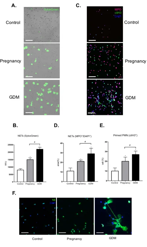
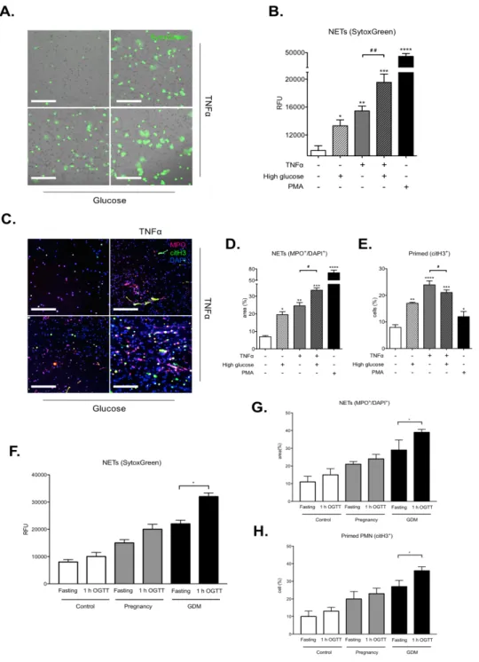
APOPTOSIS ASSAY ... 42 ASSAY FOR REACTIVE OXYGEN SPECIES (ROS) ... 43 MTT ASSAY ... 43 RADIOMETRIC GLUCOSE UPTAKE ASSAY ... 44 STATISTICAL ANALYSIS ... 45 RESULTS ... 46 SKEWING OF PMNS TOWARD A PRO-NETOTIC PROFILE DURING PREGNANCY ... 47 INCREASED LEVELS OF NETOSIS MARKERS DURING GDM ... 49 NEUTROPHILS FROM GDM WOMEN EXHIBIT EXCESSIVE NETS FORMATION ... 51 GLUCOSE AND TNF-ALPHA ACT ADDITIVELY TO DRIVE NETS RELEASE ... 53
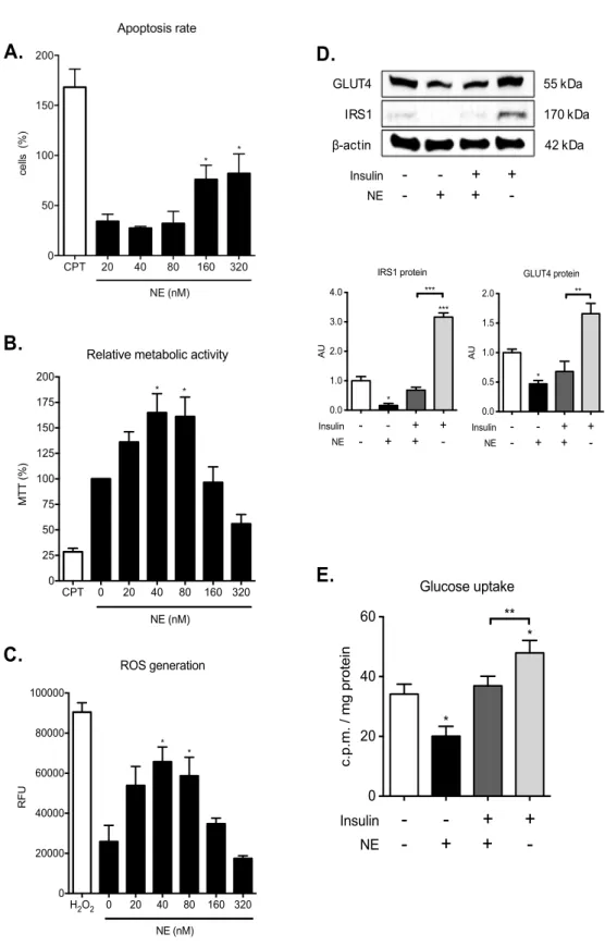
PMNS LEADS TO EXCESSIVE NE RELEASE AND IRS1 DEGRADATION IN PLACENTA ... 55
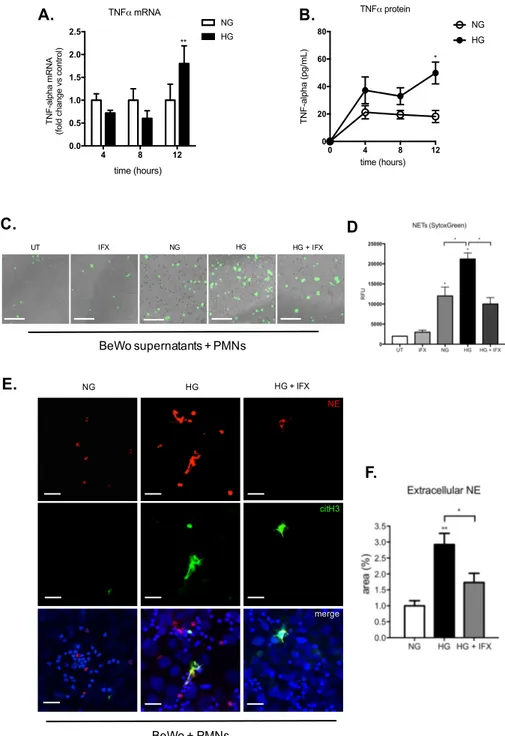
NE IMPAIRS GLUCOSE UPTAKE IN BEWO CELLS ... 57 HYPERGLYCEMIA TRIGGERS TNF-MEDIATED ACTIVATION OF PMNS IN BEWO CELLS ... 59 DISCUSSION ... 61 CONCLUSIONS ... 68 REFERENCES ... 70
A1AT Alpha-1 Antitrypsin Akt Protein kinase B APC Antigen-Presenting Cell CEACAM Carcinoembryonic Antigen Related Cell Adhesion Molecule CitH3 Citrullinated Histone H3 CXCL Chemokine (C-X-C motif) Ligand DCs Dendritic Cells dN Decidual Neutrophils G-CSF Granulocyte Colony-Stimulating Factor G-CSFR Granulocyte Colony-Stimulating Factor Receptor GDM Gestational Diabetes Mellitus GLUT4 Glucose Transporter Type 4 h Hours HDNs High-Density Neutrophils HFD High Fat Diet HG High Glucose HLA-C Major Histocompatibility Complex, Class I, C hPGH Human placental Growth Hormone hPL Human Placental Lactogen IFN Interferon IFX Infliximab IGF-1 Insulin Like Growth Factor 1 IIIT Third Trimester IIT Second Trimester IL Interleukin IRS1 Insulin Receptor Substrate 1 IT First Trimester LDNs Low-Density Neutrophils m Minutes MHC Major Histocompatibility Complex MOs Macrophages MPO Myeloperoxidase NADPH Nicotinamide Adenine Dinucleotide Phosphate NE Neutrophil Elastase NETs Neutrophil Extracellular Traps NF Nuclear factor NG Normal Glucose NK Natural Killer NLR Neutrophil to Lymphocyte Ratio
NO Nitric Oxide NOX Nicotinamide Adenine Dinucleotide Phosphate Oxidase OGTT Oral Glucose Tolerance Test PADI4 Protein Arginine Deiminases PDGFR Platelet-Derived Growth Factor Receptor PDK1 3-phosphoinositide dependent protein kinase-1 PI3K Phosphoinositide 3-kinase PKC Protein kinase C PMA Phorbol 12-myristate 13-acetate PMNs Polymorphonuclear Leukocytes PPROM Preterm Premature Rupture Of Membranes RFU Relative fluorescent units ROM Rupture of Membranes ROS Reactive oxygen species RT Room Temperature s Seconds T2DM Type 2 diabetes mellitus Th Helper T cells TLR Toll-Like Receptor TNF Tumor Necrosis Factor Tregs Regulatory T cells uNK Uterine Natural Killer Cells
This page is intentionally left blank
ABSTRACT
Human pregnancy is associated with a mild pro-inflammatory state characterized by activation of circulatory neutrophils (PMNs). Skewing of PMNs responses toward to neutrophil extracellular traps generation (NETs) is reflected in an increased of circulating nucleosomes and myeloperoxidase with advancing gestational age. Our data indicated that this pro-NETotic profile is enhanced in women with gestational diabetes mellitus (GDM). Maternal hyperglycemia and increased levels of TNF-a are a hallmark of GDM and we show a synergistic effect of both factors on the priming and release of NETs. Moreover, we hypothesized that systemic activation was associated with activated PMN in placenta. Indeed, we observed a massive infiltration of pro-NETotic PMNs and neutrophil elastase (NE) accumulation along chorionic villi of GDM placentas. To further explore whether hyperglycemia predisposes to exaggerated inflammatory response in placenta we incubated trophoblast BeWo cells in high glucose conditions and we next tested the TNF-a production capacity. Interestingly, TNF-a level was incresed and exert a pro-NETotic effect on PMN with consequent NE release. Recent studies in cancer tissues and diabetes models have described that released NE induce profound changes in the surrounding cells, altering the signal transducing cascade and promoting insulin resistance via degradation of insulin receptor substrate 1 (IRS1). Our in-vitro data indicate that addition of NE to trophoblast cell line BeWo causes degradation of IRS1 with consequent glucose uptake impairment. IRS1 is reduced in GDM placentas when compared to control placentas, suggesting that the presence of NE might be the causal factor. Taken together, our data showed that GDM is characterized by excessive NET formation and by a massive influx of pro-NETotic PMN into placentas. These findings underline the competence of NETs as a highly relevant diagnostic biomarker for GDM and NE as a new potential therapeutic target.
La gravidanza è da considerarsi una condizione pro-inflammatoria dove si osserva un’attivazione dei neutrofili circolanti. Con l'avanzare della gravidanza la concentrazione nel sangue di nucleosomi e mieloperossidasi aumenta e riflette la produzione delle trappole extracellulari dei neutrofili (NETs). Abbiamo dimostrato che in corso di diabete mellito gestazionale (GDM) tale produzione è aumentata in confronto ad una gravidanza fisiologica. Elevati livelli di glucosio e TNF-a, segni tipici presenti in corso di GDM, in vitro agiscono in modo sinergico e sono in grado di pre-attivare i neutrofili ed indurre il rilascio delle NETs. Abbiamo ipotizzato che, lo stato di iperattivazione osservato a livello sistemico possa essere associato ad una aumentata attività leucocitaria a livello placentare. A sostegno della nostra ipotesi, si osserva nei villi coriali isolati da placente GDM un’aumentata infiltrazione di PMNs pro-NETotici in associazione ad un accumulo della neutrofili elastasi (NE). Per valutare un possibile effetto pro-infiammatorio del glucosio a livello placentare, abbiamo incubato cellule di trofoblasto (BeWo) in presenza di un’elevata concentrazione di glucosio e successivamente valutato la produzione di TNF-a. È stato interessante rilevare un aumento nel rilascio di TNF-a tale da indurre un effetto pro-NETotico sui PMN con consequente rilascio della NE. Da recenti ricerche è emerso come in corso di diabete e tumore la NE possa essere internalizzata dalle cellule ed alterare la trasduzione del segnale insulinico attraverso la degradazione del substrato 1 del recettore insulinico (IRS1). Esperimenti in vitro hanno dimostrato che in presenza della NE si osserva una riduzione di IRS1 nelle cellule BeWo ed una diminuzione dell’internalizzazione del glucosio. Poichè l’espressione di IRS1 risulta ridotta nelle placente GDM é verosimile ipotizzare che la massiva presenza di NE ne può essere la causa. In conclusione, i nostri dati suggeriscono che in corso di GDM si verifica un’elevata produzione di NETs ed una massiva infiltrazione di pro-NETotici PMN nella placenta. Queste scoperte dimostrano come la NETosi abbia una significativa utilità diagnostica in corso di GDM e la NE un nuovo potenziale bersaglio terapeutico.
INTRODUCTION
Peeking into the novels aspect of neutrophil behavior
Neutrophil are the most abundant members of the leucocyte population, comprising between 40 and 60% of total white blood cells in normal healthy individuals. They play a crucial role in the immune defence against bacterial and fungal pathogens, and they also participate in the development of the inflammatory reaction (Nathan 2006). Neutrophils develop in the bone marrow from haematopoietic stem cells in a process called “granulopoiesis” and mature neutrophils are characterised by their segmented nucleus and granules that are filled with >700 proteins (Rørvig et al. 2013). Since their nucleus is organised into three to five lobules they are often called polymorphonuclear leukocytes (PMNs).
They were typically considered to be short-lived (few hours) but recently it has been shown that non-activated neutrophils have a longer lifespan (>5 days) (Pillay et al. 2010). Furthermore, neutrophils can not only migrate into tissues in response to inflammatory stimuli, but they are present in non-inflamed tissues where they have an important role in maintaining tissue homeostasis and shaping the immune responses (Ng et al. 2011).
Neutrophil biology has been studied extensively in recent years highlighting additional and unexpected functions of these cells. Neutrophils have been proposed to participate in protection against intracellular pathogens such as viruses and mycobacteria, but also as instructors of the immune system in the context of cancer and autoimmune disorders (Mócsai 2013; Amulic et al. 2012). Finally, neutrophils were found to be involved in physiological and pathological processes beyond the immune system, such as wounds healing, diabetes, atherosclerosis, and thrombus formation (Mócsai 2013; Amulic et al. 2012). The role of PMNs in reproduction is still a largely neglected topic, despite their involvement in various stages of the reproductive cycle and pregnancy-related disorders.
Heterogeneity in function and phenotype
PMNs are an essential component of the human innate immune system since they have a well-established role during fungal and extracellular bacterial infections where they promote bacterial clearance through phagocytosis, degranulation, exocytosis and the release of neutrophil extracellular traps (NETs) (Kruger et al. 2015; Kolaczkowska and Kubes 2013a). Neutrophils are one of the first blood cells to respond to infection and are recruited from the circulating blood into the tissue by chemoattractants. There are numerous host derived factors that trigger recruitment of neutrophils, among them one of the most potent is the interleukin (IL)-8 (Nathan 2006).
Neutrophils exist in one of three states: resting, primed or active (Hallett et al. 1995). In healthy condition, neutrophils exist in circulation in resting state, which ensures that their toxic intracellular contents are not accidentally released to damage host tissue. Resting neutrophils can become primed by agents that include bacterial products, cytokines and chemokines, e.g. TNF-α, G-CSF and IL-8 (Cowburn et al. 2008). This process leads to dramatic changes which enhance neutrophil function and lifespan (Wright et al. 2010). The process of priming alters the properties of the endocytic compartment in neutrophils enhancing dynamin-II-dependent and clathrin-mediated endocytosis (Lamb et al. 2012). Furthermore, the intracellular pools of CD11b and CD66b translocate from the intracellular granules to the plasma membrane to enhance cellular adhesion to integrin ligands (Wittmann et al. 2004). Additionally, phosphorylation of Ser-345 on p47phox is a necessary event in priming of neutrophils by TNF-α and G-CSF (Dang et al. 2006). The primed neutrophils are “ready to go” but awaiting further stimulus before activity is triggered (Hallett et al. 1995). Upon activation, neutrophils produce ROS in a process called the respiratory burst and use their extensive arsenal to ensnare and kill microbes (Cowburn et al. 2008; Amulic et al. 2012). ROS are generated almost exclusively by an NADPH oxidase that belongs to the family of NOX proteins. The leukocyte specific NADPH oxidase is a multisubunit entity with membrane-bound and soluble components that assemble into a heteromeric complex when the cells are stimulated. ROS are clearly important for neutrophil antimicrobial activity: neutrophils from chronic granulomatous disease (CGD) patients kill microbes poorly, making these patients susceptible to many infections (Segal et al. 2000). By definition, CGD results from failure of activity of the NADPH oxidase caused by defects in the components of this system (Segal et al. 2000). In addition to direct antimicrobial action, ROS can modify host molecules. A well-studied example of ROS in signalling is the reversible regulation of various targets (including phosphatases, metalloproteinases, and caspases) by direct oxidation of cysteine residues (Paulsen and Carroll 2010).
Phagocytosis is the major mechanism used by PMNs to remove pathogens and cell debris. It is an active, receptor-mediated process during which a particle is internalized by the cell membrane into a vacuole called the phagosome. Because particles of widely different natures can be taken up by phagocytosis, it is not surprising that numerous receptor types can mediate this process. Foreign particles such as bacteria, fungi, and parasites display many molecules that are never found in higher organisms. These pathogen-associated molecular patterns (PAMPs) can be detected by several receptors, in particular the Toll-like receptors (TLRs) (Flannagan, Jaumouillé, and Grinstein 2012). Foreign bodies can also be recognized by soluble molecules circulating in the blood and in interstitial fluids. For example immunoglobulins recognize extraneous antigens, and components of the complement cascade deposit on foreign surfaces. Following their deposition on particles, these molecules, termed opsonins, are in turn engaged by specific receptors on the membrane of PMNs, including receptors Fc R-I, -II, and -III and fragments of the third component of complement (Flannagan, Jaumouillé, and Grinstein 2012).
Importantly, besides ROS and phagocytosis, neutrophils possess an array of granules that contain a large number of cytotoxic and immune regulatory molecules that can be released by degranulation or exocytosis. As a neutrophil proceeds through activation, granules are mobilized and fuse with either the plasma membrane or the phagosome, releasing their contents into the respective environment (Borregaard and Cowland 1997). The different classes of granules demonstrate varying propensities for mobilization in response to inflammatory signals (Sengeløv, Kjeldsen, and Borregaard 1993). Because of this varying mobilization propensity, each granule subset has been traditionally associated with a particular stage of neutrophil activation (Borregaard and Cowland 1997). The primary granules are the main storage site of the most toxic mediators, including elastase, myeloperoxidase, cathepsins, and defensins. (Borregaard and Cowland 1997).
Neutrophils are no longer seen as leukocytes with a sole function of being the essential first responders in the removal of pathogens at sites of infection but they can positively or negatively regulate the generation of adaptive immune responses (Jaillon et al. 2013; Leliefeld, Koenderman, and Pillay 2015). Neutrophils were shown to promote the maturation of human monocyte-derived DC through interaction between CD18 and CEACAM1 expressed by neutrophil and DC-SIGN receptor on DC (van Gisbergen et al. 2005). The interaction of neutrophils with DC can also result in NK cell activation increasing the release of IL-12p70 (Costantini et al. 2011). Recently a new population of neutrophils was described as residing around the marginal zone of the spleen called “B-cell helper neutrophils” (NBH) (Puga et al. 2012). These neutrophils colonize the marginal zone during foetal life and become more prominent after postnatal mucosal colonization by bacteria. NBH express higher levels of B
cell-stimulating molecules and B cell chemoattractant molecules such as CXCL12 and CXCL13 compared to circulating neutrophils (Puga et al. 2012). Neutrophils can also modulate T-cells by proteases released during degranulation. These proteases are able to cleave and inactivate essential cytokine receptor on T-cells, such as IL-2 and IL-6 receptor (PMID: 11272269). Further, neutrophils can inhibit T-cell by production of hydrogenperoxide (H2O2) through cell-cell contact (Narasaraju et al. 2011).
Interestingly, PMNs use a set of membrane and intracellular molecules to sense signals from their local environment and polarize their phenotype towards a pro-inflammatory (N1) or anti-inflammatory (N2) programs (Fridlender et al. 2009; Sagiv et al. 2015). It was also shown that subpopulations of circulating neutrophils in cancer can be distinguished according to their densities (Sagiv et al. 2015), one with “normal,” HD characteristics (HDNs) having antitumor properties, previously described as N1 (Fridlender et al. 2009) , whereas the other (LDNs) being of lower density with features associated with protumor activity (N2). The cytokine involved in neutrophils polarization is TGF-β that can mediate a shift between the different neutrophils subtypes. HDNs can switch to become LDNs in the presence of increasing concentrations of TGF-β, both in vivo and ex vivo (Sagiv et al. 2015). In light of the immature state of a subset of LDNs and the mature state of all HDNs, the low- to high-density transition raises the possibility of neutrophil maturation in the circulation (Galli, Borregaard, and Wynn 2011). Neutrophil plasticity and diversity in humans is a recent discovery that needs further investigation to define their role in different human pathologies.
Dynamics of Neutrophil Extracellular Traps (NETs) formation
Neutrophil extracellular traps (NETs) formation is a unique form of cell death (NETosis) that is characterized by the release of decondensed chromatin and granular contents to the extracellular space (Brinkmann et al. 2004b). The mechanism of NET formation is clearly distinct from apoptosis because the morphological characteristics of these two forms of active cell death are very different, there is no DNA fragmentation and phosphatidylserine is not exposed before cell death (Fuchs et al. 2007). NETs are produced by neutrophils in contact with pathogens and with a variety of host factors such as activated platelets or inflammatory stimuli (Fuchs et al. 2007). NETs are released particularly in response to large microbial structures that cannot be easily phagocytosed such as Candida albicans hyphae and Mycobacterium bovis aggregates (Branzk et al. 2014). Only recently, it was recognized that NETs are also generated during viral infection (Saitoh et al. 2012) .
The molecular mechanisms behind NETs formation are still poorly understood but it was shown that chromatin decondensation is an essential step (Y. Wang et al. 2009b). The exact sequence of events leading to NEtosis is not known, although it was confirmed that is dependent upon neutrophil elastase (NE), myeloperoxidase (MPO) and the enzymes peptidyl arginine deiminase 4 (PAD4). NE translocates first to the nucleus, where it digests nucleosomal histones and promotes extensive chromatin decondensation and the late binding of MPO to chromatin enhances the NE activity (Papayannopoulos et al. 2010b; Metzler et al. 2014). NE is essential for chromatin decondensation, indeed elastase KO mice are NETosis incapable (Papayannopoulos et al. 2010b). On the other hand, the role of MPO seems to depend on the nature of the stimulus (Parker et al. 2012b). MPO-deficient neutrophils had no effect on NETs induction by P. aeruginosa, S. aureus or E. coli, but fail to respond to PMA (Parker et al. 2012a).
Initially, it was shown that NETs is a process that is dependent on ROS production. In fact, humans with NADPH-oxidase deficiency could not make NETs (Bianchi et al. 2009). On the contrary, there is now growing evidence suggesting that microbial-specific molecular patterns recognized by Pattern Recognition Receptors (PRRs) induce NETs independently of NADPH oxidase 2 (Pilsczek et al. 2010a). Therefore, based on their requirement for NADPH oxidase (NOX2), in the recent literature, NETs are classified in two types: NOX-dependent and NOX-independent (Pilsczek et al. 2010a; Zhao, Fogg, and Kaplan 2015).
NOX-dependent NETosis occurs following stimulation by PMA, a strong protein kinase C (PKC) agonist. Raf-MEK-ERK pathway is involved through activation of NADPH oxidase and upregulation of antiapoptotic proteins (Hakkim et al. 2011a). To release
NETs, activated neutrophils undergo dramatic morphological changes. During the 1 hour of stimulation the nucleus loses its lobules, the chromatin decondenses, and the inner and outer nuclear membranes progressively detach from each other. After 1 h, the nuclear envelope disaggregates into vesicles and the nucleoplasm and cytoplasm form a homogenous mass. After 2 h, the cell membrane ruptures and the interior of the cell is ejected into the extracellular space, forming NETs (Brinkmann and Zychlinsky 2012).
Besides PMA, a synthetic activator of the PKC family of enzymes, there are several natural inducers lead to PKC activation, such as TNF-a, IL-1ß, IL-8, IL-1 or G-CSF (Newton and Dixit 2012).
NOX-independent NETosis has been reported following both direct microbial exposure and lipopolysaccharide (LPS). For gram-negative bacteria, NETs are induced via Toll-like receptor (TLR) 4 activation of platelets followed by direct neutrophil-platelet interaction via CD11a, whereas both complement receptor 3 and TLR2 are required for vital NETosis following gram-positive infection (Pilsczek et al. 2010b). The regulators of NOX-independent NETosis are largely unknown. Douda et al showed that calcium activated NOX-independent NETosis is fast and mediated by a calcium-activated small conductance potassium (SK) channel member SK3 and mitochondrial ROS (David Nobuhiro Douda et al. 2015). Although mitochondrial ROS is needed for NOX-independent NETosis, it is not mandatory for NOX-dependent NETosis (David Nobuhiro Douda et al. 2015).
Unlike NOX-dependent NETosis, NOX-independent NETosis is accompanied by a substantially lower level of activation of ERK and moderate level of activation of Akt, whereas the activation of p38 is similar in both pathways (David Nobuhiro Douda et al. 2015). ERK activation is essential for the NOX-dependent pathway, whereas its activation is not essential for the NOX-independent pathway. Despite the differential activation, both NOX-dependent and -independent NETosis require Akt activity (David Nobuhiro Douda et al. 2015). Indeed, suppression of Akt switches NETosis to apoptosis (David N Douda et al. 2014). During the NOX-independent NETosis, NETs are extruded during the first few minutes (<30 min) of the neutrophils activation process (Pilsczek et al. 2010a). Moreover, the extrusion of DNA from within the nucleus to the extracellular space involves vesicular trafficking without requiring membrane perforation (Pilsczek et al. 2010a; Zhao, Fogg, and Kaplan 2015). This mechanism spares the cells from self-lysis, thereby allowing the PMN to continue to function, even to the point of becoming anuclear (Pilsczek et al. 2010a). Because these opposing cellular strategies, one causing cell death and the other leaving the cell alive, NOX-dependent netosis is called suicidal and NOX-independent NETosis is called vital (Pilsczek et al. 2010a; Zhao, Fogg, and Kaplan 2015).
In addition, histone citrullination by PAD4 is a prominent posttranslational modification in vital NETosis. Inhibition of PAD4 by Cl-amidine, a PAD inhibitor, disables NET formation (Y. Wang et al. 2009a). Like other PAD isotypes, PAD4 is a Ca2+ dependent enzyme, but only PAD4 possesses a classical nuclear localization signal (NLS) (Mastronardi et al. 2006a). The requirement for PAD4 is not universal to all NETotic pathways, as evidenced by the fact that there is a lack of PAD4 activity and histone hypercitrullination in PMA-mediated NETosis (David Nobuhiro Douda et al. 2015)
The research performed by Obermayer et al. has significantly highlighted the structure and possible functions of NETs (Obermayer et al. 2014). NETs have a unique ultrastructure, whose main components are DNA fibres, of diameter 15–17 nm and spherical protein domains of diameter 25–50 nm. Histone proteins such as H1, H2A, H2B, H3, H4 constitute about 70% of all proteins connected with NETs (Obermayer et al. 2014). Because of post-translational modifications that affect histones after the NETosis process, histones which construct neutrophil extracellular traps have a larger cell molecular mass of 2–5 kDa compared with the histones present in the cell nucleus (Papayannopoulos et al. 2010b; Metzler et al. 2014) . Among non-histone proteins present in NETs, neutrophil elastase (NE) and MPO are the most common, with a content of over 5% (Urban et al. 2009b). However, apart from NE and MPO, NETs structure also contains other proteins originating in different cellular organelles of neutrophils (Urban et al. 2009b). Even at low concentrations, they appear to play an important role in host defense (Brinkmann and Zychlinsky 2007), among them we can find (Urban et al. 2009b):
• cathepsin G, defensins, BPI-bactericidal substance-increasing permeability – originating from the primary neutrophil granule;
• alcaic phosphatase, lactoferrins, lysozyme, cathelicidins, collagenase – deriving from the granularity of secondary neutrophils;
• gelatinase, (MMP-9) – matrix metalloproteinase 9 – deriving from the granularity of tertiary neutrophils;
• catalase – deriving from peroxisomes,
• cytokeratin-10, α-actinin 1 and 4, β-actin, γ-actin, myosin-9 – constituting the cytoskeleton of neutrophils;
• some cytoplasmic proteins, among others: proteinase 3, cathelicidin LL-37, tryptase, S100 (S100A8, S100A9, S100A12) group proteins.
Impact and implications of NETs
NETs provides important biological advantage for the host to fight against microbial infections (Branzk and Papayannopoulos 2013a). A variety of gram-negative as well as gram-positive bacteria have been shown to induce NETs formation. S. flexneri is trapped and killed by NETs, which contain NE that degrades virulence factors such as IcsA and IpaB (Brinkmann et al. 2004a). Moreover, NETs specifically degrade S. aureus virulence factors and thereby help to contain tissue damage (Brinkmann et al. 2004a). Interestingly, the gram-negative bacterium K. pneumoniae is not sufficient to induce NETosis in isolated neutrophils ex vivo, but is a good inducer of NETosis in a mouse lung infection model (Papayannopoulos et al. 2010a). These observations suggest that microbial virulence may be an important stimulus in NET induction, as it may induce additional inflammatory cytokines that may act as co-activators.
PMNs play a crucial role in containing fungal infections and NETs appear to be an important part of the neutrophil antifungal arsenal. Since hyphae are too large to be phagocytosed, extracellular killing by release of NETs is an ideal strategy to contain the hyphal form and a number of studies have demonstrated that NETs are sufficient to kill C. albicans yeast and hyphae (Urban et al. 2006). Calprotectin is a cytoplasmic protein that is released via NETosis and is found associated with C. albicans in NETs (Urban et al. 2009a). The importance of calprotectin in antifungal defense is crucial since calprotectin chelates Mn2+ and Zn2+ which are required for C. albicans growth
(Urban et al. 2009a). NETs are also able to trap and neutralize the negatively charged HIV virions, significantly decreasing HIV infectivity. However, in order to suppress NETs formation, HIV engages CD209 on dendritic cells (DCs), leading to production of IL-10 by DCs, which suppresses NETs formation (Saitoh et al. 2012). The role of NETs has also been explored in parasites causing leishmaniasis. Entrapment in NETs leads to decreased viability of the parasites, although authors of different studies conclude that the main function of NETs in Leishmania infection is the immobilization of the parasite and containment of the infection (Gabriel et al. 2010) . NETs are also implicated in Toxoplasma gondii infection. NETs kill approximately 25 % of the entangled parasites, which indicates that the primary function of NETs may be to physically contain the infection (Abi Abdallah et al. 2012).
These studies suggest that NETs play an important role in the defense against wide range of pathogens (Branzk and Papayannopoulos 2013a). However, too much of a good thing can be a bad thing. NETosis dysregulation has been implicated in severe autoimmune and autoinflammatory disease. For example, systemic lupus erythematosus (SLE) patients exhibit elevated levels of anti-neutrophil cytoplasmic antibodies (ANCAs), antibodies against histones, DNA (ANAs) and ribonucleoproteins (RNP) that target host tissues [81]. NETs are a major source of extracellular chromatin,
neutrophil proteins and microbial co-stimulatory adjuvants, and are therefore suspected as an antigenic source in SLE (Branzk and Papayannopoulos 2013b).
Maintaining the right balance of NETs formation that accumulates in tissues is essential for preventing damage to the hosts. For example it has been observed that in Lewis lung carcinoma and Ewing sarcoma there are large necrotic area of dead neutrophils and NET-like structures (Berger-Achituv et al. 2013). Another example was recently published by Wong et al., they reported that large quantities of NETs were found in excisional skin wounds of diabetic mice and that DNase1, which dismantled NETs, accelerated wound healing (Wong et al. 2015b). The authors attributed the cause of the large NETs quantity to PAD4 and revealed a fourfold upregulation of PAD4 protein expression in the neutrophils from individuals with diabetes compared to healthy controls (Wong et al. 2015b). In a different pathology Gupta et al observed the presence of large numbers of NETs directly in the intervillous space of preeclamptic placentas (A. K. Gupta et al. 2005). Since excessive fibrin deposition and infarction are frequently observed in preeclamptic placentas, NETs are likely to be responsible for exacerbating the occlusion of blood flow through the intervillous space (A. K. Gupta et al. 2005). Neutrophils through the release of NETs may also contribute to the widespread systemic damage to the maternal endothelium observed in preeclampsia since endothelial cells are susceptible to cell death induced by NETs (A. K. Gupta et al. 2010).
Pregnancy and Myth of Fetal Allograft
The myth of “fetal allograft” has created the concept that pregnancy is associated with immune suppression (Ober 1998) and a substantial body of literature exists describing the mechanisms fetuses use to escape the maternal immune surveillance (Warning, McCracken, and Morris 2011). This concept, unfortunately, does not underline that pregnancy represents the most important period for the conservation of the species. It is fundamental for the immune system to strengthen all the means to protect the mother and the offspring from pathogens. Therefore, it is appropriate to refer to pregnancy as a unique immune condition that is “modulated”, but not “suppressed” (Mor et al. 2011).
Maternal mortality is surprisingly high and about 830 women die from pregnancy- or childbirth-related complications around the world every day (Kleppel et al. 2016). Moreover, the world’s number one killer of young children is not an infectious disease but rather is preterm birth. Each day, 3000 children under 5 years of age die from direct complications of being born before 37 completed weeks of pregnancy (Liu et al. 2015). Furthermore, other pregnancy complications can have severe short- and long-term health effects for both the mother and foetus. For example, women who had decreased haemoglobin levels during pregnancy were found to have two times the chance of eventual death by heart disease compared to their counterparts whose haemoglobin levels remained stable (M. Lee et al. 2011; Cirillo and Cohn 2015). And those who had preeclampsia were found to be almost 6 times more at risk of cardiovascular diseases and related death (M. Lee et al. 2011; Cirillo and Cohn 2015). In addition, gestational diabetes mellitus (GDM), besides all of the adverse effects that go along with pregnancy, affects health later in life for both mother and child increasing the risk of developing obesity, diabetes type 2 and metabolic diseases (Bellamy et al. 2009; Lauenborg et al. 2004).
Immunological phases during pregnancy
Although pregnancy is characterized by an enhanced inflammatory state, it is known that inflammation has to be fine modulated at both placenta and systemic level to promote fetal development and avoid pregnancy complications. A common mistake in the field of reproductive medicine is considering from the immunological point of view pregnancy as a single event. In reality, women go through at least three distinct immunological phases during gestational advancement (Mor et al. 2011). Noteworthy, immunological phases at the implantation site are not fully revealed at systemic level in term of circulating serum cytokines (Christian and Porter 2014).
The first trimester (week 1-week 12) of pregnancy is considered as pro-inflammatory at the implantation site. In fact, during implantation the blastocyst has to break through the epithelial lining of the uterus in order to implant. An inflammatory environment is required in order to secure the adequate repair of the uterine epithelium and the removal of cellular debris. A high level of uterine-specific natural killer (NK) cells (65-70%), antigen presenting cells (APC) such as macrophages (MOs) and dendritic cells (DCs) (10-20%), T helper (Th)-1 cells and pro-inflammatory cytokines like TNF-a, IFN-γ, IL-1, IL-1β, IL-6, IL-12 secreted by the endometrial cells characterize the site of implantation (Dekel et al. 2010).
The second trimester (week 13-week 28) is a period of rapid fetal growth and development and is considered an anti-inflammatory state. At the implantation site immune cells that produce IL-4 and IL-10 largely mediate the resolution of inflammation. IL-4 aids in the polarization of antigen-stimulated naïve Th cells into Th2 effector cells and the presence of TGF-b facilitate the polarization of inducible Tregs from naïve CD4+ T cells (Chatterjee et al. 2014). IL-10 primarily exerts its anti-inflammatory effect by inhibiting pro-anti-inflammatory cytokines and blocking antigen presentation by reducing MHC class II expression (Chatterjee et al. 2014).
During the 3rd trimester (week 29-week 40), the fetus complete its development and the mother needs to deliver the baby. Parturition can only be achieved through renewed inflammation to promotes the contraction of the uterus and the expulsion of the baby (Romero et al. 2006).
Besides the immunological phases at the implantation site, maternal immune system undergoes systemically changes characterized by significant increase in serum pro-inflammatory mediators (Christian and Porter 2014). Few studies have examined longitudinal changes in circulating serum cytokines as pregnancy progresses. Although, women showed elevations in TNF-α from early to late pregnancy, both IL-8 and IL-1β showed a U-shaped curve in the serum, with decreases in mid or late
pregnancy compared to early pregnancy. This pattern of response suggest in early pregnancy an initial inflammatory response that is then down-regulated over time (Christian and Porter 2014). On the other hand, trophoblast and macrophage-like cells constitutively produce IL-8 during pregnancy, showing maximal production of IL-8 by second trimester and term placentas (Shimoya et al. 1992).
Future studies are needed to identify the functional effects of specific cytokines during each stage of gestation, both at the peripheral and local level, as these functions should provide insight into mechanisms underlying adverse perinatal health outcomes.
Contributions of PMNs to pregnancy
Immune cells residing in the reproductive tract are T cells, macrophages/dendritic cells, natural killer (NK) cells, mast cells and neutrophils (S. K. Lee et al. 2015). The leukocytes in the reproductive tract play different roles depending from the site they reside: they maintain immunity against vaginal pathogens in the lower tract and establish immune tolerance for sperm and an embryo/fetus in the upper tract. The role of neutrophils in reproduction is still a largely neglected topic, despite the evidence of their importance at various stages of the female reproductive cycle such as menstrual cycle, implantation and pregnancy (Hahn et al. 2012).
Neutrophils generally are barely detectable in normal human endometrium but the number rises up to 6–15% of the total cell number during the menstrual period (Salamonsen and Woolley 1999). In murine systems, the migration of PMNs into the vaginal vault appears to play a crucial role in the continued progression of the oestrous cycle, as their depletion leads to a blocking of the cycle in diestrus (Salamonsen and Woolley 1999). This neutropenic abrogation of the oestrous cycle was accompanied by reduction in the levels of the sex hormones oestradiol and progesterone, implying some form of feedback mediated by endometrial presence of infiltrating PMNs (Sasaki, Nagata, and Kobayashi 2009). During pharmacological miscarriage, Milne et al. (Milne et al. 2005) showed that neutrophil infiltrate into human decidua after administration of a progesterone antagonist (RU-486) and were responsible for the vascular breakdown and decidual shedding. In a recent study, Amsalem and colleagues revealed that PMNs were present in the human decidua during 2nd trimester and that PMNs clusters were frequently located close to spiral arteries (Amsalem et al. 2014). Co-culture experiments determined that decidual neutrophils (dN) were able to promote angiogenic sprouting by uterine microvascular endothelial cells (Amsalem et al. 2014).
In order to maintain an environment favorable to implantation, neutrophils are also recruited into the female reproductive tract following insemination where they play a major role in the removal of excess sperm (Strzemienski 1989; Alghamdi et al. 2009). Indeed, sperm can be antigenic to the recipient female and immunization against them can result in infertility (Alghamdi et al. 2009).
In pregnant women the number, activation state and migratory capacity of PMNs are increased compared to non-pregnant women (Hahn et al. 2012; G. P. Sacks et al. 1998). Interestingly, in non-pregnant women neutrophil apoptosis was significantly delayed and was further delayed in pregnancy with intrauterine growth restriction and preeclampsia (von Dadelszen et al. 1999). The activation of circulating neutrophils in preeclampsia was similar to the activation state of neutrophils found in sepsis (G. P. Sacks et al. 1998). Since preeclampsia is associated with endothelial cell activation and dysfunction (Noris, Perico, and Remuzzi 2005), the increased neutrophils activation may cause the damage of vascular endothelium. Endothelial damage in vitro by complement activated PMNs was originally described by Sacks et al. (T. Sacks et al. 1978). There is now abundant evidence that PMNs mediate endothelial cell/tissue damage in vivo by a wide variety of mechanisms, such as release of damaging oxidants and proteolytic enzymes (Gao, Neff, and Ward 2006).
Despite several studies have aimed to examine the role of circulating PMNs in pregnancy, little is known regarding their effects in human placenta. However, the presence of neutrophils in reproductive tissues at term and their ability to migrate to this region during labor have been well documented in both humans and rodents (Sharp et al. 2016; Giaglis, Stoikou, Grimolizzi, et al. 2016). Neutrophils migration into tissues includes the following steps: tethering, rolling, adhesion, crawling and transmigration. Tethering and rolling of neutrophils, which greatly facilitate subsequent arrest and recruitment, are mediated by selectins (Yago et al. 2010). Neutrophils adhesion and crawling is facilitated by increased expression of integrin at the cell surface such as CD11b and CD66b (Luo et al. 2015). Many molecules are implicated in transmigration (Bayat et al. 2010) and in order to transmigrate across the membranes, neutrophils release specific proteases such as MMPs and serine proteases. These enzymes are able to affect neutrophil migration by the degradation of elastin and collagen, thereby increasing the vascular permeability (Monaco et al. 2006). Interestingly, these proteins are under hormonal regulation during pregnancy (Nekrasova and Shirshev 2013) and participate in the process of labor (Helmig et al. 2002). In human decidual tissues, the number of neutrophils was higher in women with preterm labor associated with chorioamnionitis than in women with term gestations (with and without labor) and in women with spontaneous preterm labor without chorioamnionitis (Hamilton et al. 2012). These observations suggest that neutrophils contribute to the physiological rupture of the fetal membranes (ROM) and
pathological preterm premature rupture of membranes (PPROM) during term and preterm labor (Hamilton et al. 2012).
The precise mechanism leading to the neutrophils migration into the placenta is currently unclear. It may involve the migration of circulatory neutrophils into this placental tissue via chemokines attraction (e.g. IL8) (Christian and Porter 2014). Trophoblast and macrophage-like cells constitutively produce a certain amount of IL-8 during pregnancy. IL-8 increased with development aging of the placenta, showing maximal production of IL-8 by second trimester and term placentas (Shimoya et al. 1992). The placenta: how to study a highly complex organ? In 2015 the National Institutes of Health (NIH) has dedicated $41.5 million in research awards for an initiative to understand the development, structure and functions of the human placenta during pregnancy with the goal of improving the health of mothers and children.
Many of the objectives of the Human Placenta Project, launched by the NIH will necessitate pre-clinical studies and testing in appropriately designed animal models that can be readily translated to the clinical setting.
The placenta is a complex and fascinating organ because during the course of a pregnancy, it acts as the lungs, gut, kidneys and liver of the fetus. There are two placenta components, a fetal and a maternal one that must interact successfully for a healthy pregnancy. The maternal component of the human placenta is the endometrium, which undergoes transition to form the decidua in early pregnancy. The two principal fetal sources are the trophectoderm that forms the wall of the blastocyst, and the underlying extraembryonic mesoderm. The trophectoderm differentiates into trophoblast, which in turn forms the epithelial covering of the placenta and the subpopulation of invasive extravillous trophoblast cells (Graham J Burton and Fowden 2015). Extravillous cells migrate from the tips of the placental anchoring villi into the wall of the uterus as far as the inner third of the myometrium. Uterine natural killer (uNK) cells play a key role in this process that depends on the combination of HLA-C ligands on the trophoblast cells and killer immunoglobulin-like receptors expressed by the uNK cells (Moffett, Hiby, and Sharkey 2015). Far from killing trophoblast cells, the uNK cells encourage migration of the extravillous trophoblast cells into the endometrium through the release of cytokines and chemokines (Moffett, Hiby, and Sharkey 2015). This is one remarkable example of the importance of trophoblast-immune interactions for a proper placentation.
The extraembryonic mesoderm forms the stromal core of the placenta, from which originate the fibroblasts vascular network and resident macrophage population
(Hofbauer cells) (Graham J Burton and Fowden 2015). The placenta has two surfaces, the chorionic plate that faces the fetus where the umbilical cord is attached, and the basal plate that comes into the maternal endometrium. Between these plates is the intervillous space that, combined with the villi, is the functional unit of the human placenta: here maternal fetal metabolic exchange occurs. The epithelial covering of the villous tree is the syncytiotrophoblast, a multinucleated syncytium that is involved in many of the functions of the placenta, such as the synthesis and secretion of large quantities of steroid and peptide hormones (G J Burton and Tham 1992).
In the light of recent debate at the 11th Congress of the European Society for Reproductive Immunology (Budapest, Hungary) it was agreed that no other mammal has a placenta identical to that of the human (Chaouat and Clark 2015). Although, there is an ongoing discussion regarding what represents the best animal model for placenta and pregnancy research, often the discussion is based on availability, housing conditions or financial considerations and not necessarily scientific merit (Chaouat and Clark 2015; Schmidt et al. 2015). Therefore, more representative models are required, such as primary trophoblast, villous explants and cell lines (Orendi et al. 2011).
Primary trophoblasts are routinely isolated in many laboratories and additionally stored to use cells from a single placenta for different projects over time. A major disadvantage of isolated primary trophoblasts is that they do no longer proliferate in culture. Hence, only short term cultures can be performed. At the same time these cells spontaneously syncytialize in culture and thus may be of use to study the release of syncytial fragments and factors in vitro (Tannetta et al. 2008). Villous explants are mostly used to dissect cellular processes such as proliferation, differentiation, apoptosis and syncytial fusion (Ahmed et al. 2016). One of the obvious drawbacks in studies using villous explants is the deficient availability of fresh material for explant cultures. Different to isolated primary trophoblasts, villous explants are not regularly stored. Hence, placental tissues can only be used directly after delivery, and explant cultures using tissues from a single placenta cannot be repeated at a later date. Several cell lines are available for in vitro analysis of placental cell function. The choriocarcinoma cell line BeWo is the most extensively used model since it reveals most of the characteristics of villous trophoblast, including syncytial fusion and secretion of hormones (Omata et al. 2013; Wolfe 2006). The cells were isolated at autopsy from a cerebral metastasis of a choriocarcinoma (Hertz 1959b). Then, the cells were transplanted to the cheek pouch of a hamster and maintained in hamsters through 304 serial transfers over a period of 8 years (Hertz 1959b). Only then Pattillo & Gey (Pattillo and Gey 1968b) removed cells from tumors in the cheek pouch of hamsters, cocultured them with decidual tissues and finally established the BeWo cell line.
In conclusion, many aspects of human placenta can only be understood on the basis of experiments on human cell lines and original placental tissues in combination with human subject studies (Chaouat and Clark 2015; Schmidt et al. 2015). Immunomodulation of PMNs by placenta Many factors are released from the placenta that can influence the PMNs behavior. The human placenta constitutively produces IL-8 during pregnancy and enhances its production in chorioamnionitis (Shimoya et al. 1999). Since IL-8 is chemotactic and activating factor for PMNs, placental IL-8 may act as a mediator to recruit and accumulate neutrophils into the feto–maternal area.
G-CSF, known to enhances key functions of PMNs such as superoxide production, phagocytosis and bacteriocidal killing (Cella et al. 2006), is also produced by the placenta (Umesaki et al. 1995). Interestingly, this cytokine has recently been implicated in a murine tumour model where it predispose neutrophil to the generation of NETs (Demers et al. 2012).
Immunomodulatory signals are also released as membrane-associated factors in the form of extracellular vesicles (EVs) (Sarker et al. 2014). Placental EVs are highly stable and contain a wide array of proteins, microribonucleic acids and retroviral proteins with stimulatory or inhibitory activities on immune cells (Tong and Chamley 2015). They are released in large quantities from the syncytiotrophoblast layer and include microparticles (0.2–1 µm) and exosomes (40–150 nm) (Tong and Chamley 2015). In general, microparticles may activate immune effector mechanisms, while exosomes lead towards an anti-inflammatory state. It is speculated that the physiological range of the microparticles/exosomes ratio is disturbed in various pregnancy complications (Mincheva-Nilsson and Baranov 2014). Gupta et al. showed that placentally-derived microparticle efficiently activated neutrophils and triggered NETs formation (A. K. Gupta et al. 2005).
Gestational Diabetes Mellitus:
The Dark Side of Glucose
The term Gestational Diabetes Mellitus (GDM) was first used by O'Sullivan in 1961 to describe any degree of glucose intolerance with onset or first recognition during pregnancy (O’Sullivan 1961). The background rate in pregnancy varies between 2 and 14% with incidences reported to be as high as 40% in obese populations (Chu et al. 2007; Kampmann et al. 2015a). GDM is the lacks specific symptoms, therefore, the pregnant woman is usually unaware of her condition until it is diagnosed at routine prenatal screening. Despite being symptom free, serious pregnancy complications are associated with hyperglycemia in pregnancy (Kampmann et al. 2015a). The seriousness of GDM and the dramatically increasing incidence of this condition make it one of the most urgent health challenges of this century. Screening, diagnosis and treatment: where do we stand? Screening for GDM is usually done between 24 and 28 weeks of gestation since insulin resistance increases during the second trimester and glucose levels rise in women who do not have the ability to produce enough insulin. Interestingly, the timing of the diagnosis of GDM is significantly associated with preterm delivery. Women who received a diagnosis of GDM before 24 weeks’ gestation were 10 times more likely to deliver before 37 weeks’ gestation than women who received a GDM diagnosis after 24 weeks’ gestation (Catalano and Sacks 2011). Precise level of glucose intolerance characterizing GDM has been controversial over three decades. However during the non-fasting Oral Glucose Tolerance Test (OGTT) the following ranges are recommended: fasting plasma glucose ≥ 5.1 mmol/L (≥ 92 mg/dL), 1 h ≥ 10.0 mmol/L (180 mg/dL), or 2 h ≥ 8.5 mmol/L (153 mg/dL) (International Association of Diabetes and Pregnancy Study Groups Consensus Panel et al. 2010). It has been estimated that with these new diagnostic criteria the prevalence of GDM will increase to nearly 18% (Webber, Charlton, and Johns 2015). From an economic perspective, GDM is associated with an excess of €1549.56 per patient in costs of care during pregnancy and €411.31 per patient in annual healthcare costs post pregnancy (Danyliv et al. 2015).
The main goal of GDM treatment is to prevent macrosomia and pregnancy complications. As a primary treatment of GDM an optimal diet combined to physical activity is generally recommended, aiming to maintain sufficient energy intake and ensure glycaemic control of the mother. This approach has been found to be sufficient in 70–85% of the GDM cases (American Diabetes Association 2016). If these measures are insufficient in terms of achieving optimal glycemic control subcutaneous insulin therapy is the therapy of choice (Vääräsmäki 2016). It is effective for the mother and
safe for the fetus, as it does not cross the placenta. However, insulin is relatively expensive and difficult to administer. It requires education to ensure a safe administration and it is associated with an increased risk of hypoglycemia and weight gain (Kelley, Carroll, and Meyer 2015). Therefore new oral glucose-lowering agents have been studied in the treatment of GDM, such as metformin and glibenclamide (Balsells et al. 2015). The benefits of these treatments during pregnancy include no risk of hypoglycemia or excessive maternal weight gain, are low cost and easy to use. However, both glibenclamide and metformin cross the placenta and their long-term effects on the child have yet to be considered. Short and long-term consequences of GDM
During GDM, the placenta undergoes a variety of structural and functional changes (Gernot Desoye and Hauguel-de Mouzon 2007). One of the characteristic features of a placenta from GDM is its increased weight, which is accompanied by edematous stroma, increase in the number of syncytial knots and perivillous fibrin deposition (Meng et al. 2015). Further, in GDM, the overexpression of placenta TNF-a is associated with increased fetal adiposity (Radaelli et al. 2003). Noteworthy, in diet-treated GDM patients, the amount of trophoblast insulin receptors is lower than in nondiabetic pregnancies, whereas in insulin-treated GDM, the placenta contains more insulin receptors (Al-Attas 1995).
GDM implies several risks on both mother and baby; for example increases the risk of cesarean and operative vaginal delivery, macrosomia, neonatal hypoglycemia and hyperbilirubinemia (Kampmann et al. 2015b). Moreover, women with GDM are at higher risk of hypertensive disorders including gestational hypertension, preeclampsia, and eclampsia (Kampmann et al. 2015b). GDM is also associated to long term complications for mothers: for example up to 10-fold increased risk for development of T2DM, even though most women return to a euglycaemic state shortly after delivery (Bellamy et al. 2009; Lauenborg et al. 2004). The specific biological link between GDM and T2DM remains unclear. In addition, studies provide evidence that several of the known T2DM risk associated genes are more frequent in women with previous GDM, and many of the risk factors are the same, such as a raised body-mass index, high age, family history of diabetes and Asian and black ethnicity (Chu et al. 2007; Kampmann et al. 2015a). It thus appears plausible that the pathogenesis is overlapping, and GDM may serve to identify women at high risk of future T2DM. Offspring of women with a history of GDM are also at increased long-term risk of developing metabolic diseases such as obesity, metabolic syndrome and up to 6-fold increased risk of T2DM (Bellamy et al. 2009; Lauenborg et al. 2004).
Interestingly, already 36 years ago, Freinkel (Freinkel 1980) proposed the concept of “fuel-mediated teratogenesis”, postulating that increased nutrients have both
immediate and long-lasting adverse consequences for the offspring. One plausible mechanism underlying fetal programing by GDM is that nutrient and hormonal levels in utero affect epigenetic modifications (El Hajj et al. 2014). Epigenetic, i.e. in form of DNA methylation and histone modifications, can cause changes in gene expression, which are transmitted to daughter cells during somatic cell division. It is well known that the epigenome is highly plastic during early development and susceptible to internal and external environmental factors (El Hajj et al. 2014). Epigenetic changes in GDM placentas were reported in several genes involved in metabolic pathways, such as leptin, adiponectin and ATP-binding cassette transporter A1 (Bouchard et al. 2010; Bouchard et al. 2012; Houde et al. 2013).
Noteworthy, although breast feeding is usually recommended, early neonatal ingestion of milk from diabetic mothers appears to increase the risk for obesity and impaired glucose tolerance in the offspring (Plagemann et al. 2002), whereas breast feeding after the first week of life does not affect childhood risk of obesity and diabetes (Plagemann et al. 2002). These observations suggested that the early neonatal period is a particularly critical window for therapeutical interventions to break the “unhealthy” epigenetic programming in utero.
Post-receptor defect in Insulin Receptor Substrates
Insulin is a peptide hormone, secreted by the β cells of the pancreatic islets of Langerhans, responsible to maintain normal blood glucose levels by facilitating cellular glucose uptake, regulating carbohydrate, lipid and protein metabolism and promoting cell division and growth through its mitogenic effects. Interestingly, the increased maternal estrogen and progesterone in early pregnancy promote pancreatic β cells hyperplasia causing an increased insulin release (Rieck and Kaestner 2010).
The first step by which insulin increases energy storage or utilization involves the regulated transport of glucose into the cell that depends on GLUT4 translocation to the plasma membrane (Leto and Saltiel 2012). The stimulation of glucose uptake by insulin is mediated by activation of insulin receptor (IR), which results in receptor auto-phosphorylation on cytoplasmic tyrosine residues and the tyrosine phosphorylation of IR substrates (IRS). This allows association of IRS with the regulatory subunit of phosphoinositide 3-kinase (PI3K), activating protein kinase 1 (PDK1) which in turn phosphorylates Akt, resulting in translocation of GLUT4 by promoting exocytosis of storage vesicles (Leto and Saltiel 2012). There are two main IRS proteins in humans (IRS1 and IRS2) and both are widely expressed. IRS1 is phosphorylated by both the insulin receptor and insulin-like growth factor 1 (IGF-1) receptor. The latter is proposed to be the major IRS phosphorylation in skeletal muscle. IRS2, proposed to be the main IRS in liver, is phosphorylated by the insulin receptor and mediates peripheral actions of insulin and growth of pancreatic β cells (Kido, Nakae, and Accili 2001). Physiologically, insulin sensitivity is influenced by the interplay of several hormones. A decrease in insulin sensitivity or increased insulin resistance is normally seen during the second and third trimester of pregnancy to spare the glucose for the fetus. This is attributed to the effects of placental hormones, particularly human placental lactogen (hPL) and human placental growth hormone (hPGH) that increases dramatically during gestation (Barbour et al. 2007; Bandyopadhyay et al. 2005). The molecular mechanisms that decrease insulin sensitive in response to elevated hPL or hPGH are not clear but it was suggested that these hormones inhibit PI3-kinase activity while increasing the expression of the p85 subunit of PI3-K resulting in a dominant-negative effect to form a PI3-K heterodimer with the p110 subunit (Bandyopadhyay et al. 2005).
Pregnancy with GDM present β-cell dysfunction (Kautzky-Willer et al. 1997) and extra post-receptor defects in insulin signaling due to decreased IRS1 expression (Friedman et al. 1999; Catalano et al. 2002). In skeletal muscle and adipose tissue of GDM affected women a reduction in IRS1 protein expression was measured (Friedman et al. 1999; Catalano et al. 2002). The decreased insulin sensitivity result in the decreased ability of insulin to mobilize GLUT4 from the interior of the cell to the cell surface
(Catalano 2010). Furthermore, it has recently been shown that a reduction in IRS-1 protein expression is also present in the placenta of women with pregnancies complicated by GDM (Colomiere et al. 2009). Neutrophil elastase: a new player in the immuno-metabolism field Immuno-metabolism is an emerging field of investigation at the interface between the historically distinct disciplines of immunology and metabolism. It is now becoming clear that immune cells have direct roles in regulating the cellular metabolism and metabolites can impact directly on immune cell function (Loftus and Finlay 2016).
There is increasing evidence that neutrophil elastase (NE) is a key modulator of chronic metabolic inflammation, insulin sensitivity and glucose metabolism in obesity (Talukdar et al. 2012; Houghton et al. 2010). NE is one of the major protease stored in primary granules of neutrophils, the total amount in a single cell has been estimated at up to 3 pg (Liou and Campbell 1995). High-level transcription of its gene (Takahashi et al. 1988) is limited to the promyelocytic stage of granulocyte development when NE is produced and stored in cytoplasmic azurophilic granules. NE is a powerful enzyme playing a crucial role in killing and degrading the engulfed microorganisms present in the phagolysosome. It has also been implicated in the release of pro-inflammatory cytokines such as IL-6 and IL-8 (Bédard et al. 1993) and in the activation of the pro-form of tumour-necrosis factor (pro-TNF) and interleukin-1beta (pro-IL-1beta) (Wiedow and Meyer-Hoffert 2005). However, in addition to its antimicrobial activity, NE has also been implicated in sterile inflammation (Pham 2006).
Similar to microbial induced inflammation, sterile inflammation is marked by the recruitment of neutrophils and the release of NE. There are several examples of sterile inflammatory diseases (G. Y. Chen and Nuñez 2010) such as obesity-related insulin resistance and type 2 diabetes (Olefsky and Glass 2010; Gregor and Hotamisligil 2011). A number of experimental and clinical data have clearly established that adipose tissue, liver, muscle and pancreas are sites of chronic low grade tissue inflammation in presence of obesity and T2DM (Olefsky and Glass 2010; Gregor and Hotamisligil 2011). Neutrophils are the first immune cells to respond to inflammation, and can promote a chronic inflammatory state by helping to recruit macrophages and other immune cells (Kolaczkowska and Kubes 2013b). Talukdar et al showed that feeding a high fat diet to mice causes an increase in neutrophil recruitment into adipose tissue and liver characterized by an accumulation of NE (Talukdar et al. 2012). Treatment of hepatocytes and 3T3–L1 adipocytes with NE causes cellular insulin resistance and deletion of NE in obese mice leads to decreased tissue inflammation associated with reduced adipose tissue neutrophil and macrophage accumulation (Talukdar et al.
2012). Talukdar et al. showed that NE can gain access to the intracellular space and directly causes insulin resistance through degradation of IRS1 (Talukdar et al. 2012; Houghton et al. 2010) that results in non association of the PI3K complex leading to free PI3K in cytoplasm. Interestingly, in vitro and in vivo tumour models showed that the free form of PI3K can associate with platelet-derived growth factor receptor (PDGFR) skewing the PI3K axis toward cell proliferation (Houghton et al. 2010).
Under physiological conditions, NE activity is tightly regulated by endogenous protease inhibitors, such as A1AT (Mansuy-Aubert et al. 2013) able to block the catalytic function of NE through covalent binding. Notably, NE activity was significantly increased in HFD-induced obese mice, consistent with the reciprocal reductions in the level of the NE inhibitor A1AT (Mansuy-Aubert et al. 2013).
Link between hyperglycemia and TNF-mediated inflammatory response
Hyperglycemia, as one of the key abnormalities in GDM, plays an important role in the development of inflammation in diabetes complication. In both clinical and experimental conditions, this metabolic state is associated with the generation of pro-inflammatory cytokines, such as IL-1ß, IL-6 and TNF-a (Shanmugam et al. 2003; J. Wang et al. 2012; Gonzalez et al. 2012). Although, high glucose has been shown to trigger p38 mitogen-activated protein kinase, protein kinase C (PKC) and nuclear factor-κB (NF-κB) activity (Yang et al. 2008; Ceolotto et al. 1999), the central mechanism underlying the inflammatory effect of glucose is through generation of ROS (Yu, Jhun, and Yoon 2011). Mitochondria are the major source of ROS during hyperglycemia, as high concentration of glucose increases metabolic input into mitochondria, overwhelming the electron transport chain, resulting in mitochondrial hyperpolarization and ROS overproduction (Yu, Jhun, and Yoon 2011).
A number of studies have demonstrated that pro-inflammatory cytokines deleteriously influence insulin sensitivity and β-cell function (Ruotsalainen et al. 2006). One of the most important cytokines during inflammation is TNF-a. It is produced as a membrane-bound pro-form, which needs to be cleaved proteolytically to be released in its major biological active form (Kim, Kwon, and Jue 1993). TNF-a exhibits a pro-apoptotic action on pancreatic β cells (Parkash, Chaudhry, and Rhoten 2005) and it blocks insulin action by inducing serine phosphorylation of IRS1 (Hotamisligil 2003). TNF-a can also affect insulin signaling independent of IRS1. Indeed, treatment of cultured 3T3-L1 adipocytes with TNF-a leads to reduced expression of the insulin receptor, IRS1 and Glut4 mRNA, as well as a decrease in insulin stimulated glucose uptake (Stephens, Lee, and Pilch 1997). The association between GDM and TNF-a concentrations was provided by a meta-analysis which revealed markedly elevated TNF-a in serum of GDM vs. normal pregnancies (Xu et al. 2014). From early developmental stages onward, the secretory activity of placenta cells clearly contributes to increased local and systemic levels of cytokines (Hauguel-de Mouzon and Guerre-Millo 2006; Ma et al. 2006). The Hofbauer cells, syncytiotrophoblast and the cytotrophoblast cells of the placenta produce nearly all known cytokines, including TNF-a (H. L. Chen et al. 1991). Most of the TNF-a produced by the placenta is delivered to maternal circulation and by comparison only a small amount is produced by the fetal compartment (Kirwan et al. 2002; H. L. Chen et al. 1991). There is an overproduction of placental TNF-a during GDM associated with increased fetal adiposity (Radaelli et al. 2003). Furthermore, in vitro experiment placentas from women with GDM released more TNF-a in response to a glucose stimulus than placentas from women with normal glucose tolerance (Coughlan et al. 2001a). Cowburn et al showed that in human neutrophils, TNF-α causes a robust activation of NF-κB promoting neutrophils survival and IL-8 release (Cowburn et al. 2004).