Università Politecnica delle Marche
Ph.D. School in Science
Curriculum Biomolecular Sciences
PhD Thesis:
Molecular and cytogenetic characterisation
of repetitive DNAs in squamate reptiles (Lacertidae)
PhD candidate: Academic tutor:
Dr. Paola Nisi Cerioni Prof. Vincenzo Caputo Barucchi
Cycle XIV 2012-2015
Index
INTRODUCTION
1. Tandem Repeats – Definitions and Characteristics ……….………1
1.2 Satellite DNA (satDNA) ………...….3
1.3 Evolutionary dynamics of satellite DNA ………...…4
1.4 Concerted evolution of satellite DNA ………...….6
1.5 Functional potential of satellite DNAs ………...……9
2. Sex Determining Mechanisms in Vertebrates ……….……11
2.1 Distribution of sex determining mechanisms among vertebrate taxa ………..…11
2.2 Origin sex chromosomes ……….……….13
2.3 Evolution of sex chromosomes in squamate reptiles ………...………16
3. The studied species……….………...….19
3.1 The family Lacertidae (Reptilia, Squamata) ………19
3.2 The main groupings of lacertids ……….…..20
3.3 Historical biogeography of Lacertidae ……….…22
3.4 The Tribe Lacertini Oppel, 1811 ………..23
3.5 Present assessments of relationships within the Lacertini ………...29
3.6 The genus Iberolacerta Arribas, 1997 ……….…31
3.7 The genera Lacerta (Linneus, 1758) ………....33
4. References ...37
Chapter I..
………...56Isolation and Characterization of Two Satellite DNAs in Some Iberian Rock Lizards (Squamata, Lacertidae).
Chapter II
..………..71Evolutionary dynamics of two satellite DNA families in rock lizards of the genus Iberolacerta (Squamata, Lacertidae): different histories but common traits.
Chapter III
: ………..……….………93Characterisation of a satellite DNA involved in the W chromosome differentiation in the genus Lacerta Linnaeus, 1758 (Reptilia, Lacertidae).
1
Introduction
1.
Tandem Repeats – Definitions and Characteristics
Repetitive DNA sequences represents a substantial portion of the eukaryote genome (Dover, 1982; John and Miklos, 1988). In the 1960s, scientists identified these repetitive elements as the explanation for the negative correlation between an organism’s phenotypic complexity and Its genome size (Hartl, 2000). For instance, repeats constitute almost 46% of the entire human genome and prokaryotic genomes contain roughly 10% repetitive regions, a significant amount considering their small sizes (Van Belkum et al., 1998).
However, since no protein coding function could be primarily associated with repetitive DNAs, early hypothesis considered them as useless genomic elements accumulated as junk (Ohno, 1972), or alternatively, as sequences that represent genomic parasites proliferating for their own sake (Orgel and Crick, 1980). In many cases these sequences seem to be maintained solely by their ability to replicate within the genome ( the ‘selfish DNA’ hypothesis) (Doolittle and Sapienza, 1980; Orgel and Crick, 1980). Far from conferring benefits, their behaviour can sometimes result in a fitness loss to the host (Mackay, 1986). Some human genetic diseases are known to be caused in this way, including mutations due to insertions of transposable element (Wallace et al., 1991 and Holmes et al., 1994), to chromosomal rearrangements induced by recombination between repeated sequences (Lakich et al., 1993), or to the amplification of microsatetellite sequences (Kuhl et al., 1993). It has often been proposed that repetitive sequences are functionnally important for the lost
organism or rare maintained because their mutagenic activities contribute to long-term evolutionary potential of the population. But these may be consequences rather than causes of the presence of repead squences (Charlesworth et al., 1994).
Two categories of repetitive sequences exist: interspersed repeats and tandem repeats (TRs).
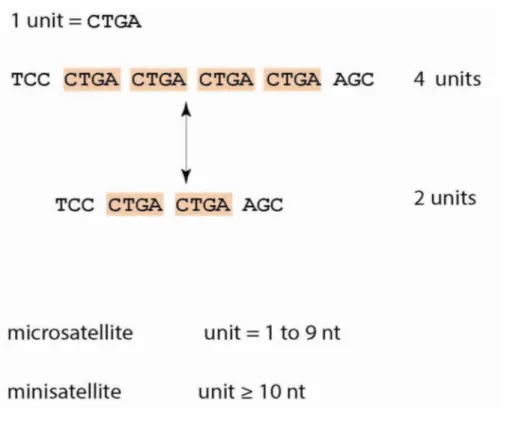
Interspersed repeats, the more predominant type of repeat, are remnants of transposons dispersed throughout the genome. Such elements are responsible for the diverse array of genome sizes amongst various species (Feschotte et al., 2009). On the other hand, TRs are repetitive DNA sequences, which exist directly adjacent, or in tandem, to one another (Figure 1). TRs are often referred to as satellite DNA because they were first identified as sequences constituting the second or “satellite” band that is detected after density-gradient centrifugation of DNA (Kit, 1961).
On the basis of unit length (unit= repeated sequence of DNA - see Figure 1), TRs are further divided into three subcategories-microsatellites, minisatellites and satellite.
2
Microsatellites, or simple sequence repeats (SSRs), are short TRs with unit length between one to five nucleotides, found in vertebrate, insect and plant genome, located in the euchromatin. Copy numbers are characteristically variable within a population, typically with mean array sizes of the order of 100 but with multiple array size classes distributed around the mean (Bruford, 1993; Di Renzo, 1994).
Minisatellites are TRs with unit length larger than ten nucleotides, generally involving mean array lengths of 0.5-30 Kb. They are found in euchromatic regions of the genome of vertebrates, fungi and plants,and are also highly variable in array size (Armour, 1992).
Satellite are TRs that can be similar in length (5-10bp) to micro- or ninisatellites, or much larger (~100pb). They are typically organized as large [up to 100 megabases (Mb)] clusters in the heterochromatic regions of chromosomes, near centromeres and telomeres or on the Y chromosome ( Cavalier-Smith, 1985; John and Miklos, 1988; Tyler-Smith and Willard, 1993; Lohe et al., 1993). They are apparently notas variable in array size within populations as micro- and minisatellitea (Jabs et al., 1989; Wevrick et al., 1989).
Figure 1. Main definitions and characteristics of tandem repeats (TRs). TRs are unstable due to frequent changes in the number of repeat units. TRs with short unit length are also called microsatellites and those with long units are called minisatellites. nt= nucleotides (from Gemaley et al., 2012)
3
1.1 Instability of Repeats
Tandem repeats are evolutionarily pertinent due to their instability; they mutate at rates between 10 -3 and 10-6 per cellular generation (i.e., 1 to 10 orders of magnitude greater than point mutations)
(Verstrepen et al., 2005 ). Variation in the numbers and lengths of tandemly repeated units occurs on many different scales. Several genetic mechanisms can affect the number of repeating units in tandem arrays, usually occur from the addition or deletion of repeat units, rather than nucleotide substitutions. For instance, in a CTGA tract, most mutations occur by the addition or deletion of an entire CTGA unit as opposed to rare cases in which only a part of the repeat unit is altered (e.g., deletion of two nucleotides GA) (Figure 1)(Gemaley et al., 2012).
A set of major models have been proposed to explain TR expansions and contractions:
Strand-slippage replication (slipped-strand mispairing or DNA slippage) is a DNA replication error by which mispairing occurs between the template and nascent strands. As such, the template strand can loop out, causing contraction; the nascent strand can also loop out, leading to repeat expansion. Recombination events, such as unequal crossing over and gene conversion may additionally lead to contractions and expansions of TR sequences (Verstrepen et al., 2005; Paques et al., 1998).
Rolling circle amplification (Feliciello et al., 2006) here, circular plasmids created by intrastrand exchange integrate (at some low level) into arrays by homologous recombination, either on the same chromosome or possibly on non-homologous chromosomes. If the circular plasmids contain the replication origins, rolling circle replication can greatly expand a short array sequence on a plasmid, allowing for rapid amplification (Hourcade et al., 1973; Flavell, 1982).
1.2 Satellite DNA (satDNA)
Satellite DNAs can be defined as highly reiterated noncoding DNA sequences, organized as long arrays of head-to-tail linked repeats located in the constitutive heterochromatin, the part of eukaryotic genomes that remain condensed throughout the cell cycle (Heitz, 1928).
The term “satellite DNA” is historical, because this kind of sequences was initially isolated from satellite bands in experiments with gradient centrifugation, due to the difference in A+T content from the rest of genomic DNA (Szybalski, 1968).
The basic repeating units, satDNA monomers, are often A+T rich and range in length from only few bp up to more than 1 kb, building up to 100 Mb long arrays. The preferential monomer length of 150–180 bp and 300–360 bp detected in many satellites in both plants and animals is often considered to mirror requirements of DNA length wrapped around one or two nucleosomes (Schmidt and Heslop-Harrison, 1998; Henikoff et al., 2001).
4
The copy number is substantially conserved within the populations, but the monomeric unit may shows several variants regarding the nucleotide sequence (Charlesworth et al., 1994; Ugarkovic and Plohl, 2002).
Satellite DNA contribution to total genomic content varies significantly among species, exceeding sometimes 50% of total DNA (Elder and Turner, 1995; Schmidt and Heslop-Harrison, 1998), and consequently they are involved in the enormous variation of genome size in eukaryotes (Doolittle and Sapienza, 1980; Cavalier-Smith, 1985; Gregory et al., 2007).
Satellite sequences are the main constituent of centromeric and pericentromeric heterochromatin, two epigenetically determined regions responsible for correct pairing and disjunction of eukaryotic chromosomes in cell divisions (see for example Arney and Fisher, 2004; Hall et al., 2004; Bloom, 2007).
It is observed that principal DNA components underlying the majority of centromeres in plants and animals are satellite repeats, as corroborated by chromatin immunoprecipitation (ChIP) data (e.g. Nagaki et al., 2003; Zhong et al., 2002; Lee et al., 2005). Centromere is a multidomain locus necessary for poleward chromosomal segregation in mitosis and meiosis. Functional centromeres are usually embedded into large blocks of pericentromeric heterochromatin, but chromatin structure in centromeres is distinct from that in heterochromatin and in euchromatin (Sullivan and Karpen, 2004). While centromere structure and function is conserved through complex eukaryotes, DNA sequences in that region are paradoxically variable (Henikoff et al., 2001).
Satellite families in (peri)centromeric regions vary significantly in copy number, nucleotide sequence, organizational patterns, number and nature of inserted non-satellite DNA sequences (Plohl et al., 2008). Domains formed by single satellites are usually several hundreds kb, or even Mb long, such as in humans (Shiels et al., 1997; Mahtani and Willard, 1998; Schueler et al., 2001). Among individuals, array length of a single satellite can be highly polymorphic. For example, array length in alpha-satellite from human X varies almost 3 times (Mahtani and Willard, 1990).
It has been hypothesized that low abundance of satellite repeats in the centromere represents an early stage in the centromere evolution, characterized by progressive accumulation of satellite repeats in mature centromeres. (Wu et al., 2004; Nagaki et al., 2004).
1.3 Evolutionary dynamics of satellite DNA.
As discussed above, satDNAs in (peri)centromeric heterochromatin, represent rapidly evolving genomic components. Consequently, even among the most closely related species, they differ in
5
nucleotide sequence, copy number, and/or composition of satellite families (reviewed in Schmidt and Heslop-Harrison, 1998; Ugarković and Plohl, 2002).
Rapid evolution of satDNA sequences is possible owing to the accumulation of nucleotide divergences, usually with a high rate and in a gradual manner (Bachmann and Sperlich, 1993). Gradual accumulation of mutations follows phylogeny at different hierarchical ranks. At the species level, centromeric satDNAs were informative in phylogenetic studies of the Drosophila obscura group (Bachmann and Sperlich, 1993), or in the study of the fish family Sparidae (Garrido-Ramos et al., 1999). Even within a genome, distinct forms of satellite DNAs can accumulate mutations at different rates, thus producing diversity of sequence patterns in (peri)centromeric areas. Interestingly, centromerically located higher-order units diverge more rapidly than pericentromerically located monomeric repeats (Rudd et al., 2006).
Accumulation of mutations in satellite families is not the only way to alter specific profiles of satellite repeats in short evolutionary periods. Since more than one satellite family exists in a genome, expansions and contractions of satellite arrays can efficiently change a landscape of DNA sequences in heterochromatin by replacing one dominant (major) satellite repeat with another one less represented (reviewed in Ugarković and Plohl, 2002) (fig.2). Unequal crossover is proposed to be the major mechanism responsible for dramatic fluctuations in the copy number of satellite DNAs (Smith, 1976).
6
Figure 2. The library model. In a genome, several satellite DNA families are coexisting on chromosomes, one family being often at high copy number as the major satellite. Different families can be preferentially amplified in the derived chromosomes, therefore changing the relative contribution of each family to the (peri)centromeric chromatin and leading to species-specific profiles of satellite repeats (from Plohl et al., 2008).
Satellite repeats may be the preferred form of DNA sequences in functional centromeres and their flanking regions just because of their unique characteristic to maintain sequence homogeneity over long stretches of DNA, and simultaneously to change rapidly in evolution.
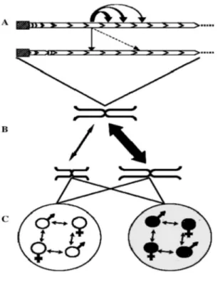
This characteristic is achieved by non-independent evolution of monomers. It is a consequence of molecular drive, a two-level process in which mutations are homogenized throughout members of a repetitive family, and concomitantly fixed within a group of reproductively linked organisms (Dover, 1982, 1986). The consequence is concerted evolution of monomers constituting a satellite DNA family.
1.4 Concerted evolution of satellite DNA
Evolution of satDNA sequences is governed by principles of concerted evolution, in which mutations are homogenized throughout members of a repetitive family and fixed within a group of reproductively linked organisms in a stochastic process of molecular drive (Dover, 1986).
Sequence homogenization is due to diverse molecular mechanisms of nonreciprocal transfer, such as unequal crossover, gene conversion, rolling circle replication and reinsertion, and transposon-mediated exchange (Stephan, 1986; Dover, 2002; Glinka et al., 2006). While it is not clear which of the above reported mechanisms is preferentially involved in sequence homogenization, it is generally acknowledged that these mechanisms act more efficiently within localized subsets of satellite repeats, while efficiency drops progressively when changes are homogenized between arrays on the same chromosome, homologous and heterologous chromosomes (Fig. 3; Dover, 1986). Because of differences in rates of local and global sequence homogenization, adjacent monomers show a higher degree of sequence similarity than those retrieved at random, and can be often grouped into subsets or subfamilies, defined by diagnostic mutations (Willard and Waye, 1987; Durfy and Willard, 1989; Schindelhauer and Schwarz, 2002; Hall et al., 2005; Roizes, 2006). Distinctive groups of monomer variants are usually chromosome-specific. As predicted by theoretical models (Smith, 1976; Stephan, 1989), monomers at array ends are more divergent than those located centrally due to the low efficiency of homogenization mechanisms (predominantly
7
unequal crossover) in bordering regions of the satellite array (Mashkova et al., 1998; McAllister and Werren, 1999; Bassi et al., 2000; Schueler et al., 2005). Adjacent monomer variants can be sometimes homogenized together and form a new, composite higher-order repeat (HOR) unit in which former monomers became subrepeats or subunits (Willard and Waye, 1987; Warburton and Willard, 1990). Since a HOR is a homogenization unit, HORs generally show high level of sequence identity, while substantial sequence divergence is accumulated among constituent subunits (Willard and Waye, 1987; Roizes, 2006 and references therein). While homogenization depends on mechanisms of genomic turnover, fixation results from random chromosomal assortment in sexual reproduction through meiosis and amphimixis, depending thus on population factors (Plohl et al., 2008).
Figure 3. Molecular drive and concerted evolution. A) Homogenization of mutated variant throughout the members of a repetitive family within an array and between sister chromatids. Arrow appearance is correlated with the homogenization efficiency. B) Efficiency of homogenization between homologous and non-homologous chromosomes. C) Variant spread among individuals (fixation) depends on bisexuality and population factors. Reproductive isolation leads to fixation of different repeat variants in genomes of different evolutionary units (white and black circles) (from Plohl et al., 2008).
These mechanisms induce high turnover of satellite sequences and rapid changes in copy number, nucleotide sequence and composition of satellites in the genome, resulting in high within-species homogenity of satellite repeats and alterations in satellite profiles between species.
8
Some satellite sequences indeed change rapidly in evolution and accumulate mutations even at the population level, for example in the pupfish (Elder and Turner, 1994). On the other hand, some are widely distributed across species, providing evidence of nucleotide sequence conservation for long-term evolutionary periods. A study on Pimelia (Coleoptera, Tenebrionidae) suggested that a satDNA, highly abundant in all examined species, has persisted for more than 8 million years (Pons et al., 2002). Satellites in species from the Drosophila virilis group remained conserved for about 20 Myr (Heikkinen et al. 1995), and cetacean satellite DNA persisted for at least 40 Myr (Arnason et al. 1992). Low-copy number repeats indistinguishable in their nucleotide sequence from high-copy
Palorus ratzeburgii satDNA have been detected in distant species Pimelia elevata, although these
taxa diverged 60 Myr ago (Mravinac et al., 2002). Conservation or “freezing” of repeat families for long evolutionary periods can be explained as a consequence of mechanisms involved in concerted evolution, if a small bias in turnover mechanisms is anticipated (Dover and Flaveli, 1984). Such preference to maintain some putative “optimal” set of monomer variants may be due to functional constraints imposed on the nucleotide sequence (Mravinac et al., 2005). Slow rates of sequence change and of concerted evolution in some satellites were explained as specificity of slow general genomic evolution in sturgeons (Robles et al., 2004) and in whales (Arnason et al., 1992). Therefore, forces (i.e., gene conversion) leading to concerted evolution might be acting in these species, but that speciation by hybridization and/or polyploidization events played an important role in forming of their phylogeny. Thus, such a trend could be leading to the apparent failure of the concerted evolution process (Robles et al., 2004).
Furthermore, the final outcome of an extreme conservation of nucleotide sequence can be also predicted by the model, if “non-desirable” mutations are preferentially eliminated instead of being spread throughout a satellite family. The nucleotide sequence of some satellite families indeed remained “frozen” for long periods, even for tens of Myrs. Although the basis for favouring one sequence variant over another is usually not known, it might mirror constraints imposed on satellite sequences by some functional interactions. In that case, the evolution of at least some satellites seems to be driven by an interplay of selective constraints and stochastic events (by Plohl et al., 2008 and references therein).
Accumulation of mutations in satellite families is not the only way to alter specific profiles of satellite repeats in short evolutionary periods. In addition to sequence changes, satDNAs are permanently altered in copy number by expanding and contracting arrays of satellite monomers (Ugarković and Plohl, 2002; Plohl et al., 2012). Because usually more than one satellite family exists in a genome, fluctuations in their copy numbers can change very efficiently and rapidly any
9
profile of genomic satDNA. The library model of satDNA evolution explains the occurrence of species-specific satellite profiles as a result of differential amplifications and/or contractions within a collection, or library, of satellite sequences shares by related species (Fry and Salser, 1977; Mestrović et al., 1998; Ugarković and Plohl, 2002). Not only distinct satDNAs, but also monomer variants or subfamilies from a single family can be distributed in genomes in the form of a library (Cesari et al., 2003).
1.5 Functional potential of satellite DNAs
The possible role of this fraction of the genome has been long discussed. Various evidences led scientists to formulate some hypotheses on the involvement of satellite DNAs in a series of functions ranging from centromere formation and function, heterochromatin assembly, regulation of gene expression and in epigenetic regulatory processes (reviewed in Ugarković, 2009; Pezer et al., 2012).
Some studies supported the role played by heterochromatic genomic compartments in a proper chromosomal behaviour in mitosis and meiosis (Csink and Henikoff, 1998). Indeed, satellite DNAs appear to be major constituents of functional centromeres, as shown in detail in Drosophila
melanogaster (Sun et al., 1997) and in humans (Schueler et al., 2001). Satellite repeats should
contain sequence motifs recognized by protein components. The best known is CENP-B box, the 17-bp long sequence motif found in its functional form in a subset of higher-order alpha satellite monomers. The motif binds the CEN-B protein, suggested to facilitate kinetochore formation (Ikeno et al., 1994; Masumoto et al., 2004; Schueler et al., 2005). Motifs resembling to CENP-B box were observed in diverse satellite families from various species, but their true functional significance is not known (Canapa et al., 2000; Lorite et al., 2004).
Transcriptional activity was not expected for repetitive DNA sequences residing in the transcriptionally suppressive heterochromatin environment. However, satellite transcripts have been so far in many animal and plant taxa indicating that satellite transcription might be a general phenomenon (for example, Varadaraj and Skinner, 1994; Lorite et al., 2002; Rudert et al., 1995; Pathak et al., 2006; Lee et al., 2006). Satellite DNAs never show any prominent open reading frame, and accordingly, transcript translation has never been demonstrated (Plohl et al., 2008). However, various functional roles have been hypothesized for these transcripts. The strand or tissue or stage specificity observed in some cases suggests the involvement of satellite transcripts in regulatory functions (Varadaraj and Skinner, 1994; Lorite et al., 2002; Pathak et al., 2006).
10
The most striking example is the sequence of human a satellite DNA, which is found highly conserved in chicken and zebrafish (Li and Kirby, 2003). This sequence is transcribed during early embryogenesis of both species and it is proposed that it may serve as a control element in gene regulation. It has recently been shown that transcripts of satellite DNAs and other repetitive sequences are functional in the form of small interfering RNAs which act as signals necessary for establishment and/or maintenance of heterochromatin in different eukaryotes (Volpe et al., 2002; Aravin et al., 2003).
Some satellite DNAs from insects, nematodes and amphibians produce hammerhead structures with a possible ribozymic activity (Rojas et al., 2000 and references therein). Transcripts of centromeric satellite in maize were shown to remain tightly bound within centromeric chromatin and contribute to initiation and stabilization of kinetochore chromatin structure (Topp et al.,2004).
Another proposed role of satellite DNA transcripts attracted particular attention in recent years. It was observed that satellite DNA transcripts are involved in the initiation of histone H3 methylation, a necessary prerequisite in heterochromatin formation and maintenance (Volpe et al., 2002; Martienssen, 2003). Transcripts from centromeric satellites are processed to produce small interfering RNAs (siRNA) that mobilize a number of proteins and specifically target their coding sequence. This sequence is then packed into the transcription-inhibiting heterochromatin structure (reviewed in Grewal and Elgin, 2007). This mechanism requires low-levels of transcription and may be universal, since siRNAs processed from centromeric satellite repeats were identified in several eukaryotic species (Lee et al., 2006 and references therein). However, the relationship between transcription of centromeric satellite repeats and centromeric silencing/centromere function is still unclear.
Furhtermore, a potential indirect role of satellite DNAs in chromosomal repatterning should be mentioned. This role would be important in that karyotype rearrangements can be important in promoting reproductive isolation between populations, ultimately leading to speciation (Coghlan et al., 2005). For example, a link between satellite DNAs and chromosomal instability was studied in the genus Ctenomys, one of the most specious and karyotypically diverse mammalian taxon. The high karyotypic variability was associated with amplifications and deletions of the major Ctenomys satellite DNA and with the number of species (Slamovits et al., 2001; Hartmann and Scherthan, 2004; Ellingsen et al., 2007). Satellite DNAs appear also involved in genome restructuring during development in different organisms. The process of chromatin diminution is known to occur during development in different organisms, such as in the nematodes Parascaris univalens and Ascaris suum, in copepods and in a hagfish. The quantity of lost DNA ranges up to 94%, and is mainly
11
composed of satellite sequences (Stanley et al., 1984; Drouin, 2006, and references therein). The hypothesis that RNAi-related mechanisms are involved in chromatin diminution has been put forward (Drouin, 2006).
2 Sex Determining Mechanisms in Vertebrates
Sexual reproduction is a nearly universal feature of eukaryotic organisms. Given its ubiquity and shared core features, sex is thought to have arisen once in the last common ancestor to all eukaryotes. Vertebrates have various sex determining mechanisms. These have been broadly classified as either environmental sex determination (ESD) or genotypic sex determination (GSD). The term environmental sex determination (ESD) implies that the sex of an individual is
determined by the environmental conditions experienced during early development. The key environmetal factor can be pH (e.g., in fish; Römer and Beisenherz, 1996), or social conditions or relative juvenile size (.g., in fish; Francis and Barlow, 1993; Holmgren and Mosegaard, 1996), and the temperature. This latter is the most common and most studied environmental parameter inducing sexual determination and it is generally referred to as TSD (Temperature-dependent Sex Determination) system.
In genotypic sex determination (GSD) the sex of a zygote is determined entirely by its genotype, and the sex of an individual is fixed at fertilisation. The most common type of GSD involves sex chromosomes. Two main systems of sex chromosomes can be found in vertebrates. One type is termed as XX/XY system and male (XY) is the heterogametic sex. The other type is indicated as ZZ/ZW system with the heterogametic sex represented by female (ZW). In polygenic sex determination, which is less common than the preceding one, sex is determined by a number of genes, each with minor effect, distributed throughout the chromosome complement (Liew et al., 2012)
2.1 Distribution of sex determining mechanisms among vertebrate taxa
ESD is common among reptiles, and occurs also in amphibians and fish. TSD is the form of ESD found in reptiles. In some lizards and in alligators, eggs incubated at low temperature(less than 27°C) give rise to 100% females, and eggs incubated at high temperatures (above 30°C) give rise to 100% males. In many turtles it is the other way round: 100% males at low temperatures and 100% females at high temperatures. In other turtles and in crocodiles, incubation at intermediate temperatures leads to 100% males, whereas both low and high temperatures lead to females only (reviewed in Bull,
12
1983). In all these cases there is only a very narrow temperature range in which both sexes are produced. This range, however, may vary within a species and is heritable (Bull et al., 1982, Janzen, 1992). In fishes, the sex ratios usually vary less extremely with temperature, but nevertheless temperature-dependent sex determination has been foun in various species (e.g. Conover and Heins, 1987, reviewed in Francis 1992, Römer and Beisenherz, 1996, Goto et al., 2000). The molecular mechanism by which temperature triggers sex determination in these taxa is completely unknown, although an influence temperature on hormones (estrogens) and thir receptors has been proposed (Sarre et al., 2004 ).
In mammals, birds, amphibians, and many reptiles and fish, sex is determined genetically. GSD mechanisms range from those that depend on allelic variation at a single locus to those in which the sex-determining gene is borne on a pair of differentiated sex chromosomes (Ezaz et al., 2005). In all these systems, one sex (homogametic sex) produces gametes that are homozygous for the same sex allele or chromosome and the other sex (heterogametic sex) produces equal proportions of two kinds of gametes (one containing the X or Z and the other the Y or W) that specify equal numbers of males and females in the offspring. There is a fundamental distinction between species with male heterogamety (XY male, XX female, such as mammals), and female heterogamety (ZZ male, ZW female such as birds). However, the two systems share many parallels; for instance, the X and Z chromosomes are usually larger and contain many more active genes than the sex-specific Y or W. Environmental and genetic sex determination have traditionally been thought to constitute completely different triggers, but these systems sometimes interact, as in some reptile species, amphibians, and fish where we find have elements of both (Quinn et al., 2007). Genotypic and environmental sex determination have been viewed as two ends of a continuum of sex-determining mechanisms (Shine et al., 2002; Sarre et al., 2004). For example, XX/XY sex chromosomes were described in a scincid lizard (Bassiana duperreyi) that presumably possessed TSD mechanisms (Shine et al., 2002). The mechanism for long-term maintenance of these sex chromosomes in a population with ESD is not clear (Bull, 2008). The continuum between ESD and GSD can be explained by the existence of GSD with environmental effects (Valenzuela et al., 2003), where environmental conditions can influence the observed sex ratio of hatchlings, but sex of an individual is still determined by its genotype (e.g. by differential fertilization of gametes producing particular sex or sex-specific mortality of embryos), or by the thermal induction of sex revertants (i.e. production of individuals with the wrong gonad type for their genotypes). Sex revertants induced by extreme developmental temperatures are well known in many GSD vertebrate lineages (e.g. Witschi, 1929) and were recently documented in two species of lizards as well (Quinn et al., 2007 in the ZZ/ZW agamid Pogona vitticeps, and Radder et al., 2008 in the XX/XY skink Bassiana
13
duperreyi). In both cases, the existence of thermally induced sex reverted individuals was interpreted by the authors as evidence for the co-occurrence of GSD and TSD or for a transitional state between TSD and GSD. Nevertheless, gonadally and phenotypically reverted individuals still possess sex chromosomes corresponding to their genotypic sex, and when crossed with non reverted mates, they produce progeny with a skewed sex ratio. We understand GSD and TSD as two dichotomous sex determining systems that do not differ in thermal dependency of sex ratios, but basically in the presence or absence of sex chromosomes (Pokorná et al., 2009).
2.2 Origin of sex chromosomes
Sex chromosomes are very atypical of the genome. Evidently the acquisition of a sex determining allele confers on a chromosome special properties and a special fate. Sex chromosomes are considered to be the most variable region of the genome. However, this is true only for the sex-specific element (the Y or W). The mammal X and the bird Z are extremely conserved (Graves, 2008).
Sex chromosomes differ from autosomes in that the two members of the sex chromosome pair typically vary in morphology, size, number, staining, and gene content. They are highly specialized and appear to have evolved independently many times in vertebrates (for review see Graves, 2008). Sex chromosomes are thought to evolve from autosomes (Ohno, 1967). However, vertebrate XY and ZW sex chromosomes are not homologous, suggesting independent evolution of sex chromosomes in different lineages from non-homologous ancestral autosomes (Fridolfsson et al., 1998; Nanda et al., 2000, 2002). The autosomal origin of sex chromosomes is also supported by the fact that sex chromosomes of one group of organisms are autosomal in another group (e.g., Matsubara et al., 2006; Pokorná et al., 2011), which would be consistent with an independent origin of sex determination in vertebrates (e.g. Graves, 2008).
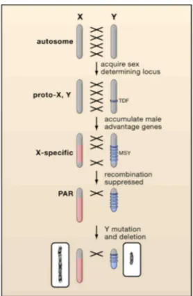
The trajectory from autosomes to sex chromosomes may start with the emergence of a mutation that confers a sexual advantage. Additional sex-linked mutations in other genes then accumulated on the same homologue. Recombination between the primordial sex chromosomes was suppressed by chromosomal rearrangements such as inversions to preserve the block of sex-linked genes. The absence of recombination fostered the accumulation of mutations and repetitive sequences with subsequent ‘heterochromatization’ of the sex-specific chromosome. Deletions of heterochromatin account for the smaller sizes usually observed for the Y or W chromosomes compared with the X or Z chromosomes, respectively (by Modi et al., 2005) (Fig. 4).
14
Figure 4. Differentiation of an X and Y chromosome from an ancestral autosome.
This process is initiated when one partner acquires a sex-determining locus such as the testis-determining factor (TDF). Accumulation of male-specific alleles selects for repression of recombination (represented by crosses), creating an X-specific region on the X and a male-X-specific region on the Y (MSY). Exclusion from recombination leads to rapid degradation of the MSY leaving only a small pseudoautosomal region (PAR). Active genes are lost, leaving largely genes that have, or acquire, a male advantage. This model accounts for the differences in size and gene content of the human X (left) and Y (right) (Graves 2006).
The initiating mechanisms of recombination suppression are not yet clear. However, two hypotheses have been proposed to explain how restriction of recombination spreads along sex chromosomes. Stepwise model highlights the role of inversions in suppression of recombination, which is supported by evolutionary strata of different age, i.e. sex chromosome regions of different levels of divergence, observed in mammals (Kohn et al. 2004, Macha et al. 2012), birds (Nam and Ellegren 2008), snakes (Vicoso et al. 2013a), papaya (Wang et al. 2012), and white campion, Silene
latifolia (Bergero et al. 2007, 2013, Hobza et al. 2007). However, it was shown that the multiple
inversions on the Y chromosome are a consequence rather than a cause of suppressed recombination in S. latifolia (Bergero et al. 2008). Thus, other mechanisms such as heterochiasmy, i.e. sex-specific differences in recombination, are probably involved in a gradually proceeding cessation of recombination in the early stage of sex chromosome differentiation (Perrin 2009, Bachtrog 2013, Natri et al. 2013).
15
The suppression of recombination between the heterochromosome and its homologue would start the gradual erosion of the heterochromosome (Y or W) itself because genes that are not essential for males (in XY systems) or females (in ZW systems) show accelerated rates of mutation and deletion. Consequently, the heterochromosome becomes progressively gene-poor (see Handley et al., 2004) and in the extreme case the simplification process can lead to the complete loss of the heterochromosome (e.g., Just et al., 2007).
Concerning the heterochromatinization of heterochromosomes (W or Y), it is still debated whether repetitive sequences are the cause of the suppression of recombination or whether these elements accumulate on the Y or W chromosome as a consequence of the block of genetic exchange between sex-chromosomes. The observation of an evolving incipient Y chromosome in the fish families Gasterosteidae (Peichel et al., 2004) and Cichlidae (Griffin et al., 2002) supports the first hypothesis. Indeed, despite sex chromosomes are not clearly cytogenetically distinct, a molecular analysis revealed an accumulation of heterochromatin in the sex-determining region of the “proto-Y” resulting in a reduction in recombination. On the other hand, accumulation of heterochromatin could be a mere effect of cessation of recombination. In fact, the impossibility of recombination between the unpaired sex chromosomes (W or Y) opens the door to the “invasion” of various repetitive sequences, like transposons, microsatellites and tandem repeats, on sex chromosomes. In this respect, it has been hypothesized that the heterochromatinization may be a mechanism for the defense against invasive transposable elements (Kidwell, 2002; Steinemann & Steinemann, 2005). However, little is known about how this occurs or about how the absence of recombination affects the subsequent evolutionary fate of the repetitive sequences in the W or Y chromosome.
The repetitive DNA sequences or TEs accumulation and expansions on one hand and contractions on the other hand are stepwise or are occurring simultaneously (Kejnovsky et al. 2009). However, the lifetime of an old Y chromosome is often prolonged by the addition of segments transferred from autosomes (Graves, 2005). Acquisition of new genes from autosomes mediated by retrotransposition has been shown in humans (Lahn and Page, 1999), and a similar duplicative transfer has also been shown in the young Y chromosomes of Silene latifolia (Matsunaga et al., 2003). Kejnovsky et al. (2008) have recently discussed the potential for junk DNA accumulation to start at an early stage in the evolution of sex chromosomes. Both past cytogenetic analyses and recent genome projects have revealed that many animal Y chromosomes have more abundant heterochromatin derived from repetitive sequences compared with X chromosomes and autosomes. Accumulation of repetitive sequences induces abnormal recombination and chromosome breaks. Thus, junk DNA accumulation may well be a factor in the generation of differences in morphology and size observed between X and Y chromosomes; for example, in both the fruit fly, Drosophila
16 melanogaster, and in humans, the Y chromosome is drastically smaller than the X chromosome (Adams et al., 2000; Skaletsky et al., 2003). These findings in animal species show that the accumulation of junk DNA is an important step in promoting the morphogenesis of sex chromosomes.
Junk DNA accumulation on Y chromosomes has been believed to be a symptom of Y-chromosome degeneration. Insertion of repetitive sequences into coding genes and regulatory regions induces alteration in the genes’ functions and results in gene loss. However, there is no correlation between the insertion of the transposable element and gene dysfunction on the Y chromosome of D. miranda (Bachtrog et al., 2008). The contradiction between gene acquisition and accumulation of highly repetitive sequences on the Drosophila Y chromosomes, indicates that junk DNA accumulation is not always directly connected with Y chromosome degeneration (Matsunaga 2009).
Then the Y chromosome either remains as a genetic entity or can be lost entirely. A new autosomal pair can then be chosen to become a new pair of sex chromosomes and the cyclic process can continue. The persistence of the Y chromosome indicates that it can repeatedly arise de novo, for example, by the fusion between an autosome and an X chromosome followed by the fixation of the neo-X and the neo-Y chromosomes as was shown in grasshopper Podisma pedestris (Westerman and Hewitt, 1985; Veltsos et al., 2008).
2.3 Evolution of sex chromosomes in squamate reptiles
The study of reptilian genome is of great interest because reptiles occupy a pivotal position in the phylogeny of vertebrates — they are the direct ancestor to birds and mammals — and because they also possess several unique biological attributes that, if better understood, could contribute significantly to understanding basic evolutionary biology and the molecular mechanisms behind human health and disease (Modi and Crews, 2005). Reptiles exhibit some of the most extraordinary variability in sex chromosome structure and patterns of sex determination modes seen among vertebrates (Fig. 5) (Valenzuela and Lance, 2004). These modes include gonochorism (separate sexes) and parthenogenesis, oviparity, viviparity, and ovoviviparity, genotypic sex determination (GSD), with either male (XX/XY) or female (ZZ/ ZW) heterogamety, and temperature-dependent sex determination (TSD) (Ezaz et al., 2009a).
17
Figure 5. Vertebrate phylogeny illustrating sex determination modes in different taxa. ‘‘Female’’ and ‘‘Male’’ represent genetic sex determination with female and male heterogamety, respectively. TSD represents temperature-dependent sex determination. An unanswered question in contemporary reptilian phylogenomics regards the relationships of turtles to other reptiles (from Modi e Crews, 2005).
The variability seen among reptilian sex chromosomes suggests that sex chromosome and sex determination systems have evolved independently in different lineages (Modi e Crews, 2005). Lizards (order Squamata, suborder Sauria) are particularly fascinating because the distribution of sex determining mechanisms shows no clear phylogenetic segregation. This implies that there have been multiple transitions between TSD and GSD, and between XY and ZW sex chromosome systems (Ezaz et al., 2009a). Lizards with GSD display remarkable diversity in sex chromosome differentiation, ranging from cryptic or homomorphic to highly differentiated. Much of this variation occurs within families, often among closely related species and even within the various races or populations of the same species. For example, the gekkonid lizard Gehyra purpurascens displays two Z chromosome and six W chromosome morphs, primarily as the result of centromeric inversions (Moritz, 1984) ( fig. 6 ). Variation in the morphology of sex chromosomes among closely-related taxa, or populations of one taxon, indicate that morphological evolution of sex chromosomes, and perhaps also sex-determining mechanisms in lizards may occur relatively easily in comparison to mammals and birds (Ezaz et al., 2009a).
18
Figure 6. A snapshot of morphological diversity of sex chromosomes in lizards. Ch: Chromosome. ACO: Anolis
conspersus AMO: Anolis monesis,BDU: Bassiana duperreyi,], BTR: Bipes tridactylus,CEN: Claireascincus entrecasteauxii, CLE: Calyptommatus leiolepis], CLI: Cnemidophorus littoralis, CTI: Cnemidophorus tigris, DIN: Delma inornata, DNO: Dibamus novaeguineae, GCE: Gonatodes ceciliae, GGE: Gekko gecko, GHO: Gekko
hokouensis, GPL: Gymnophthalmus pleei, GPU: Gehyra purpurascens, LBU: Lialis burtonis, LVI: Lacerta vivipera, MAL: Micrablepharus allicolus, PLA: Phyllodactylus lanei, PSI: Podarchis sicula, PVI: Pogona vitticeps, PVL:
Phrynocephalus vlangalii, SCZ: Saproscincus czechurai, SLA: Scincella lateralis, SLU: Sceloporus lundelli, SMA:
Sceloporus maculosus, VAC: Varanus acanthurus (from Ezaz et al., 2009a).
Like birds, turtles and snakes, most lizards have a karyotype composed of macrochromosomes (ranging from 2n = 10–46) and microchromosomes (ranging from 2n = 0–26) (Olmo and Signorino, 2005). Microchromosomes have been found to be gene rich in birds with 2–3 times the number of genes contained in macrochromosomes (Smith et al., 2000) and to have higher recombination rates (Rodionov et al., 1992). In addition, microchromosomes are GC and CpG-rich and contain few repetitive elements (Hillier et al., 2004) and therefore, are likely to be important for generating genetic variation (Organ et al., 2008). Importantly, in some species of lizards, sex chromosomes
19
have been found to be microchromosomes (Gorman and Atkins, 1966; Gorman, 1973; Bull, 1983; Ezaz et al., 2005; Ezaz et al., 2009a). The patterns of differentiation of these sex microchromosomes are highly variable within and among groups (fig. 6), and have evolved primarily via the accumulation and amplification of heterochromatin (Ezaz et al., 2009a). Sex microchromosomes appear to be highly labile in at least one family of lizards, with agamids exhibiting a substantial array of forms among closely related species. In this group, the W chromosomes are highly to moderately heterochromatic, whereas the Z chromosomes are euchromatic and can be detected only by mapping sex chromosome specific DNA sequences (Ezaz et al., 2005, 2009a) or by mapping sex chromosome specific BAC clones (Ezaz et al., 2009a). These patterns of heterochromatic variability suggest various stages of sex chromosome differentiation within closely related species.
The accumulation of repetitive sequences during sex chromosome evolution in reptiles has been studied only in snakes (Jones and Singh, 1985; O’Meally et al., 2010). Pythons, considered basal in snake phylogeny, show homomorphic sex chromosomes, without accumulation of repetitive DNAs. On the contrary, in many advanced snakes like colubrids or elapids (Colubroidea) the heteromorphic W sex chromosome exhibit a strong accumulation of repeats (Jones and Singh, 1985; O’Meally et al., 2010). For example, the W chromosome in the elapid Notechis scutatus is composed almost entirely of repetitive sequences, including 18S rDNA and the banded krait minor-satellite (Bkm) repeats (Lee et al., 2007). The Bkm repeats consist of tandem arrays of 26 and 12 copies, respectively, of two tetranucleotides, GATA and GACA (Epplen et al., 1982). Bkm-related repeats are also accumulated on the heterogametic sex chromosomes in many vertebrates and also in plants (Jones and Singh, 1981; Parasnis et al., 1999), suggesting their possible role in the transcriptional activation of sex chromosome heterochromatin (Singh et al., 1976).
One interesting reptilian group in which sex chromosomes evolution involves heterochromatinization is represented by the lizards belonging to the Lacertidae family.
3. The study species
3.1 The family Lacertidae (Reptilia, Squamata)
The family Lacertidae consist of about 42 genera including 321 species widespread in the Palaearctic region (Uetz and Hošek, 2015). Recent molecular analyses strongly support the monophyly of lacertids, and suggest that Lacertidae may be the sister-group of Amphisbaenia, the worm lizards (Townsend et al. 2004; Vidal and Hedges 2004), otherwise its nearest relatives are Teiioidea, a group of squamates currently exclusive to the American continent (Arnold et al., 2007).
20
Lacertids are defined as a clade by a number of mainly exclusive synapomorphies:
• lack of downgrowths on the parietal bone (Estes et al., 1988);
• supratemporal fenestra largely or wholly filled by postfrontal bone (a feature shared with
Scincidae);
• usual presence of sexual variation in the number of presacral vertebrae;
• bodenaponeurosis divided into two lobes caudally, and a parasagittal vertical sheet
connecting the quadrate aponeurosis to the temporal fascia (Rieppel, 1980);
• abdominal fat-bodies largely outside the peritoneum (Arnold, 1989a);
• either the lobes of the hemipenis invested by the retractor penis magnus muscle, or the lobes
usually omplexly folded;
• the erect hemipenis supported by an elaborate cartilaginous supporting structure, termed
armature (Arnold, 1973, 1986, 1989a).
To these features presence of a microornamentation on the epidermis of the hemipenial lobes consisting of individual cells that are typically hook-shaped spines or crown-shaped tubercles can probably be added (Klemmer 1957; Böhme 1971; Arnold 1973, 1986, 1989a). Additional putative synapomorphies of the Lacertidae, involving the scaling of the posterior dorsal surface of the head, such as widespread presence of an occipital scale, have also been put forward (Borsuk-Bialynicka et al. 1999).
3.2 The main groupings of lacertids
In recent years this family has been the subject of several taxonomical studies, considering both molecular and morphological characters.The analysis of a relatively large morphological data set (84 characters, equivalent to 112 binary characters) allowed the recognition of a Palaearctic and Oriental group of relatively primitive forms, and a monophyletic group consisting of Afrotropical and advanced Saharan and Eurasian taxa (Arnold 1989a).
Afterwards a series of studies based on sequences of mitochondrial DNA genes (cytochrome b, 12S rRNA, 16S rRNA and Cytochrome Oxidase subunit I) (Harris et al. 1998a, Fu 2000) and two nuclear genes (RAG-1 and c-mos) (Mayer and Pavličev 2007) explored the relationships of lacertid lizards. All data sets strongly indicate that the family Lacertidae is phylogenetically arranged in two subfamilies, Gallotiinae and Lacertinae. The Gallotiinae, which includes two genera, Gallotia and
Psammodromus, is sister to the Lacertinae. This latter comprises two monophyletic tribes, the
Eremiadini of Africa and arid southwest and central Asia, and the Lacertini of Europe, northwest Africa and southwest and east Asia (Kapli et al., 2011) ( Fig. 7).
21
Figure 7. 95% majority rule consensus tree for Lacertidae with divergences estimated under an Uncorrelated Lognormal relaxed molecular clock, based on a concatenated data set of 3 mitochondrial and 2 nuclear genes. Gray bars represent mean divergence dates ± 1 standard deviation. Nodes are numbered consecutively and correspond to node numbers in the Additional file 1. A geological time scale in millions of years is shown below. (from Hipsley et al., 2009).
22
In all Bayesian phylogenetic analysis carried out by Hipsley et al. (2009), the Amphisbaenia-Lacertidae split was dated to the Cretaceous, about 75 million years ago, while within Amphisbaenia-Lacertidae, the split between Gallotinae and Lacertinae, is estimated to have occurred in the Paleocene (56–58 Mya), with the initial radiation of the African clade occurring in the mid-Eocene (44–46 Mya). Within the Eremiadini, the separation of the Saharo-Eurasian and Ethiopian clades occurred after their split from the Lacertini, about 40–43 Mya. The subfamily Gallotinae diverged into its component genera, Gallotia and Psammodromus, during the Oligocene, 29–32 Mya (Fig. 7). Within the Lacertidae, the majority of divergences occur in the mid- to late Eocene, after the Eremiadini split from their palearctic sister clade, giving rise to a large number of species (Hipsley et al., 2009 and references therein).
3.3 Historical biogeography of Lacertidae
Most authors agree that lacertids originated in Europe, as indicated by the mainly European
distribution of the basal Gallotinae (Arnold et al., 2007). According to most reliable model (ULN), the majority of the lacertid radiation occurred in the mid-Eocene, 43–46 Mya. In that epoch, Europe was an archipelago composed of larger and smaller islands separated by shallow bodies of water (Hipsley et al., 2009 and references therein). The appearance of land bridges in the Eocene as well as increasing aridity are thought to have played an important role in terrestrial vertebrate migration, and evidence for faunal exchange between Europe and Africa can be seen in the fossil records of mammals and alligators (Hipsley et al., 2009 and references therein).
Three hypotheses about how this migration took place have been proposed:
1. A first hypothesis is that lacertids entered North Africa at its northwestern edge via a chain of islands and diversified as they moved towards the southern tip of the continent (Fig. 8). A primarily western migration for African lacertids is supported by modern biogeography, since the basal most taxa of both the European and African radiations are found along the western edges of the continents. The basal-most palearctic genus in our analysis (Podarcis; ULN, DM, CPP 50% consensus trees) occurs primarily in the western Mediterranean region and Atlantolacerta andreanskyi, which morphologically and genetically appears basal in the African radiation (Arnold et al., 2007) is restricted to the Atlas Mountains in northern Africa.
2. According to a second hypothesis,proposed from Mayer and Benyr 1994, and Arnold et al. 2007, the colonization of Africa by Lacertidae occurred in the Miocene over the land bridge connecting Arabia and Africa, which remained up until the early Pliocene. However, these dates for the initial radiation of African lacertids conflict with hypothesis Hipsley et al., 2009.
23
3. An alternative colonization scenario proposes that the African lineage split from the Lacertini in Europe prior to migrating to Africa, and then only later radiated into its component lineages after reaching the African continent. The discovery of a fossil lacertid in Europe with African-like trait would support this hypothesis. Indeed, the Baltic amber lizard
Succinilacerta from mid-Eocene Poland was for some time assigned to the south African genus Nucras, suggesting that it resembles an African lacertid, at least superficially (Hipsley et al., 2009 and references therein).
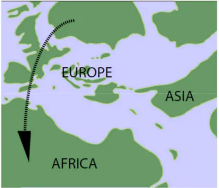
Figure 8. Paleogeographic map of Europe and North Africa in the Late Eocene.
Arrow indicates possible lacertid migration route to Africa between southwestern Europe and northwestern Africa via
small island chains (by Hipsley et al., 2009).
3.4 The Tribe Lacertini Oppel, 1811
Morphological and chromosomal features (Arnold et al., 2007 and references therein):
Tribe Lacertini exhibits a distinctive syndrome of morphological features, including flattened heads and bodies, fenestrated supraocular osteoderms in adults, often unkeeled dorsal body scales and little posterior overlap of the ventral ones, slender and fragile tails and, frequently, dorsal patterns in which longitudinal striping is reduced or absent.
24
• Body size. Adults of most Lacertini species are around 55–90 mm from snout to vent
(exceptionally over 90 mm in Podarcis). A few forms are smaller including some
Algyroides (A. fitzingeri not more than about 45 mm and A. moreoticus often under 50 mm).
In contrast, Lacerta and Timon are generally much larger than other Lacertini, with respective adult sizes of about 70–175 mm and 100–210 mm from snout to vent or more. Adult males are usually larger than females, exceptions are represented by some Darevskia,
Lacerta, Iberolacerta, Takydromus and Zootoca.
• Body shape. The head and body is fairly deep in most Lacertini, particularly in Lacerta,
Timon and Zootoca. In contrast, a number of species that regularly use crevices as refuges are moderately to very depressed.
• Dorsal body scales. The body is usually covered above and on its flanks by small scales that
are not as large as those on the tail. The number of scales in a transverse row across the mid-body is often 40–80 but figures may reach or even exceed 100 in some populations of the
Timon lepidus group, or be as low as 25 in some Zootoca. Dorsal scales are frequently
lightly keeled and more strongly so in forms like Lacerta, Parvilacerta parva, Podarcis
taurica, Timon princeps and many Zootoca species. However, keeling is absent in many
forms and, in ones that regularly use crevices as refuges, such as Dalmatolacerta,
Dinarolacerta and Iberolacerta horvathi, the scales themselves are flattened. Algyroides and
most Takydromus differ from all other Lacertini in having dorsal scales that are much bigger than those on the tail.
• Femoral pores. The femoral pores under the thigh are arranged in most Lacertini in a row
numbering 7– 31, beginning close to the midline of the body. Normally the row extends to the knee, but it is sometimes shortened distally, for example in Darevskia derjugini and some members of the Lacerta trilineata group. Takydromus is exceptional in often having just one or two femoral pores on each side or sometimes none at all; the maximum number found in this genus is five.
• Toes. The toes of Lacertini usually have one or two rows of unkeeled tubercular lamellae
beneath, but the lamellae bear a single row of subdigital keels in Apathya and sometimes a faint double row in Parvilacerta parva.
• Tail. The tail scales are arranged in regular whorls, two to each vertebra. The whorls may be
subequal in length but sometimes are alternatively markedly longer and shorter. The scales bordering the ventral mid-line of the tail are usually about the same width as neighbouring ones, but they are markedly expanded in Dalmatolacerta (Fig. 18) and to a lesser extent in some Hellenolacerta.
25
• Colouring. Lacertini exhibits considerable variation in their colouring and there are often
extensive intraspecific differences, both between and within different populations.
• Sexual dimorphism. Some species show little difference between males and females in their
dorsal colouring, but sexual dimorphism is sometimes well developed, like in Podarcis. It also occurs in varying extents in Algyroides moreoticus, some Lacerta populations (particularly well developed in L. schreiberi and L. agilis), and some populations of
Hellenolacerta, Darevskia, Iberolacerta and Teira. Some Lacertini have broad dorsolateral stripes that are lighter than the flanks and mid-dorsum but sometimes consist of ground colour rather than being lighter than this; they occur in some Anatololacerta, Apathya,
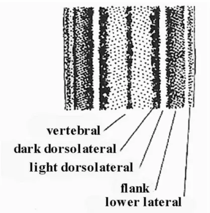
Scelarcis, Takydromus and Teira. Absence of striping in juveniles is uncommon but is found in Archaeolacerta, Dalmatolacerta, Timon and some Dinarolacerta, Podarcis, Scelarcis and many Takydromus. In these cases, animals are spotted or reticulated from hatching. Dorsal ground colour is often various shades of brown, buff or grey but it may be at least transiently bright green in many Lacerta and Timon and a wide though sporadic range of other forms. Melanism occurs sporadically in some taxa, but is especially common in Podarcis populations on small islands and in Dalmatolacerta at high altitudes. It arises in three different ways: general darkening of the ground colour (the commonest), increase in the number of dark markings, and the spread of those markings already present. These conditions were respectively named melanismus, abundismus and nigrismus, by Reinig (1937).
26
Figure 9. Common elements of patterning in lacertines: mid-back showing frequent positions of longitudinal stripes or rows of spots. From Arnold & Burton (1978).
• Hemipenis. Lobes having especially thin walls, which are complexly folded when the
hemipenis is retracted.
Chromosomes:
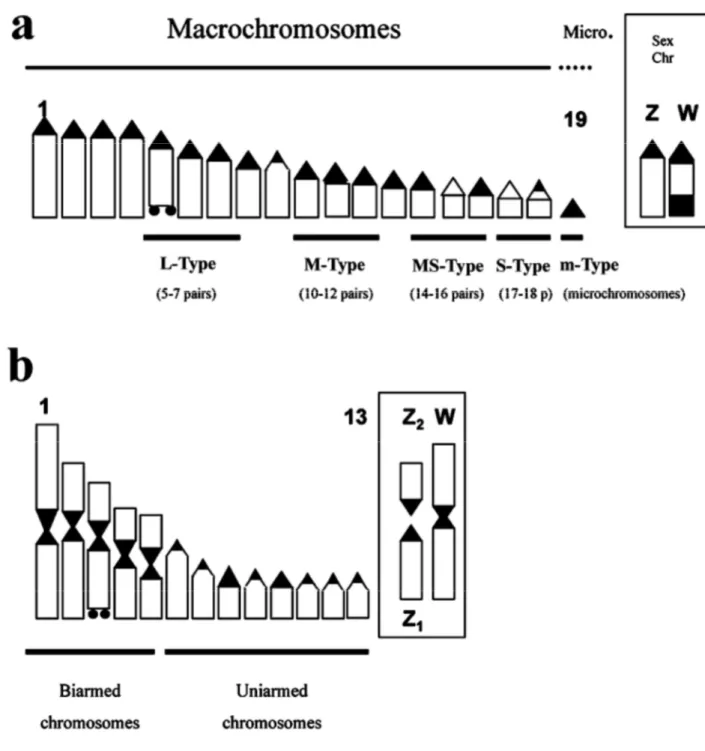
The diploid number of chromosomes in Lacertini is usually 38, consisting of 36 single-armed macrochromosomes (otherwise known as uniarmed, acrocentric or subtelocentric) and two microchromosomes. The total number of chromosome arms is termed the Fundamental Number (FN), which is usually 38 in Lacertini (figure 10) (Arnold et al., 2007).
27
Figure 10. Diagramatic representations of lacertine karyotypes (a single chromosome of each homologous pair is represented). (a) Common haploid condition with 18 single armed (that is uniarmed or acrocentric macrochromosomes) and one microchromosome, producing a diploid number of 38. The boxes in figure 10 illustrate the sex chromosomes, which are part of the usual complement of chromosomes illustrated to the left but revealed in cytological preparations by specific dyes; in this case they are of the ZW type, with ZZ males and ZW females. Chromosomes where the nucleolar organiser (NOR) may be situated are marked by horizontal bars and organisers are assigned to categories based on the size of these: if the NOR is situated in a macrochromosome this may be large (L-type), medium (M-type), medium-small (MS-type) or small (S-type); nucleolar organisers may also occur on the microchromosomes (m-type). In the present case the nucleolar organiser, indicated by two small black dots, is L-type. (b) Derived haploid condition with many double armed (that is biarmed, metacentric or submetacentric) chromosomes, based here on Iberolacerta aurelioi, but similar conditions occur in other Pyrenean Iberolacerta and in Parvilacerta. The five double-armed chromosomes
28
appear to have each been produced by fusion of two chromosomes (Robertsonian fusions). The sex chromosome system
is Z1Z2W, in which males have Z1Z1Z2Z2 and females Z1Z2W. Here the W chromosome is bi-armed as a result of
Robertsonian fusion of two chromosomes, giving females one fewer chromosome than males (by Arnold et al. 2007).
Conditions in other lacertids suggest this overall pattern is the primitive state in this tribe.
Iranolacerta brandtii is distinctive in having a different arrangement of macrochromosomes, with
34 single-armed ones and a pair that are double-armed (metacentric). With the two microchromosomes, this may indicate a fundamental number of 40 (2n =38, FN =40), which is also found in some Gallotiinae (Gallotia - 2n =40, FN =40; most Psammodromus algirus - 2n = 38, FN =38, but others are reported as 2n = 40, FN = 40). Takydromus has diploid numbers from 38 to 42, with 36 macrochromosomes and 2–6 microchromosomes. The diploid number is reduced to no more than 36 in Zootoca and Iberolacerta by loss of the microchromosomes, A similar degree of reduction in chromosome number occurs in Timon but here there has apparently been fusion (Robersonian fusion) of two pairs of the single-armed macrochromosomes resulting in one pair of two-armed macrochromosomes. A greater degree of reduction is found in some Pyrenean
Iberolacerta, resulting in diploid numbers of 26 or 24 in males and 26, 25 or 23 in females, the last
being the lowest chromosome number known in lacertid lizards. In male Iberolacerta with reduced chromosome numbers, there are 16 or 12 single-armed macrochromosomes and 10 or 12 double-armed ones. Similarly, in Parvilacerta the diploid number is reduced to 24 by seven fusions so that there are eight single-armed macrochromosomes, 14 double armed ones and two microchromosomes. In Darevskia, hybrids between sexual males and parthenogenetic females may be triploid with 3n=57 chromosomes. Sex chromosomes. Specific chromosomes among the total complement described above determine the sex of individual lizards. In the widespread ZW system, males have two Z chromosomes (ZZ in the diploid cells) and females one Z and one W chromosome (ZW in the diploid cells). In Iberolacerta with reduced chromosome numbers, the sex chromosome system is Z1Z2W, in which males have Z1Z1Z2Z2 and females Z1Z2W. Populations of Zootoca exhibit a range of conditions, including the primitive ZW one in Z. vivipara carniolica and some Hungarian populations still considered as Z. v. vivipara (Odierna et al. 2004), and the Z1Z2W system across most of the vast distribution of the genus, from the egg-laying Iberian populations to the Pacific coast of Siberia and Sakhalin island (Z. v. sachalinensis). In the Z1Z2W system, females have a total chromosome number of 35 chromosomes rather than the 36 usual in males. The W chromosome of Zootoca is doublearmed in some populations but has single-armed by heterochromatinization and loss of chromosome fragments in others.
29 Nucleolar organiser. This is situated in chromosomes of different sizes in different species. It may occur
in large (L-type), medium (M-type), medium-small (MS-type) or small (S-type) macrochromosomes, or in a microchromosome (m-type). Details are given in Fig. 24. As, L-type nucleolar organisers are known in the Lacertini but are so far unrecorded from other lacertids, they are likely to represent a derived condition. In some individuals or populations of Timon lepidus group, a second nucleolar organiser may occur (Arnold et al., 2007 and references therein).
3.5 Present assessments of relationships within the Lacertini
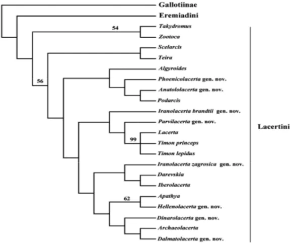
Taxonomic treatment of the species now assigned to Lacertini has varied considerably over time, but a number of assemblages are now commonly recognised. The fact that taxonomy was initially based on morphology and now it relies more on molecular data has inevitably led to confusion and instability in nomenclature. As a foundation for a more rational and comprehensive system of names for groups of Lacertini, we use new and old evidence for relationships within the group, based both on DNA sequences and morphology. The mitochondrial studies of Harris et al.(1998) and Fu (2000), and reanalyses of data presented by Arnold et al. (2007), support many of the recognised groupings within the Lacertini that have more than one species to corroborate the clade status of 19 groups (see Fig. 11) (Arnold et al. 2007).
30
Figure 11. Phylogeny of Lacertini based on a total of 64 morphological characters (58 parsimony-informative); equivalent to 83 binary characters.. Figures above the nodes indicate bootstrap support (from Arnold et al. 2007).
In all, 19 units were recognised which molecular clocks suggest separated 12–15 My ago (by Arnold et al., 2007).
If Europe is the source area for modern lacertids, there must have been several invasions of other regions. The molecular clock used here indicates that the Lacertini split into most of its component living genera 12–16 My ago, so they underwent quite rapid speciation at this time (but see Hipsley et al., 2009 for older divergence time estimates, around 43-46 mya). Most genera in the Lacertini have largely allopatric and often disjunct ranges, which may mean that initial spread of the group was followed or accompanied by multiple vicariance (see Fig. 12) (Arnold et al., 2007). A few units do not fit this pattern and have large ranges that overlap with several other taxa, although these too may have began as vicariant isolates with small ranges and then spread (Arnold et al., 2007 and references therein).