Dipartimento di Chimica e Tecnologie Chimiche
Study of Enzymatic Mechanisms Using
Density Functional Theory and
QM/MM Methods
José Gaspar Rangel Pamplona Pizarro Pinto
11/12/2015
Il lavoro presentato in questa tesi riguarda il meccanismo catalitico di tre enzimi naturali e di un biomimetico. All'inizio del documento è riportata una breve introduzione sul ruolo svolto in natura dalle proteine biologiche in generale, e in particolare dagli enzimi. Poche righe sono state dedicate all’importanza di alcuni metalli di transizione che si trovano più spesso nei siti attivi degli enzimi, con un particolare riferimento ai metalli presenti nei sistemi oggetto di questo studio. Nel capitolo successivo sono stati descritti i principi base delle varie metodologie teoriche utilizzate (QM e QM/MM) ed è stata fatta una breve descrizione della teoria cinetica enzimatica. Data la complessità dei sistemi in studio, dovuto alle loro dimensioni e alla presenza in essi dei metalli di transizione, è stato ritenuto utile dedicare la terza parte di questa tesi ai protocolli computazionali maggiormente usati per trattare argomenti di questo tipo che permettono di ottenere risultati affidabili.
Nei capitoli successivi sono stati riportate le singole problematiche affrontate e i risultati finora ottenuti che hanno riguardato in particolare il meccanismo di lavoro dei seguenti enzimi:
-modello biomimetico del sito attivo dell’enzima 3-Hydroxyanthranilate-3,4-dioxygenase (HAD).
-Tyrosine Ammonia-Lyase (TAL) -Human Pancreatic Amylase (HPA)
The work presented in this thesis concerns the catalytic mechanism of three natural enzymes and a biomimetic. At the beginning of the document provides a brief introduction on the general role of biological proteins, and in particular on that of enzymes. A few lines are dedicated to the importance of some transition metals that are found more often in enzyme active sites, with a special emphasis to metals present in the systems object of this study.In the next chapter were described the basic principles of the various theoretical methodologies used (QM and QM/MM), and a brief description of enzymatic catalysis theory.Given the complexity of the studied systems, due to their size and the presence in them of the transition metals, it was deemed useful to devote the third part of this thesis to the computational protocols mainly used to treat such arguments that allow us to obtain reliable results.
In the following chapters were given individual issues addressed and the results so far obtained involving in particular the action mechanism of the following enzymes:
-Biomimetic model of the active site of the enzyme 3-Hydroxyanthranilate-.4-3 dioxygenase (HAD).
Tyrosine Ammonia-Lyase (TAL) -Human Pancreatic Amylase (HPA)
O trabalho apresentado nesta tese refere-se ao mecanismo catalítico de três enzimas naturais e um mecanismo biomimético. No início do documento ha uma breve introdução ao papel desempenhado pelas proteínas de natureza biológica, em geral, e em particular pelas enzimas. Algumas linhas foram dedicadas para a importância de alguns metais de transição que são encontrados com mais frequência nos centros ativos de enzimas, com uma referência especial para os metais presentes nos sistemas em estudo. No capítulo seguinte, têm sido descritos, os princípios básicos das diversas metodologias utilizadas teóricos (QM e QM / MM) e também uma breve descrição da teoria de cinética enzimática. Dada a complexidade dos sistemas em estudo, devido ao seu tamanho e à presença neles de metais de transição, foi considerado útil para dedicar a terceira parte desta tese protocolos computacionais mais comumente usadas para tratar este tipo de argumentos que permitem a obtenção de resultados confiáveis . Os capítulos seguintes são dirigidos aos resultados obtidos até agora. Estes resultados centram-se no mecanismo de funcionamento das seguintes enzimas:
-modelo biomimetico do centro ativo da enzima 3-Hydroxyanthranilate-3,4-dioxygenase (HAD).
-Tyrosine Ammonia-Lyase (TAL) -Human Pancreatic Amylase (HPA)
Título e composição do Júri ... ii
1 Introduction ... 1
1.1 Enzymatic Catalysis generalities ... 3
2 Theoretical Background ... 7
2.1 Quantum mechanics (QM) ... 7
2.1.1 Density Functional Theory ... 7
2.1.2 Density Functionals ... 9
2.2 Basis set ...10
2.3 Molecular Mechanics (MM) ...11
2.3.1 General Force Field Equation ...12
2.3.2 AMBER ...14
3 Modeling Reactions ...17
3.1 Cluster Model Approach ...17
3.2 QM/MM Approach – ONIOM ...18
4 A QM/MM study and MD simulation to uncover the reaction mechanism of the Dipeptidyl Peptidase III enzyme ...21
4.1 Abstract ...21
4.2 Introduction ...21
4.3 Methods ...24
4.3.1 Models ...24
4.3.2 Molecular Dynamics Simulations ...24
4.3.3 QM/MM Calculations ...25
4.4 Results ...26
4.5 Conclusion ...33
5.1 Abstract ...37
5.2 Introduction ...38
5.3 Methods ...41
5.4 Results ...43
5.4.1 Phosphotriesterase mechanism: ...43
5.4.2 Promiscuous phosphodiesterase mechanism: ...49
5.5 Bibliography ...57
5.6 Supporting Information ...61
6 Scope - Tyrosine Ammonia-Lyase given by a QM/MM model ...73
7 Scope – Human pancreatic -amylase ...75
1 Introduction
Proteins can be found virtually in every body part, cells and tissue. They are large and complex molecules and are required for functionality, structure and regulation of the body organs and tissues. They are responsible for most of metabolic processes that sustain life, so the correct work of enzymes is of extreme importance in every human organisms but also in all kinds of organisms, from bacteria to animals.
Enzymes work by lowering the activation energy of a given reaction. The enzyme may do its job by stabilizing the transition state or even by distorting the substrate to the point that the transition state does not require an impossible high Gibbs free energy. Another way in which an enzyme may lower the activation energy of a reaction is by changing its pathway. -galactosidase deficiency is an example of the importance a single enzyme has in the normal life of a person. Lactose intolerant people do not produce enough -galactosidases or the ones they do produce work deficiently. This makes lactose impossible to digest at normal enzymatic digestive velocities leaving lactose to be metabolized by bacteria resulting in the fermentation and its product gases.
The active site of an enzyme may be surrounded by just amino acids, or amino acids and co-factors, which help the reaction by stabilizing substrates, transition states or products. In the active site of enzymes, we can also find metal atoms (one or two) or even a cluster of metal atoms. The iron-sulfur cluster enzymes are a good example of the latter.
Enzymes with non-metal active sites are the most common biologically. The substrate enters the pocket where there areelectrostatic interactions or covalent bonds fixing the substrate in the correct position and place so catalysis may happen.
About a third of all proteins are metalloproteins in which the metal atom participates directly or indirectly in the catalysis.
There are ten metals which are commonly found in proteins, either in their cationic form or associated with amino acids. Of the alkali metals, sodium (Na) and potassium (K) and alkaline earth metals, magnesium (Mg) and calcium (Ca) are among the ten
most common metals typically found in the cationic form. The transition metals, manganese (Mn), iron (Fe), cobalt (Co), nickel (Ni), copper (Cu) and zinc (Zn), complete the group of metals commonly found in proteins, and are normally associated with amino acids. In addition, manganese, iron and cobalt are also found as components of cofactors such as chlorophyll and heme groups.
Iron is the most common transition metal in biological systems; it is also the most common metal on earth’s crust. A complex system, in humans, regulates iron equilibrium. Both excess and scarcity of iron on the human organism has some kind of disease as a consequence. One well known effect of iron deficiency is anemia.
Iron is most known for heme and heme like ligands. But iron-sulfur complexes are also well known and very interesting. Such clusters are found on nitrogenase enzymes with the biological role of nitrogen fixation. Because of a high iron presence on earth it is only natural to try and use iron when synthesizing a biomimetic catalyst. In this document we will show a biomimetic inorganic catalyst, mimicking a ring cleaving enzyme.
Zinc has a stable oxidation state of 2+ with its d-orbital full. In this oxidation state every electron is paired and as a result it has a low spin configuration. After Iron, Zinc it’s the second most common transition metal in proteins. In biological systems its commons ligands are mainly histidine residues, aspartic and glutamic acids. Its coordination spheres my go from three to six ligands, though four is the most common number of ligands in a zinc coordination sphere.(1; 2)
Cobalt is not as common in biological systems as both previous referred metals. It may be found in proteins in three different oxidation states, from Co(I) to Co(III). In vitamin B12, a vitamin that plays a key role in controlling the normal functioning of the brain and the nervous system, cobalt has an oxidation state of Co(III). It may also work as a red-ox agent changing from Co(I) to Co(II) and vice versa. It is also find in binuclear centers as we’ll see farther in this document.
1.1 Enzymatic Catalysis generalities
A catalyst is a substance that increases the rate of a reaction without itself being consumed by the process. It lowers the Gibbs energy of activation by providing a different mechanism for the reaction. This mechanism enhances the rate and it applies to both the forward and the reverse directions of the reaction.(3)
The catalyst forms an intermediate with the reactant(s) in the initial step of the mechanism and is released in the product-forming step, thus does not appear in the overall reaction. Regardless of the mechanism and the energetics of a reaction, a catalyst cannot affect the enthalpies or Gibbs energies of the reactants and products. Thus, catalysts increase the rate of approach to equilibrium, but cannot alter the thermodynamic equilibrium constant.
There are three types of catalysis: heterogeneous, homogeneous, and enzymatic. Enzyme catalysis is also mostly homogeneous in nature. However, because it is of biological origin is the most complex of the three types of catalysis, and it is treated as a separate category.
An enzyme usually contains one or more active sites, where reactions with substrates take place. An active site may comprise only a few amino acid residues; the rest of the protein is required for maintaining the three-dimensional integrity of the network. The specificity of enzymes for substrates varies from molecule to molecule. Many enzymes exhibit stereochemical specificity in that they catalyze the reactions of one conformation but not the other. Some enzymes are catalytically inactive in the absence of certain metal ions. Although enzymes are evolved to catalyse a particular reaction on a particular substrate with a high catalytic efficiency (kcat/KM, Michaelis–Menten
kinetics), they can often perform side reactions in addition to their main, native catalytic activity. These promiscuous activities are usually slow relative to the main activity and are under neutral selection.(4) Despite ordinarily being physiologically irrelevant, under new selective pressures these activities may confer a fitness benefit therefore prompting the evolution of the formerly promiscuous activity to become the new main activity. Promiscuity comes in many different forms. We can have the so called “enzyme condition promiscuity” that is shown by enzymes with catalytic
activity in various reaction conditions different from their natural ones, such as anhydrous media, extreme temperature or pH and so on. Another type of promiscuity is known as “enzyme substrate promiscuity” and it is shown by enzymes with relaxed or broad substrate specificity. Finally, we have the “enzyme catalytic promiscuity” which is shown by enzymes catalyzing distinctly different chemical transformations with different transition states. The latter can be either:
(i) accidental – a side reaction catalyzed by the wild-type enzyme;
(ii) induced – a new reaction established by one or several mutations rerouting the reaction catalyzed by the wild-type enzyme.
In the 1890s the German chemist Emil Fischer (1852–1919) proposed a lock-andkey theory of enzyme specificity. According to Fischer, the active site can be assumed to have a rigid structure, similar to a lock. A substrate molecule then has a complementary structure and functions as a key. Although appealing in some respects, this theory has been modified to take into account the flexibility of proteins in solution. We now know that the binding of the substrate to the enzyme results in a distortion of the substrate into the conformation of the transition state. At the same time, the enzyme itself also undergoes a change in conformation to fit the substrate.
The enzyme and substrate interact to form an enzyme-substrate complex. The interactions between the substrate and active site are weak, noncovalent interactions (i.e. the substrate does not covalently bind to the active site but weakly interacts with it through interactions like hydrogen-bonding, van der Waals interactions, etc).(5)(6) The orientation in which the two interact is highly favorable for facilitating conversion of the substrate to product. In the enzyme-substrate complex, the substrate molecule binds to a very specific region of the enzyme molecule called the active site. The active site is usually found in a 3-D groove or pocket of the enzyme, lined with amino acid residues (or nucleotides in RNA enzymes). These residues are involved in recognition of the substrate. Residues that directly participate in the catalytic reaction mechanism are called active site residues. After an active site has been involved in a reaction, it can be used again.Substrates bind to the active site of the enzyme through hydrogen bonds, hydrophobic interactions, temporary covalent interactions (van der Waals) or a
combination of all of these to form the enzyme-substrate complex. Residues of the active site will act as donors or acceptors of protons or other groups on the substrate to facilitate the reaction. In other words, the active site modifies the reaction mechanism in order to change the activation energy of the reaction. An enzyme binding to a substrate will lower the energy barrier that normally stops the reaction from happening. The product is usually unstable in the active site due to steric hindrances that force it to be released and return the enzyme to its initial unbound state.
As we will see, the active site in an enzyme is of crucial importance to study the catalytic behaviuor from a computational point of view.
2 Theoretical Background
2.1 Quantum mechanics (QM)
The first approximation, applied in any theoretical method, is certainly the Born-Oppenheimer approximation, according to which, given the difference in mass between the nuclei and electrons, it is possible to decouple the motion of the nuclei from that of the electrons whilst the nuclei are considered as fixed. By breaking down the molecular system into two subsystems (electronic and nuclear), the expression of the Schrödinger equation is simplified, as it is possible to neglect the kinetic energy of the nuclei but still maintain the nucleus-nucleus repulsive interaction. The search for theoretical methods apt to provide approximate solutions of Schrödinger equation without resorting to parameters obtained experimentally has led to the development of Hartree-Fock (HF), post-Hartree-Hartree-Fock (post-HF) and Density Functional Theory (DFT) methods. With the methods previously mentioned we are able to run computational studies of a few hundred atoms. Despite the higher theoretical level of the HF and post-HF methods, in this thesis we use and present only the DFT method for its lower computational costs while maintaining a high theoretical level and accuracy. The density functional theory states it is possible to extract the properties of a given system from its electronic density. (7)
2.1.1 Density Functional Theory
The density functional theory foundation lies in the two Hohenberg-Kohn theorems(8). Hohenberg and Kohn showed that the density of the ground state of a non-degenerate electron system makes it possible to uniquely determine all the properties of the ground state. So each physically observable ground state, such as the total energy of the ground state, can be expressed as a functional of the density, E []. The energy E [] is in fact a function of the electron density (r), which is in turn a function of the spatial coordinates (x, y, z). The electronic energy functional E[] is generally divided into several contributions which take into account separately the electronic kinetic energy T
[], the potential energy of electron-electron repulsion interaction Vee [] and the
potential energy of electron-nuclei interaction VNe []. The first two contributions are
independent of the nuclear positions and can therefore be grouped together in the universal density functional F []:
E [] = F [] + VNe [ρ] (6)
The exact determination of the functional F[] is in fact not possible. Because of the limitations of this equation Kohn and Sham introduced a set of orbitals (analogous to HF orbitals) in which the kinetic energy can be divided into two parts. [9]
In their theorem, Kohn and Sham, represented the functional of the kinetic energy of a system in two parts, one which could be exactly calculated assuming that the electrons do not interact with each other, and another part which was a correction to account for the interaction between them.
The Kohn-Sham formalism states that the ground state electronic energy in a system, with n electrons and Z nuclei is calculated as follows:
𝐸[𝜌] = −1 2∑ ∫ 𝛹𝑖 ∗(𝑟 1)𝛻𝑖2𝛹𝑖(𝑟1)𝑑𝑟1− ∑𝑁𝑋=1∫𝑟𝑍𝑋𝑖𝑋𝜌(𝑟1) 𝑛 𝑖=1 𝑑𝑟1+ 12∫ ∫𝜌(𝑟1𝑟)𝜌(𝑟12 2)𝑑𝑟1𝑑𝑟2+ 𝐸𝑋𝐶[𝜌] (7)
The four terms in the equation are respectively kinetic energy of electrons where there interactions, Ψ represents the orbital Kohn-Sham, the attraction between electron density and nuclei, the Coulomb repulsion distribution of the total load r1 and r2 and the
term exchange - correlation which represents the correction made to the first term where there are no interactions between the nuclei. Here the exchange functional describes the exchange interaction between the electrons with the same spin
multiplicity and functional correlation describes the interaction of electrons with opposite spin multiplicity.(9; 10)
2.1.2
Density Functionals
The density functionals first developed were very simplistic and two different types of density functional existed at the time. The big difference between them was the inclusion of spin effects in one type (LSDA – Local Spin-Density Approximation)(11; 12; 13; 14) and the other type of density functionals had no inclusion of spin effects (LDA – Local Density Approximation)(15). Density functionals such as the GGA (Generalized Gradient Approximation) (e.g. BLYP and PBE)(16; 17) that perform better than the earlier LDA and LSDA are now in use. There are also some further developments of this type of orbital, when calculating the second derivative of electron density, which is the kinetic energy density, the functional Meta - GGA (e.g. TPSS and M06-L)(18) results can be obtained with higher accuracy. There are also functional hybrids can be H-GGA (e.g. B3LYP) or HM-GGA (e.g. TPSSh)(19), based on whether or GGA to M-GGA respectively(20; 21; 22; 23).
The development of density functionals is still ongoing with new functionals every year.
2.1.2.1 B3LYP
B3LYP is a hybrid density functional that was obtained from the combination of other the exchange and correlation functionals. In addition to the introduction of B88, LYP and VWN it was also included 20% of exact HF exchange(22; 24).
EXCB3LYP= (1 − a)EXLSDA+ aEXHF+ bEXB88+ cECLYP+ (1 − c)ECVWN (8)
In equation 8 the coefficients a=0.20, b=0.72 and c=0.81were obtained from the B3PW91hybrid density functional.
In this work we chose B3LYP to all optimizations given that it has been broadly tested and used and is considered to yield good geometries. The parameters introduced in this functional were fitted to energies of atoms from the three first rows of the periodic table, however, studies have shown that geometries of various transition metals very small mean absolute deviation.
2.2 Basis set
In the case of the method that was briefly described above as well as in other ab-initio and DFT methods, basis-set functions are needed for this calculation. Basis-set functions allow us to describe approximately an unknown function.
Including basis-set is not in itself an approximation, assuming that the unknown function is fully described, but for that to be true an infinite number of basis-set, which is an impossibility in practical terms. Obviously the smaller the number of basis-set functions, the worse will be the representation of an unknown function. Other than the number of set used to describe an unknown function the correct choice of basis-set used is also of the utmost importance.
There are two common types of basis-set functions used: STO - Slater Type Orbitals and GTO - Gaussian Type Orbitals.
The Slater-type orbitals describe more accurately the electron- nucleus interaction.(25; 26) However, for non-hydrogenoid molecules, this orbital is not suitable as it computational expensive to use it for large molecules.
The Gaussian type orbitals have a simpler mathematical resolution. However their description of the behavior of electrons is not done as well, especially when the electrons are near the core. Nonetheless, for large molecules their use is considered to be the better choice.
Whatever the chosen basis-set is, they shall not describe with accuracy only one atom, but be flexible enough so that they can describe the molecule as a whole and not a particular atom. This good description on the molecular level can be obtained with the inclusion of polarization and diffuse functions. The polarization functions have a
higher angular momentum, thus describing the distortion of atomic orbital in a molecular environment. On charged or highly polarizable molecules is advisable to use diffuse functions because they describe best electron density away from the core.
2.2.1.1 Pseudopotentials
When studying systems of considerable dimensions or even transition metals it is wise to use basis-set with pseudopotentials. The introduction of pseudopotentials has the purpose to lower the computational cost of the calculations by lessening the number of functions needed to describe all the electrons present in a calculation.(27)
This methodology is based on the assumption that core electrons and other lower energetic level electrons are mainly inactive and do not disturb valence electrons that are responsible for bond properties.
2.3 Molecular Mechanics (MM)
Molecular mechanics is one of the most used methods in the study of biochemical systems but as for other methods there are advantages and disadvantages. Molecular mechanic calculations are much faster than the quantum mechanical calculations and are therefore used in systems with many atoms. However their major drawback is to be unable to explicitly describe electrons. This method can’t be applied directly to study a system in which electrons are exchange between atoms or even a study of how a particular chemical connection is made. The methods used in the calculation of molecular mechanics (MM) are quite different from those used in quantum mechanics (QM). Unlike QM methods that are based in the Schrödinger equation, MM methods describe the energy of a system just from the coordinates of its nuclei. As the MM can’t explicitly describe electrons, it considers atoms to be balls connected to each other through springs, where the balls have different sizes and springs have different loads depending on the atom they are representing. Here the energy of the system is a sum of several different processes such as stretching of bonds and bending of angles.
MM methods also require parameters that are obtained from smaller model calculations using a much higher theoretical level such as Hartree-Fock methods. Alternatively, experimental data can also be used. The accuracy of these parameters is therefore of utmost importance to have a high degree of confidence in molecular mechanics studies.
2.3.1 General Force Field Equation
A force field is a set of parameters used to define the potential energy of a system. The set of parameters of a force field comes from experimental work and calculations at a quantum mechanics level. The energy of a force field is usually described as the sum of several energy terms with different physical meaning and contribution. An example of the general equation is as follows:
𝐸𝑇 = 𝐸𝑠𝑡𝑟 + 𝐸𝑏𝑒𝑛𝑑 + 𝐸𝑡𝑜𝑟𝑠 + 𝐸𝑉𝐷𝑊 + 𝐸𝑒𝑙𝑒 (9) 𝐸𝑠𝑡𝑟 = 12𝑘𝑙(𝑙 − 𝑙0)2 (10) 𝐸𝑏𝑒𝑛𝑑 = 1 2𝑘𝜃(𝜃 − 𝜃0)2 (11) E𝑡𝑜𝑟𝑠 = ∑ 1 2 𝑗 𝑉𝑗 [ 1 − cos (jω)] (12) 𝐸𝑉𝐷𝑊 = ∑ [𝐴𝑖𝑗 𝑟𝑖𝑗12− 𝐵𝑖𝑗 𝑟𝑖𝑗6] 𝑖𝑗 (13) 𝐸𝑒𝑙𝑒 = ∑ 𝑞𝑖𝑞𝑗 𝜀𝑟𝑖𝑗 𝑖𝑗 (14)
In the equation (9) the first term corresponds to the stretching of bonds, the second term to the bending of angles, the third term describes the torsion of dihedral angles. These are the three terms with bonding interactions. The two following terms describe the non-bonding interactions and correspond to the energy of van der Waals and
electrostatic energy respectively. There are some force fields that include a correlation term.
In equation (10) kl is the force constant and l0 is the constant that describes the
reference distance between the atoms considered. The approximation resulting from the use of Hooke’s formula is usually accepted for bond length variations near the reference value associated with the minimum energy. Even though the Morse potential gives a better description of the energy of a given bond, the harmonic potential yields similar results in the bond range that chemists normally work and it is computationally cheaper. The difference is in the extremes, where the Morse potential describes the bond energy better than the harmonic potential.
As the stretching of the binding energy, the energy of an angle can be described in a harmonic way. In equation (11) k is the force constant of the angle and the
reference value of the angle at the balance position.
The torsion energy is the energy required for the rotation of the bond AB and CD in a group of atoms designated ABCD. Unfortunately, unlike the other two terms of binding interactions, this term is more difficult described by a harmonic potential. This is due to the fact that torsion energy presents a cyclic behavior and the variation between each minima requires very low energy. In equation (12), ω is the dihedral angle, i. e., the angle that AB makes with CD, j is its multiplicity, the minimum number of function while the link is rotated 360° (e.g. if j = 6, the function will have five minima and each energetic minima will be separated by 60°) and Vj is the torsional
constant force.
The van der Waals term describe interactions between atoms which are not bonded, regardless of their charge. These interactions are attractive for small distances but tend to null very quickly when the distance between atoms increases. At small distances, i.e., when the electronic clouds overlap there is van der Waals repulsion. This interaction is commonly calculated using the Lennard - Jones potential. When seeing equation (13) we may be lead on to think that the use of 12 and 6 on the repulsion – attraction potencies is the one that describes best the van der Waals term. It is just
another approximation to lower the computational cost. In the equation 8, A and B are constants, and rij is the distance between the two atoms.
The electrostatic term in the total energy equation, equation (14), describes the non-bonding interactions between positively and negatively charged atoms; usually this interaction is described by the Coulomb equation. Electrostatic energy is a function of atomic charges (qi qj), the distance between these (rij) and dielectric constant (ε).
These terms may differ slightly from force field to force field.
2.3.2 AMBER
The AMBER force field(28; 29; 30; 31; 32) (Assisted Model Building and Energy Refinement) was initially thought to be used in proteins and nucleic acids. Nowadays the AMBER family has a variety of parameterization schemes that describe proteins and nucleic acids (FF## libraries). There are also parameters to small organic molecules, especially within GAFF(33; 34) (General Amber Force Field) and also carbohydrates parameters in GLYCAM##.(35)
The AMBER energy calculation is described by this mathematical equation:
𝑈 (𝑅⃗ ) = ∑𝑏𝑜𝑛𝑑𝑠𝑘𝑏(𝑏 − 𝑏0)2+ ∑𝑎𝑛𝑔𝑙𝑒𝑠𝑘𝜃(𝜃 − 𝜃0)2+ ∑𝑑𝑖ℎ𝑒𝑑𝑟𝑎𝑙𝑠𝑉2𝑛 (1 + cos( 𝑛𝜙 − 𝛾)) + ∑ [𝐴𝑖𝑗 𝑟𝑖𝑗12− 𝐵𝑖𝑗 𝑟𝑖𝑗6 + 𝑞𝑖𝑞𝑗 ε𝑟𝑖𝑗] 𝑛𝑜𝑛𝑏𝑜𝑛𝑑𝑖𝑛𝑔 (15)
Where kb, kθ and Vn are the force constants, b, θ and 𝜙 are the bond distance, angle and
dihedral angle, with subscript zero indicating the reference values for these quantities. In this equation γ is the phase angle can have values between 0 and 180. In non-bonding terms Aij Bij are the van der Waals dispersion, qi qj are the partial charges of
atoms and εl is the dielectric constant. The nonbonding interactions terms of the
equation are calculated for atoms separated by three or more atoms.
From the computational point of view, metalloproteins are a very particular case. The presence of a metal atom can cause problems in applying some methods that are
already well established and proven to give good results in proteins without metal active sites. These problems can be very easily seen when needing to perform a molecular dynamics simulation on a metalloprotein, where the lack of specific parameters makes their computational study various steps behind the computational study of other proteins.
This is therefore a major limitation of the force fields and also obviously of the AMBER force field.
3 Modeling Reactions
Given that there are two different set of physical laws for the computational study of catalysis mechanisms there are also two different approaches for doing it. Depending on what one is studying it is better to use on approach or the other. There is not one better than the other though there is those who defend one over the other. For instance, because DFT methods are based on quantum mechanics laws, the electrons are taken into account, but the computation of these methods take a very long time, and the size of the model used does not usually exceeds two hundred atoms. On the other hand the MM methods are based on classical mechanics laws, which do not take electrons into account. The smallest unity in MM methods is the atom and it is connected to other atoms with a spring coil. Though they cannot describe chemical reactions they are much faster than DFT calculations and as consequence the size of the model used may be in the thousands. When used alone both approaches have different goals. Since classical mechanics do not take electrons into account, classical mechanic laws cannot be used to describe a chemical reaction with atomistic detail. Though they can and are used in molecular dynamics which gives us an insight into how a given protein moves in time. With quantum methods alone and today’s technology it would be impossible to study the dynamics of a protein.
So given the limitations of each approach we know that we have to do some compromises.
3.1 Cluster Model Approach
In the cluster model approach we use ab-initio methods (DFT in our case). As we said before we cannot use many atoms and it follows that the whole protein is out of question. The compromise here is using a small part of a given protein to study its catalytic mechanism. This small model of the enzyme will be of the active site of the enzyme, where the catalysis occurs.
When we have a crystallographic file of the whole enzyme, the active site has to be identified as well as the most important amino acids, those that have an important role in the catalytic mechanism. Ideally the model will have the amino acids that participate
in the catalytic mechanism and some others that stabilize the ones that do participate in the reaction. Some may ask if the rest of the enzyme is not important to the mechanism. The idea behind the cluster model approach is that the chemical reaction is concentrated in the active site and that the long range interactions are too small to have such a big contribution for the overall energy.
Nevertheless, after the geometry optimizations for all the stationary states along the reaction path are finished and the enzymatic mechanism is found, single point calculations are done with a polarizable continuum model to simulate the rest of the enzyme or the presence of a solvent. The compromise here is having the same dielectric constant embracing the cluster model instead of having different charges depending on the amino acid present in a given spot of the enzyme, thus eliminating any possible long-range interactions that could stabilize or destabilize the active site. Also in a cluster model approach the amino acids present in the model should have an atom frozen in space so as not to assume conformations it would not assume with the constrains imposed by the rest of the protein. Normally the atom that stands the farthest away from the active site should be frozen in order to allow as much freedom as possible to the amino acid.
When building a model one must take very careful into account the ratio of the model size per computation time. The model must be consistent enough that the mechanism is well described but also small enough so the computational cost is not exaggerated.
3.2 QM/MM Approach – ONIOM
Other than use only quantum mechanics with a small part of the enzyme (even if it is the most important part for the catalytic mechanism) we can use classical mechanics. For the study of catalytic mechanisms it is possible to combine computationally, quantum mechanics and classical mechanics. With this approach we use ab-initio methods in the active site and at the same time maintain the rest of the enzyme to be treated with molecular mechanics. In doing this the active site has to be identified and the amino acids that participate in the reaction mechanism have to be known so they
are included in the QM part of the calculation. In an ONIOM(36) calculation the QM part is usually smaller than it would be if we were doing a cluster model calculation. With ONIOM it is not needed to maintain the supporting amino acids in the QM part of the calculation for they will be present just the same but will be treated with MM. The interface of the layers represents a problem. It would not do to sever the bond and treat one part with QM and the other with MM and after the optimization of the geometry sum the energies.
What happens at the interface of both treatments is that we have to say to the program that the atom that is to be treated at a QM level of theory is bonded to a hydrogen atom. This way there are no atoms with a deficit of bonds. To facilitate the explanation the active site which is treated with ab-initio methods will be called the “small model” and the whole system will be called “real system”.
What an ONIOM calculation does is actually divide the job into three steps. On the first step an ONIOM calculation treats the real system with a low level of theory, meaning it uses the parameters in the AMBER libraries to calculate the energy of the enzyme’s conformation. After obtaining the energy for the real system, the energy of the small model is calculated, still using a low level of theory. The third step is the calculation of the energy in the small model with a high level of theory, normally and in our case, DFT level of theory is used.
In the end the energy in an ONIOM calculation is obtained as follows:
EONIOM=E low (RS) – E low (SM) + E high(SM) (16)
4 A QM/MM study and MD simulation to uncover the
reaction mechanism of the Dipeptidyl Peptidase III
enzyme
4.1 Abstract
In this work, the catalytic mechanism of dipeptidyl peptidase III (DPPIII) was studied with atomistic detail, using a hybrid quantum mechanical/molecular mechanical method at the ONIOM (B3LYP/6-31G(d):Amber) level. The hydrolytic reaction proceeds via a general acid/base mechanism, in which the first mechanistic step involves a proton transfer from the zinc-bound water molecule to the Glu508. The second step involves the remaining hydroxyl zinc-bound group that performs a nucleophilic attack on the scissile carbonyl bond leaving the zinc connection to the substrate oxygen atom. The third step involves a proton transfer from Glu508 to the peptide nitrogen and a subsequent cleavage of the peptide bond to yield the products in their neutral forms. The conserved residue Glu508 is ideally aligned and has the ability to slightly rearrange its conformation to act as a highly effective proton shuttle. Our results so far indicate that the nucleophilic attack agrees with the data available for other enzymes with reaction mechanism similar to the DPPIII, with activation energy for the second step being close to 16 kcal/mol.
4.2 Introduction
Zinc containing metallopeptidases constitute an expanding list of structurally related proteases which are widely distributed in nature. They are involved in highly important biochemical events, such as deformylation in bacterial protein synthesis, the case of peptide deformylases, extracellular matrix degradation and tissue remodelling (matrix metalloproteinases), digestion (carboxypeptidase A, astacin), enzyme hypertension management (human angiotensin-converting) and blood-pressure regulation (neprilysin), to name a few examples. Within the big family of enzymes that contain a Zn2+ in its active site there is a branch with a special HEXXH binding motif, where the
XX stands for different residues in different enzymes. For this reason, different enzyme active sites may even be superimposed and matched based on these similarities. 1–6
The similarity among the metallopeptidases is such that a standard orientation has been proposed for the visualization of the active site. Since the active site cleft is large enough for a polypeptide to enter, it is proposed that the substrates of these enzymes’ family align somewhat horizontally in the cleft. They bound in the active site N to C-terminal.7
catalysis capabilities of the enzymes with the two histidine residues being in the first sphere of coordination and the glutamate residue participating in the mechanism as a general acid/base. Bonded to the zinc atom is also another glutamate residue (E508).8,9
The enzyme of our study, dipeptidyl peptidase III (DPP III), was first discovered in the bovine anterior pituitary gland. It has in the active site a zinc atom bonded to two histidine residues (from the motif HELLGH), a glutamate residue and a water molecule. Stabilizing the water molecule is a glutamate residue (from the HELLGH motif).
Later DPP III was shown to be present in several animal tissues, such as rat and human. Both the rat and the human DPP III were proven to have a Zn2+ ion per mol of
protein. In 2008 the yeast DPP III structure was resolved, showing a different protein fold. Nevertheless the zinc coordination three-dimensional structure and full HELLGH motif is the same as seen in the human and rat DPP III.
This motif is so important that promotes metal promiscuity to the enzyme. A previous mutagenic study as shown that substituting the Zn2+ for other metal atoms such as copper (Cu2+), cobalt (Co2+) and nickel (Ni2+) maintains the ability of the enzyme to hydrolyse the substrate Arg-Arg-NA (two arginine residues and a naphthalene group bonded). On the other hand when a leucine residue (L453) is deleted from the enzyme the catalytic ability of the enzyme disappears and the Arg-Na peptide bond is not hydrolysed.10,11
DPP III is an important encephalin-degrading enzyme associated with the human pain modulatory system. Inhibition studies show that a neuropeptide named spinorphin formed by amino acids (Leu-Val-Val-Tyr-ProTrp-Thr) is effective in the inhibition of monkey purified DPP III.12 This neuropeptide acts differently than morphine and is
widely used as an analgesic in morphine-resistant cases. 13
In recent years the study on DPP III has grown, with overwhelming results. In the last 6 years structural data on inhibitors and substrate are available, kinetic studies were done to yeast, 14,15 rat10 and human16 DPP. Because of the works and results in
literature we now have more data to compare to computational studies.
Computational studies have also been done on the DPP III and other enzymes with similar proposed mechanism. As said before the active site cleft is large enough for a polypeptide to enter and accommodate, this means that DPP III hydrolyses a variety of peptides. The conformation that the enzyme acquires when a substrate is bonded to the active site is then, very important for the activity of the enzyme. This data is already in the literature which helped us with our study, as we will show in the results.17
Also of importance to our work, was a previous QM/MM study on ACE (angiotensin-converting enzyme). This work shows a very similar active site of different enzyme, with a reaction mechanism similar to what we expected to obtain.
Thermolysin, another zinc containing enzyme, was also studied computationally at a DFT level of theory, showing a reaction mechanism very close to the reaction mechanism shown on the ACE work. Moreover, the reaction mechanisms of both the ACE (QM/MM) study and the thermolysin (Cluster Model) study yield very similar energetic results for similar mechanistic steps.
These two reaction mechanisms initiate with a nucleophilic attack of a water molecule on the carbon of the peptide bond (scissile bond). In this process the water molecule
loses a proton that will eventually bond to the nitrogen to break the C – N bond. In both cases the first sphere of the zinc coordination is exactly the same we found in our enzyme, i.e., two histidine residues, a glutamate and a water molecule. These ligands adopt a tilted trigonal pyramidal geometry (scheme 1). The substrate is on top of this pyramid with a residue (histidine or tyrosine) stabilizing the oxygen of the carbonyl group, Opep. Stabilizing the water molecule is a glutamate residue from the HELLGH
motif, confirming its importance on the overall catalytic capability of the enzyme.
4.3 Methods
4.3.1 Models
The DPPIII:substrate complex was modeled from two different X-ray crystallographic structures that were obtained from the PDB databank18 (ID: 3FVY13 and 3T6B with a resolution of 1.90 and 2.00 Å respectively) . We first had to superimpose both structures by their catalytic residues because the 3FVY has a zinc atom in active site and the 3T6B does not. On the other hand, the 3T6B structure has an inhibitor from which we modelled the substrate Arg-Arg-Na. 14 After the substrate was modelled, we proceeded to delete the 3T6B structure. The resulting model was used as a starting point for the computational studies. Molecular dynamics (MD) simulations were performed to evaluate the effectiveness of modelling and to confirm that it was stable near the active site. Parametrization was need for the substrate. The zinc ligands were not needed to parametrized since that work had been done before by Brás et al. 3
For the MD simulations we used the amber force field (ff99sb) 19 parameters for the protein. Explicit solvent TIP3P water was used in a cubic box with a minimum of 12 Å around the protein, and a total of nineteen (19) sodium (Na+) ions were added to neutralize the charge of the system. The resulting models consisted of X atoms.
4.3.2 Molecular Dynamics Simulations
We ran three minimization steps, the first one with the protein fixed and the water molecules free to move, in the second step we kept the water molecules free of constraints and freed also the substrate, since it was modelled it had to minimize its energy before the whole system was free of constraints. Finally we proceeded to the third step in which the whole system had no constraints. The minimized structures were then submitted to a further 200 ps long warm-up simulations, from 0 till 300K, at constant volume and using periodic boundary conditions. Further 200 ns Langevin dynamics with 2 fs integration step were carried out. Non-bonded interactions were treated with the PME algorithm and the cut-off was set to 10 Å, real part also truncated at 10 Å with pressure of one atmosphere at 310 K with ensemble NTP. All calculations were performed using the Amber12 20 simulations package. Bonds involving hydrogen atoms were constrained with the SHAKE algorithm 21.
Since the conformations obtained from the MD simulation were in agreement with the conformations said to be most apt for the mechanism to occur 17 we continue our study with the next step.
4.3.3 QM/MM Calculations
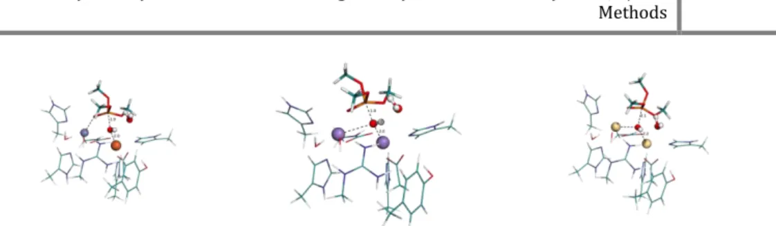
We performed QM/MM calculations to determine the potential energy surface (PES), using the Gaussian 09 software 22. We took from the last minimization the model used for the calculations. We froze the residues that were farther than 15 Å from the active site and most of the water molecules from the solvent were cut off yielding a model with a total of 11914 atoms and 898 residues. We divided our system in two layers and the ONIOM formalism 23 was used in order to calculate the corresponding PES. In the high layer there are 100 atoms, i.e. one zinc atom, the naphthalene group from the substrate, the backbone of the arginine residues of the substrate and two CH2 of each
side chain, one water molecule, two histidine residues side chains and two glutamate residues side chain (until the beta carbon). The high layer was treated with density function theory (DFT) at the B3LYP/6-31G (d) level 24–27. The low layer (the rest of the enzyme together with the solvent water molecules) was treated at the molecular mechanics level with the parameters of the amber force field package. We used electrostatic embedding to treat the coupling between both layers.
Transition states were searched for using flexible scans. Scans were made along the reaction bond in each step of the mechanism. After knowing the approximated geometry of each transition state, an optimization was carried out to find the geometry of the transition state and respective frequencies. To ensure that the minima found corresponded to same reaction coordinate as the transition state, IRC calculations were made.
The zero point energies were calculated at the same QM/MM level as the geometry optimizations, within the harmonic approximation. Thermal corrections (at 310 K) and entropy were calculated within the ideal gas/rigid rotor/harmonic oscillator model. Single point energy calculations were performed subsequently with the B3LYP density functional 28,29 with the larger 6-311++G(2d,2p) basis set.
4.4 Results
Given the importance of DPP III in the pain modulatory system it is underwhelming the amount of data available in the literature. The can be found experimental studies on it. Such as kinetic studies on the bacteria, rat and human DPP III, nevertheless computational studies on the mechanism itself, be them at DFT level or at QM/MM level, are not found, to the best of our knowledge. In the PDB databank for instance, we can only find four DPP III crystallographic structures. Three originated from human and one from Saccharomyces cerevisiae. Which is very few compared to the hundred and twenty eight (128) crystallographic structures of thermolysin present in the same databank.
Because of the quantity of Zinc-metallopeptidases and their importance, it is expectable that some of them are not as studied as others at a given time. The example of thermolysin is apparent, but also -lactamases and angiotensin converting enzymes have been studied and their catalytic mechanisms have been proposed and supported by computational studies.
This study as not yet reached its final stage. The last mechanistic step has not yet been uncovered. However, the first and second steps have been uncovered and the rate limiting step energetics is in agreement with the data obtained experimentally.
Because of the discussion and conclusions about the conformation of DPP III we decided to run a rather long molecular dynamics simulation. In figure 2 we show the RMSd of our MD of 150 nanoseconds, though the RMSd is rather high once it is stabilized, it is explainable through the high mobility of both arginine residues in the substrate. The side chain of arginine is rather long and has six single bonds in a row, meaning it is very movable. The overall conformation and position of the substrate in the active site is stable throughout the MD simulation.
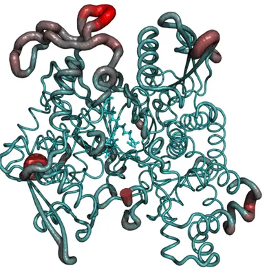
On the other hand the RMSd of the protein’s backbone stabilizes around 2.2 angstroms and stays that way throughout the simulation. Nevertheless we decided to see where the higher fluctuations on the protein were. Unsurprisingly we learned that the outer shell of the protein had the highest fluctuations. The active site and the substrate have very low fluctuations (figure 3).
Figure 2- RMSd values for the protein backbone and the substrate.
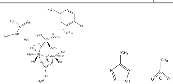
The reactants model we used to start the calculations (scheme 1) has the Zinc bonded to four ligands. Adopting a trigonal pyramidal conformation are two histidine residues, a glutamate residue and a water molecule. The water molecule forms a hydrogen bond with a glutamate residue. Directly above the water molecule is the scissile carbon of the substrate. Close to the substrate and stabilizing it in the active site is a histidine residue.
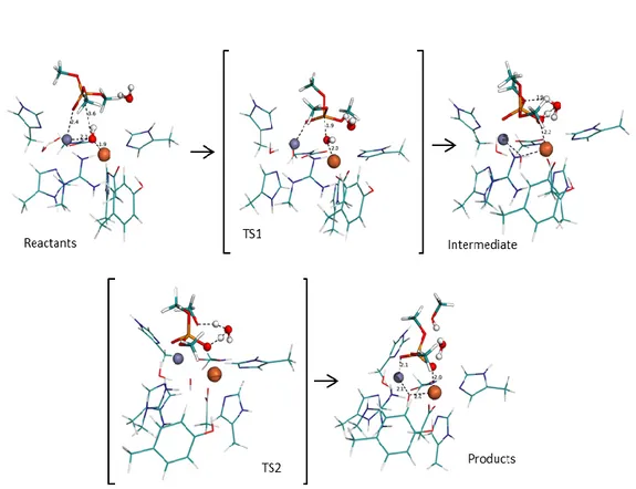
From this initial model we knew from similar enzymes that the oxygen of the water molecule would attack the scissile carbon. After trying to scan the attack directly with the water molecule we saw that it was not strong enough to attack and that every time, one proton from the water molecule would dissociate from the water to bond to the glutamate residue (figure 1).
This first step is energetically very cheap, with the transition state and the successive intermediate having virtually the same energy of the reactants. The energy of the three
states is so close to one another because there is not much change in the active site. In the reactants the proton is at 1.437 Å from the glutamate residue and still bonded (1.078 Å) to the nucleophilic oxygen. The transition state shows the proton almost midway between the Glu508 residue (1.211 Å) and the nucleophilic oxygen (1.234 Å). At the end of this mechanistic step, the first intermediate is achieved and the proton is now bonded to the Glu508 residue, with a bond length of 1.075 Å and is forming a hydrogen bond with the nucleophilic oxygen, at a distance of 1.460 Å. Because the nucleophilic oxygen is bonded to the zinc the bond it formed with the proton in the reactants conformation was already elongated for a normal O – H bond in a water molecule (0.998 Å) in liquid phase.30 This elongation of the bond, allows the transition of the proton from one oxygen atom to the other to have no energetic expense (figure 4).
Figure 3- Root mean square fluctuations of the enzyme’s backbone and the substrate. Cyan color is lowest RMSf and red color is highest. Values of RMSf range from 0 to 4.46 Å.
The first step transforms the water molecule in a hydroxide group. This transformation allows the hydroxide to have enough strength to attack and bond with the scissile
carbon of the substrate. The nucleophilic attack, in similar mechanisms, is the rate limiting step. In our study the trend seen in other enzymes is also seen here. The second transition state has an energy difference of 18.6 kcal/mol comparing with the reactants and an energy difference of 19.4 kcal/mol in comparison with the previous intermediate. Initially the Onuc is at 1.948 Å from the zinc and at 2.871 Å from the Csci.
The Opep of the substrate is being stabilized by a hydrogen bond of the His566 residue
and is distant from the zinc at this point (3.799 Å). At the same time that the Onuc is
approaching theCsci and distancing itself from the zinc, the Opep is doing the opposite,
approaching the zinc atom. At the second transition state (TS2), the Onuc is at 2.037 Å
This approximation the substrate to the Onuc means that the Opep is also closer to the
zinc atom. In fact, at the TS2 the distance of the Opep to the zinc atom is now only
2.148 Å, where before it was 3.799 Å. Continuing from the transition state to the second intermediate, the trend continues as before, with the Onuc detaching from the
zinc and the Opep binding to zinc, maintaining the former conformation of the zinc.
From the available data from other studies we knew how the reaction should evolve from here. We did a scan from the second intermediate where the Onuc transfers its
proton to the amino group on the naphthalene side. The Glu508 residue should also transfer its proton to the Onuc, reprotonating the Onuc. Even though in previous works
we saw that this double transfer of protons may occur at the same time, in this study we did not see that happen.
The proton being transfer from the Onuc to the NH-Na is at 1.309 Å from the nitrogen
and at 1.186 Å from the Onuc at the TS3 conformation. This transfer has an energetic
cost of 6.7 kcal/mol, and the transition state has been confirmed through frequency calculations.
After the transition state is passed and the bonds are formed, the NH2-Na group,
distances from the rest of the substrate. The Onuc, being deprotonated, binds again with
the zinc atom, while the other oxygen of the carboxyl group unbinds, returning to the conformation seen in the first intermediate.
By protonating the amino group of the naphthalene the bond between nitrogen and carbon no longer exists and the NH2-Na distances itself from the rest of the substrate.
The amino group establishes a hydrogen bond N – 1.262 Å – H – 1.730 Å – O from Glu508) with the Glu508. Because the naphthalene is so big and rigid once it is stabilized, the difference in energy is somewhat higher than we expected, with the minimum after TS3 being 28.8 kcal/mol more stable than the transition state that precedes it.
From this stationary point, the next logic step would be the proton transfer from the Glu508 to the Onuc, however we have not, at this time, completed the calculations for
Figure 5- Potential energy surface obtained for the DPP III reaction mechanism obtained using the B3LYP density functional and the 6-311++G(2d,2p) basis set.
4.5 Conclusion
Because of the lack of studies on this enzyme, we believe that this work is important to further our knowledge about the mechanism of the DPP III.
We found in this study a mechanism that is in agreement with the experimental data. Our rate limiting step has as a relative energy of 19.7 kcal/mol while the experimental data tells us that the reaction has an energetic cost around 16.1 kcal/mol.
The DPP III study is not yet completed. We still want to confirm the last step of the reaction mechanism. There may be other questions worth to answer about this mechanism.
As future work we intend to finish the current mechanism and address other possibilities. One of the questions we would like to answer is if the first and second steps that we present here occur at the same time, since the proton transfer of the first step is so energetically cheap.
Another question that interests us is about the intermediate 2. We would like to see if it is possible that instead of the Opep bonding to the zinc when the Onuc breaks its bond, the
4.6 Bibliography
1. Musilek, K., Dolezal, M., Gunn-Moore, F. & Kuca, K. Design, evaluation and structure-activity relationship studies of the AChE reactivators against
organophosphorus pesticides. Med. Res. Rev. 31, 548–75 (2011).
2. Sturrock, E. D., Natesh, R., van Rooyen, J. M. & Acharya, K. R. Structure of angiotensin I-converting enzyme. Cell. Mol. Life Sci. 61, 2677–86 (2004). 3. Brás, N. F., Fernandes, P. A. & Ramos, M. J. A QM/MM study and MD
simulations on the hypertension regulator Angiotensin-Converting Enzyme. (2014).
4. Dive, V., Chang, C.-F., Yiotakis, A. & Sturrock, E. D. Inhibition of zinc metallopeptidases in cardiovascular disease--from unity to trinity, or duality?
Curr. Pharm. Des. 15, 3606–21 (2009).
5. Zhang, T. et al. Theoretical insights into the functioning of metallopeptidases and their synthetic analogues. Acc. Chem. Res. 48, 192–200 (2015).
6. Pelmenschikov, V., Blomberg, M. R. A. & Siegbahn, E. M. A theoretical study of the mechanism for peptide hydrolysis by thermolysin.
doi:10.1007/s007750100295
7. Gomis-Rüth, F. X., Botelho, T. O. & Bode, W. A standard orientation for metallopeptidases ☆. BBA - Proteins Proteomics 1824, 157–163 (2012). 8. Khaket, T. P., Redhu, D., Dhanda, S. & Singh, J. In Silico Evaluation of
Potential DPP-III Inhibitor Precursors from Dietary Proteins. Int. J. Food Prop. 18, 499–507 (2015).
9. Jajč Anin-Jozi, N., Tomi, S. & Abrami, M. Importance of the three basic residues in the vicinity of the zinc-binding motifs for the activity of the yeast dipeptidyl peptidase III. (2013). doi:10.1093/jb/mvt093
10. Fukasawa, K. M., Hirose, J., Hata, T. & Ono, Y. In rat dipeptidyl peptidase III, His568 is essential for catalysis, and Glu507 or Glu512 stabilizes the
coordination bond between His455 or His450 and zinc ion. BBA - Proteins
Proteomics 1804, 2063–2069 (2010).
11. Hirose, J. et al. Characterization of the metal-substituted dipeptidyl peptidase III (rat liver). Biochemistry 40, 11860–5 (2001).
12. Arolas, J. L., Botelho, T. O., Vilcinskas, A. & Xavier Gomis-Rüth, F. Peptide-Bond Synthesis Structural Evidence for Standard-Mechanism Inhibition in Metallopeptidases from a Complex Poised to Resynthesize a Peptide Bond**. doi:10.1002/anie.201103262
13. Bezerra, G. A. et al. Entropy-driven binding of opioid peptides induces a large domain motion in human dipeptidyl peptidase III.
doi:10.1073/pnas.1118005109
14. Jajčanin - Jozić, N., Deller, S., Pavkov, T., Macheroux, P. & Abramić, M. Identification of the reactive cysteine residues in yeast dipeptidyl peptidase III.
Biochimie 92, 89–96 (2010).
15. Chemica, C., Ccacaa, A., Jajčanin-Jozić, N., Macheroux, P. & Abramić, M. Yeast Ortholog of Peptidase Family M49: the Role of Invariant Glu. Croat.
16. Tomi, A. et al. Human dipeptidyl peptidase III: insights into ligand binding from a combined experimental and computational approach.
doi:10.1002/jmr.1115
17. Tomi, A., Berynskyy, M., Wade, R. C. & Tomi, S. Molecular simulations reveal that the long range fluctuations of human DPP III change upon ligand binding. Mol. BioSyst. Mol. BioSyst 11, 3068–3080 (2015).
18. Bernstein, F. C. et al. The Protein Data Bank: a computer-based archival file for macromolecular structures. J. Mol. Biol. 112, 535–42 (1977).
19. Hornak, V. et al. Comparison of multiple Amber force fields and development of improved protein backbone parameters. Proteins 65, 712–25 (2006).
20. D. A. Case, T. A. Darden, T. E. Cheatham, C. L. Simmerling, J. Wang, R. E. D.
et al. No Title. (2012). at <http://ambermd.org/>
21. Ryckaert, J.-P., Ciccotti, G. & Berendsen, H. J. . Numerical integration of the cartesian equations of motion of a system with constraints: molecular dynamics of n-alkanes. J. Comput. Phys. 23, 327–341 (1977).
22. Frisch, M. J. et al. Gaussian 09. 2009 (2009). doi:10.1159/000348293
23. Dapprich, S., Komáromi, I., Byun, K. S., Morokuma, K. & Frisch, M. J. A new ONIOM implementation in Gaussian98. Part I. The calculation of energies, gradients, vibrational frequencies and electric field derivatives. J. Mol. Struct.
THEOCHEM 461-462, 1–21 (1999).
24. Lee, C., Yang, W. & Parr, R. G. Development of the Colle-Salvetti correlation-energy formula into a functional of the electron density. Phys. Rev. B 37, 785– 789 (1988).
25. Becke, A. D. Density-functional thermochemistry. III. The role of exact exchange. J. Chem. Phys. 98, 5648 (1993).
26. Stephens, P. J., Devlin, F. J., Chabalowski, C. F. & Frisch, M. J. Ab Initio Calculation of Vibrational Absorption and Circular Dichroism Spectra Using Density Functional Force Fields. J. Phys. Chem. 98, 11623–11627 (1994). 27. Vosko, S. H., Wilk, L. & Nusair, M. Accurate spin-dependent electron liquid
correlation energies for local spin density calculations: a critical analysis. Can.
J. Phys. 58, 1200–1211 (1980).
28. Becke, A. D. Density-functional thermochemistry. IV. A new dynamical correlation functional and implications for exact-exchange mixing. J. Chem.
Phys. 104, 1040 (1996).
29. Schmider, H. L. & Becke, A. D. Optimized density functionals from the extended G2 test set. J. Chem. Phys. 108, 9624 (1998).
30. Silvestrelli, P. L. & Parrinello, M. Structural, electronic, and bonding properties of liquid water from first principles. J. Chem. Phys. 111, 3572 (1999).
5 Assessing the promiscuity of the Organo-phosphate
degrading enzyme from Agrobacterium radiobacter
5.1 Abstract
Organo-phosphate degrading enzyme from Agrobacterium
radiobacter exhibits promiscuity, not only in the reaction it catalyzes, but also in the
metals it uses to catalyze those reactions. Here, we studied three different pairs of binuclear metal centers, Cd – Cd, Zn – Fe and Mn – Mn, for both the hydrolysis of trimethylphosphate and the successive phosphodiester hydrolysis. Both mechanisms have been studied at DFT level of theory using a cluster model approach. For the case of the pair Mn – Mn, calculations were made also with antiferromagnetic coupling, for both high and low spin. For the three different pairs, various spin states were studied to assess the spin state with the lowest initial energy.
The study confirms that the hydrolysis reaction of the phosphotriester is faster than the phosphodiester and in some cases the phosphodiester reaction seems to be too slow to be present in nature.
Our activation energies are in agreement with previous experimental results for the phosphotriesterase mechanism.
5.2 Introduction
Binuclear metallohydrolases such as purple acid phosphatase, urease and the organophosphate degrading enzyme from agrobacterium radiobacter have similar active sites with a pair of metal atoms. It may be a pair of homovalent metal atoms, such as the two nickel atoms (Ni2+) of urease , or a pair of heterovalent metal atoms as in the case of purple acid phosphatase, with one conserved iron (Fe3+) atom and a second Zn2+, Fe2+, or Mn2+atom. When a binuclear metallohydrolase has homovalent metal atoms, these may be the same metal or different metal atoms.
In the purple acid phosphatase active site, both metal atoms are pentacoordinated in a trigonal bipyramid way. The iron atom has a first coordination sphere composed of two aspartate residues, one histidine, one tyrosine and one hydroxide. The zinc coordination sphere consists of one asparagine, two histidine residues, and two residues shared with the iron atom: an aspartate and a hydroxide molecule.. In the case of urease, which has a very different substrate, we see that the active site is nonetheless very similar. Each nickel atom binds to two histidine residues, one water molecule and bridging the two metal atoms is a hydroxide and a carboxyl group, in this case is a naturally modified lysine residue.
With the revolution of agriculture 70 years ago, the usage of organophosphate esters pesticides grew suddenly. Not only are these organophosphates still used in agriculture but there are also some organophosphate compounds that are used as weapon, such as sarin and VX gases. Nerve agents like sarin are toxic for their ability to hinder acetyl cholinesterase (AChE) normal activity. This enzyme plays a critical role in the normal function of nerve cells. For this reason there is a wide range of organisms that may be endangered in the presence of organophosphate triesters[1], [2].
Besides their use as a weapon, organophosphate compounds are still used to protect crops from insects, leading ultimately to the death of the insects through paralysis. The wide use of these compounds in agriculture means that insects are not the only beings being affected[3], efforts to diminish the usage of these compounds in agriculture are already being made.
The general chemical structure of organophosphate compounds is a phosphate center with three esters bonded and a hydroxide group. Two of the ester groups in the phosphate are fairly stable, while the remaining ester group is more labile, as we shown in this work, and have been proposed before.[4]
There are, however, some strains of bacteria that have shown the ability to hydrolyze organophosphates, which hydrolyze these compounds. Pseudomonas diminuta,
Flavobacterium, Enterobacter aerogenes and Agrobacterium radiobacter are examples
of bacteria with organophosphate degrading enzymes. The enzymes in these bacteria have a sequence identity with over 90% similarity. This similarity shows that different enzymes evolved to reach a similar or the same result[5], [6].