original
research
article
Introduction
A technique for bone regeneration in maxillary posterior deficit (TGSL) was already codified by the Authors Pozzi and Moy (1), our study de-scribes the ability to perform a technique TGSL without the use of bone grafting materials, using a highly minimally invasive protocol.
Due to the amount of pneumatization of the
max-illary sinus, loss of teeth in the posterior quadrant is usually associated to the alveolar bone resorp-tion (2) with a consequent reducresorp-tion of the resid-ual height of the alveolar ridge; this does not favor the insert of standard length implants (3, 4). Moreover, the low quality of bone in the posterior maxilla, has a negative influence on the survival rate of implants placed in this site (5). Nowadays, the treatment planning of implant-prosthetic reha-bilitation in the atrophic rear maxilla offers
vari-t
ranscrestal guided sinus lift
without grafting materials
:
a
36
months clinical prospective study
D. SPINELLI
1, G. DE VICO
1, R. CONDÒ
2, L. OTTRIA
2, C. ARCURI
21 Private practice in Rome, Italy
2 Department of Clinical Science and Translational Medicine, University of Rome “Tor Vergata”, Rome, Italy
SUMMARY
Purpose. This study describes the ability to perform a technique for bone regeneration in maxillary posterior deficit (TGSL)
without the use of bone grafting materials using a highly minimally invasive protocol.
Materials and methods. Sixty six implants have been inserted in the sinus floor of a total of 39 patients through the
tran-screstal guided sinus lift technique (TGSL). All patients have been followed for at least three years in function. The drilling protocol was adapted on the basis of bone density of each implant site to achieve a torque between 45 and 55 Ncm. Heal-ing titanium abutments tightened to 35 Ncm have been used. A CAD/CAM metal ceramics final prosthetic restoration has been generated a six months after the tissues healing and the provisional functionalization of the occlusion. Survival rate of implants and prostheses, biological and biomechanical complications, changes in marginal bone levels, and total height of alveolar crest bone before and after surgery have been evaluate and measured by the results obtained in this prospective study. It was also measured the periodontal parameters as well as levels of perception of pain by the patient during the entire recovery period.
Results. The result of the data of follow-up was 41.96 (24 to 36) months. Cumulative implant survival was 98.53% at 3
years. There were no biological and mechanical complications and there were no prosthetic failures during the whole pe-riod of follow-up. The Marginal Bone Loss (MBL) average during the first year of operation was from 0.33 to 0.36 mm, while the 3-year follow-up, the MBL average was 0.51 to 0.29 mm. The average of residual bone height of alveolar ridge before treatment was 6.7 to 1.6 mm (range 5.1 to 9.2 mm), while the average bone height was gained 6,4 - 1.6 mm (range 3.2 to 8.1 mm). All patients reported lower pain levels and found to have normal periodontal parameters.
Conclusions. This study suggests that the use of guided surgery to perform transcrestal maxillary sinus lift to increase
the sub-antral crestal height is a minimally invasive technique of success for the short and medium-term of follow-up, thus avoiding the extended treatment time and reducing the morbidity associated with the lifting of the floor of the maxillary si-nus with traditional technique using bone grafting materials. Furthermore, this protocol without the use of graft materials does not vary the final outcome that have demonstrated the presence of newly formed bone around implants offering al-ways predictable results, and giving a further reduction in the costs of the procedure rehabilitation.
(6) rather than angled implants, or enhance the amount of residual bone with techniques to in-crease the sinus floor (7). In attempt to avoid a bone graft procedure, the choice to insert short im-plants often is the result of compromised biome-chanical situations, in which implants are located in poor bone sites with low quality and high strength of occlusal loading (8-10); short implants have been associated with lower success rates compared with most of standard length implants (11).
Currently, in absence of an adequate bone height, the results obtained by the use of short implants may be comparable to those of longer ones placed in regenerated bone, (6) but further investigations are needed to confirm the long-term follow-up da-ta at 5 months published to date.
The severely atrophic maxilla is still a challenge, many clinical approaches have been recommend-ed to increase the amount of available bone (12-14). Schneider’s membrane elevation can be achieved through a lateral (15) or transcrestal (8) surgical approach to increase the residual sub-antral bone (15-19). The bone grafts regeneration of maxillary atrophies and of the sinus floor is a proven long-term option of treatment (20-24); however, patients may refuse such procedure for the invasiveness of the technique, the increased time of healing and additional costs.
Lateral antrostomy can result in significant post-surgical morbidity and presents an increased risk of tearing of the membrane (21). Moreover, to achieve predictable results, the surgical experi-ence along with a two-step procedure with delayed placement of implants is recommended (10, 25, 26). To overcome these limitations and potential complications of the sinus lift, the literature has suggested the use of inclined implants located in anatomical areas anteriorly or posteriorly to the si-nus and, if it is present in the sisi-nus septum, in the palate, and in pterygoid process to avoid implants endo-sinus (27-36). The inclined positioning of the implants in those areas allows the choice of greater lengths, thus improving the bone anchor-ing, by increasing the inclination angle and there-fore the support for the prosthesis, thus avoiding
plants were splinted with axial implants placed in the anterior maxilla, they have shown success rates in line with the results of previous studies that have used similar techniques with individual inclined implants (29, 32, 36). The lifting of the si-nus floor with a transcrestal approach is a “mini-mally invasive” technique because the surgical breach is run and greatly reduced by avoiding the lateral access, maintaining the vestibular wall of the sinus and significantly reducing post-operative morbidity. Pjetursson et al., in a study on the tech-nique with osteotomes to increase trans-alveolar bone in the maxillary sinus, recorded a high rate of patient satisfaction at more than 9% (10). The transcrestal sinus lift was introduced for the first time by Summers (17). Subsequently, various modifications to the original technique have been performed in order to improve the reliability and safety, such as: the introduction of the osteotome for the sinus lift (17), the osteotome for position-ing of bone (37) and the use of a balloon catheter for the membrane lifting (38-41). Fractures or per-foration of the sinus floor are the main complica-tions related to the transcrestal approach with technique of osteotomes (14, 16, 17) and drills with (18, 19) or without (15) depth stop; the limi-tations of this surgical approach are represented by: impossibility of a direct visualization of the sinus cavity and of the Schneider membrane, lim-ited amount of bone increase and high risk of ac-cidental perforation of the membrane, without the possibility of surgical repair than the lateral ap-proach. In patients with severely maxillary bone resorption is recommended a conventional ap-proach with surgical lateral window because of perceived limitations with the transcrestal ap-proach (42). Nowadays the computer assisted im-plant-prosthesis, is gaining popularity among doc-tors and patients. The advent of 3D technology (CAD)/(CAM) optimizes the planning of implant treatment, allowing the doctor to place dental im-plants with high accuracy. The conversion of the data generated by the computer allows a minimal-ly invasive procedure with a consequent low mor-bidity and reduction of the total time of treatment (43-46).
original
research
article
The purpose of this work is to verify the effective-ness of the minimally invasive technique TGSL even without the use of graft materials and with immediate implant placement. The bone increase was performed with a transcrestal guided ap-proach of sinus lift (TGSL) (1), using a surgical technique or in combination with drills and os-teotomes foam-compactors.
The review of the international literature allows to consider this work as the second prospective study with TGSL technique but unlike its predecessor does not resort to grafting materials as simply us-es the same implant as a space maintainer (47, 48).
Materials and methods
The clinical study examines data collected from 39 patients with single or multiple edentulous sites located in the posterior maxilla; treatments were performed using an upward transcrestal sinus, with computer-guided implant surgery based on radiological and surgical templates (NobelClini-cian, Nobel Biocare AG, Zurich, Switzerland) and a protocol of foam-compactors osteotomes: the TGSL technique (1). Patients were selected and treated between October 2010 and February 2013. All patients were followed for a minimum period of three years in function (range 24-48 months, average 36.96 months). All the procedures were conducted in accordance with the rules for bio-medical research involving human subjects. In the preliminary visit, the patients were informed about the procedures, the benefits, potential risks and complications, as well as on the assessments of the follow-up needed for clinical research. Patients were included into the study after obtaining a con-sent module written and signed. For screening and initial evaluation, preoperative radiographs in-cluding periapical and panoramic X-rays, Com-puted Tomography (CT), and cone beam CT were performed. A residual alveolar ridge of at least 3 mm in height and 5 mm in width distal to the ca-nine, the need for an increase in bone of the max-illary sinus and the refusal to undergo a sinus lift with the conventional method, insertion torque ofthe implant between 45-55 Ncm and good peri-odontal health, means in absence of bleeding on probing and plaque index less than or equal to 25% were the main inclusion criteria of the study. Were considered exclusion criteria: stroke, recent heart attack, severe bleeding disorder, uncon-trolled diabetes, cancer, psychiatric therapy, preg-nancy, breastfeeding, untreated periodontitis, the presence of infection in the tissues adjacent to the implant sites provided, previous facial radiothera-py, edentulous, acute infection and inflammation (sinusitis) in the area intended increase bone and implant placement, severe bruxism, and poor oral hygiene. Radiographic dimes were produced in acrylic resin for models with diagnostic wax-up, which represent functional and aesthetic parame-ters of the ideal designed prosthesis. Eight ra-diopaque markers with 1.5 mm in diameter were placed in the flanges of the buccal and palatal tem-plate, away from metal restorations in order to avoid the effects of back scatter metal or struis-sero-view of the markers. To stabilize the mask against radiological teeth antagonists during the CTscan was produced centric occlusion in vinyl -polysiloxane drive.
A CT-scan is obtained by each patient using the technique of double-scan: the first scan of the maxilla was taken in position by the planning di-ma, while the second one was performed by the only radiographic dima (48). The DICOM file (Digital Imaging and Communication in Medi-cine) of the two sets of scans were acquired and transferred in a 3D planning software (NobelClin-ician, Nobel Biocare AG) and the images were su-perimposed and assembled together (49).
The virtual three-dimensional positions and angles of the plants were determined on the basis of the emergence profile prosthetic established on X-ray pattern (Figure 1).
The proportion of available bone (ABH) was cal-culated on the three-dimensional planning soft-ware (NobelClinician, Nobel Biocare AG) as the distance between the bone crest and the lower point of the sinus floor, measured on the long axis of the planned implant (Figure 2).
The working length of each drill was equal to the ABH less than 1.0 mm, in order to avoid the
pen-etration in the antrum sinus. Once the treatment plan has been assessed and approved by the den-tist, the data is sent digitally to a central worksta-tion producworksta-tion (Nobel Biocare AB, Kloten, Switzerland) for the manufacture of stereolithog-raphy surgical mask, which records the positions of the provided implants. An index of a surgical occlusion is to help to adjust the vertical dimen-sion of the occludimen-sion between the surgical tem-plate/mask and the opposing dentition in order to allow the correct insertion and positioning of the surgical template during the surgery.
It was administered a drug therapy to prophylaxis (thiamphenicol glycinate acetylcysteinate 810 mg/4 ml) and cortisone (betamethasone 1 mg) twice a day from the day before surgery and
con-tinued for 10 days after surgery. The day of sur-gery, a single dose of antibiotic (2 g of amoxicillin and clavulanic acid or clindamycin 600 mg in case of allergy to penicillin) one hour before the sur-gery and continued for 7 days (1 g of amoxicillin and clavulanic acid climdamicina or 300 mg twice a day) after surgery. Before the surgery, the pa-tients performed ablutions with 0.2% chlorhexi-dine mouthwash for 1 minute. Local anesthesia was achieved with a solution of articaine 4% with epinephrine 1:100,000 (Ubistein, 3M / ESPE, Mi-lan, Italy). It was used a technique without setting up a flap with the introduction of a rotary tissue punch through the stereolithography guided tem-plate (Nobel Biocare AB) was employed. In some cases it ran a mini-flap with partial thickness to
Preoperative three-dimensional planning and vir-tual surgery with implant placement according to prosthetic needs.
Figure 2
Periapical preoperative radiograph with aBH measurement (aBH = available bone height).
original
research
article
preserve and increase the amount of keratinized tissue, thereby improving the quality of the soft tissue around the implant. The drilling protocol recommended by the manufacturer draws a cus-tomized using drills designed for the specific im-plant, and using the protocol previously discussed, leaving the depth braking 1 mm shorter than the length aBH (Figure 2).
The receiving site was prepared according to the bone density, measured on the three-dimensional software planning (NobelClinician, Nobel Biocare AG) for the primary stability of the system to get a torque input between 45 and 55 Ncm. The initial counterbore drill recommended by the drilling protocol was not used in order to preserve the anatomy of the remaining crestal bone. Each drill was used by the surgical template/mask under co-pious irrigation and bringing the tip in and out of the guide to avoid heating until the desired depth. A depth stop has been applied to each tip to con-trol the working length of each passage. They have been used of the foam-compactor osteotomes with a length of calibrated work up to 26 mm, compat-ible with the surgical dima NobelGuide, of in-creasing diameter from 3.1 mm to 3.2 mm drill ameter and 4.1 mm for the drill from 4.2 mm
di-ameter, allowing a good margin of tolerance be-tween the two different diameters. The depth stops for tips and the scarce cutting ability osteotomes has helped to avoid damaging the sinus membrane fact osteotomes once performed the first drilling are able to perform safely a slight bone fracture si-nus floor and provide the best tactile feedback for this important step to minimize the risk of perfo-ration of the membrane. The incidence of perfora-tion of the membrane was evaluated by the Valsal-va maneuver immediately after the fracture of the floor of the sinus, in the case where the Valsalva maneuver was positive for an injury to the Schnei-der membrane is proceeded to the positioning of 0.5 ml of fibrin glue (Tisseal, Baxter-Healthcare Corporation, Wien, Austria) in the apical portion of the implant site preparation, using a needle with a plastic flexible cap the programmed depth, and subsequently using the osteotome as a final plug-ger (Figures 3, 4).
The lifting of the sinus membrane was obtained thanks to the hydraulic pressure created by the system and the blood squeezed into the prepared site by osteotomes (50). Implant placement was performed by inserting the implant through the guide sleeve of the surgical template after
deposit-Figure 3
ing another 0.5 ml of fibrin sealant (Tisseal, Bax-ter-Healthcare Corporation) to the new depth of the apical site prepared (1). All implant platforms (shoulder/neck) were placed at bone crest level. Two different types of implants (NobelSpeedy Groovy and NobelActiveInternal, Nobel Biocare AB) have been employed; all implants had the same porous anodized surface (TiUnite, Nobel Biocare AB). Healing screws were positioned to promote the maturation of the soft tissue and 5 months after healing, and a precision spoon opened impression was taken individualized with a polyether impression material (Impregum, 3M ESPE, Seefeld, Germany). Abutments CAD / CAM zirconium or titanium, have been linked to the implants with prosthetic screws tightened to 35 Ncm and the final restoration cemented after tight-ening the prosthetic abutment.
The patients were evaluated clinically with fol-low-up controls scheduled to 1, 2, 6, and 16 weeks, and then every 6 months after implant placement. The patients were recalled and pro-grammed for oral hygiene every 3-4 months after surgery.
The primary objective was to intercept the failures that required the removal of the implant and meas-ure changes in the peri-implant bone or any ad-verse event (biological or mechanical complica-tions that had taken place during the period of fol-low-up) (51, 52). In addition, it assessed the per-ception of pain by the patient as well as the meas-ures of periodontal parameters (bleeding on prob-ing and plaque scores).
The success criteria used in this survey are the success criteria proposed by Van Steenberghe (52).
The success of a plant is when: the implant does not cause allergic reactions locally or systemati-cally infectious or toxic; it provides anchoring functional denture; shows no signs of fracture or bending; shows no mobility when tested for per-cussion; shows no sign of a radiolucent intraoral radiography using a parallel technique strictly per-pendicular to the interface bone-implant. A sur-vivor facility is a facility that remains in the jaw and is stable, even if all the individual success cri-teria were not met, while a failed system is a sys-tem that has been removed.
Figure 4
original
research
article
The marginal bone level changes were revalued annually using the intraoral radiographs taken with a parallel technique. The distance from the edge most coronal implant collar to the point of contact most coronal bone-implant was measured and compared with bone crest level. The values of the radiographic measurements mesial and distal were taken for each facility at the time of implant placement and then annually for a minimum of three years. The Marginal Bone Loss (MBL) for each interval was calculated by subtracting the Bone Crest Level (BCL) recorded at each follow-up visit by measuring BCL starting. X-rays have been used only orthogonal to record ABH. The in-crease of available bone [(Inin-creased Height Of The Bone (IBH)] was calculated as the distance
between the bone crest and the sign radiopaque more superior to the system, measured along the long axis of the implant (Figure 5).
In order to avoid distortions, bone increase was calculated as the difference between IBH and ABH.
Perception of pain the patient was assessed by a questionnaire. Each patient was asked to score the intensity of the perception of pain in the first week after implant located, and the number of analgesic tablets taken after surgery. Pain was assessed us-ing a scale numbered 0-10, 0 beus-ing no pain and 10 as maximum perceived pain. The questionnaires were collected and analyzed one week after im-plant located.
The last visit of follow-up were recorded the plaque index (PI, defined as the presence of plaque/no) bleeding on probing (BOP) using a pe-riodontal probe Hu-Friedy (Chicago, IL, USA) (Figures 6, 7).
The measurement of the complex PI abutment/ restoration was obtained with a periodontal probe around the implant, probing parallel to the abut-ment surfaces. The BoP was obtained, 20 seconds after a careful insertion of a periodontal probe 1 mm in the mucosa parallel to the wall of the groove abutment, (0 = no bleeding; 1 = visible bleeding) in 6 sites for implantation. The peri-odontal parameters of recording were measured immediately before maintenance therapy by den-tal hygienist.
Figure 5
Postoperative periodical radiograph with IBH measure-ment (IBH = increased bone height).
Figure 6
Preoperative intraoral view.
Figure 7
Results
A total of 39 patients (22 men and 17 women) par-tially edentulous in the maxillary posterior sectors, with height of the alveolar bone crest residual be-tween 3 and 9 mm, were consecutively inserted in this study and treated with guided technical of transcrestal sinus lift by using software to generate surgical masks, guided implant surgery and foam-compactors osteotomes (50). All patients were fol-lowed for a minimum of 3 years, allowing the col-lection of data in the short term. The mean age of patients was 54.5 years (range 33-76). 31 of 39 pa-tients were non-smokers, while 8 papa-tients were smoking less than 10 cigarettes/day.
No patients has been lacking for the whole period of follow-up and there has been no change from
in October 2010 and the last in February 2013. Overall, 66 implants (22 NobelSpeedy Groovy, and 44 NobelActive, Nobel Biocare AB) with sur-face (TiUnite, Nobel Biocare AB) were placed in 44 maxillary posterior quadrants, with a torque be-tween 45 and 55 Ncm (Table 1).
All the implants were between 10 and 13 mm long with regular platform and large diameters of 4.0, 4.3, mm, respectively (Table 2).
All patients achieved the follow-up of 3 years (av-erage 42.96, 36-48 months). An implant (Nobel Speedygroove 4.0 mm in diameter and 13 mm in length) has failed before the step of final prosthe-sis, determining a cumulative success rate system to 3 years of follow-up of 98.83%. The system failed was immediately replaced and loaded after 3 months of healing.
Table 1 - Implants and site anatomic features.
Available bone height (aBH) aBH > 3 < 5,5 aBH > 6 < 9
Total number of inserted implants (n=66) 35 31 Total number of treated posterior sextants (n= 44) 24 20 Total number of posterior sextants treated with one implant (n= 35) 20 15 Total number of posterior sextants treated with two implants (n = 9) 5 4
Table 2 - Implant distribution.
Type of Implant Nobel Speedy groovy RP 4,0 Nobel Active RP 4,3
Total number of inserted implants (n=66) 22 (33,5%) 44 (66,5%) 4/4.3 mm width and 10 mm long n = 15 (22,3%) 5 10 4/4.3 mm width and 11.5 mm long n = 29 (44,5%) 11 18 4/4.3 mm width and 13 mm long n = 22 (33.2%) 8 14
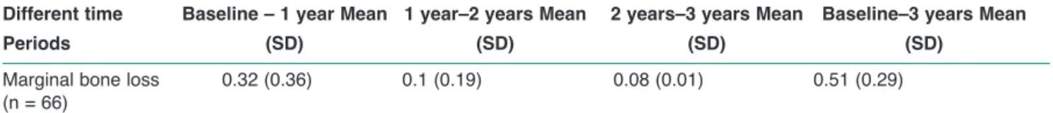
Table 3 - Mean marginal bone loss at different time periods.
Different time Baseline – 1 year Mean 1 year–2 years Mean 2 years–3 years Mean Baseline–3 years Mean
Periods (SD) (SD) (SD) (SD)
Marginal bone loss 0.32 (0.36) 0.1 (0.19) 0.08 (0.01) 0.51 (0.29) (n = 66)
original
research
article
All implants were included in the analysis. It was classified a total of 66 implants with single crowns. No prosthetic complication has occurred during the study period, which represents a suc-cess rate of prosthetic of 100%. No biological or mechanical complication was identified during the whole period of follow-up (such as mobility, pain or discomfort, no screw loose or pillars fracture of the structures in titanium or zirconium).
The MBL average during the first year of opera-tion was of 0.33 ± 0.36 mm.
Between 1 and 2 years of follow-up, the MBL av-erage was 0.1 ± 0.19 mm, and between 2 and 3 years of follow-up, the mean MBL was 0.08 ± 0.1 mm , indicating an average level marginal bone stable after the second year of operation. The MBL cumulative average between implant place-ment, at 3-year follow-up, was 0.51 ± 0.29 mm (Table 3).
All procedures have been as successful as expect-ed. ABH average of the alveolar crest was 6.7 ± 1.6 mm (range 5.1 to 9.2 mm), while the average of gained height bone was 6.4 ± 1.6 mm (range 3.2- 8.1 mm).
All patients reported lower pain levels. The mean pain score in the first week after implantation was 3.17 ± 1.82 (median, 3.00; 95% CI: 2.51 to 3.49), while the average number of analgesic pills taken was 3.6 ± 1.31 (median 3.00; 95% CI: 2.65-3.35). All patients showed clinical measurements of suc-cess of periodontal parameters (PI and BoP <25%). In particular, 1-year follow-up, the score PI showed plaque buildup of 9.01% of the 66 im-plants analyzed. BoP showed a peri-implantation bleeding in 5.09% of the 224 sites analyzed. A 3-year follow-up, the PI was 10.04%, while the BoP was 4.98.
Discussion
This prospective study was performed to evaluate the clinical and radiographic outcome of 66 im-plants placed consecutively, in the posterior max-illa using TGSL, guided implant surgery, and os-teotomes.
This clinical research provides evidence that the use of osteotomes combined with implant place-ment guided of individual implants can make us reach high success rates implant when the im-plants are placed in the alveolar crest with limited bone height (ABH > 3 <5.5 mm).
The main limitation of this study was the lack of a control group, as well as the lack of randomization which is obtained by controlled clinical trials. The clinical and radiographic results of this survey are similar to those reported by Wells et al. in a re-cent, multicentric, medium-term (48.2 months) of follow-up prospective study of 136 implants placed in elevation sinus floor transcrestal with TGSL technique (1). In their study, the Authors re-ported a rate of implant survival of 98.53%, with an average height of residual bone 3.46 ± 0.91 mm and a radiographic bone gain of 6.4 ± 1.6 mm. The significant difference of our study compared to other similar was the surgical technique which included the use of a planning CT three-dimen-sional, with minimally invasive surgery guided through the use of a surgical mask generated by a software CAD/CAM, and the non use of bone grafting materials in thicknesses of residual bone crest less than 5 mm. The predictability of the technique TGSL with immediate implant place-ment is strictly dependent on the ABH to obtain adequate primary stability. The success and the survival rate of dental implants decrease with re-duced height of residual bone (8, 53, 54).
In a retrospective multicentric study (53) was evaluated the result of the Summers technique for placement of implants below the floor of the max-illary sinus: the success rate was 96% when the residual bone height was equal or exceeding to 5 mm, but dropped significantly to 85% when the height of the crestal bone was less than 4 mm. Pjetursson et al. have reported a survival rate of 91.3% when the residual bone height was between 4 and 5 mm (10). The procedure investigated in this study was also performed in the implant sites with a ABH less than 3 mm, even if the initial im-plant stability was not optimum in all cases, so the minimum recommended ABH was 3 mm for this study.
than 5 mm (19). The amount of bone gain report-ed with TGSL technique usreport-ed in this study was 6.4 ± 1.6 millimeters and this gain was maintained for all 3 years of radiographic examinations.
The main contribution to the success of the tech-nique TGSL is the use of a surgical template that guides the placement of the implant so that the membrane is raised and kept on by the implant, that’s used as a space maintainer to stabilize the clot and to start the physiological intrasinusal bone regeneration (62).
Furthermore, the procedure TGSL assisted by the surgical mask CAD/CAM has enabled the physi-cian to carry out the elevation of the Schneider membrane without sinus penetration which in-creases the potential for tearing of the membrane itself. In an in vivo study of the implementation of a similar procedure with guided surgery, Pommer and Watzek (55) reported a bone gain in average of 10.6 ± 1.6 mm with the technique of gel pres-sure.
Vasak et al. (45) evaluated the accuracy of the planning wizard with the same software used in previous research, and have found that the mean deviations measured were 0.43 millimeters (buc-co-lingual), 0.46 mm (mesio-distal), and 0.53 mm (depth) at the level of the implant shoulder, while slightly higher than the average values of 0.7 mm (buccolingual), 0.63 mm (mesio-distal), and 0:52 mm (depth) in the apex of the implant.
However, all investigative procedures reported have been executed in partially edentulous pa-tients with surgical guides entirely docked to the teeth.
This is known because accuracy is significantly higher when the model has a dental support rather than a mucosal one (45, 56-59). In addition, it af-fects the learning curve demonstrating that the sur-geon has more or less familiar with the surgical procedures.
It has been shown that the lifting of Schneider’s membrane is possible through the introduction of liquids into which the volume remains constant. Pascal’s Law states that the pressure exerted on a portion of a liquid is unaltered transmitted through the entire volume of the liquid itself.
actually lifts the sinus membrane transmitting the pressure generated by a careful and precise move-ment of the osteotome.
However, it should be noted that the force exerted by the graft material in the compaction can not be easily controlled, and this can sometimes be harm-ful to the integrity of the sinus membrane (60). In order to minimize the risk of tearing the mem-brane with this technique, a fibrin sealant glue was positioned deep apically to the prepared site to minimize the risk of injury.
Membrane perforation can occur as soon as the forces exceed the elastic properties of the sinus membrane. The cushioning effect of the highly viscous fibrin absorbs the hydraulic pressure, min-imizing the risk of perforation of the membrane. Tilted (44) and short implants (6) have been pro-posed as alternative methods of sinus grafting. The use of short implants with roughened surfaces showed acceptable clinical results in the treatment of posterior maxilla, after a healing period of 6 months, with a success rate of 90% after 5 years (61). The use of short implants in the premolars and molars maxillary areas, poor of bone quality and exposed to high support forces, generally it causes a biomechanical situation compromised with insufficient crown-implant ratio. More fol-low-up studies are necessary to assess the short implants prognosis in the posterior maxilla. Tilting individual implants to the palate, or placing them in the septa, inclined mesial or distal may in-volve compromises emergency prosthetic profiles with unfavorable moments load and long-term prognosis of the final restoration unpredictable be-cause of difficult hygiene maintenance.
The TGSL technique represents a transcrestal minimally invasive procedure that avoids the set-ting up of flaps extended and the removal of the side wall of the maxillary sinus.
The main advantages of the TGSL technique in-clude less bone resorption as there is a full thick-ness skeletonization, keeping the blood supply to the alveolar process (62), minimal postoperative discomfort, and greater patient acceptance for this surgical procedure.
original
research
article
with the minimum use of analgesics during the first few days after surgery, and low postoperative morbidity. The treatment time has reduced be-cause of the combined approach of the grafting procedure with immediate implant placement (the same period of healing for both procedures). The reduction in the total time of treatment mini-mizes the number of surgical procedures, medica-tions for pain required, recovery time and lower total cost of treatment for the patient.
The main indication of the TGSL procedure is im-plant minimally invasive treatment of the single missing tooth in the rear area of the maxilla with insufficient height of alveolar bone, where the conventional lateral approach would have a high cost for these patients, both in terms of post-oper-ating morbidity, discomfort, and treatment costs.
Conclusions
The results at three years, in the medium term of the present study, suggest that the use of guided surgery, CAD/CAM, in the elevation of the sinus floor by transcrestal, with protocols of immediate implant placement without the use of graft materials, it’s a predictable procedure. Within the limits of this study, the results can expand the indications of the tradi-tional transcrestal approach. In addition it’s necessary to confirm these preliminary multicentre, random-ized, and prospective clinical trials results to compare the TGSL with the known, conventional approach, for the lateral sinus elevation, obviously without grafting materials.
References
1. Pozzi A, Moy Peter K. Minimally Invasive Transcrestal
Guided Sinus Lift (TGSL): A Clinical Prospective Proof-of-Concept Cohort Study up to 52 Months Clin Implant Dent Relat Res. 2014;16(4):582-93. doi: 10.1111/cid.12034. Epub 2013.
2. Wallace SS, Froum SJ. Effect of maxillary sinus aug-mentation on the survival of endosseous dental im-plants. A systematic review. Ann Periodontol. 2003;8: 328-43.
3. Pramstraller M, Farina R, Franceschetti G, Pramstraller C, Trombelli L. Ridge dimensions of the edentulous posterior maxilla: a retrospective analysis of a cohort of 127 patients using computerized tomography data. Clin Oral Implants Res. 2011;22:54-61.
4. Winkler S, Morris HF, Ochi S. Implant survival to 36 months as related to length and diameter. Ann Peri-odontol. 2000;5:22-31.
5. Sogo M, Ikebe K, Yang TC, Wada M, Maeda Y. As-sessment of bone density in the posterior maxilla based on Hounsfield units to enhance the initial stability of implants. Clin Implant Dent Relat Res. 2012;14 (Suppl 1):e183-7.
6. Felice P, Soardi E, Pellegrino G, et al. Treatment of the atrophic edentulous maxilla: short implants versus bone augmentation for placing longer implants. Five-month post- loading results of a pilot randomised controlled trial. Eur J Oral Implantol. 2011;4:191-202.
7. Esposito M, Grusovin MG, Rees J, et al. Effectiveness of sinus lift procedures for dental implant rehabilitation: a Cochrane systematic review. Eur J Oral Implantol. 2010;3:7-26.
8. Emmerich D, Att W, Stappert C. Sinus floor elevation using osteotomes: a systematic review and meta-analy-sis. J Periodontol. 2005;76:1237-51.
9. Ferrigno N, Laureti M, Fanali S. Dental implants place-ment in conjunction with osteotome sinus floor eleva-tion: a 12-year lifetable analysis from a prospective study on 588 ITI implants. Clin Oral Implants Res. 2006;17:194-205.
10. Pjetursson BE, Rast C, Brägger U, Schmidlin K, Zwahlen M, Lang NP. Maxillary sinus floor elevation using the (transalveolar) osteotome technique with or without grafting material. Part I: implant survival and patient’s perception. Clin Oral Implants Res. 2009; 20:667-76.
11. Renouard F, Nisand D. Short implants in the severely resorbed maxilla: a 2-year retrospective clinical study. Clin Implant Dent Relat Res. 2005;7:104-10. 12. Esposito M, Hirsch JM, Lekholm U, Thomsen P.
Bio-logical factors contributing to failures of osseointe-grated oral implants. (I). Success criteria and epidemi-ology. Eur J Oral Sci. 1998;106:527-51.
13. Cricchio G, Lundgren S. Donor site morbidity in two different approaches to anterior iliac crest bone har-vesting. Clin Implant Dent Relat Res. 2003;5:161-9. 14. Nkenke E, Schultze-Mosgau S, Radespiel-Troger M,
Kloss F, Neukam FW. Morbidity of harvesting of chin grafts: a prospective study. Clin Oral Implants Res. 2001;12:495-502.
15. Tatum H Jr. Maxillary and sinus implant reconstruc-tions. Dent Clin North Am. 1986;30:207-29. 16. Bruschi GB, Scipioni A, Calesini G, Bruschi E.
Lo-calized management of sinus floor with simultaneous implant placement: a clinical report. Int J Oral Max-illofac Implants. 1998;13:219-26.
18. Cosci F, Luccioli M. A new sinus lift technique in con-junction with placement of 265 implants: a 6-year ret-rospective study. Implant Dent. 2000;9:363-8. 19. Trombelli L, Franceschetti G, Rizzi A, Minenna P,
Mi-nenna L, Farina R. Minimally invasive transcrestal si-nus floor elevation with graft biomaterials. A random-ized clinical trial. Clin Oral Implants Res. 2012;23:424-32.
20. Lundgren S, Moy P, Johansson C, Nilsson H. Aug-mentation of the maxillary sinus floor with particulated mandible: a histologic and histomorphometric study. Int J Oral Maxillofac Implants. 1996;11:760-6.
21. Moy PK, Lundgren S, Holmes RE. Maxillary sinus augmentation: histomorphometric analysis of graft ma-terials for maxillary sinus floor augmentation. J Oral Maxillofac Surg. 1993;51:857-62.
22. Clavero J, Lundgren S. Ramus or chin grafts for max-illary sinus inlay and local onlay augmentation: com-parison of donor site morbidity and complications. Clin Implant Dent Relat Res. 2003;5:154-60. 23. Bernardello F, Righi D, Cosci F, Bozzoli P, Carlo MS,
Spinato S. Crestal sinus lift with sequential drills and simultaneous implant placement in sites with <5 mm of native bone: a multicenter retrospective study. Im-plant Dent. 2011;20:439-44.
24. Rocci A, Rocci M, Lattanzio D, Gargari M. Treating maxillary horizontal atrophies with particulate homo-logus bone grafts: a case report. Oral Implantol. 2009; 2(1):11-8.
25. Lundgren S, Nystrom E, Nilson H, Gunne J, Lindhagen O. Bone grafting to the maxillary sinuses, nasal floor and anterior maxilla in the atrophic edentulous maxilla. A two-stage technique. Int J Oral Maxillofac Surg. 1997;26:428-34.
26. Lundgren S, Rasmusson L, Sjostrom M, Sennerby L. Simultaneous or delayed placement of titanium im-plants in free autogenous iliac bone grafts. Histologi-cal analysis of the bone graft-titanium interface in 10 consecutive patients. Int J Oral Maxillofac Surg. 1999;28:31-7.
27. Fortin T, Isidori M, Bouchet H. Placement of posterior maxillary implants in partially edentulous patients with severe bone deficiency using CAD/CAM guidance to avoid sinus grafting: a clinical report of procedure. Int J Oral Maxillofac Implants. 2009;24:96-102. 28. Krekmanov L. Placement of posterior mandibular and
maxillary implants in patients with severe bone defi-ciency: a clinical report of procedure. Int J Oral Max-illofac Implants. 2000;15:722-30.
29. Calandriello R, Tomatis M. Simplified treatment of the atrophic posterior maxilla via immediate/early func-tion and tilted implants: a prospective 1-year clinical study. Clin Implant Dent Relat Res. 2005;7(Supp1):S1-12.
30. Mattsson T, Köndell P-A, Gynther GW, Fredholm U, Bolin A. Implant treatment without bone grafting in
se-Surg. 1999;57:281-7.
31. Aparicio C, Perales P, Rangert B. Tilted implants as an alternative to maxillary sinus grafting: a clinical, radi-ologic, and periotest study. Clin Implant Dent Relat Res. 2001;3:39-49.
32. Aparicio C, Arévalo JX, Ouazzani W, Granados C. Retrospective clinical and radiographic evaluation of tilted implants used in the treatment of the severely re-sorbed edentulous maxilla. Appl Osseointegrat Res. 2002;3:17-21.
33. Zampelis A, Rangert B, Heijl L. Tilting of splinted implants for improved prosthodontic support: a two-di-mensional finite element analysis. J Prosthet Dent. 2007;97 (Suppl 6):S35-43.
34. Agliardi E, Panigatti S, Clericò M, Villa C, Malò P. Im-mediate rehabilitation of the edentulous jaws with full prostheses supported by four implants: interim results of a single cohort prospective study. Clin Oral Im-plants Res. 2010;21:459-65.
35. Malò P, de Araújo Nobre M, Lopes A, Moss SM, Molina GJ. A Longitudinal study of the survival of All-on-Four implants in the mandible with up to 10 years of follow-up. J Am Dent Assoc. 2011;142:310-320.28. 36. Agliardi E, Francetti L, Romeo D, Del Fabbro M.
Im-mediate rehabilitation of the edentulous maxilla: pre-liminary results of a single-cohort prospective study. Int J Oral Maxillofac Implants. 2009;24:887-95. 37. Boyne PJ, James RA. Grafting of the maxillary sinus
floor with autogenous marrow and bone. J Oral Surg. 1980;38:613-6.
38. Soltan M, Smiler DG. Antral membrane balloon ele-vation. J Oral Implantol. 2005;31:85-90.
39. Kfir E, Kfir V, Mijiritsky E, Rafaeloff R, Kaluski E. Minimally invasive antral membrane balloon elevation followed by maxillary bone augmentation and implant fixation. J Oral Implantol. 2006;32:26-33.
40. Chen L, Cha J. An 8-year retrospective study: 1,100 pa-tients receiving 1,557 implants using the minimally invasive hydraulic sinus condensing technique. J Peri-odontol. 2005;76:482-91.
41. Suguimoto RM, Trindade IK, Carvalho RM. The use of negative pressure for the sinus lift procedure: a tech-nical note. Int J Oral Maxillofac Implants. 2006;21: 455-8.
42. Engelke W, Capobianco M. Flapless sinus floor aug-mentation using endoscopy combined with CT scan-designed surgical templates: method and report of 6 consecutive cases. Int J Oral Maxillofac Implants. 2005;20:891-7.
43. Malo P, de Araujo Nobre M, Lopes A. The use of com-puter guided flapless implant surgery and four im-plants placed in immediate function to support a fixed denture: preliminary results after a mean follow-up period of thirteen months. J Prosthet Dent. 2007;97 (Supp1 6):S26-34.
original
research
article
of preoperative planning of an image-guided system for oral implant placement based on three-dimensional images: an in vivo study. Int J Oral Maxillofac Im-plants. 2003;18:886-93.
45. Vasak C, Watzak G, Gahleitner A, Strbac G, Schemper M, Zechner W. Computed tomography-based evalua-tion of template (NobelGuide)-guided implant posi-tions: a prospective radiological study. Clin Oral Im-plants Res. 2011;22:1157-63.
46. Yong LT, Moy PK. Complications of computer-aided-design/computer-aided-machining-guided (Nobel-Guide) surgical implant placement: an evaluation of early clinical results. Clin Implant Dent Relat Res. 2008;10:123-7.
47. Cricchio G, Sennerby L, Lungren S. Sinus bone for-mation and implant survival after sinus membrane el-evation and implant placement: a 1 to 6 years follow-up study. Clin Oral Impl Res. 2011;22(10):1200-12. 48. Cricchio G, Imburgia M, Sennerby L, Lungren S.
Im-mediate loading of implant simultaneously whit sinus membrane elevation in the posterior atrophic maxilla: a two years follow-up study on ten patients. Clin Impl Dent Rel Res. 2013.
49. Marchack CB, Moy PK. The use of a custom template for immediate loading with the definitive prosthesis: a clinical report. J Calif Dent Assoc. 2003;31:925-9. 50. Pozzi A, De vico G, Sannino G, Spinelli D, Schiavetti
R, Ottria L, Barlattani A. Flapless transcrestal maxillary sinus floor elevation: computer guided implant sur-gery combined with expanding-condensing osteotomes protocol. Oral Implantol. 2011;4(1-2):4-9.
51. Van Steenberghe D, Glauser R, Blomback U, et al. A computed tomographic scan-derived customized gical template and fixed prosthesis for flapless sur-gery and immediate loading of implants in fully eden-tulous maxillae: a prospective multicenter study. Clin Implant Dent Relat Res. 2005;7(Suppl 1):S111-20. 52. Van Steenberghe D. Outcomes and their measurement
in clinical trials of endosseous oral implants. Ann Pe-riodontol. 1997;2:291-8.
53. Rosen PS, Summers R, Mellado JR, et al. The bone added osteotome sinus floor elevation technique: mul-ticenter retrospective report of consecutively treated pa-tients. Int J Oral Maxillofac Implants. 1999;14:853-8. 54. Tan WC, Lang NP, Zwahlen M, Pjetursson BE. A
sys-tematic review of the success of sinus floor elevation and survival of implants inserted in combination with sinus floor elevation. Part II: transalveolar technique. J Clin Periodontol. 2008;35(8 Suppl):241-54. 55. Pommer B, Watzek G. Gel-pressure technique for
flap-less transcrestal maxillary sinus floor elevation: a pre-liminary cadaveric study of a new surgical technique. Int J Oral Maxillofac Implants. 2009;24:817-22. 56. Ersoy AE, Turkyilmaz I, Ozan O, McGlumphy EA.
Re-liability of implant placement with stereolithographic surgical guides generated from computed tomography: clinical data from 94 implants. J Periodontol. 2008;79: 1339-45.
57. Van Assche N, van Steenberghe D, Guerrero ME, et al. Accuracy of implant placement based on pre-surgical planning of three-dimensional conebeam images: a pi-lot study. J Clin Periodontol. 2007;34:816-21. 58. Van Assche N, Quirynen M. Tolerance within a
surgi-cal guide. Clin Oral Implants Res. 2010;21:455-8. 59. Ozan O, Turkyilmaz I, Ersoy AE, McGlumphy EA,
Rosenstiel SF. Clinical accuracy of 3 different types of computed tomography-derived stereolithographic sur-gical guides in implant placement. Int J Oral Maxillo-fac Surg. 2009;67:394-401.
60. Nkenke E, Schlegel A, Schultze-Mosgau S, Neukam FW, Wiltfang J. The endoscopically controlled os-teotome sinus floor elevation: a preliminary prospective study. Int J Oral Maxillofac Implants. 2002;17:557-66. 61. Perelli M, Abundo R, Corrente G, Saccone C. Short (5
and 7mm long) porous implants in the posterior at-rophic maxilla: a 5-year report of a prospective single-cohort study. Eur J Oral Implantol. 2012;5:265-72. 62. Cricchio G1, Palma VC, Faria PE, de Olivera JA,
Lundgren S, Sennerby L, Salata LA. Histological out-comes on the development of new space-making de-vices for maxillary sinus floor augmentation. Clin Im-plant Dent Relat Res. 2011;13(3):224-30. doi: 10.1111/j.1708-8208.2009.00208.x. Epub 2009.
Correspondence to:
Dr. Dario Spinelli Private practice in Rome
E-mail: [email protected]; [email protected]