REVIEW
Current concepts in the prevention of pathogen transmission via
blood/plasma-derived products for bleeding disorders☆
Giovanni Di Minno
a,⁎
, Carlo Federico Perno
b, Andreas Tiede
c, David Navarro
d, Mariana Canaro
e,
Lutz Güertler
f, James W. Ironside
ga
Dipartimento di Medicina Clinica e Chirurgia, Regional Reference Centre for Coagulation Disorders, Federico II University, Via S. Pansini 5, 80131 Naples, Italy
b
Department of Experimental Medicine and Surgery, University of Rome Tor Vergata, Via Montpellier 1, 00133 Rome, Italy
c
Department of Hematology, Hemostasis, Oncology and Stem Cell Transplantation, Hannover Medical School, Carl-Neuberg-Str. 1, D-30625 Hannover, Germany
d
Department of Microbiology, Microbiology Service, Hospital Clínico Universitario, School of Medicine, University of Valencia, Av Blasco Ibáñez 17, 46010 Valencia, Spain
eDepartment of Hemostasis and Thrombosis, Son Espases University Hospital, Carretera de Valdemossa, 79, 07120 Palma de Mallorca, Spain f
Max von Pettenkofer Institute for Hygiene and Medical Microbiology, University of München, Pettenkofer Str 9A, 80336 Munich, Germany
g
National Creutzfeldt–Jakob Disease Research and Surveillance Unit, School of Clinical Sciences, University of Edinburgh, Western General Hospital, Edinburgh EH4 2XU, UK
a b s t r a c t
a r t i c l e i n f o
Keywords: Pathogen Transfusion Blood safety Clotting HaemophiliaThe pathogen safety of blood/plasma-derived products has historically been a subject of significant concern to the medical community. Measures such as donor selection and blood screening have contributed to increase the safety of these products, but pathogen transmission does still occur. Reasons for this include lack of sensitiv-ity/specificity of current screening methods, lack of reliable screening tests for some pathogens (e.g. prions) and the fact that many potentially harmful infectious agents are not routinely screened for. Methods for the purifica-tion/inactivation of blood/plasma-derived products have been developed in order to further reduce the residual risk, but low concentrations of pathogens do not necessarily imply a low level of risk for the patient and so the overall challenge of minimising risk remains. This review aims to discuss the variable level of pathogenic risk and describes the current screening methods used to prevent/detect the presence of pathogens in blood/ plasma-derived products.
© 2015 The Authors. Published by Elsevier Ltd. This is an open access article under the CC BY-NC-ND license (http://creativecommons.org/licenses/by-nc-nd/4.0/).
1. Introduction
Acute bleeding episodes can arise either because of inherited bleed-ing disorders (e.g. haemophilia, von Willebrand disease), acquired de fi-ciency of haemostatic components (e.g. due to infection, malignancy or autoimmune disease), trauma, surgery or as a result of infection with an organism that causes haemorrhagic disease (e.g. Ebola or Marburg virus)[1]. Various treatment options exist for preventing or treating acute bleeding episodes, including fresh-frozen plasma/cryoprecipitate, platelets and plasma-derived/recombinant clotting factor concentrates
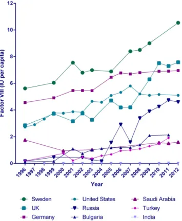
[2,3]. The use of blood-derived and recombinant haemostatic products has increased markedly over recent years, as exemplified by the global use of factor VIII products (Fig. 1)[4]. This increased use has been driven by improved availability of clotting factors, increased life expectancy of people with bleeding disorders[5,6], increased use of prophylaxis for severe bleeding disorders[7,8]and decreased risk of transmission of in-fectious agents.
Historically, the risk of transmission of infectious agents via blood/ plasma-derived products has been of great concern to the medical
community. This risk has reduced dramatically since the implementa-tion of stricter donaimplementa-tion screening/donor selecimplementa-tion procedures and im-proved purification procedures, but cannot be fully eradicated. Furthermore, the implementation of pathogen inactivation technology for blood/plasma-derived products has further reduced the risk of trans-mission of both known and emerging pathogens, although results can be variable according to the methods used[9,10]. However, it is impor-tant to note that patient risk is highly dependent on the circumstances under which blood products are collected, handled and used. In general, clinicians assess the level of risk associated with the use of blood/ plasma-derived products by evaluating factors such as patient charac-teristics (e.g. age, immune status, geographical location, lifestyle) and the nature of the pathogen (e.g. physical characteristics, level of viru-lence, chronicity of infection, prevalence). The presence of a particular pathogen within blood/plasma-derived products may pose a significant threat to specific patient groups (e.g. the elderly or immunocompro-mised), while being of low risk to the general population (e.g. HEV).
While the clinical assessment of risk is based on a variety of factors, the virological assessment of risk is based solely on the presence or ab-sence of pathogens. The preab-sence of pathogens implies the possibility of infection, so only pathogen-free products can be described as entirely risk-free. Adopting the virological approach (i.e. discarding all products
⁎ Corresponding author. Tel.: +39 081 746 2060; fax: +39 081 5466152. E-mail address:[email protected](G. Di Minno).
http://dx.doi.org/10.1016/j.blre.2015.07.004
0268-960X/© 2015 The Authors. Published by Elsevier Ltd. This is an open access article under the CC BY-NC-ND license (http://creativecommons.org/licenses/by-nc-nd/4.0/).
Contents lists available atScienceDirect
Blood Reviews
which may contain infectious agents) is effective in reducing the rate of pathogen transmission, but may result in the unnecessary wastage of blood/plasma-derived products.
Since recombinant clotting factors are not derived from blood or plasma, they present a minimal risk of pathogen transmission (particu-larly third generation factors, which have no contact with blood/ plasma-derived components whatsoever) and can therefore be consid-ered safer than using plasma-derived clotting factor concentrates[11]. However, there has been concern that the use of recombinant clotting factors may be associated with an increased rate of inhibitor formation in patients who regularly receive these products[12]; this is another factor that may influence the overall clinical assessment of patient risk. Recombinant clotting factors are available for the treatment of haemophilia A (FVIII) and B (FIX) and factor VII/XIII deficiency, while plasma-derived concentrates are available for most other clotting fac-tors (including von Willebrand factor,fibrinogen and the vitamin K de-pendent clotting factors). In some cases only fresh frozen plasma and cryoprecipitate are available for replacement therapy (e.g. factor V de fi-ciency). Ideally, plasma and plasma-derived products would be completely replaced by recombinant products in order to minimise the risk of pathogen transmission; however, this is not always possible. A recent article comprehensively reviewed the pathogen safety of plasma-derived and recombinant clotting factors[13].
This review examines the potential for transmission of infectious agents that might be present in blood/plasma-derived products used to treat haemostatic disorders. We focus on the identities of these agents and the screening procedures used for their detection, as well as the limitations of screening. Current unmet needs in thefield of path-ogen safety of blood/plasma-derived products are also discussed. 2. Methodology
The PubMed database was interrogated from 1 January 2000 to the present using the search strings‘bleeding disorders OR haemophilia’
and‘pathogens AND blood safety’. The search terms ‘virus’, ‘bacteria’, ‘haemorrhagic disorders’, ‘von Willebrand disease’, ‘FVII deficiency’ and‘FXIII deficiency’ were added to this search but did not yield any ad-ditional pertinent result. The bibliographies of reviews were also used to identify relevant references and individual searches were conducted for information on specific pathogens. Information and opinions were also provided by the authors.
3. Infectious agents present in blood/plasma-derived products: lessons from the past and current concerns
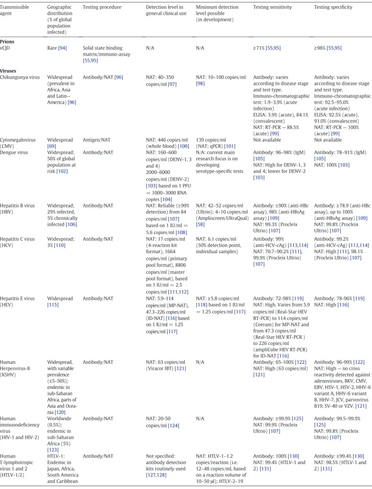
A large number of pathogenic agents (including viruses, protozoan parasites and prions) can be transmitted via blood/plasma-derived products and are capable of causing disease in humans (Table 1). The presence of viruses in plasma-derived products became a concern in the 1980s, when 60–70% of patients with severe haemophilia became infected with human immunodeficiency virus (HIV-1)[6]. This concern continued with the discovery that 80% of patients treated with plasma-derived products prior to 1992 had become infected with hepatitis C virus (HCV)[5]. Current donor selection and screening practices have improved our ability to detect or reduce the presence of pathogens in blood/plasma-derived products; for example, the residual risk of transfusion-transmitted infection (TTI) with HIV/HBV/HCV has fallen to near or less than 1 per million transfused units[14,15]. Despite this success, however, a residual risk still remains.
3.1. Potential risk
The pathogenic agents shown inTable 1(and the Supplementary Appendix) do not form an exhaustive list. Many microorganisms that are normally non-pathogenic have the potential to cause disease when responding to changes in the biological environment, or when transfused to an immunosuppressed patient. In addition, there is still a risk that new and emerging pathogens may enter the blood supply (Table 2).
4. Screening for pathogens
The standard assays commonly used for blood screening are nucleic acid amplification technology (NAT) and immunoassays for detection of antibody and/or antigen. Immunoassays are frequently used for screen-ing purposes as multiple samples can be processed together and they may yield semi-quantitative results. NAT assays allow earlier pathogen detection than with immunoassays, but they are also more costly and complex. Assay selection is generally determined by the level of accura-cy/speed required, but factors such as the resources available (e.g. staff, infrastructure), assay complexity and cost considerations (e.g. consum-ables) must also be considered. Most assays for blood donation screen-ing are mandatory (particularly in Europe and North America) and the World Health Organization (WHO) recommends that all whole blood (and blood which has been processed by apheresis) should undergo pathogen screening before it is used for clinical or manufacturing pur-poses. Screening for HIV-1, HIV-2, HBV, HCV and Treponema pallidum subspecies pallidum (T. pallidum; the causative agent of syphilis) is strongly advised. The WHO and World Federation of Hemophilia (WFH) suggest that countries should carry out individual routine screening for further pathogens based on epidemiological information for their region e.g. HTLV-1 and Trypanosoma cruzi[16]. The WFH also acknowledges the positive impact of HIV, HBV and HCV screening on global blood safety and recommends that these screening tests be im-plemented whenever possible[2]. Details of the serological tests carried out on individual donor plasma and NAT testing of plasma mini-pools are shown inTables 3 and 4 [17].
It is recommended that the minimum evaluated sensitivity/speci fic-ity level of any assay used for blood donation screening should be 99.5% or higher[16]. However, not all assays fulfil these criteria as sensitivity/
Fig. 1. Use of plasma-derived and recombinant factor VIII products in selected countries. Results are from Stonebraker et al. (1996–2006)[4]and WFH Annual Global Surveys (2007–2012)[196].
specificity levels vary according to assay type and type of micro-organism being tested for (seeTable 1for details). Therefore, the gener-al advice is to screen donated blood to as high a standard as possible
[16].
4.1. Blood donation screening: assays for key pathogens 4.1.1. HIV, HBV and HCV testing
In the early stages of infection with HIV, HBV or HCV, viraemia oc-curs in the host's bloodstream at variable levels. Viral antigens may ap-pear at the same time as DNA/RNA, but more often become detectable several weeks later. Specific antibodies are measurable 2 to 6 weeks after infection; the time between initial infection and the appearance of detectable parameters of infection (e.g. viral nucleic acid/antigens/ antibodies) is known as the‘window period’[18–20].
4.1.1.1. HIV. When screening blood for the presence of HIV-1/HIV-2, the use of a combined antigen/antibody assay is advised (combined HIV p24 Ag and anti-HIV-1 + anti-HIV-2 antibodies) as it allows earlier detec-tion of infecdetec-tion. The performance of NAT testing is mandatory in many countries and further reduces the window period (from around 20 to 11 days)[20–22].
4.1.1.2. HBV. The majority of diagnostic laboratories focus on the detec-tion of HBsAg, which is thefirst detectable serological marker of infec-tion. However, there is a risk that the HBsAg concentration may decline to undetectable levels during the course of infection, yielding a false negative result[23]. Screening for antibodies to HBc is the most conservative approach for identifying potentially exposed donors, as this identifies all individuals who have ever immunologically experi-enced any type of HBV infection (either current, chronic or resolved) and who may experience viral reactivation during their lifetime (partic-ularly under conditions of immunosuppression). However, assays to measure HBc antibodies are relatively non-specific and do not always correlate with the presence of HBV virus in plasma[18]. Also, we cannot exclude the possibility that donors who test positive for anti-HBc do not have pulsed recurrences of virus replication, resulting in the presence of low levels of HBV-DNA in plasma. For these reasons, national transfu-sion services do not always routinely screen donations for anti-HBc. NAT assays can be carried out, but their use is restricted by potentially low levels of viral DNA[16,18]. The combined use of HBsAg screening tests and NAT assays has reduced the window period for detection of HBV infection from approximately 60 to 35.5 days[20,24]. Mutant HBV strains (escape variants) should also be considered, as they may oc-casionally escape serological detection (although most can be detected by NAT assays)[24,25]. These HBV-variants are rare, but therefore more likely to enter and contaminate the blood supply as they are more difficult to detect[25]. In summary, the screening of blood sup-plies for the presence of HBV is effective, but an optimal screening sys-tem has not yet been defined.
4.1.1.3. HCV. HCV (both recent and chronic infection) can be detected by screening blood for the presence of both HCV antigen and HCV antibody (anti-HCV). Seroconversion occurs at approximately 6–8 weeks post-infection; however, steady improvements in screening technology (in-cluding the adoption of NAT assays) have reduced the window period to approximately 1–3 weeks[20,26]. As with HIV, NAT assays are more useful for detecting early infection, although the issue of low viral RNA concentration persists[27,28].
4.1.2. HTLV-1 testing
HTLV-1 and HTLV-2 are endemic in some regions, but very rare in others, and therefore screenings are conducted on a geographical or at-risk basis. The presence of virus is mostly inferred by the detection of virus-specific antibodies, using sensitive immunoassays[16].
4.1.3. Syphilis testing
In this instance, screening involves detection of non-specific, non-treponemal or specific treponemal antibodies. NAT assays are generally not used[29]. Specific antibody tests identify all individuals who have ever been exposed to this bacterium (and may continue to yield positive results for more than ten years following initial infection), while the non-specific tests (e.g. VDRL or cardiolipin tests) are primarily of use in identifying donors who may have an active infection. Since T. pallidum is heat-sensitive and cannot readily withstand extended storage at low temperatures, storage at 4 °C for more than three days is sufficient to render the pathogen non-infectious. However, blood components (e.g. platelets) that are stored at temperatures of around 20 °C do present a risk of T. pallidum persistence. Therefore screening for antibodies of this organism is recommended[16].
4.2. Testing for emerging viral pathogens 4.2.1. West Nile virus
Blood can be screened for further pathogens as appropriate, accord-ing to geographical location, seasonal activity of the vector and also pa-tient risk factors. A current viral pathogen of interest is the mosquito-borneflavivirus West Nile virus (WNV), which was confirmed to have been transmitted via transfusion in 2002[30]. An immediate screening policy was put in place in the USA in order to reduce the risk of further transmission. This policy included deferral of any individual displaying symptoms of infection, quarantine of plasma collected during periods of high mosquito activity (when WNV is most prevalent) and the rapid development/use of WNV-specific NAT and serological assays. These measures were highly effective and caused a significant reduction in the number of confirmed cases of WNV transfusion-related transmis-sion. However, WNV outbreaks still occur within the Americas, indicat-ing a potential need for seasonal blood screenindicat-ing for WNV[31]. WNV outbreaks have also occurred in Europe (including Italy and Greece), prompting the implementation of seasonal blood screening procedures in the affected regions of those countries[32].
4.2.2. Chikungunya virus
This is another mosquito-borne pathogen that could potentially pose a risk to transfusion safety, although to date reports of transfusion-related transmission of this virus are rare[33]. A mutated form of the Chikungunya virus has been responsible for several epidemics in the past decade, spreading to the Reunion Islands in the Indian Ocean (2005), Italy (2007) and the Caribbean area (2012/2013)[34–36]. The virus may be detected in blood donors by NAT, which will help to reduce the level of transmission risk[35].
4.2.3. Parvovirus B19
There is also concern about the possibility of parvovirus B19 in the blood supply. B19 is prevalent worldwide, with seroprevalence in blood donors varying from between 0.2–1.3% in the USA, Europe and Africa and 25–40% in Asia[37]. The risk of parvovirus transmission is higher when units of blood are pooled (e.g. to create batches of clotting factor concentrates, albumin etc.) and so individuals with bleeding dis-orders are at a higher risk of infection. B19 DNA was detected in 26% of clotting factor concentrates in a recent German study[38], and another study found that populations receiving blood-derived products were 1.7 times more likely to display antibodies to B19 than populations who had not received blood products[39]. B19 lacks a lipid envelope, which renders it highly resistant to some methods of pathogen inactiva-tion[40]. Screening of blood donations for B19 DNA is not currently rou-tine, but many manufacturers only process plasma that has been screened for the absence of B19 DNA in order to reduce the risk of trans-mission[41]. Given the prevalence of B19 in different populations, it is difficult to define the residual TTI risk of this virus. However, it is clear that a transmission risk does exist.
Table 1 Blood-transmissible pathogens. Transmissible agent Geographic distribution (% of global population infected)
Testing procedure Detection level in general clinical use
Minimum detection level possible (in development)
Testing sensitivity Testing specificity
Prions
vCJD Rare[94] Solid state binding matrix/immuno-assay
[55,95]
N/A N/A ≥71%[55,95] ≥98%[55,95]
Viruses
Chikungunya virus Widespread (prevalent in Africa, Asia and Latin--America)[96] Antibody/NAT[96] NAT: 40–350 copies/ml[97] NAT: 10–100 copies/ml [98] Antibody: varies according to disease stage and test type.
Immuno-chromatographic test: 1.9–3.9% (acute infection) ELISA: 3.9% (acute), 84.1% (convalescent) NAT: RT-PCR— 88.5% (acute)[99] Antibody: varies according to disease stage and test type.
Immuno-chromatographic test: 92.5–95.0% (acute infection) ELISA: 92.5% (acute), 91.0% (convalescent) NAT: RT-PCR— 100% (acute)[99] Cytomegalovirus (CMV) Widespread [69]
Antigen/NAT NAT: 446 copies/ml (whole blood)[100]
139 copies/ml (NAT; qPCR)[101]
Not available Not available Dengue virus Widespread;
50% of global population at risk[102] Antibody/NAT NAT: 160–600 copies/ml (DENV-1, 3 and 4) 2000–6000 copies/ml (DENV-2) [103]based on 1 PFU = 1000–3000 RNA copies[104]
N/A: current main research focus is on developing serotype-specific tests
Antibody: 96–98% (IgM)
[105]
NAT: High for DENV-1, 3 and 4; lower for DENV-2
[103] Antibody: 78–91% (IgM) [105] NAT: 100%[103] Hepatitis B virus (HBV) Widespread; 29% infected, 5% chronically infected[106]
Antibody/NAT NAT: Reliable (≥99% detection) from 84 copies/ml[107] based on 1 IU/ml = 5.6 copies/ml[108] NAT: 42–52 copies/ml (Ultrio); 4–10 copies/ml (Ampliscreen/UltraQual) [58] Antibody:≥90% (anti-HBc assay), 98% (anti-HBsAg assay)[109] NAT: 99.3% (Procleix Ultrio)[107] Antibody:≥78.9 (anti-HBc assay), up to 100% (anti-HBsAg assay)[109] NAT: 99.8% (Procleix Ultrio)[107] Hepatitis C virus (HCV) Widespread; 3%[110]
Antibody/NAT NAT: 17 copies/ml (4-reaction kit format), 1664 copies/ml (primary pool format), 8806 copies/ml (master pool format), based on 1 IU/ml = 2.5 copies/ml[111,112] NAT: 6.1 copies/ml (50% detection point, individual samples) Antibody: 99% (anti-HCV-cAg)[113,114] NAT: 70.7–90.2%[111], 99.9% (Procleix Ultrio) [107] Antibody: 99.2% (anti-HCV-cAg)[113,114] NAT: High[111], 98.1% (Procleix Ultrio)[107] Hepatitis E virus (HEV) Widespread [115] Antibody/NAT NAT: 5.9–114 copies/ml (MP-NAT), 47.3–226 copies/ml (ID-NAT)[116]based on 1 IU/ml = 1.25 copies/ml[117] NAT:≥5.8 copies/ml [118]based on 1 IU/ml = 1.25 copies/ml[117] Antibody: 72-98%[119]
NAT: High. Varies from 5.9 copies/ml (Real-Star HEV RT-PCR) to 114 copies/ml (Ceeram) for MP-NAT and from 47.3 copies/ml (Real-Star HEV RT-PCR ) to 226 copies/ml (ampliCube HEV RT-PCR) for ID-NAT[116] Antibody: 78-96%[119] NAT: High[116] Human Herpesvirus-8 (KSHV) Widespread, with variable prevalence (≤5–50%); endemic in sub-Saharan Africa, parts of Asia and Ocea-nia[120]
Antibody/NAT NAT: 63 copies/ml (Viracor IBT)[121]
N/A Antibody: 65-100%[122]
NAT: High (63 copies/ml)
[121]
Antibody: 96-99%[122]
NAT: High— no cross reactivity detected against adenoviruses, BKV, CMV, EBV, HSV-1, HSV-2, HHV-6 variant A, HHV-6 variant B, HHV-7, JCV, parvovirus B19, SV-40 or VZV.[121] Human immunodeficiency virus
(HIV-1 and HIV-2)
Worldwide (0.5%); endemic in sub-Saharan Africa (5%) [123] Antibody/NAT NAT: 20-50 copies/ml[124] N/A Antibody:≥99.9%[125] NAT: 99.9% (Procleix Ultrio)[107] Antibody: 99.5–99.9% [125] NAT: 99.8% (Procleix Ultrio)[107] Human T-lymphotropic virus 1 and 2 (HTLV-1/2) HTLV-1: Endemic in Japan, Africa, South America and Caribbean
Antibody/NAT Not specified: antibody detection kits routinely used
[127,128] NAT: HTLV-1–1.2 copies/reaction (i.e. 12–48 copies/ml, based on a reaction volume of 10–50 μl); HTLV-2–19 Antibody: 100%[130] NAT: 99.4% (HTLV-1 and 2)[131] Antibody:≥99.4%[130] NAT: 98.5% (HTLV-1 and 2)[131]
Table 1 (continued) Transmissible agent Geographic distribution (% of global population infected)
Testing procedure Detection level in general clinical use
Minimum detection level possible (in development)
Testing sensitivity Testing specificity
HTLV-2: Prev-alent in Central/West Africa, the Americas and Europe[126] copies/reaction (i.e. 76–191 copies/ml, based on a reaction volume of 10–50 μl)[129] Parvovirus B-19 Widespread, variable prevalence (0.2–40% in European and Asian blood donors)[37]
Antibody/NAT NAT: 668 copies/ml (based on 1 IU/ml = 3.34 copies/ml) [132,133] NAT: 10 copies/reaction (i.e. 100–500 copies/ml, based on a reaction volume of 10–50 μl) [134] Antibody: 89.1%[135] NAT:≥93%[132] Antibody: 99.4%[135] NAT:≥97%[132] Parvovirus 4 Widespread, variable prevalence (2–35%) [136–138] NAT NAT: 200–500 copies/ml[139,140] NAT: 10 copies/reaction (i.e. 100–500 copies/ml, based on a reaction volume of 10–50 μl) [134]
Not available Not available
Torque-tenovirus (TTV) Present worldwide at variable prevalence (higher in developing countries) [141]
NAT NAT: no general test — PCR used to confirm presence. NAT:≥100 copies/reaction (i.e. 1000–5000 copies/ml, based on a reaction volume of 10–50 μl) [142] NAT: Up to 100%[142] NAT:≥96%[142]
West Nile virus (WNV)
Widespread (Africa, West Asia, Middle East, Europe and North and South America)[90]
Antibody/NAT NAT: 100 copies/ml
[143] NAT:≥9.8 copies/ml [144] Antibody: 50% (IgM), 86% (IgG)[145] NAT: High[144] Antibody: 95% (IgM), 69% (IgG)[145] NAT: High[144] Bacteria Treponema pallidum subspecies pallidum Widespread; (0.5% of global population infected) [146] Antibody/NAT/dark ground microscopy DGM) NAT: 32,000 copies/ml [147] NAT: 82%[148] Antibody: 78–88% (primary syphilis), 100% (secondary), 95–100% (latent), 71–96 (late) [149] DGM: 79–97%[147] NAT: 95%[148] Antibody: 96–99% DGM: 77–100%[147,149] Yersinia enterocolitica, Salmonella enterica, Listeria monocytogenes, Coxiella burnetii (only during outbreaks) Rare, donor selection dependent
Not done N/A N/A N/A
Protozoa
Babesia spp Widespread; predominantly in United States and (less frequently) Europe[150] Antibody/NAT Antibody: 10000–100000 parasites/ml (Giemsa staining)[151] NAT: 5000–10000 parasites/ml[151] Antibody:≥97%[152] NAT: High[151] Antibody:≥97%[152] NAT: 100%[151] Leishmania spp Widespread; 1.3 million new cases per year[153] Antibody/NAT NAT:≥10 parasites/ml (PCR; sensitivity varies according to method)[154] NAT:≥10 parasites/ml [154] Antibody: 75-95%[155] NAT: 98.7% (kinetoplast DNA PCR), 91% (ITS1 PCR), 53.8% (SLME PCR)[156] Antibody: 70-98%[155] NAT: 57.1% (kinetoplast DNA PCR), 100% (ITS1 PCR and SLME PCR)[156] Plasmodium spp Widespread (most common in sub-Saharan Africa). 30% at risk[157] Antibody/NAT Microscopy: 50000 parasites/ml[158] NAT:b10 parasites/ml [158] Antibody:≥95%[159] NAT: 76.1–100% [158] Antibody:≥97.7%[159] NAT: 89.6–100% [158] Trypanosoma brucei gambiense/rhodiense Regional in Africa, 10% at risk[160] Direct microscopic visualisation/Antibody/NAT NAT:b100 trypanosomes/ml [161] NAT: 1–10 trypanosomes/ml [162] Antibody: CATT— 87–98% [161] NAT: 88%[163] Antibody: CATT— 95% [161] NAT: 99.2%[163]
4.2.4. Hepatitis E virus
The potential presence of hepatitis E virus (HEV) in blood or blood-derived products is relevant. Currently there seems to be a discrepancy between the number of HEV-RNA positive blood donations in Europe (ranging from 1 in 1240 in Germany to 1 in 1761 donations in the Netherlands)[42], and the low number of confirmed cases of hepatitis E in blood transfusion recipients (one confirmed case in the UK [2006], one in France [2007] and two in Germany [2014])[43–45]. This indicates that the subject of HEV infectivity and pathogenicity needs to be investigated further[46–49]. There is also a debate over the necessity of introducing screening blood for the presence of HEV; the virus is not currently screened for in the UK and other European countries, although one study of English donors found HEV to be wide-spread (1 in 2848 donations) within the donor population[50]. Since in-fection with this virus can be harmful to immunocompromised individuals, the potential need for introducing HEV screening should be considered[42].
The residual risk for TTI of HBV, HCV, HIV and HEV in selected coun-tries is given inTable 5.
4.3. Prions
Despite recent advances in methods for the detection of prions, no single method has been developed as a screening test for blood, al-though several methods in animal models show great promise
[51–53]. In humans, a protocol for the evaluation of a blood-based test for its suitability in the diagnosis of variant Creutzfeldt–Jakob disease (vCJD) has been established, but no test yet appears to satisfy the re-quirements of sensitivity and specificity[54]. The only report so far of a blood-based diagnostic test for vCJD claimed an assay sensitivity of 71.4% and a specificity of 100% in symptomatic patients and its potential applicability as a screening test to detect asymptomatic vCJD infection has recently been investigated[55]. There is currently no strategy for confirming a positive screening result, although the protein misfolding cyclical amplification technique has recently been shown to yield
positive results in buffy coat/white blood cell samples from a small number of patients with vCJD[56].
4.4. Interpretation of test results and practicalities of screening 4.4.1. Assay specificity and sensitivity
Extensive use of blood donor selection and testing does not always guarantee a safe product. If the test is insufficiently sensitive, then false negatives may occur. Alternatively, tests which are not sufficiently specific (e.g. anti-HBc assay for HBV) may cause false positive results, leading to an unnecessary decrease in the number of clotting products available[16]. Torque Tenovirus (TTV) is an example where testing specificity has been an issue, as this virus exists in various divergent forms (23 distinct genotypes have been identified thus far)[57]. Since TTV is often detected in healthy individuals and is not associated with any particular disease, routine screening for this virus is not considered to be necessary; even a test with excellent sensitivity/specificity would not contribute to increase the level of safety of blood/plasma-derived products with regard to TTV.
Insufficient assay sensitivity remains a rare but potential problem when blood donations are screened during the window period of initial HBV, HCV or HIV infection. An increase in testing sensitivity threshold is needed to prevent HBV transmission via blood/plasma-derived prod-ucts and by blood transfusion, as extremely low concentrations of HBV (e.g. 1.6 copies/ml) is capable of viral transmission (see above section on anti-HBc positive patients)[58]. In the case of HIV and HCV, even NAT testing may not always be sufficient to ensure sufficiently high levels of safety as virus transmission has previously occurred after trans-fusion of blood with undetectable levels of viraemia[59].
Recent reports have highlighted concerns about the inability of NAT assays to detect different variants of HIV. There have been at least four cases in which the presence of HIV-1 RNA was undetected by NAT assay screening, potentially putting transfusion recipients at risk[21]. Two of these false-negative results occurred due to genetic mutation in the viral RNA regions targeted by NAT assay primers (although in
Table 1 (continued) Transmissible agent Geographic distribution (% of global population infected)
Testing procedure Detection level in general clinical use
Minimum detection level possible (in development)
Testing sensitivity Testing specificity
Trypanosoma cruzi 0.1–0.2% (mostly Latin and South America)[164] Antibody/NAT NAT:≥0.5 parasites/ml[165] NAT: 0.05 parasites/ml [165] Antibody:75–100% (majorityN 90%)[166] NAT: 83.3–94.4%[165] Antibody:≥97%[166] NAT: 85–95%[165]
In cases where no overallfigures are available, specificity/sensitivity has been described as low, medium or high (as appropriate).
CATT, card agglutination test for Trypanosomiasis; ID-NAT, individual donation NAT; MP-NAT, mini-pool NAT; NAT, nucleic acid testing; PCR, polymerase chain reaction; PFU, plaque-forming unit.
Table 2
Emerging blood-transmissible pathogens. Recently emerging pathogen Pathogen structure/classification Year of emergence
Mode of transmission References
Bas Congo virus Enveloped; rhabdovirus 2009 Direct contact (human–human and zoonotic), nosocomial [167]
Huaiyangshan Bunyavirus
Enveloped; bunyavirus 2010 Direct contact (human–human), vector-borne, blood-borne, airborne (potentially) [168,169]
Influenza H5N1 Enveloped; orthomyxovirus 2005 Airborne, direct contact (human–human and zoonotic) [170–172]
Influenza H7N9 Enveloped; orthomyxovirus 2012 Direct contact (zoonotic and potentially human–human),airborne [172,173]
Lujo virus Enveloped; arenavirus 2008 Airborne, direct contact (human–human and zoonotic), nosocomial, blood-borne (potentially) [174,175]
Marseillevirusa
Marseillevirus 2010 Blood-borne, faecal–oral (?) [176,177]
MERS coronavirus Enveloped; coronavirus 2013 Airborne, direct contact (human–human and zoonotic), nosocomial [178,179]
SARS coronavirus Enveloped; coronavirus 2003 Faecal/oral, airborne, direct contact (human–human and zoonotic), nosocomial [180–182]
NJ polyomavirus Non-enveloped polyomavirus 2014 Direct contact, saliva, blood? [183]
aUnconfirmed — may have been the result of laboratory contamination
the majority of cases serology yielded a positive result)[60,61]. False-negative results can be avoided by designing NAT assays that target a minimum of two amplification regions; such testing will be mandatory in Germany from 2015[21,62]. It is hoped that as the sensitivity and specificity of NAT tests continue to improve, cases of undetected infec-tion may become less of an issue in the future.
4.4.2. The need for multiple methods of screening
A recent report suggests that it is not necessary to carry out serolog-ical screening for multiple HBV markers, and that NAT based screening is preferred[58]. However, there is a risk that the practice of relying upon a single method of screening may lead to a higher incidence of false-negative results. In one case of transfusion-associated parvovirus B19 transmission, donated blood was screened for the presence of B19
antigen and deemed to be safe since no antigen was detected. Since the recipient subsequently developed B19 infection, the donation was re-analysed and found to contain B19 DNA[63]. Reports such as this support the argument for multiple parameter testing.
The localisation of pathogens within blood may also influence ease of detection. For example, WNV is present at 10-fold higher levels in whole blood than in plasma in viraemic seropositive donors. The situa-tion is reversed in viraemic seronegative donors, who display higher WNV levels (4-fold) in plasma than in whole blood[64]. Also, cell-associated viruses such as CMV and HTLV-1 are less frequently trans-mitted with the use of leukoreduced products[65,66], indicating that these viruses are less likely to be found in blood/plasma-derived prod-ucts. The minimum infective dose (MID: the lowest number of patho-genic particles required to successfully infect a host) of a particular pathogen is more likely to be reached in whole blood or blood-derived components, making the use of plasma-derived or recombinant clotting factors the safest option.
4.4.3. Infective dose
To date, studies attempting to measure human MID values have generally determined the viral concentration needed to infect a particu-lar percentage of the exposed population (e.g. 50%). This value (the human infective dose for 50% of the population) is referred to as HID50
and is often described as the human MID[67]. The HID50value varies
greatly between pathogens (even if they are physically similar)[68]
and also varies depending upon the immune status of the recipient, as immunocompromised individuals, neonates and the elderly are at greater risk of infection than healthy individuals[67]. When thisfinding and the prevalence of immunocompromised patients receiving blood products are both taken into account, it implies a need for screening tests to have the highest sensitivity possible.
4.4.4. Patient characteristics
The impact of transmission on morbidity and mortality is dependent on patient characteristics. For example, although the vast majority of cases of CMV infection are not clinically important, influencing factors such as genetic predisposition, malnutrition and pre-existing infection can lead to the development of severe disease[69]. National screening programmes do not currently screen for CMV as standard, but NAT as-says exist and may be carried out if required, e.g. if blood is specifically intended for vulnerable recipients, such as pregnant women or trans-plant patients[70]. It is the opinion of the authors that both serological assays and NAT tests should be used in order to reach the highest level of safety possible, particularly when in the case of immunosuppressed patients.
5. Other means of reducing and managing pathogen risk
As well as blood donation testing, a range of other measures are used to increase the pathogen safety of blood- and plasma-derived products. These include donor selection and screening, recipient vaccination and the use of blood product purification/inactivation methods. The choice of inactivation method also impacts upon the level of risk.
5.1. Selection and screening of donors
Questionnaires are often used to attempt to assess donors' health status and their potential exposure to various risks. Donors can be ac-cepted or rejected on the basis of these answered questionnaires, or al-ternatively their blood may be put through additional screening tests as appropriate[16,31].
Table 3
Summarised table of serologic tests on individual donor plasma (WFH 2012)[17]. Plasma source Serologic tests performed on individual
donor plasmaa,b
US paid apheresis (Talecris, Grifols, others)
Syphilis, HIV-1/2, HBsAg, HCVAb, ALT United States, recovered,
unpaid
Syphilis, HIV-1/2, HTLV-1&2, HBcAb, HBsAg, HCVAb, ALT
Baxter BioScience: United States, Austria, Germany, Sweden, Czech Republic, Switzerland, Norway
Syphilis, HIV-1/2, HBsAg, HCVAb
CSL Behring: Austria, Denmark, Germany, United States
Syphilis (US), HIV-1/2, HBsAg, HCVAb Biotest: Austria, Belgium, Germany,
United States, Switzerland
HIV-1/2, HBsAg, HCVAb Intersero: Austria, Belgium,
Germany
Syphilis, HIV-1/2, HBsAg, HCVAb, ALT Germany unpaid Syphilis, HIV-1/2, HbsAg, HCVAb, ALT Octapharma: Sweden, Austria,
Germany
Syphilis, HIV-1/2, HBcAb, HBsAg, HBcAb, HCVAb
American Community Blood Centers, unpaid (Octapharma)
Syphilis, HIV-1/2, HTLV-1&2, HBcAb = anti-HBc, HBsAg, HCVAb
Finnish Red Cross BS: Finland, unpaid Syphilis, HIV-1/2, HBsAg, HCVAb. HTLV-1&2 1st donation.
Sanquin: The Netherlands Syphilis, HIV-1/2, HTLV-1&2, HBsAg, HBcAb, HCVAb
LFB: France Syphilis, HIV-1/2, HTLV-1&2, HBcAb, HBsAb, HBsAg, HCVAb
Grifols: Spain, Czech Republic, Slovakia
Syphilis, HIV-1/2, HBsAg, HCVAb Kedrion: Italy Syphilis, HIV-1/2, HBsAg, HCVAb, ALT National Bioproducts Institute:
South Africa
Syphilis, HIV-1/2, HBsAg, HCVAb Australian Red Cross Blood Service Syphilis, HIV-1/2, HTLV-1&2, HBsAg,
HCVAb
New Zealand Blood Service Syphilis, HIV-1/2, HBsAg, HCVAb. HTLV-1&2 (1st donation only) Centre for Transfusion Medicine,
Singapore
Syphilis, HIV-1/2, HBsAg, HCVAb National Blood Center, Malaysia Syphilis, HIV-1/2, HBsAg, HCVAb Hong Kong Red Cross BTS Syphilis, HIV-1/2, HTLV-1&2, HBsAb,
HBsAg, HCVAb
Taiwan Blood Services Foundation Syphilis, HIV-1/2, HTLV-1&2, HBsAb, HBsAg, HCVAb, ALT
Japan Syphilis, HIV-1/2, HTLV-1, HBcAb, HBsAb, HBsAg, HCVAb, ALT, B-19 parvovirus Korean Red Cross: South Korea Syphilis, HIV-1/2, p-24 antigen, HBsAg,
HCVAb, ALT Shanghai RAAS Blood Products Co:
China
Syphilis, HIV-1/2, HBsAg, HCVAb, ALT Bio Products Laboratory, UK: US paid
apheresis
Syphilis, HIV-1/2, HBsAg, HCVAb. HTLV-1&2, HBcAb (recovered only)
a Serological targets for HIV detection include p24 antigen and anti-HIV-1/2 antibodies
[185,186].
b
Nontreponemal tests are routinely used to screen blood for the presence of Treponema pallidum subspecies pallidum (T. pallidum, the causative agent of syphilis). These tests measure both proteins from the Treponema cell surface and IgG/IgM antiphospholipid antibodies which are produced by the host in response to cell damage in the early stages of infection[150].
5.2. Vaccination
Patients with bleeding disorders should be vaccinated against HAV and HBV. European studies have reported that universal HBV vaccination of blood donors could be cost-effective as this measure would reduce the risk of HBV transmission in general and might even remove the necessity for general HBV NAT testing; however, this would not reduce the risk posed by HBV escape variants (as described earlier)[71,72].
5.3. Purification and inactivation techniques
Blood/plasma-derived products typically undergo various dures designed to reduce the pathogen level, although these proce-dures are not effective against all pathogens. Plasma donations undergo quarantine (approximately 4–6 months) prior to fraction-ation and when the donor is again screened negative FFP can then
be subjected to chromatographic fractionation, solvent-detergent treatment, nanofiltration and/or heat inactivation[73,74]. Prolonga-tion of product storage time can be effective in reducing the infectiv-ity of temperature-sensitive pathogens (such as T. pallidum). Production of recombinant products also follows strict protocols to remove and inactivate any viruses that might be present, even though the risk of viral presence is minimal.
Although current purification/inactivation techniques (such as solvent-detergent treatment, nanofiltration and heat activation) do re-duce the risk of pathogen transmission, they are not always sufficient
to render blood/plasma-derived products safe [75]. Small
non-enveloped viruses (e.g. HAV, B19 and picornavirus) are often highly re-sistant to inactivation procedures and may still be infectious in some plasma-derived concentrates[75,76].
The presence of prions is also a concern. Attempts to remove prions from plasma-derived products have involved several techniques, in-cluding ion-exchange chromatography and nanofiltration [77,78].
Table 4
Plasma inventory hold and NAT testing of mini-pools (WFH 2012)[17].
Company or Fractionator Mini-Pool NAT Testsa Manufacturing Pool NAT
Testsb
NAT on Final Productc Inventory
Holdd
Mini-Pool Sizee
CSL Behring: United States, Germany HAV, HBV, HCV, HIV-1, B-19 parvovirus HAV, HBV, HCV, HIV, B-19 parvovirus
No 60+ days 512 or
fewer Baxter BioScience: United States, Austria,
Italy
HAV, HBV, HCV, HIV-1, B-19 parvovirus HAV, HBV, HCV, HIV-1/2, B-19 parvovirus
No 60+ days 512 or
fewer Talecris: United States HBV, HCV, HIV-1, B-19 parvovirus HBV, HCV, HIV-1, B-19
parvovirus
No 60+ days 96 or 480 Grifols: United States, Spain, Czech Republic,
Slovakia
HAV, HBV, HCV, HIV, B-19 parvovirus HBV, HCV, HIV, B-19 parvovirus
60+ days 512 or fewer Bio Products Laboratory, UK HAV, HBV, HCV, HIV-1/2, B-19 parvovirus HCV 60 days 512 or
fewer Biotest: Germany HAV, HBV, HCV, HIV-1/2, B-19 parvovirus HBV, HCV, HIV No 60 days 960 Intersero, Germany HAV, HBV, HCV, HIV 1, B-19 parvovirus HBV, HCV, HIV 60+ days 960 German Red Cross BSO NSTOB HAV, HBV, HCV, HIV 1, B-19 parvovirus HCV 2 months 48 Octapharma: Sweden, Austria, Germany, USA HBV, B-19 parvovirus, HAV, HCV, HIV-1 HCV No 2 months 16-512 Finnish Red Cross BS: Finland HBV, HCV, HIV (individual)
HAV, B-19 parvovirus (mini-pool)
FRC BS does not make plasma pools
1 or 96 Sanquin: The Netherlands HCV (6), HIV (6), HBV (6), B-19
parvovirus (480), HAV (480)
HBV, HCV, HIV, B-19 parvovirus
No 480 or 6
LFB: France 1: B-19 parvovirus 2: HAV, HCV HAV, HBV, HCV, HIV-1, B-19 parvovirus
80+ days 1: 300 2: 1000 Kedrion: Italy HBV, HCV, HIV 1, B-19 parvovirus (HAV if
required)
HCV 60+ days 480 or
fewer National Bioproducts Institute: South Africa HAV, HCV, HIV, B-19 parvovirus HAV, HCV, HIV 1 and 216
CSL Biotherapies: Australia HCV, HIV HCV, HIV, B-19 parvovirus 480
Australian Red Cross Blood Service Fractionated at CSL Biotherapies
HCV, HIV, B-19 parvovirus (optional) HCV, HIV, B-19 parvovirus 480/512 New Zealand Blood Service Fractionated at
CSL Biotherapies
HCV, HIV, B-19 parvovirus (optional) HCV, HIV, B-19 parvovirus 480/512 Hong Kong Red Cross BTS Fractionated at CSL
Biotherapies
HCV, HIV Blood Services Group, Singapore Fractionated
at CSL Biotherapies
HCV, HIV HCV, HIV 480/512
National Blood Centre of Malaysia Fractionated at CSL Biotherapies
HCV, HIV HCV, HIV 480/512
Taiwan Blood Services Foundation Fractionated at CSL Biotherapies
HAV, HBV, HCV, HIV, B-19 parvovirus HCV, HIV (optional HBV, HAV, B-19 parvovirus)
480/512 GreenCross: South Korea HAV, HCV HAV, HBV, HCV, HIV HAV, HBV, HCV, HIV 45 days b450 Japanese Red Cross: Japan HBV, HCV, HIV-1 HBV, HCV, HIV-1 HAV, HBV, HCV, HIV-1,
B-19 parvovirus
6 months 20 Kaketsuken: Japan 1: HBV, HCV, HIV-1 2: HAV, B-19
parvovirus HAV, HBV, HCV, HIV-1, B-19 parvovirus HAV, HBV, HCV, HIV-1, B-19 parvovirus 6 months 1: 50 2: 500 Benesis: Japan HBV, HCV, HIV-1 HBV, HCV, HIV-1 HAV, HBV, HCV, HIV-1,
B-19 parvovirus
6 months 50 Shanghai RAAS Blood Products: China HBV, HCV, HIV-1 HBV, HCV, HIV-1 HBV, HCV, HIV-1 60+ days 48 HAV, hepatitis A virus; HBV, hepatitis B virus; HCV, hepatitis C virus; HIV-1, human immunodeficiency virus-1; NAT, nucleic acid testing.
a
The viruses which are screened for by the company or fractionator using NAT at mini-pool stage. Numbers in brackets indicate the mini-pool sizes for each NAT type (if applicable).
b
The viruses which are screened for by the company or fractionator using NAT at manufacturing pool stage.
c The viruses which are screened for by the company or fractionator using NAT within thefinal product (if any). d Length of time that plasma is retained between donation and processing stages for donor information gathering. e
A mini-pool is a pool of donor samples, formed by directly pooling samples from individual donors or by pooling of samples from subpools. The numbers in the table indicate the number of samples present in the pool[187].
Several problems exist with these approaches, in particular the use of exogenous“spikes” derived from prion-infected brain homogenates to measure prion clearance, which may result in an overestimation of the amount of prion removal, and the methods used for the estimation of the reduction in prion load, which ideally should involve bioassay to measure infectivity[79]. Further developments in thisfield are required to address these issues.
A recently proposed approach for the inactivation of infectious agents in blood is whole-blood treatment with ultraviolet (UV) light in combination with a photosensitiser such as riboflavin or amotosalen[80,81]. The main disadvantage of this approach is that UV treatment has been linked to the formation of neoantigens, which may be generated via modification of the surface antigens of platelets. The presence of neoantigens may provoke a recipient immune response during transfusion of UV-treated platelets, caus-ing them to be rapidly cleared from the circulation[81]. While this approach to pathogen inactivation is currently used for platelets and is effective, it needs further refinement for the inactivation of whole blood[81,82].
5.4. Product choice
The WFH strongly recommends the use of plasma-derived or recombinant products in preference to cryoprecipitate or fresh fro-zen plasma, as the infectious load of any infectious human pathogen is lower in plasma-derived products than in cryoprecipitate, and even lower in recombinant products[2].In some European countries, recombinant products have almost completely replaced
plasma-derived products[12]. The use of recombinant products, which
have been manufactured and formulated with minimal addition of human/animal-derived materials, greatly reduces the risk of recipi-ent exposure to plasma-derived infectious agrecipi-ents and after they have undergone virus removal/inactivation processes, recombinant products can be considered to be as safe as currently possible[83]. Despite these benefits, the use of recombinant products may be lim-ited by higher costs and perceived problems of inhibitor formation
[83]. For indications where no recombinant factor concentrates are available, the use of inactivated plasma-derived concentrates is safer than fresh frozen plasma and will reduce the risk of other ad-verse effects such as hypervolemia, transfusion-related lung injury (TRALI) and hypersensitivity[84].
6. Barriers to a minimal risk approach
A minimal risk approach would ensure that patients receive effective treatment with the lowest possible risk, but this is difficult to achieve in practical terms. Regulatory needs in different European countries are usually based on recommendations from the medical community, so in order to achieve minimal risk, it would be ideal for these regulations to be standardised and mandatory in all countries. Directives issued by the European Union Commission describe the regulatory require-ments for the safety of whole blood and plasma, stating that“all pre-cautionary measures during their collection, processing, distribution and use need to be taken making appropriate use of scientific prog-ress in the detection and inactivation and elimination of transfusion transmissible pathogenic agents”[85]. However, the relative safety of different screening tests, products and processing methods is not discussed and so individual countries may adopt different ap-proaches towards minimising risk. Although recombinant products are associated with the highest level of pathogen safety, higher costs for development and production may make them too expen-sive for some healthcare systems[86]. Inhibitor formation also re-mains an important element of concern with both plasma-derived and recombinant products, particularly with regard to FVIII in haemophilia A[87]. The risk of inhibitor formation was shown to be greater with recombinant versus plasma-derived factor VIII con-centrates in some cohort studies[88], but similar in others[89].
In light of these considerations (availability, adverse reactions and cost), it appears that the issue of pathogen safety of blood/plasma-derived products is highly important but may not be the limiting factor with respect to overall patient safety. The benefits of treatment with a hypothetically‘unsafe’ plasma-derived product may outweigh the risk of a negative outcome (e.g. bleeding, inhibitor formation), although we suggest that it may be clinically simpler to deal with inhibitor forma-tion than to combat an infecforma-tion from an unknown or untreatable pathogen.
7. Current knowledge gaps and areas of unmet need
There are still significant knowledge gaps and areas of unmet need with respect to the pathogen safety of blood/plasma-derived and recombinant products. The incidence of HBV, HCV and HIV TTI has fallen to near or below 1 per million transfused units in the industrialised world, indicating that current donor selection and blood screening strategies have had a positive impact on blood safety
[14]. However, it is clear that screening both donors and donated blood cannot exclude all known pathogens or eliminate all risks from emerging pathogens[63,90]. Historical precedent indicates that the blood supply is always vulnerable to contamination by hith-erto non-prevalent/unknown pathogens, and that this risk cannot be accurately gauged[91]. As we identify new infectious agents of con-cern and develop new tests for their detection, it will also be neces-sary to clearly define the infectious dose range for each agent and use appropriately sensitive tests for their identification. For example, in suspected cases of vCJD infection, considerable challenges remain in the development of screening and confirmatory tests that have sufficient sensitivity and specificity to be of use in both a clinical set-ting and within blood banks[92].
Surveillance of people with haemophilia is required to monitor path-ogen safety issues related to blood and plasma products. The European Haemophilia Safety Surveillance system (EUHASS), which began in 2008, is a pharmacovigilance programme which spans 25 European countries and is designed to detect, monitor and investigate adverse drug reactions. Reports of adverse events (such as acute/allergic events, TTIs and inhibitors) are submitted to EUHASS by participating centres and cumulative patient and clotting factor data are recorded annually
[93].
Table 5
Approximate residual risk of HBV, HCV, HIV and HEV TTI in selected countries. Virus Country Residual risk, or prevalence in blood
donations (dependent on testing) per 100,000 Reference HBV Congo-Kinshasa 390 [188] Brazil 289 [189] USA 5.9–7.5 [58] England/North Wales b3.7 [190] Australia 2 [191] Germany 0.4 [192] Portugal 0.2 [193] HCV Brazil 191 [189] Germany 0.9 [192] Portugal 0.03 [193] HIV Congo-Kinshasa 22 [188] Germany 0.9–2.4 [21,192] HEV Germany 81 [42] Netherlands 57 [42] England 35 [50] Scotland 6.9 [194] Portugal 0.6 [193] USA b2 [195]
HBV, hepatitis B virus; HCV, hepatitis C virus; HIV-1, human immunodeficiency virus-1; TTI, transfusion-transmitted infection.
A formal coordinated risk-assessment and management action plan, in addition to a task force, should be developed to respond quickly to any emerging infections. Such a plan should include long-term storage of samples from produced batches (for retesting in the case of outbreaks with known or recently emerged infectious agents) and guidance on re-sponsibility for developing/performing tests for emerging pathogens (industry vs. regulatory agencies). Guidance on approaching patients who have potentially been infected and surveillance strategies for pa-tients at high risk would also be beneficial.
8. Conclusions
The majority of evidence indicates that the concept of clinical safety of blood/plasma derivatives does not necessarily correspond to the con-cept of pathogen safety; blood can only be classed as microbially safe in reference to the infectious agents that are known and have been screened for. Establishing whether the presence of undetected microbes in the blood is clinically relevant will require further long-term, detailed studies. It should also be noted that even though the risk of transmission of key detectable viruses (such as HIV, HBV and HCV) via transfusion has fallen significantly, transmission does still occur.
In general, balancing safety, efficacy and practicalities is a difficult goal to achieve— patient safety is typically the key driver, but striv-ing for near-complete safety at the expense of the patient's health or quality of life may not be the best course of action for patients or cli-nicians. The lack of a cohesive international strategy for blood dona-tion and screening is a pressing concern that needs to be addressed. Furthermore, a formal coordinated continuous risk-assessment and management action plan needs to be developed to deal with the con-stant potential risk of emerging infections. Establishing an interna-tional registry (or harmonising data collection in Nainterna-tional Registries) and dedicated task force may help to identify newly emerging pathogens more rapidly than in the past and to further im-prove pathogen safety of blood/plasma-derived products and the blood supply in general.
Practice points
• The use of blood/plasma-derived products for the treatment of bleed-ing disorders carries a risk of pathogen transmission.
• Blood donations are screened for key pathogens such as HBV, HCV, HIV and the causative agent of syphilis, but other screening tests should be conducted as required according to geographical location and patient risk factors.
• Screening tests for pathogens may lack sensitivity/specificity and so false negatives may occur, resulting in a residual pathogen risk to patients.
• In terms of pathogen safety, recombinant products (products which have had minimal exposure to blood/plasma-derived proteins) are con-sidered to pose the lowest level of risk to patients.
Research agenda
• Regional and international rates of transfusion-transmitted infection for key pathogens and emerging pathogens
• Safety and efficacy of blood/plasma-derived products and recombi-nant products for treatment of bleeding disorders.
Conflict of interest statement
A.T. has received grants and personal fees from Bayer, grants and personal fees from Baxter, grants and personal fees from Biotest, grants and personal fees from CSL Behring, grants and personal fees from Novo
Nordisk, grants and personal fees from Pfizer, and grants and personal fees from Octapharma, during the conduct of the study.
C.F.P. has received grants as bureau speaker, consultant, or advi-sor, from Gilead, Merck Sharp and Dohme, Roche, Pfizer, Abbott, Bristol-Myers Squibb, VIIV, and Boehringer-Ingelheim. None of these personal activities is in conflict with the opinions he expressed in this manuscript.
D.N. has received honoraria for conferences from Pfizer, Roche Pharma, Roche Diagnostics, Abbott, MSD, and Astellas.
G.D.M. has disclosed the followingfinancial relationships — speaker or a member of a speaker bureau for: Boehringer-Ingelheim, Sano fi-Aventis, Bayer, Novo Nordisk, Pfizer,
Biotest, and Grifols. Consultant or ad hoc speaker/consultant for: Boehringer-Ingelheim, Eli-Lilly, Sanofi-Aventis, Bayer, CSL Behring, Novo Nordisk, Pfizer, Biotest, and Grifols.
J.W.I. has received personal fees from Piramal and grants from the Department of Health, UK, outside the submitted work.
L.G. reports no potential conflicts of interest.
M.C. has received research grants, lecture fees, and honoraria for consultancy from Baxter, Bayer, and Pfizer.
Role of funding source
Funding for medical writing assistance and two author meetings were provided by Pfizer. Pfizer had no editorial influence regarding the contents of this manuscript and did not comment on the outline or any drafts prior to journal submission.
Acknowledgements
We wish to thank Professor Hermann Eichler for his appraisal of this manuscript and helpful suggestions. Medical writing assistance was provided by Hanna Mourad-Agha of inScience Communications, Springer Healthcare. This assistance was funded by Pfizer.
Appendix A. Supplementary data
Supplementary data to this article can be found online athttp://dx. doi.org/10.1016/j.blre.2015.07.004.
References
[1]Paessler S, Walker DH. Pathogenesis of the viral hemorrhagic fevers. Annu Rev Pathol 2013;8:411–40.
[2]Srivastava A, Brewer AK, Mauser-Bunschoten EP, Key NS, Kitchen S, Llinas A, et al. Guidelines for the management of hemophilia. Haemophilia 2013;19:e1-47.
[3]Benjamin RJ, McLaughlin LS. Plasma components: properties, differences, and uses. Transfusion 2012;52(Suppl. 1):9S–19S.
[4]Stonebraker JS, Brooker M, Amand RE, Farrugia A, Srivastava A. A study of reported factor VIII use around the world. Haemophilia 2010;16:33–46.
[5]Plug I, Van Der Bom JG, Peters M, Mauser-Bunschoten EP, De Goede-Bolder A, Heijnen L, et al. Mortality and causes of death in patients with hemophilia, 1992-2001: a prospective cohort study. J Thromb Haemost 2006;4:510–6.
[6]Franchini M, Mannucci PM. Past, present and future of hemophilia: a narrative re-view. Orphanet J Rare Dis 2012;7:24.
[7]Franchini M, Tagliaferri A, Mannucci PM. The management of hemophilia in elderly patients. Clin Interv Aging 2007;2:361–8.
[8]Trimble SR, Parker CS, Grant AM, Soucie JM, Reyes N. Assessing emerging infectious threats to blood safety for the blood disorders community. Am J Prev Med 2010;38: S468–74.
[9]Hardwick J. Blood processing. ISBT Sci Ser 2008;3:148–76.
[10] World Health Organisation (WHO). WHO Technical Report: Guidelines on viral inactivation and removal procedures intended to assure the viral safety of human blood plasma products. 2004. Accessible from:http://www.who.int/ bloodproducts/publications/WHO_TRS_924_A4.pdf.
[11]Di Minno G, Canaro M, Ironside JW, Navarro D, Perno CF, Tiede A, et al. Pathogen safety of long-term treatments for bleeding disorders: still relevant to current prac-tice. Haematologica 2013;98:1495–8.
[12]Gringeri A. Factor VIII safety: plasma-derived versus recombinant products. Blood Transfus 2011;9:366–70.
[13]Di Minno G, Canaro M, Ironside JW, Navarro D, Perno CF, Tiede A, et al. Pathogen safety of long-term treatments for bleeding disorders: (un)predictable risks and evolving threats. Semin Thromb Hemost 2013;39:973.
[14]Stramer SL, Dodd RY. Transfusion-transmitted emerging infectious diseases: 30 years of challenges and progress. Transfusion 2013;53:2375–83.
[15]Hourfar MK, Jork C, Schottstedt V, Weber-Schehl M, Brixner V, Busch MP, et al. Experience of German Red Cross blood donor services with nucleic acid testing: results of screening more than 30 million blood donations for human immunode-ficiency virus-1, hepatitis C virus, and hepatitis B virus. Transfusion 2008;48: 1558–66.
[16] World Health Organisation (WHO). Recommendations: Screening Donated Blood for Transfusion-Transmissible Infections. 2009. Accessible from:http://www.who. int/bloodsafety/ScreeningTTI.pdf.
[17] World Federation of Hemophilia (WFH). Registry of Clotting Factor Concen-trates. 2012. Accessible from: http://www1.wfh.org/publications/files/pdf-1227.pdf.
[18]Gerlich WH. Medical virology of hepatitis B: how it began and where we are now. Virol J 2013;10:239.
[19]Ananworanich J, Fletcher JL, Pinyakorn S, van Griensven F, Vandergeeten C, Schuetz A, et al. A novel acute HIV infection staging system based on 4th generation immu-noassay. Retrovirology 2013;10:56.
[20]Klamroth R, Groner A, Simon TL. Pathogen inactivation and removal methods for plasma-derived clotting factor concentrates. Transfusion 2014;54:1406–17.
[21]Muller B, Nubling CM, Kress J, Roth WK, De Zolt S, Pichl L. How safe is safe: new human immunodeficiency virus Type 1 variants missed by nucleic acid testing. Transfusion 2013;53(Suppl. 3):2422–30.
[22]Bruhn R, Lelie N, Custer B, Busch M, Kleinman S, International NATSG. Prevalence of human immunodeficiency virus RNA and antibody in first-time, lapsed, and repeat blood donations acrossfive international regions and relative efficacy of alternative screening scenarios. Transfusion 2013;53:2399–412.
[23]Datta S, Chatterjee S, Veer V. Recent advances in molecular diagnostics of hepatitis B virus. World J Gastroenterol 2014;20:14615–25.
[24]Lieshout-Krikke RW, Molenaar-de Backer MW, van Swieten P, Zaaijer HL. Surface antigen-negative hepatitis B virus infection in Dutch blood donors. Eur J Clin Microbiol 2014;33:69–77.
[25]Teo CG, Locarnini SA. Potential threat of drug-resistant and vaccine-escape HBV mutants to public health. Antivir Ther 2010;15:445–9.
[26]Gupta E, Bajpai M, Choudhary A. Hepatitis C virus: Screening, diagnosis, and inter-pretation of laboratory assays. Asian J Transfus Sci 2014;8:19–25.
[27]Kleinman S, Busch MP, Korelitz JJ, Schreiber GB. The incidence/window period model and its use to assess the risk of transfusion-transmitted human immuno-deficiency virus and hepatitis C virus infection. Transfus Med Rev 1997;11: 155–72.
[28]Stramer SL, Krysztof DE, Brodsky JP, Fickett TA, Reynolds B, Dodd RY, et al. Compar-ative analysis of triplex nucleic acid test assays in United States blood donors. Transfusion 2013;53(Suppl. 3):2525–37.
[29]Sena AC, White BL, Sparling PF. Novel Treponema pallidum serologic tests: a paradigm shift in syphilis screening for the 21st century. Clin Infect Dis 2010; 51:700–8.
[30]Pealer LN, Marfin AA, Petersen LR, Lanciotti RS, Page PL, Stramer SL, et al. Transmis-sion of West Nile virus through blood transfuTransmis-sion in the United States in 2002. N Engl J Med 2003;349:1236–45.
[31]Glynn SA, Busch MP, Dodd RY, Katz LM, Stramer SL, Klein HG, et al. Emerging infec-tious agents and the nation's blood supply: responding to potential threats in the 21st century. Transfusion 2013;53:438–54.
[32]Pupella S, Pisani G, Cristiano K, Catalano L, Grazzini G. West Nile virus in the transfusion setting with a special focus on Italian preventive measures adopted in 2008-2012 and their impact on blood safety. Blood Transfus 2013;11: 563–74.
[33]Parola P, de Lamballerie X, Jourdan J, Rovery C, Vaillant V, Minodier P, et al. Novel chikungunya virus variant in travelers returning from Indian Ocean islands. Emerg Infect Dis 2006;12:1493–9.
[34]Liumbruno GM, Calteri D, Petropulacos K, Mattivi A, Po C, Macini P, et al. The Chikungunya epidemic in Italy and its repercussion on the blood system. Blood Transfus 2008;6:199–210.
[35]Petersen LR, Epstein JS. Chikungunya virus: new risk to transfusion safety in the Americas. Transfusion 2014;54:1911–5.
[36]Tsetsarkin KA, Vanlandingham DL, McGee CE, Higgs S. A single mutation in chikungunya virus affects vector specificity and epidemic potential. PLoS Pathog 2007;3:e201.
[37]Norja P, Lassila R, Makris M. Parvovirus transmission by blood products - a cause for concern? Br J Haematol 2012;159:385–93.
[38]Modrow S, Wenzel JJ, Schimanski S, Schwarzbeck J, Rothe U, Oldenburg J, et al. Prevalence of nucleic acid sequences specific for human parvoviruses, hepatitis A and hepatitis E viruses in coagulation factor concentrates. Vox Sang 2011;100: 351–8.
[39]Soucie JM, Monahan PE, Kulkarni R, De Staercke C, Recht M, Chitlur MB, et al. Evi-dence for the continued transmission of parvovirus B19 in patients with bleeding disorders treated with plasma-derived factor concentrates. Transfusion 2013;53: 1143–4.
[40]Heegaard ED, Brown KE. Human parvovirus B19. Clin Microbiol Rev 2002;15: 485–505.
[41]Ragni MV, Sherman KE, Jordan JA. Viral pathogens. Haemophilia 2010;16(Suppl. 5):40–6.
[42]Pawlotsky JM. Hepatitis E screening for blood donations: an urgent need? Lancet 2014;384:1729–30.
[43]Boxall E, Herborn A, Kochethu G, Pratt G, Adams D, Ijaz S, et al. Transfusion-transmitted hepatitis E in a 'nonhyperendemic' country. Transfus Med 2006; 16:79–83.
[44]Colson P, Coze C, Gallian P, Henry M, De Micco P, Tamalet C. Transfusion-associated hepatitis E, France. Emerg Infect Dis 2007;13:648–9.
[45]Huzly D, Umhau M, Bettinger D, Cathomen T, Emmerich F, Hasselblatt P, et al. Transfusion-transmitted hepatitis E in Germany, 2013. Euro Surveill 2014;19.
[46]Xu C, Wang RY, Schechterly CA, Ge S, Shih JW, Xia NS, et al. An assessment of hep-atitis E virus (HEV) in US blood donors and recipients: no detectable HEV RNA in 1939 donors tested and no evidence for HEV transmission to 362 prospectively followed recipients. Transfusion 2013;53(Suppl. 3):2505–11.
[47] Dreier J, Juhl D. Autochthonous Hepatitis E Virus Infections: A New Transfusion-Associated Risk? Transfus Med Hemother 2014;41:29–39.
[48]Takeda H, Matsubayashi K, Sakata H, Sato S, Kato T, Hino S, et al. A nationwide sur-vey for prevalence of hepatitis E virus antibody in qualified blood donors in Japan. Vox Sang 2010;99:307–13.
[49]Mansuy JM, Bendall R, Legrand-Abravanel F, Saune K, Miedouge M, Ellis V, et al. Hepatitis E virus antibodies in blood donors, France. Emerg Infect Dis 2011;17: 2309–12.
[50]Hewitt PE, Ijaz S, Brailsford SR, Brett R, Dicks S, Haywood B, et al. Hepatitis E virus in blood components: a prevalence and transmission study in southeast England. Lancet 2014;384:1766–73.
[51]Orru CD, Wilham JM, Vascellari S, Hughson AG, Caughey B. New generation QuIC assays for prion seeding activity. Prion 2012;6:147–52.
[52]Morales R, Duran-Aniotz C, Diaz-Espinoza R, Camacho MV, Soto C. Protein misfolding cyclic amplification of infectious prions. Nat Protoc 2012;7: 1397–409.
[53]Segarra C, Bougard D, Moudjou M, Laude H, Beringue V, Coste J. Plasminogen-based capture combined with amplification technology for the detection of PrP(TSE) in the pre-clinical phase of infection. PLoS One 2013;8:e69632.
[54]Cooper JK, Andrews N, Ladhani K, Bujaki E, Minor PD. Evaluation of a test for its suitability in the diagnosis of variant Creutzfeldt-Jakob disease. Vox Sang 2013; 105:196–204.
[55]Jackson GS, Burk-Rafel J, Edgeworth JA, Sicilia A, Abdilahi S, Korteweg J, et al. Population Screening for Variant Creutzfeldt-Jakob Disease Using a Novel Blood Test: Diagnostic Accuracy and Feasibility Study. JAMA Neurol 2014;71: 421–8.
[56]Lacroux C, Comoy E, Moudjou M, Perret-Liaudet A, Lugan S, Litaise C, et al. Preclin-ical detection of variant CJD and BSE prions in blood. PLoS Pathog 2014;10: e1004202.
[57]Irshad M, Joshi YK, Sharma Y, Dhar I. Transfusion transmitted virus: A review on its molecular characteristics and role in medicine. World J Gastroenterol 2006;12: 5122–34.
[58]Stramer SL, Notari EP, Krysztof DE, Dodd RY. Hepatitis B virus testing by minipool nucleic acid testing: does it improve blood safety? Transfusion 2013;53(Suppl. 3):2449–58.
[59]Salles NA, Levi JE, Barreto CC, Sampaio LP, Romano CM, Sabino EC, et al. Human im-munodeficiency virus transfusion transmission despite nucleic acid testing. Trans-fusion 2013;53(Suppl. 3):2593–5.
[60]Schmidt M, Korn K, Nubling CM, Chudy M, Kress J, Horst HA, et al. First trans-mission of human immunodeficiency virus Type 1 by a cellular blood product after mandatory nucleic acid screening in Germany. Transfusion 2009;49: 1836–44.
[61]Chudy M, Weber-Schehl M, Pichl L, Jork C, Kress J, Heiden M, et al. Blood screening nucleic acid amplification tests for human immunodeficiency virus Type 1 may re-quire two different amplification targets. Transfusion 2012;52:431–9.
[62] Paul-Ehrlich-Institut. Instruction to implement measures for risk minimisation in using HIV-1 NAT test systems. 2012. Available from:http://www.pei.de/ SharedDocs/Downloads/vigilanz/haemovigilanz/bescheide/2012-06-15-imple-mentation-hiv-1-nat-test-systems-dual-target.pdf?__blob=publicationFile&v=3. [63]Tsukada Y, Yokoyama K, Ishida A, Handa M, Mori T, Kizaki M, et al. Erythroid crisis
caused by parvovirus B19 transmitted via red blood cell transfusion. Intern Med 2011;50:2379–82.
[64]Lai L, Lee TH, Tobler L, Wen L, Shi P, Alexander J, et al. Relative distribution of West Nile virus RNA in blood compartments: implications for blood donor nucleic acid amplification technology screening. Transfusion 2012;52:447–54.
[65]Goncalves DU, Proietti FA, Ribas JG, Araujo MG, Pinheiro SR, Guedes AC, et al. Epi-demiology, treatment, and prevention of human T-cell leukemia virus type 1-asso-ciated diseases. Clin Microbiol Rev 2010;23:577–89.
[66]Lindholm PF, Annen K, Ramsey G. Approaches to minimize infection risk in blood banking and transfusion practice. Infect Disord Drug Targets 2011;11: 45–56.
[67]Yezli SaJAO. Minimum Infective Dose of the Major Human Respiratory and Enteric Viruses Transmitted Through Food and the Environment. Food Environ Virol 2011; 3:1–30.
[68]Leggett HC, Cornwallis CK, West SA. Mechanisms of pathogenesis, infective dose and virulence in human parasites. PLoS Pathol 2012;8:e1002512.
[69]Boeckh M, Geballe AP. Cytomegalovirus: pathogen, paradigm, and puzzle. J Clin In-vest 2011;121:1673–80.
[70]Souza MA, Passos AM, Treitinger A, Spada C. Seroprevalence of cytomegalovirus an-tibodies in blood donors in southern, Brazil. Rev Soc Bras Med Trop 2010;43: 359–61.
[71]Ringwald J, Mertz I, Zimmermann R, Weisbach V, Strasser E, Achenbach S, et al. Hepatitis B virus vaccination of blood donors–what costs may be expected? Transfus Med 2005;15:83–92.
[72]Fischinger JM, Stephan B, Wasserscheid K, Eichler H, Gartner BC. A cost-benefit analysis of blood donor vaccination as an alternative to additional DNA testing for reducing transfusion transmission of hepatitis B virus. Vaccine 2010;28: 7797–802.