Review Article
The reticulons: Guardians of the structure and function of the
endoplasmic reticulum
Federica Di Sano
a, Paolo Bernardoni
a, Mauro Piacentini
a,b,⁎
aDepartment of Biology, University of Rome“Tor Vergata”, Via della Ricerca Scientifica 1, 00133 Rome, Italy bNational Institute for Infectious Diseases, IRCCS“L. Spallanzani”, Via Portuense, 00149 Rome, Italy
A R T I C L E I N F O R M A T I O N A B S T R A C T Article Chronology:
Received 20 December 2011
Revised version received 1 March 2012 Accepted 2 March 2012
Available online 9 March 2012
The endoplasmic reticulum (ER) consists of the nuclear envelope and a peripheral network of tubules and membrane sheets. The tubules are shaped by a specific class of curvature stabilizing proteins, the reticulons and DP1; however it is still unclear how the sheets are assembled. The ER is the cellular compartment responsible for secretory and membrane protein synthesis. The re-ducing conditions of ER lead to the intra/inter-chain formation of new disulphide bonds into poly-peptides during protein folding assessed by enzymatic or spontaneous reactions. Moreover, ER represents the main intracellular calcium storage site and it plays an important role in calcium signaling that impacts many cellular processes. Accordingly, the maintenance of ER function rep-resents an essential condition for the cell, and ER morphology constitutes an important preroga-tive of it. Furthermore, it is well known that ER undergoes prominent shape transitions during events such as cell division and differentiation. Thus, maintaining the correct ER structure is an es-sential feature for cellular physiology. Now, it is known that proper ER-associated proteins play a fundamental role in ER tubules formation. Among these ER-shaping proteins are the reticulons (RTN), which are acquiring a relevant position. In fact, beyond the structural role of reticulons, in very recent years new and deeper functional implications of these proteins are emerging in re-lation to their involvement in several cellular processes.
© 2012 Elsevier Inc. All rights reserved. Keywords: Reticulons ER stress Apoptosis Neurodegeneration
Contents
Introduction . . . 1202Reticulons in membrane trafficking . . . 1203
Reticulons in cell death . . . 1204
Conclusions . . . 1205
Acknowledgments . . . 1205
References . . . 1205
⁎ Corresponding author at: Department of Biology, University of Rome “Tor Vergata”, Via della Ricerca Scientifica 1, 00133 Rome, Italy. Fax: +39 062023500.
E-mail address:[email protected](M. Piacentini). 0014-4827/$– see front matter © 2012 Elsevier Inc. All rights reserved. doi:10.1016/j.yexcr.2012.03.002
A v a i l a b l e o n l i n e a t w w w . s c i e n c e d i r e c t . c o m
Introduction
Reticulons (RTNs) are a novel class of membrane-bound proteins present in all eukaryotic organisms. RTNs not only associate with ER but also with other cellular constituents including plasma membrane and the Golgi apparatus[1].
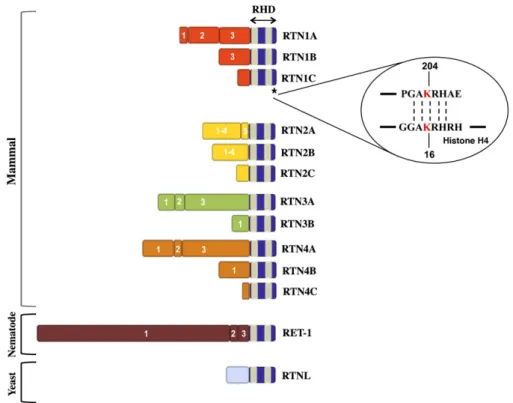
In mammals, four independent reticulon genes have been identified and named as rtn1, 2, 3, and rtn4/nogo. Each of them en-codes for different protein isoforms by using alternative splicing sites or different promoter regions[2]. All members of RTN family are characterized by a C-terminal reticulon homology domain (RHD) of 150–200 amino-acid residues, a common structural fea-ture which is evolutionarily preserved from plants to yeasts to humans (Fig. 1). The RHD consists of two hydrophobic regions, that assess the membrane association of the protein, separated by a 66 amino-acid hydrophilic loop and followed by a short C-terminal tail[3]. The conservation of RHD confers common func-tions among RTNs, such as subcellular localization and/or protein interactions. In fact, unlike ER-resident proteins, reticulons do not display the canonical ER-localization signal in the amino-terminal part but the retention in the ER membrane compartment could be due to the structural conformation of RHD. In fact, disrup-tion of the second transmembrane segment has been observed to affect the ER localization of RTN4[4], and mutations in the first transmembrane domain of RTN3 are able to compromise its mem-brane insertion[5]. Moreover, RHD hydrophobic regions are un-usually long, around 28–36 amino-acids, in comparison to the alpha-helix domain of typical transmembrane proteins that are
only about 20 amino-acids in length. Therefore, the topology of these hydrophobic regions within membranes diverges from the usual integral membrane proteins[6], and still remains partially elusive. In one of the most qualified model the reticulon trans-membrane domains do not fully cross the trans-membrane but each of them forms a hairpin-like structure into the outer leaflet of the lipid bilayer with both N- and C-terminal ends facing in the cyto-solic side (“W” topology). These hairpin-like topologies are typical of proteins involved in vesicle budding/membrane-shaping such as caveolin-1 and reggie-1[7]. Because RTNs are present in cell membranes characterized by high positive curvature, such as ER, it was hypothesized that reticulon hairpin-like structures are re-sponsible for this membrane bending. Consistent with this hy-pothesis, in Arabidopsis thaliana it was demonstrated that the lengths of the transmembrane segments in RTNLB13 are likely to be responsible for its ER membrane curvature[8]. Subsequently, together with RTNLB13, four other members of the reticulon fam-ily, RTNLB1-4, have shown a similar effect on the ER structure in A. thaliana[9].
It seems that RTNs not only individually bend the membrane but can also form multimeric, arc-like structures which may ulti-mately shape and determine the diameter of ER tubules[10].
Moreover, it has been demonstrated that the yeast RTN Rtn1p is sufficient to induce tubule formation in vitro when mixed with lipids[11]. Furthermore, similar results from Hu et al. have previ-ously been obtained for RTN4A where its ability to form ER tubule structures was demonstrated in association with DP1, an integral membrane protein orthologues of yeast Rtn1p-binding protein Yop1[12]. On the other hand, a growing line of evidence supports
Fig. 1– Schematic representation of mammal, nematode and yeast RTNs. Numbers indicate the different exons for each reticulon at the N-terminus. The reticulon homology domain is indicated by a gray box and the blue boxes represent the hydrophobic regions. Asterisk refers to RTN1C C-terminal domain (a.a. 201–208) sharing the GAKRH motif with Histone H4. The acetylated lysine is marked in red.
other different reticulon topologies from the hairpin-like model that might mirror a non-structural role for these isoforms. Thus, it is plausible to think that reticulons can have different topologies when associated to different cellular membrane sites and/or pro-tein partners reflecting the multiple functions exerted by these proteins.
In contrast to the closely conserved C-terminus that may con-fer common functions among reticulons, the N-terminal regions show little or no sequence homology (Fig. 1)[13]. These variable domains are likely to interact with distinct proteins and to confer specific biological functions to the various reticulon isoforms. In particular, the specific RTN4/Nogo function has been well charac-terized in mammalian central nervous system (CNS), where it ex-erts its inhibitory effect in neurite outgrowth[14]. Albeit a great pool of this reticulon protein localizes in the endomembrane sys-tem of the cell (ER and Golgi complex), as well as the other RTN family members, a little fraction of Nogo is also expressed in oligo-dendrocyte plasma membrane where it may function as a CNS-myelin specific inhibitor of axonal regeneration[15]. Unlike the myelin of the peripheral nervous system, the myelin from the CNS prevents neuronal regeneration after injury, and Nogo repre-sents one of the myelin-associated inhibitory factors implicated in this phenomenon. Indeed, in vivo neutralizing antibody against Nogo results in axonal growth of corticospinal tract and enhanced functional recovery following spinal cord injury, as well as in sen-sory motor cortex lesioned rats[16,17].
In particular, two extracellular exposed regions of Nogo, such as a stretch at N-terminus and 66 amino-acid loop of RHD (Nogo-66), are responsible for neurite growth-inhibiting effect
[14]. It is now known that Nogo-66 binds NgR, a corresponding re-ceptor present on neuron surface. The activation of NgR and its co-receptors induces the inhibitory signal in neurons via RhoA-ROCK signaling pathway that leads to cytoskeleton rigidification and growth cone collapse[3,18].
Numerous studies have largely demonstrated that blocking Nogo/NgR1 signaling could be a way to promote regeneration
[18], and thus Nogo could be a therapeutic target for CNS injuries and neurodegenerative disorders.
Further, more recently four WW domains that can bind to E3 ubiquitin-ligases WWP1 through its PPxY motifs have been identi-fied in the N-terminus of NogoA [19]. On the basis of these
speculations, ubiquitinylation likely represents an important mech-anism by which NogoA expression and activity may be regulated.
Reticulons in membrane trafficking
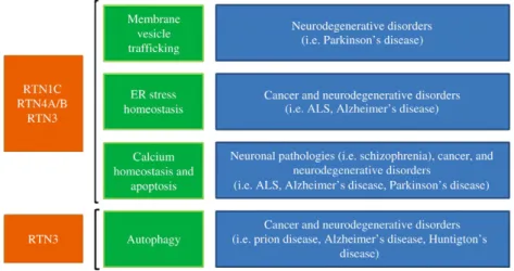
Although reticulons have mainly an ER localization, these proteins are also localized with the Golgi and plasma membrane. This pleio-tropic localization, in addition to the reticulon membrane-bending property, suggests an important involvement of RTNs in mem-brane vesicle trafficking.
Consistent with the implication of reticulons in cellular traffick-ing, RTN3-overexpression in HeLa cells prevents the retrograde transport of proteins from Golgi to ER[20]. Furthermore, by yeast two-hybrid screening RTN1-A/B have been identified to bind AP50, a component of the AP-2 adaptor complex that mediates en-docytosis[21]. Conversely, RTN1C may be involved in exocytosis. In fact, ectopic expression of RTN1C in PC12 cells induces an increase in the secretion rate of the growth hormone[22]. Further-more, RTN1C has been observed to co-immunoprecipitate with components of the SNARE complexes such as syntaxin-1, -7, -13, and VAMP2[22](Fig. 2). Moreover, the Caenorhabditis elegans reti-culon interacts with the protein RME-1, a regulator of endocytic recycling, thus suggesting an early evolution of the reticulon func-tion in vesicle trafficking of eukaryotic cells (Fig. 2).
Interestingly, it is well known that abnormalities in exocytosis and endocytosis may be one of the mechanisms involved in neuro-degenerative diseases. In keeping with this hypothesis, altered expression levels of reticulons have been observed in several neu-rological disorders (Fig. 2)[23,24]. For instance, RTN3 expression is increased in cerebral tissue obtained from Alzheimer disease patients[25]. Moreover, RTN3 and RTN4B/C have been identified to interact and negatively modulate beta-secretase BACE1, a pro-tease directly involved in amyloidogenic pathway in Alzheimer pathology[26]. However, the molecular basis of how RTN3 and other reticulons may function as a negative regulator of amyloid-β production is unclear. Interestingly, Shi et al. have observed a reduced level of amyloid-β deposition in RTN3-overexpressing mice due to a major retention of BACE1 in the ER compartment where this enzyme is less active. Considering the involvement of reticulons in vesicle trafficking, the authors hypothesize that the
association of RTN3 with BACE1 alters its trafficking itinerary to the cell surface[27].
Recently, it has been proposed that during cell mitosis, the nuclear envelope is regenerated from the ER and it is not assem-bled from the fusion of separate vesicles[28]. Notably, Anderson and Hetzer in their recent work have demonstrated that the dis-placement of ER shaping proteins, such as reticulons, is rate-limiting for nuclear assembly, suggesting a critical transition step from tubular to sheet nuclear membrane. In fact, overex-pression of RTN3, RTN4 and DP1 in U2OS cells delays nuclear envelope formation, whereas their knockdown by siRNAs ac-celerates this process[29]. Moreover, yeast RTN1 and Yop1 are required for nuclear pore complex (NPC) distribution, structural stability and biogenesis. In fact, rtn1Δ yop1Δ double mutant results in clusters of aberrant NPC-like structures. Then, in a cell-free assay based on Xenopus laevis egg extracts, a specific antibody against N-terminal domain of RTN4A is able to inhibit de novo nuclear pore assembly without affecting the nuclear envelope expansion. This effect could suggest a putative distinct function of the reticulon protein in the nuclear envelope flat-tening during mitotic telophase[30]independently by its role in the nuclear pore formation. In keeping with RTNs involvement in NPC biogenesis and stability, Chadrin et al. found a little pool of RTN1 copurifying with Pom33, a novel transmembrane nucleoporin protein identified in yeast. This data might suggest a dynamic interaction between RTN1 and Pom33, or could be explained by hypothesizing that only the NPC-associated fraction of reticulon takes part in this interaction[31]. In the future, it will be interesting to investigate whether the RTN proteins interact with other NPC components, and to evaluate the possibility that this functional connection is conserved in mammals considering the complexity in the redundant and specific functions of the wide set of RTN isoforms present in these organisms.
Reticulons in cell death
Among the proposed specific functions of RTNs, there are several evidences indicating their implications in the cell death pathways, in particular in the regulation of ER-stress-induced apoptosis[3]
(Fig. 2). Considering the role played by the reticulon proteins in ER morphogenesis and homeostasis, it was obvious to think about the important implications of these proteins under stressful conditions of this cellular organelle. It is well known that cell death can be triggered from various organelles, for instance via endoplasmic reticulum. Moreover, ER maintains intimate liaison with other organelles, such as nucleus, Golgi apparatus and mito-chondria, and thus represents a central player in cell physiology
[32]. In particular, several important regulators of apoptosis, in-cluding Bcl2 family members are localized in ER, and their pro/ anti apoptotic functions are mediated by an altered signaling from ER to mitochondria, which induces cell death by modulating the release of calcium from the ER store[33].
Interestingly, both RTN1C and RTN4B were identified as pro-teins interacting with Bcl-XL, a member of the Bcl2 family with
anti-apoptotic activity. Conversely, RTN4B is able to interact with another apoptosis inhibitor, Bcl2. Thus both the subcellular locali-zation, and consequently the activity of these anti-apoptotic factors are regulated by reticulons. In fact, RTN1C and RTN4A have been observed to reduce the anti-apoptotic ability of their
respective interactors in mammal cells[34]. However, the ectopic expression of this RTN family member triggers extensive apopto-sis in CGL4 and SaOS-2 cancer cell lines. Moreover, NogoB mRNA expression is abrogated in small cell lung cancer, suggesting a potential involvement in cancer development [35]. Additional studies indicate that NogoB overexpression causes ER stress-mediated apoptosis accompanied by ER lumenal Ca2+depletion
[36]. On the other hand, these results contrast with other data showing any sensitivity to apoptosis induced by chemotherapeu-tic drugs, like staurosporine or tunicamycin, in SaOS and CHO cell lines characterized by the stable NogoB overexpression[37]. This putative NogoB antiapoptotic function is also supported by the fact that this reticulon is cleaved by caspase-7 target during apoptosis upon dephosphorylation of its Ser16[38], suggesting a
highly regulated mechanism for NogoB proteolysis. On the basis of this evidence, it is tempting to speculate that NogoB processing might contribute to apoptosis phenotype by affecting the interac-tions with Bcl2 and Bcl-XL. It would be interesting to investigate
whether the other RTNs are also targets for caspases as well as RTN4B/NogoB.
We have demonstrated a role for RTN1C in the modulation of ER stress-induced apoptosis. In particular, RTN1C-overexpressing neuroblastoma cells show a significant increase in their suscepti-bility to cell death upon ER stress induction [39]. Moreover, RTN1C overexpression induces per se apoptosis via ER stress in-duction characterized by a significant and toxic ER store calcium depletion [40]. Similarly to RTN1C, RTN3 overexpression also causes apoptosis accompanied by depletion of ER Ca2+ stores
[41]. However, it is important to note that controversial results have been reported about the pro-apoptotic role of RTN3. In fact, it has been shown that ER stress-mediated RTN3 upregulation can be protective against several insults, contributing to the Bcl2 translocation to the mitochondria [42]. The molecular mecha-nisms by which RTN1C and RTN3 affect the intracellular Ca2+ ho-meostasis still remain elusive. In the past Oertle and Schwab hypothesized that RTN oligomerization could lead to the forma-tion of pores in the ER[13]. This hypothesis could explain the in-trinsic ability of RTNs to induce the Ca2+efflux from ER, since no
interactions of RTN1C or other RTNs involved in the ER Ca2+ de-pletion with the inositol trisphosphate (IP3) receptors or
sarco-plasmic/endoplasmic reticulum Ca2+pump (SERCA) have been
demonstrated. It is well known that other proapoptotic Bcl2 fam-ily members, like Bak and Bax, are also localized in ER membrane where they may trigger ER-stress induced apoptosis[43], by regu-lating the Ca2+efflux through type I IP
3receptor[44]. Based on
these findings it is tempting to hypothesize that the reticulons might induce Ca2+ depletion from ER by indirectly activating
Bax/Bak molecules or alternatively by reducing their inhibitory in-teractions with Bcl2/Bcl-XL.
It is noteworthy that the second hydrophobic domain at NogoB C-terminus is important for apoptotic function of this protein[35]. As described above the different RTN isoforms share a C-terminal reticulon homology domain containing two hydrophobic seg-ments and a 66-amino acid hydrophilic loop. We have recently identified in the C-terminal region of RTN1C, a unique consensus sequence (GAKRH) showing 100% identity with the DNA-binding domain of histone H4 (Fig. 1). Interestingly, we showed that this sequence is essential for RTN1C-mediated ER-stress induced apoptosis, although it does not affect its ER-localization[45]. It is noteworthy that the lysine 204 present in this region is
post-translationally modified by acetylation and that this event is associated with a significant decrease in histone deacetylase activity contributing RTN1C binding to DNA. These data demon-strate a molecular mechanism by which RTN-1C might control apoptosis.
Chen et al. recently showed that RTN3 overexpression en-hances apoptosis in N2a mouse neuroblastoma cells treated with an ER stress inducing drug. Interestingly, in this work an im-portant link between reticulons and autophagy has been shown, the latter being the most important degradative pathway of the lysosomal system for the removal of cellular organelles and protein aggregates that are too large to be delivered to the pro-teasome[46]. They demonstrated that RTN3 plays an important role in autophagy induced by disease-associated prion proteins; in particular, the knockout of RTN3 protein by increasing autop-hagy induction in human prion-overexpressing N2a cells and enhances the clearance of the prion aggregates. The authors found that RTN3 inhibits beclin-1-dependent autophagy by pro-moting the formation of the inactive Bcl2–Beclin-1 complex
[47]. Despite the molecular mechanism by which RTN3 promotes the interaction between Bcl2 and Beclin-1 is unknown, previous findings showed that RTN3 is able to bind Bcl2 and that RTNs act in the transport of constituents from ER to other membrane compartments, thus it is reasonable to believe that RTN3 could promote Bcl2 mobilization upon ER stress induction to regulate bcl2-dependent cellular processes such as autophagy or apopto-sis. Based on these findings it will be interesting to investigate whether other RTNs interacting with Bcl2 are implicated in autophagy and whether their action is associated to their capacity to mobilize Ca2+from the ER. In keeping with this hypothesis, the
release of Ca2+from ER has been recently postulated to be an
important early regulatory event during autophagy induction
[48].
Conclusions
Several recent studies have shown the importance of RTNs in ER homeostasis. In particular, their proapoptotic function as a novel regulator of ER stress-mediated cell death is well characterized
[39]. However, the molecular mechanism by which the reticulon family members orchestrate different cellular responses is still poorly understood. Future studies should define the molecular de-tails of how the RTN action is translated at ER level into a death stimulus. The ability to respond to perturbations in ER function is a key property of all cells. In fact, in settings of chronic ER stress, the associated apoptosis may contribute to pathophysiological processes involved in a number of prevalent diseases, including neurodegenerative diseases, diabetes, atherosclerosis and renal disease[32]. By using microarray analysis of the whole human genome, we have recently demonstrated that RTN-1C is able to specifically regulate gene expression, modulating transcript clus-ters which have been implicated in the onset of neurodegenera-tive disorders. Interestingly, we show that some of the identified genes were also modulated in vivo in a brain-specific mouse model overexpressing RTN-1C[49].
In keeping with this, the reticulons may act as modulator of PDI subcellular distribution. PDI is an ER resident protein fundamental for correct protein maturations. Therefore they act as important cellular defense against misfolded protein accumulations, typical
of neurodegenerative disorders. Noteworthy, this property has been associated with a neuroprotective effect in ALS mice models of amyotrophic lateral sclerosis (ALS) [50], and thus reticulon-mediated ER changes, including PDI relocalization, could repre-sent a potential therapeutic target in ALS. In line with these findings, RTNs have been shown to play an important role in the nervous system both in normal and pathological settings. In fact, studies carried out on post-mortem brain tissues have revealed changes in reticulons expression in the temporal and frontal cortex of patients with Alzheimer's disease and schizophrenia
[24]. In keeping with these data, human association studies have implicated mutations in RTN4 in the development of schizophre-nia[51](Fig. 2). Taken together, all these new findings provide a basis for further investigation of RTNs as a potential molecular target for use in therapy and as a specific marker for neurological diseases.
Acknowledgments
This work was supported by grants from Compagnia di San Paolo, the Ministry of Health of Italy “Ricerca Corrente” and “Ricerca Finalizzata”, the Ministry of University Research “FIRB” and AIRC. The support of the EU grant“Transpath” Marie Curie project is also acknowledged.
R E F E R E N C E S
[1] F.Y. Teng, B.L. Tang, Cell autonomous function of Nogo and reticulons: the emerging story at the endoplasmic reticulum, J. Cell. Physiol. 216 (2008) 303–308.
[2] T. Oertle, M. Klinger, C.A. Stuermer, M.E. Schwab, A reticular rhapsody: phylogenic evolution and nomenclature of the RTN/Nogo gene family, FASEB J. 17 (2003)
1238–1247.
[3] Y.S. Yang, S.M. Strittmatter, The reticulons: a family of proteins with diverse functions, Genome Biol. 8 (2007) 234.
[4] T. Oertle, M.E. Van der Haar, C.E. Bandtlow, A. Robeva, P. Burfeind, A. Buss, A.B. Huber, M. Simonen, L. Schnell, C. Brsamle, K. Kaupmann, R. Vallon, M.E. Schwab, Nogo-A inhibits neurite outgrowth and cell spreading with three discrete regions, J. Neurosci. 23 (2003) 5393–5406.
[5] W. He, Q. Shi, X. Hu, R. Yan, The membrane topology of RTN3 and its effect on binding of RTN3 to BACE1, J. Biol. Chem. 282 (2007) 29144–29151.
[6] F. Brandizzi, N. Frangne, S. Marc-Martin, C. Hawes, J.M. Neuhaus, N. Paris, The destination for single-pass membrane proteins is influenced markedly by the length of the hydrophobic domain, Plant Cell 14 (2002) 1077–1092.
[7] M. Bauer, L. Pelkmans, A new paradigm for membrane-organizing and -shaping scaffolds, FEBS Lett. 580 (2006) 5559–5564. [8] N. Tolley, I. Sparkes, C.P. Craddock, P.J. Eastmond, J. Runions, C.
Hawes, L. Frigerio, Transmembrane domain length is responsible for the ability of a plant reticulon to shape endoplasmic reticulum tubules in vivo, Plant J. 64 (2010) 411–418.
[9] I. Sparkes, N. Tolley, I. Aller, J. Svozil, A. Osterrieder, S. Botchway, C. Mueller, L. Frigerio, C. Hawes, Five Arabidopsis reticulon isoforms share endoplasmic reticulum location, topology, and membrane-shaping properties, Plant Cell 22 (2010) 1333–1343.
[10] Y. Shibata, C. Voss, J.M. Rist, J. Hu, T.A. Rapoport, W.A. Prinz, G.K. Voeltz, The reticulon and DP1/Yop1p proteins form immobile
oligomers in the tubular endoplasmic reticulum, J. Biol. Chem. 283 (2008) 18892–18904.
[11] J. Hu, Y. Shibata, C. Voss, T. Shemesh, Z. Li, M. Couglin, M.M. Kozlov, T.A. Rapoport, W.A. Prinz, Membrane proteins of the endoplasmic reticulum induce high-curvature tubules, Science 319 (2008) 1247–1250.
[12] G.K. Voeltz, W.A. Prinz, Y. Shibata, J.M. Rist, T.A. Rapoport, A class of membrane proteins shaping the tubular endoplasmic reticulum, Cell 124 (2006) 573–586.
[13] T. Oertle, M.E. Schwab, Nogo and its partner, Trends Cell Biol. 13 (2003) 187–194.
[14] T. Oertle, M.E. van der Haar, C.E. Bandtlow, A. Robeva, P. Burfeind, A. Buss, A.B. Huber, M. Simonen, L. Schnell, C. Brosamle, K. Kaupmann, R. Vallon, M.E. Schwab, Nogo-A inhibits neurite outgrowth and cell spreading with three discrete regions, J. Neurosci. 23 (2003) 5393–5406.
[15] D.A. Dodd, B. Niederoes, S. Bloechlinger, L. Dupuis, J.P. Loeffler, M.E. Schwab, NogoA, -B, -C are found in many different cell types, J. Biol. Chem. 280 (2005) 12494–12502.
[16] K. Fouad, V. Dietz, M.E. Schwab, Improving axonal growth and functional recovery after experimental spinal cord injury by neutralizing myelin associated inhibitors, Brain Res. 36 (2001) 204–212.
[17] A.J. Emerick, E.J. Neafsey, M.E. Schwab, Functional reorganization of the motor cortex in adult rats after cortical lesion and treatment with monoclonal antibody IN-1, J. Neurosci. 23 (2003) 4826–4830.
[18] C.L. McDonald, C. Bandtlow, M. Reindl, Targeting the Nogo receptor complex in diseases of the central nervous system, Curr. Med. Chem. 18 (2011) 234–244.
[19] H. Quin, H.X. Pu, M. Li, S. Ahmed, J. Song, Identification and structural mechanism for a novel interaction between a ubiquitin ligase WWP1 and Nogo-A, a key inhibitor for central nervous system regeneration, Biochemistry 47 (2008) 13647–13658. [20] Y. Wakana, S. Koyama, K. Nakajima, K. Hatsuzawa, M. Nagahama,
K. Tani, H.P. Hauri, P. Melancon, M. Tagaya, Reticulon 3 is involved in membrane trafficking between the endoplasmic reticulum and Golgi, Biochem. Biophys. Res. Commun. 334 (2005) 1198–1205.
[21] J. Iwahashi, N. Hamada, Human reticulon 1-A and 1-B interact with a medium chain of the AP-2 adaptor complex, Cell. Mol. Biol. 49 (2003) 467–471.
[22] P. Steiner, K. Kulangara, J.C. Sarria, L. Glauser, R. Regazzi, H. Hirling, Reticulon 1-C/neuroendocrine-specific protein-C interacts with SNARE proteins, J. Neurochem. 89 (2004) 569–580.
[23] A. Fergani, L. Dupuis, N. Jokic, Y. Larmet, M. de Tapia, F. Rene, J.P. Loeffler, J.L. Gonzales de Aguilar, Reticulons as markers of neurological diseases: focus on amyotrophic lateral sclerosis, Neurodegener Dis 2 (2005) 185–194.
[24] R. Yan, Q. Shi, X. Zhou, Reticulon proteins: emerging players in neurodegenerative diseases, Cell. Mol. Life Sci. 63 (2006) 877–889. [25] T. Yokota, M. Mishra, H. Akatsu, Y. Tani, T. Miyauchi, T.
Yamamoto, K. Kosaka, Y. Nagai, T. Sawada, K. Heese, Brain site-specific gene expression analysis in Alzheimer's disease patients, Eur. J. Clin. Invest. 36 (2006) 820–830.
[26] K.S. Murayama, F. Kametani, S. Saito, H. Kume, H. Akiyama, W. Araki, Reticulons RTN3 and RTN4-B/C interact with BACE1 and inhibit its ability to produce amyloid beta-protein, Eur. J. Neurosci. 24 (2006) 1237–1244.
[27] Q. Shi, M. Prior, W. He, X. Tang, X. Hu, R. Yan, Reduced amyloid deposition in mice overexpressing RTN3 is adversely affected by preformed dystrophic neuritis, J. Neurosci. 29 (2009) 9163–9173.
[28] P. Collas, D. Poccia, Membrane fusion events during nuclear envelope assembly, Subcell. Biochem. 34 (2000) 273–302. [29] D.J. Anderson, M.W. Hetzer, Nuclear envelope formation by
chromatin-mediated reorganization of the endoplasmic reticulum, Nat. Cell Biol. 9 (2007) 1160–1166.
[30] T.R. Dawson, M.D. Lazarus, M.W. Hetzer, S.R. Wente, ER membrane bending proteins are necessary for de novo nuclear pore formation, J. Cell Biol. 184 (2009) 659–675.
[31] A. Chadrin, B. Hess, M. San Roman, X. Gatti, B. Lombard, D. Loew, Y. Barral, B. Palancade, V. Doye, Pom33, a novel transmembrane nucleoporin required for proper nuclear pore complex distribution, J. Cell Biol. 189 (2010) 795–811.
[32] I. Tabas, D. Ron, Integrating the mechanisms of apoptosis induced by endoplasmic reticulum stress, Nat. Cell Biol. 13 (2011) 184–190.
[33] D. Rodriguez, D. Rojas-Rivera, C. Hetz, Integrating stress signals at the endoplasmic reticulum: the BCL-2 protein family rheostat, Biochim. Biophys. Acta 163 (2010) 564–574.
[34] S. Tagami, Y. Eguchi, M. Kinoshita, M. Takeda, Y. Tsujimoto, A novel protein, RTN-XS, interacts with both Bcl-XL and Bcl-2 on endoplasmic reticulum and reduces their anti-apoptotic activity, Oncogene 19 (2000) 5736–5746.
[35] Q. Li, B. Qi, K. Oka, M. Shimakage, N. Yoshioka, H. Inoue, A. Hakura, K. Kodama, E.J. Stanbridge, M. Yutsudo, Link of a new type of apoptosis-inducing gene ASY/Nogo-B to human cancer, Oncogene 20 (2001) 3929–3936.
[36] E. Kuang, Q. Li, X. Xu, H. Zou, T. Qi, ER stress triggers apoptosis induced by Nogo-B/ASY overexpression, Exp. Cell Res. 312 (2006) 1983–1988.
[37] T. Oertle, D. Merkler, M.E. Schwab, Do cancer cells die because of Nogo-B? Oncogene 22 (2003) 1390–1399.
[38] R. Schweigreiter, T. Stasyk, I. Contarini, S. Frausher, T. Oertle, L. Klimashewski, L.A. Huber, C.E. Bandtlow, Phosphorylation-regulated cleavage of the reticulon protein Nogo-B by caspase-7 at a noncanonical recognition site, Proteomics 7 (2007) 4457–4467. [39] F. Di Sano, B. Fazi, R. Tufi, R. Nardacci, M. Piacentini, Reticulon-1C
acts as a molecular switch between endoplasmic reticulum stress and genotoxic cell death pathway in human neuroblastoma cells, J. Neurochem. 102 (2007) 345–353.
[40] R. Tufi, T. Panaretakis, K. Bianchi, A. Criollo, B. Fazi, F. Di Sano, A. Tesniere, O. Kepp, P. Paterlini-Brechot, L. Zitvogel, M. Piacentini, G. Szabadkai, G. Kroemer, Reduction of endoplasmic reticulum Ca2+levels favors plasma membrane surface exposure of
calreticulin, Cell Death Differ. 15 (2008) 274–282.
[41] E. Kuang, Q. Wan, X. Li, H. Xu, Q. Liu, Y. Qi, ER Ca2+depletion
triggers apoptotic signals for endoplasmic reticulum (ER) overload response induced by overexpressed reticulon 3 (RTN3/HAP), J. Cell. Physiol. 204 (2005) 549–559. [42] Q. Wan, E. Kuang, W. Dong, S. Zhou, H. Xu, Y. Qi, Y. Liu,
Reticulon 3 mediates Bcl-2 accumulation in mitochondria in response to endoplasmic reticulum stress, Apoptosis 12 (2007) 319–328.
[43] D.G. Breckenridge, M. Germain, J.P. Mathai, M. Nguyen, G.C. Shore, Regulation of apoptosis by endoplasmic reticulum pathways, Oncogene 22 (2003) 8608–8618.
[44] S.A. Oakes, L. Scorrano, J.T. Opferman, M.C. Bassik, M. Nishino, T. Pozzan, S.J. Korsmeyer, Proapoptotic BAX and BAK regulate the type 1 inositol trisphosphate receptor and calcium leak from the endoplasmic reticulum, Proc. Natl. Acad. Sci. U. S. A. 102 (2005) 105–110.
[45] B. Fazi, S. Melino, S. De Rubeis, C. Bagni, M. Paci, M. Piacentini, F. Di Sano, Acetylation of RTN-1C regulates the induction of ER stress by the inhibition of HDAC activity in neuroectodermal tumors, Oncogene 28 (2009) 3814–3824.
[46] N. Mizushima, Physiological functions of autophagy, Curr. Top. Microbiol. Immunol. 335 (2009) 71–84.
[47] R. Chen, R. Jin, L. Wu, X. Ye, Y. Yang, K. Luo, W. Wang, D. Wu, X. Ye, L. Huang, T. Huang, G. Xiao, Reticulon 3 attenuates the clearance of cytosolic prion aggregates via inhibiting autophagy, Autophagy 7 (2011) 205–216.
[48] S.G. Pfisterer, M. Mauthe, P. Codogno, T. Proikas-Cezanne, Ca2+/
calmodulin-dependent kinase contributes to the regulation of WIPI-1 at the onset of autophagy, Mol. Pharmacol. 80 (2011) 1066–1075.
[49] B. Fazi, M. Biancolella, B. Mehdawy, M. Corazzari, D. Minella, F. Blandini, S. Moreno, R. Nardacci, R. Nisticò, S. Sepe, G. Novelli, M. Piacentini, F. Di Sano, Characterization of gene expression induced by RTN-1C in human neuroblastoma cells and in mouse brain, Neurobiol. Dis. 40 (2010) 634–644. [50] Y.S. Yang, N.Y. Harel, S.M. Strittmatter, Reticulon-4A (Nogo-A)
redistributes protein disulfide isomerase to protect mice from
SOD1-dependent amyotrophic lateral sclerosis, J. Neurosci. 29 (2009) 13850–13859.
[51] R. Hsu, A. Woodroffe, W.S. Lai, M.N. Cook, J. Mukai, J.P. Dunning, D.J. Swanson, J.L. Roos, G.R. Abecasis, M. Karayiorgou, M. Gogos, Nogo Receptor 1 (RTN4R) as a candidate gene for schizophrenia: analysis using human and mouse genetic approaches, PLoS One 2 (2007) e1234.