IL-15/IL-15Rα signalling and synaptic
transmission: a crosstalk between the
immune and the nervous system?
PhD school in Clinical/Experimental Neuroscience and Psychiatry
Curriculum in Neurophysiology
Cycle XXXI
Coordinator Prof. Marco Salvetti
Candidate:
Dr. Laura Carbonari
Supervisor:
Prof. Cristina Limatola
“Una delle principali cause della miseria delle scienze sta, molto spesso, nella loro presunzione di essere ricche. Scopo della scienza non è tanto quello di aprire la porta all'infinito sapere, quanto quello di porre una barriera all'infinita ignoranza.”
B. Brecht
“The world is stranger than we can imagine and surprises are inevitable in science. Thus we found, for example, that pesticides increase pests, antibiotics can create pathogens, agricultural development creates hunger, and flood control leads to flooding. But some of these surprises could have been avoided if the problems had been posed big enough to accommodate solutions in the context of the whole.”
Richard Levins
“Far better an approximate answer to the right question, which is often vague, than an exact answer to the wrong question, which can always be made precise.”
INDEX
1. OVERVIEW ... 6
2.1 Neuroimmune system ... 8
2.1.1 Composition and interactions ... 8
2.1.2 Microglia as one of the main actors on the neuroimmune stage ... 10
2.2 The cytokines system: a common language between immune and brain cells 11 2.2.1 The huge world of cytokines ... 11
2.2.2 Cytokines and synaptic plasticity ... 12
2.3 Interleukin 15 (IL-15) ... 14
2.3.1 IL-15/IL-15Rα system and signalling transduction ... 14
2.3.2 Signal transduction of IL-15 signalling ... 16
2.3.3 The IL-15/IL-15Rα system and behaviour ... 18
2.4 Hippocampal memory and GABAergic system ... 19
2.4.1 The hippocampal connectivity ... 19
3. AIM OF THE STUDY ... 21
4. MATERIALS AND METHODS ... 23
4.1 Animals ... 23
4.2 Genotyping DNA extracted from mouse tail ... 23
4.3 Patch clamp recordings of CA1 pyramidal neurons from ex vivo hippocampal brain slices ... 24
4.4 Extracellular field recordings of CA3-CA1 circuit from ex vivo hippocampal brain slices ... 26
4.5 Isolation of CD11bþ cells and extraction of total RNA ... 27
4.6 RT–PCR ... 28
4.7 IL-15 delivery in hippocampus of WT mice with micro-osmotic pumps ... 28
4.8 Novel object recognition (NOR) ... 29
4.9 Statistical analysis ... 30
5. RESULTS ... 31
5.1 IL-15Rα KO mice show altered inhibitory synaptic transmission in CA1 hippocampal neurons. ... 31
5.2 IL-15Rα KO mice show increased GABA release in CA1 area ... 33
5.3 IL-15Rα KO mice show normal CA3-CA1 glutamatergic transmission with no alterations in short- (STP) and long-term potentiation (LTP) ... 35
5.4 IL-15Rα KO mice are impaired in Novel Object Recognition (NOR) task ... 36
5.5 Microglia extracted from hippocampi of IL-15Rα KO mice show an altered mRNA expression ... 37
5.6 Treatment with exogenous IL-15 affects inhibitory but not excitatory synaptic transmission in the hippocampus ... 39
1. OVERVIEW
Immune and nervous system have been traditionally considered separately, but from ‘90s many studies had unraveled the deep interconnection and interdependence between these two systems, enough to coin the term “neuroimmune system” to define this relationship.
While it was well known that central nervous system (CNS) actively communicates with the immune system to control immune responses both centrally and peripherally, the opposite action was just recently discovered.
Related to the role of immune system in defending and react, the interactions between immune system and CNS have been classically studied in contexts of neuroinflammation such as trauma, injury and disease [1] [2].
Recent evidences about the neuroinflammatory process in non-pathological conditions and the discovery of the important involvement of adaptive immune system in healthy brain development and activity [3], have opened many questions about physiological neuroimmune cross-talk.
In this view, the cytokine network, well known to operate in a bidirectional way affecting both immune and nervous system, has a pivotal role in neuroimmune cross-talk [4]. Traditionally seen as immunomodulators, in the last years has been evident that cytokines are also potent neuromodulators [5].
In the complex cytokine system, interleukin 15 (IL-15) is considered a bridge between adaptive and innate immune system and it is one of the first upregulated cytokines in neuroinflammation [6]. It has many bioregulatory roles which range from those of modulator of selected adaptive immune responses [7] [8] and central player in the development and homeostasis of several immunocyte populations [9] to those of a potent, general inhibitor of apoptosis in multiple systems [9].
Interestingly, has been shown that IL-15 and IL-15Rα deletions affect memory and neurotransmitters concentration suggesting a major role of this signalling in cerebral functions which cannot be compensated during the development [10] [11] [12]. IL-15Rα KO mice, in particular, show decreased retention of spatial memory and contextual fear, both related to hippocampus-dependent memory, and alteration in GABA concentration. Their hippocampal ultrastructure is, however, well preserved, suggesting that the modulatory changes may involve neural
plasticity even if the exact role of IL15 in modulating neurotransmission has not been investigated so far.
The understandings about the mechanism by which IL-15/IL-15Rα system affect the synaptic transmission may be useful to get insight into the mechanisms of cross talk between the immune and the nervous system and eventually to develop strategies to treat pathologies whose symptoms are memory impairments and neuroinflammation.
2. INTRODUCTION
2.1 Neuroimmune system
2.1.1 Composition and interactions
In recent years it has become clear that there is an extensive cross-talk between the nervous and the immune system, so that the term “neuroimmune system” has been coined to indicate all the biochemical, electrophysiological and cellular structures belonging to both nervous and immune system [17]. This cross-talk takes place in different organs, involving a wide range of cells and mediators, coordinated through sensory and effector pathways [13].
In mammals, this system involves reciprocal regulation which works in two directions in both physiological and pathological conditions: the
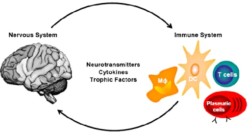
immune-mediated regulation of nervous system function and the nervous system-mediated regulation of immunity (Figure 1). Both kinds of interactions are
mediated by complex mechanisms in which participate the same groups of cells and the same kind of molecules [4] and occur both centrally and peripherally [4]. If in the past cytokines were considered as the mediators of immune cells and neurotransmitters as the mediators of nervous system cells, currently it is known that immune system cells can also communicate through neurotransmitters and that, viceversa, brain cell populations can communicate each other through cytokines. Evidences from the last decade have shown that both the systems are able both to sense and produce these molecules.
Figure 1. Neurotransmitters and cytokines mediate bidirectional cross-talk between immune and nervous cells. This inter-systemic communication may occur in both directions: the regulation of immune cells by
nervous-derived molecules as well as the modulation of nervous cells by immune-derived molecules. [4]
Traditionally, neuroimmunology research has been mostly focused on interactions in the context of diseases, injuries or traumas but starting from studies on the role of microglia in brain development, many researches have shed light on the involvement of immune cells in normal CNS function [14]. It was shown, indeed, that mechanisms reminiscent of neuroinflammation, such as the involvement of complement components and microglia in synapse pruning, also occur in healthy brain development [3]. To date, however, the mechanism and the regulation of the physiological cross-talk occurring in the neuroimmune system is still almost obscure.
The most common and studied interaction between immune system and CNS is the neuroinflammation whose concept has widened years by years up to include the response of the reactive CNS elements to altered homeostasis due to infections and other causes of cell death, as well as infiltration of the brain and spinal cord by cells of the innate and adaptive immune systems [15]. It characterizes almost every neurological disease, from developmental ones to traumatic ones passing through neoplastic and neurodegenerative diseases. Recent research has established a significant role for the immune system in several brain diseases including mental disorders further implicating dependent interrelationships between the immune system and the brain.
Any alterations in tissue homeostasis begin a tissue response by CNS-resident cells that involves mediators associated with the immune system. In these conditions glial cell responses, usually mediated by astrocytes and microglia,
include the elevated production of an array of cytokines. Conversely, in chronic inflammatory diseases or in acute infectious diseases leukocytes that invade the CNS are a major source of inflammatory mediators, including cytokines. If from a theoretical point of view we can distinguish between diseases in which cytokines are produced predominantly by CNS-resident cells (like during neurodegeneration) and disorders where cytokines are delivered by CNS-invading leukocytes (like in encephalitides and inflammatory demyelinating disorders), actually, many of the cytokines implicated in driving the pathology associated with neurodegeneration are also key players in neuroinflammation mediated by invading leukocytes like IL‑12, IL‑23 [16], IL‑6 and IL‑1β [17] [18].
2.1.2 Microglia as one of the main actors on the neuroimmune
stage
Microglial cells are the main regulators of both innate and adaptative immune response in the CNS. They are the only resident macrophages of CNS under steady-state conditions and constitute the first defence line of the immune system against any trauma, disease or injury. Their distribution is widespread but there are region dependent differences for what concern the density, phenotype and responsiveness [19].
Since relatively recent years microglia in healthy condition have been considered
“resting” believed functionally turned off, due to the low expression of
activation-associated molecules and their apparently immobile ramified morphology characterised by thin cellular processes branching off from a small soma. In the “activated” state, the microglial cells assume an amoeboid shape, enlarging the soma and shortening the processes [14] and is able to modulate several molecules to trigger the inflammatory process.
Nevertheless, it is now well ascertained that microglia is actually very active in its
“resting” state, constantly monitoring the extracellular space and participating in
many physiological processes – from synaptic modelling to scavenging of cellular debris and secretion of trophic factors - thanks to its thin branches [20]. In this state microglial cells are very reactive and can go in the activated state very quickly, changing some receptors expression and releasing a broad spectrum of inflammatory and immunoregulatory compounds (e.g., neurotoxins, cytokines, ROS).
Focusing on synapses, it has been revealed that microglia interact with neurons directly contacting axonal terminals, dendritic spines, or synaptic clefts and eliminate some of them depending on changes in neuronal activity and sensory experience, both in the developing and mature brain [21] [22]. Moreover, has been demonstrated that disruption of communication between neurons and microglial cells through CX3CL1/CX3CR1 signalling leads to defective synaptic maturation in hippocampus probably due to a transient reduction in microglia surveillance and a consequent reduction of synaptic pruning [3].
At the same time a variety of neurotransmitter receptors and transporters are expressed on microglia and help mediate the bidirectional communication between neurons and microglia. Presumably, the information about the neuronal state given through neurotransmitters release is sensed by microglia which, in turn, influences neuronal viability in positive and negative feedback loops [22]. Electrophyisiological recordings in microglial cells from acute brain slices and in culture have shown, for instance, that GABAB-Rs agonists triggered the induction
of outwardly K+ conductance and attenuate the LPS-induced release of microglial
cytokines such as IL-6 and IL-12p40 [22].
2.2 The cytokines system: a common language between immune
and brain cells
2.2.1 The huge world of cytokines
The term ‘cytokines’ derives from the fusion of two Greek words cyto, "κύτος, kytos" which means “cavity, cell" and kines, "κίνησις, kinēsis" which means “movement". It identifies a group of small (~5-20kDa) soluble peptides and pleiotropic proteins released by immune cells, representing key regulators of the prototypical innate and adaptive immune response to infection. Their pleiotropic nature by which a given cytokine can induce differential, even opposite responses in different cell types, makes the cytokine network very complex [23]. Moreover, cytokines can cross-talk with signalling from other soluble factors in a time-, concentration- and tissue-specific manner.
The cytokine family is very wide (over 300 cytokines were discover up to date) and includes interferons (IFNs), interleukins (ILs), chemokines, mesenchymal growth factors, the platelet derived growth factors (PDGF), transforming growth factors (TGF), the tumor necrosis factors family (TNFs) and adipokines classified
depending on the nature of the immune response, with individual cytokines also performing specific roles dependent upon cell type and location. Usually the cytokines were distinguished pro- and anti-inflammatory but numerous evidences showed that this classification is too simplicistic and that a given cytokine may behave as a pro- or anti-inflammatory one depending on the cytokine amount, the nature of the target cell, the nature of the activating signal, the timing, the sequence of cytokine action and even the experimental model.
2.2.2 Cytokines and synaptic plasticity
Cytokine levels in nervous tissue are relatively low in physiological conditions (≤1 pg in 1 ml or mg) but they rapidly increase after various CNS or PNS injuries, seizures or infection [5]. Nevertheless, it is clear that cytokine networks in the brain are fundamental for the dynamics and the communications between neurons, glia, endothelial and immune cells acting in complex paracrine and autocrine ways. It seems that a controlled and timely production of cytokines is required for normal tissue function. Beyond their recognized role in neuroinflammation, they are involved in several aspects of normal CNS activities: they participate in the regulation of sleep [24], in neuronal development [25] [26] [27], in neuroendocrine functions and in ageing.
The major sources of cytokine synthesis and release in the CNS is the glia (microglia and astrocytes), although blood-imported cytokines may contribute to tissue level changes.Excessive levels of cytokines can, in turn, promote both glia and BBB dysfunction, with an impact on neuronal cell excitability and viability. Neurons may release cytokines too, particularly during the acute injury phase underlying neuronal damage [28]. Anyway, inflammatory pathways may be activated even in the absence of pathogens - the so-called sterile inflammation - because of changes in the extracellular ionic milieu. In this situation cytokines and related effector molecules can act both in autocrine or paracrine manner inducing different signalling pathways depending on the target cell.
With respect to neurons, cytokines have profound effects upon synaptic plasticity, especially in the hippocampus [29] [30], and many electrophysiological studies have demonstrated a modulation of neuronal activity by a wide variety of cytokines [31] [32] [33] [34]. This modulation can occur either indirectly by promoting the release of neuroactive molecules from glia or the endothelium (e.g,
nitric oxide, glutamate, prostaglandins, neurotrophins), or directly by activating their neuronal receptors in the brain and spinal cord [35] [36] [37].
The direct activation of neuronal cytokine receptors rapidly alters their excitability via post-translational modifications of either voltage-gated or receptor-coupled ion channels, and by promoting presynaptic changes in neurotransmission (Figure
2). These alterations are very rapid in their onset (seconds/minutes) but they
persist for very long time in vivo (hours/weeks) [38].
Figure 2. Several cytokines have been recognized as modulators of synaptic transmission [5].
Cytokines have been found to modulate neuronal properties at both pre- and post-synaptic levels. Effects of cytokines on voltage-gated channels (VGCs) as on receptor operated ion channels (ROCs) have been reported. Some cytokines affect only excitatory or inhibitory transmission, but several others behave in more complex ways acting differently depending on neuronal type, brain region or dose [5].
Given that activation of peripheral inflammatory responses has been associated with mood and anxiety disorders and that peripheral immune activation can spread to the CNS, there has been great interest in the impact of cytokines and their signalling pathways on neurotransmitter systems known to be associated with depression and anxiety including serotonin, norepinephrine, dopamine and glutamate [39]. There is less knowledge about the effects of inflammatory cytokines on other neurotransmitter systems in the brain that play a role in depression and anxiety like GABA and acetylcholine. Nevertheless, there is emerging data regarding interactions between inflammatory cytokines and these neurotransmitter systems that may have profound consequences for immune
regulation. For example, studies in rodents have indicated that GABA can reduce the release of inflammatory cytokines through inhibition of NF-kB and p38 MAPK signalling pathways [40] and decrease the progression of experimental autoimmune encephalomyelitis, an animal model of MS [41]. Interestingly, CSF from MS patients with enhanced brain lesions inhibited GABA transmission in mouse brain slices, an effect that could be blocked with an IL-1-beta, antagonist [41]. These findings indicate that inflammation in the CNS may decrease GABAergic tone, which could further trigger inflammatory cytokine production. Taken together, these findings indicate that a potential decrease in release of GABA in the brain in response to inflammatory cytokines may promote inflammatory responses [42].
Finally, recent studies demonstrated that the CXCL16 chemokine can modulate, through A3 receptor and microglial cells, both inhibitory and excitatory synaptic transmission in CA1 area increasing the frequency of the miniature inhibitory synaptic currents (mIPSCs) and the paired-pulse ratio (PPR) of evoked IPSCs (eIPSCs), suggesting a presynaptic modulation of the probability of GABA release [43].
2.3 Interleukin 15 (IL-15)
2.3.1 IL-15/IL-15Rα system and signalling transduction
More than 60 cytokines are classified as interleukins and are named by interleukin number. The name "interleukin" was chosen in 1979 during the Second International Lymphokine Workshop in Switzerland and derives from (inter-) "as a means of communication", and (-leukin) deriving from the fact that many of these proteins were first seen to be produced by leukocytes and act on leukocytes. Actually, the name is a residue because has been found that interleukins are produced by a wide variety of body cells.
Among cytokines, interleukin-15 (IL-15) was discovered in 1994 by its ability to mimic IL-2–mediated T-cell proliferation but it is now well established that it is produced even by many nonimmune cells both in basal conditions and in response to viral infections, LPS, and other signals that trigger innate immunity. It is, in fact, a pleiotropic cytokine of the 4-a-helix bundle cytokine family that includes not only cytokines such ad IL-2 but also growth factors and classical hormones, including human growth hormone and prolactin.
The importance of this interleukin is suggested by the finding that it is well conserved among different species, exhibiting 73% of identity between human and murine forms.
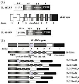
There are two alternatively spliced mRNA products which account for the existence of two distinct IL-15 isoforms, which differ only in the length of their signal peptide [9]. The two isoforms can be translated in an IL-15 precursor protein with a 48-aa long signal peptide (IL-15LSP) and a in a precursor IL-15 protein with a 21-aa short signal peptide (IL-15SSP). In both human and mouse, both alternative transcripts produce a mature IL-15 protein that differs only in the sequence of the signal peptide (Figure 3). IL-15SSP is not secreted, but rather stored intracellularly in the cytoplasm while IL-15LSP is found in the Golgi, early endosomes and the ER, possibly leading to cytokine secretion [44]. IL-15 protein is a member of the four a-helix bundle cytokine family characterized by antiparallel juxtaposed helices A, C, B, D, and 2 long end-to-end loops, loops AB and CD, which are connected by a short b-sheet packed against helices Band D. In the secondary structure there are two disulphide bonds at positions 42Cys–88Cys and 35Cys–85Cys. The C terminus of IL-15 also contains two sites for N-linked glycosylation.
Figure 3. Schematic diagram of IL-15 and IL-15Ra genes. (A) IL-15 gene consists of nine exons and eight
introns located on human chromosome 4q31 and mouse chromosome 8. Two isoforms of IL-15 containing short (21-aa) or long (48-aa) signal peptide (IL-15SSP and IL-15LSP, respectively) exist. Alternative exon 4a in human corresponds to exon 5A in the mouse IL-15 gene. (B) IL-15Ra gene consists of seven exons, and many protein isoforms were described as a result of an alternative splicing mechanism in both human and mice [6].
The IL-15 receptor (IL-15R) consists of 3 subunits, IL-15Rα chain, IL-2Rβ chain (CD122), and the common γc (CD132). The IL-15Rα subunit is unique to IL-15 but it is incapable of signalling alone. IL-2Rβ is also a receptor for IL-2, and the
common γc is shared by 2, 4, 7, 9, and 21. Unlike the 2Rα, IL-15Rα consists of 1 sushi domain which are used for ligand binding.
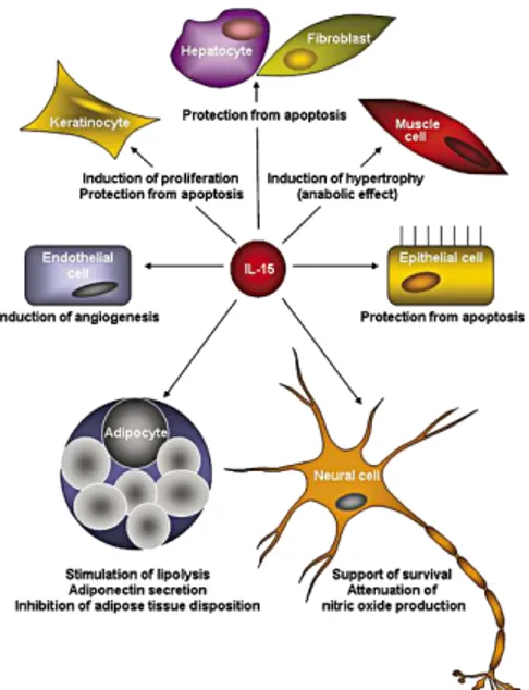
The IL-15/IL-15R system modulates functions of almost all cell populations of immune system and it is very important for their development and homeostasis. Interestingly, IL-15 is recognized as a major modulator of many different types of non-immune cells (Figure 4).
Figure 4. Functional properties of IL-15. Schematic diagram of the IL-15-mediated effects in different
non-immune cell types [9].
This is one of the most important functional difference with IL-2, whose functions are almost exclusively restricted to T cells. IL-15 mRNA is found to be highly expressed in mesenchymal stem cells and their differentiated cell types, including osteoblasts, adipocytes, endothelial cells and myoblasts. Finally, studies on fetal human brain showed IL-15 and IL-15Rα mRNA expressions in the cerebral cortex, cerebellum, hippocampus, medulla, and thalamus [19] and human cell cultures express IL-15 mRNA in microglia, astrocytes, and neuronal cell lines [20]. In the mouse brain, instead, IL-15Rα transcripts are present since P6, while IL-15 mRNA appears in hippocampal formation only since P20 [21].
The role of IL-15 in non-immune tissues is still largely unexplored.
2.3.2 Signal transduction of IL-15 signalling
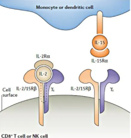
IL-15 functions through different mechanisms: the trans-presentation mechanism which is contact-dependent and consists in the presentation of the membrane-bound IL-15/IL-15Rα complex to responding cells that express IL-2/IL-15Rβ-γc complex [45]; the cis-presentation mechanism which is less common and consists
in the presentation of the membrane-bound IL-15/IL-15Rα complex to 2/15Rβ-γc complex present on the same cell surface; the binding of soluble IL-15/IL-15Rα complex to IL-2/IL-15Rβ-γc complex (Figure 5).
In vivo, IL-15 is believed to exist mainly in a complex with IL-15Rα, this could explain why the trans-presentation is the main mechanism of transduction [46].
Figure 5. The mode of interaction of interleukin-2 and interleukin-15 with the subunits of their receptors.
Interleukin-2 (IL-2) is a secreted cytokine that binds pre-formed high-affinity heterotrimeric receptors that comprise the IL-2 receptor α-chain (IL-2Rα), IL-2/15Rβ and the common cytokine-receptor γ-chain (γc). By contrast, IL-15 is a membrane-associated molecule that induces signalling at the immunological synapse between antigen-presenting cells and natural killer (NK) cells or CD8+ T cells. IL-15Rα on the surface of monocytes or dendritic cells presents IL-15 in trans to cells that express IL-2/15Rβ and γc alone, thereby allowing signalling through these complexes. The mode of interaction of interleukin-2 [46].
In T lymphocytes, IL-15, binding to its complex receptor system, initiates a cytosolic signal cascade which induces tyrosine phosphorylation of the Janus Kinase (JNKs) family members JAK-1 and JAK-3 and subsequently downstream activation of the transcription factors STAT-3 and STAT-5. Phosphorylated STATs do form homodimers, translocate in the nucleus and regulate transcription factors.
In mast cells, IL-15 utilises another receptor called IL-15RX and activates a distinct signalling pathway involving JAK-2 and STAT-5.
It is clearly demonstrated that JAK/STAT pathway is one of the most important signalling pathways involved in the regulation of neural function and that its dysregulation in brain pathologies both in human and animal models. It is involved in leptin-induced neuroprotection and in the control of food intake [47], Alzheimer's disease and memory [48].
Finally, it is known that JAK can regulate the expression or function of several neurotransmitter receptors, including NMDA, AMPA and GABA receptors [49]. These evidences suggest that IL-15 system may affect neurotransmission through its JAK/STAT effector pathway.
2.3.3 The IL-15/IL-15Rα system and behaviour
Beyond its recognized role in inflammation and diseases, several researches have recently investigated the ability of IL-15/IL-15Rα to affect behaviour. To this purpose, IL-15 and IL-15Rα KO mice have been generated. They both have a significant reduction of the peripheral and thymic NK cells, Natural Killer T (NKT), intestinal intraepithelial lymphocytes and CD8+ memory T cells which makes them more susceptible to fatal infections with microorganisms that are normally not lethal [46]. Nevertheless, the two different interruptions of IL-15 signalling result in different mouse phenotypes proving unique roles for IL-15Rα and IL-15 in vivo [50].
IL-15Rα KO mouse shows significant reduced anxiety in the “open field test” and in the “elevated plus maze test” while IL-15 KO and IL-2Rγ KO mice showed much milder changes, indicating that deficiency of a cytokine ligand or shared receptor can be partially compensated by pleiotropic cytokine pathways [10]. According to evidences from in vitro studies which demonstrate a modulation of the IL-15 during muscle function and dysfunction, the global IL-15Rα KO mice have greater cage activity during both the light and dark spans and resistance to fatigue due to a remodelling of fast skeletal muscles to a slower and more oxidative phenotype [51]. This behaviour seems to depend on the effect of IL-15Rα signalling on the hypothalamus which lead to excessive motor activation, disrupted circadian rhythm of thermoregulation, and altered metabolic phenotype [52]. This phenotype is not recapitulated by muscle-specific deficiency of IL-15Rα [53] but it is likely that it is due to an anxious state related to hippocampal functionality [12]. Hippocampus-specific IL-15Rα KO mice, in fact, show a greater peripheral locomotor activity [22] – thigmotaxis, validated as a measure of anxiogenic behaviour [54].
These evidences and the demonstration of abundance of IL-15 and its specific receptor in the hippocampus [55], suggest a role of IL-15/IL-15Rα signalling in hippocampal-dependent memory even due to its recognized involvement in hippocampal neurogenesis [56] [57].
The effect of the signalling seems limited to a specific part of memory because IL-15Rα KO mice do not have any problems in memory acquisition while show deficits in memory retention in the ”Stone T-maze test”, with a significant augment of errors [11]. Moreover, trained in “fear conditioning test”, the
IL-15Rα KO mice show the same freezing response to tone conditioning suggesting intact emotional memory but freezing to the preceding contextual fear conditioning is significantly reduced, indicating impaired contextual memory [10]. Since hippocampal activity is required for early consolidation of fear conditioning with a short trace interval [58] [59] [60], it is likely that the hippocampus is a major structural component mediating the memory deficits of the IL-15Rα knockout mice.
2.4 Hippocampal memory and GABAergic system
2.4.1 The hippocampal connectivity
The hippocampus, in the temporal lobe, is phylogenetically one of the oldest parts of the brain and forms part of the limbic system. It is one of the most studied region of the brain thanks to its relatively simple circuitation, its high plasticity and its well-recognized role in learning and memory. This plasticity is both structural and functional. For each hippocampal cell type, in fact, the size and complexity of the dendritic trees as well as the size, shape, and number of dendritic spines can change, and substantial adult neurogenesis has been demonstrated in the dentate gyrus [61].
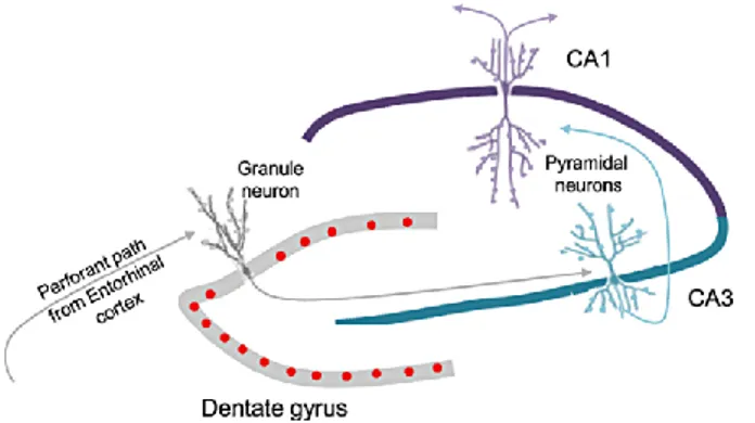
The hippocampus properly said is defined by the dentate gyrus (DG) and Cornu
Ammonis (CA) (Figure 6). While the dentate gyrus contains the fascia dentata
and the hilus, the CA is anatomically and functionally differentiated into distinct subfields named CA1, CA2 and CA3.
The hippocampal cortex has a three-layered appearance. The first layer is a deep layer, comprising a mixture of afferent and efferent fibres and interneurons. In the DG this layer is represented by the hilus, whereas in the CA regions it is referred to as the stratum oriens. More superficially there is the cell layer, which is composed of principal cells and interneurons. In the DG this layer is called the granule layer, whereas in the CA regions it is referred to as the pyramidal cell layer (stratum pyramidale). The most superficial layer is called the molecular layer (stratum moleculare) in the DG while in the CA region it is subdivided in many sublayers. In CA3, three sublayers are distinguished: the stratum lucidum, which receives input from the DG; the stratum radiatum, comprising the apical dendrites of the neurons located in the stratum pyramidale; and, most superficially, the stratum lacunosum-moleculare, comprising the apical dendrites.
The lamination in CA2 and CA1 is similar, with the exception that the stratum
lucidum is not present.
Figure 6. picture of hippocampal circuit. The main hippocampal inputs arrive through Entorhinal Cortex
(EC) to dentate gyrus. The granule cells of DG project to CA3 pyramidal neurons which, in turn, make synapses with CA1 neurons.
Most of the hippocampus’s neocortical inputs come from the perirhinal and parahippocampal cortices, through the entorhinal cortex, and most of its neocortical output is through the subiculum, which also projects back to the entorhinal cortex.
The hippocampus has an elongated and curved form which is conserved across all mammalian orders. It expands along a dorsal (septal)-to-ventral (temporal) axis in rodents corresponding to a posterior-to‑anterior axis in humans but the intrinsic circuitry is maintained throughout the long axis. Despite this conserved intrinsic circuitry, the dorsal and ventral portions have different connectivity with cortical and subcortical areas which lead to different functions.
It was proposed that the more ventral parts of the hippocampus mediate emotional responses [62] because there is denser ventral than dorsal connectivity with the amygdala [63] [64] and hypothalamic endocrine and autonomic nuclei [65]. The dorsal parts of the hippocampus seem to mediate cognitive functions, particularly memory [66]. Rodents with hippocampal lesions exhibit impairments in different kind of memories from spatial to object recognition memory [67].
3. AIM OF THE STUDY
It has long been apparent that the immune system and the brain are closely connected and that they interact in a bidirectional way but the mechanism by which this communication occurs are still poor known. The interleukin IL-15 system is considered one of the key pathways linking innate and acquired immunity and it is known to be constitutively expressed in human neural cell lines and tissues [55]. In addition, deletion of IL-15/IL-15Rα system affect hippocampal-dependent memory suggesting a role for IL15 in the regulation of synaptic transmission at the hippocampal level.
The aim of my PhD research project was to investigate how the synaptic transmission in hippocampal area was affected by modulation of IL-15/IL-15Rα system to disclose the mechanisms behind the cross-talk between the immune and the nervous system.
To this purpose I used both in vivo and ex vivo experimental approaches including electrophysiological recordings and behavioural tests, to evaluate:
• alterations in hippocampal synaptic transmission associated with deletion of IL-15/IL-15Rα signalling;
• behavioural phenotype of IL-15Rα KO mouse through the NOR task known to be related to hippocampal-dependent memory [68];
• the effects on synaptic transmission of in vivo delivery of IL-15 or IL15 application in acute slices.
Microglia represents the resident immune cell population of the brain, being one of the major sources of IL-15 in the CNS. Moreover, in previous works has been observed that IL-15Rα KO mice have hippocampal microgliosis [10].
In the attempt to get insights about the cellular pathways associated to IL-15-mediated regulation of neuronal function, I also evaluate the microglial alterations in IL-15Rα mice.
4. MATERIALS AND METHODS
4.1 Animals
Procedures using laboratory animals were in accordance with the Italian and European guidelines and were approved by the Italian Ministry of Health in accordance with the guidelines on the ethical use of animals from the European Communities Council Directive of September 20, 2010 (2010/63/UE). All efforts were made to minimize suffering and number of animals used.
Wild type (WT) C57BL/6J andB6;129X1-IL-15ratm1Ama/J (IL-15Rα KO) mice were used for electrophysiological recordings of synaptic currents and microglia isolation and data were confirmed on IL-15Rα+/+, IL-15Rα+/- and IL-15Rα
-/-littermates mice. For IL-15 treatment of brain slices and IL-15 brain delivery with osmotic pumps, adult C57BL/6J (WT) mice were used. All experiments were performed on adult male mice (6-8 post-natal weeks).
4.2 Genotyping DNA extracted from mouse tail
IL-15Rα-/- mice were mated with C57BL/6J to obtain heterozygous IL-15Rα
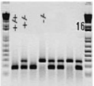
+/-mice. These heterozygous mice, in turn, were mated between them to obtain heterozygous IL-15Rα+/- and homozygous IL-15Rα+/+ and IL-15Rα-/- littermates mice. The offspring was genotyped by the polymerase chain reaction (PCR). For genotyping, PCR was used to amplify targeted regions from the IL-15Rα+/+, IL-15Rα+/- and IL-15Rα-/- gene allelesFigure 1 (Figure 7).
Figure 7. Results of PCR performed on DNA extracted from tail snips obtained in-blind from IL-15Rα
+/-mice offspring. The double band represents the IL-15Rα+/- allele, the lower band is IL-15Rα+/+ allele and the
higher band is IL-15Rα-/- allele.
Genomic DNA was extracted from tail snips (1 cm) obtained in-blind from anesthetized adult male mice before performing in-blind electrophysiological experiments and DNA was extracted through a chloroform/ethanol DNA extraction protocol. PCR reactions were performed using a 25 μl total reaction volume containing 1 μM each of forward and reverse primers, 0.2 mM each of
dNTP, 2 mM magnissium chloride, 1.5 U Taq DNA polymerase and 0.1 μg genomic DNA with a thermal cycler (iCycler, Bio-Rad). The cycling parameters were as follows: hot start 95 °C (2 min); denaturing 94 °C (30 s); annealing 58 °C (30 s); extension 72 °C (45 s) with final extension step of 7 min. Thirty-two cycles were used for these experiments. These are primer sequences used to identify the different genotypes:
Gene Forward 5’—3’ Reverse 5’—3’
IL-15Rα +/+
(171 bp) ATTGAGCATGCTGACATCCG ACTGATGCACTTGAGGCTGG IL-15Rα -/-
(280 bp) CTTGGGTGGAGAGGCTATTC AGGTGAGATGACAGGAGATC
4.3 Patch clamp recordings of CA1 pyramidal neurons from ex
vivo hippocampal brain slices
For electrophysiological recordings of GABAergic and glutamatergic transmission at CA3-CA1 synapses, acute hippocampal slices were obtained from 6-8 weeks old mice. Animals were decapitated under halothane anaesthesia. Whole brains were rapidly removed from the skull and immersed in ice-cold artificial cerebrospinal fluid (ACSF) solution containing (in mM): 87 NaCl, 75 Sucrose, 2 KCl, 7 MgCl2, 0.5 CaCl2, 25 NaHCO3, 1.2 NaH2PO4 and 10 glucose,
pH 7.3, 300–305 mOsm. The ACSF was continuously oxygenated with 95% O2 and 5% CO2 to maintain the physiological pH. Trasversal 300 μm thick slices were cut at 4°C using a Vibratome (ThermoScientific HM 650 V) and placed in a chamber filled with oxygenated ACSF containing (in mM):125 NaCl, 2 KCl, 1.2 MgCl2, 2 CaCl2, 25 NaHCO3, 1.2 NaH2PO4 and 10 glucose, pH 7.3, 300–305
mOms. Before use, brain slices were allowed to recover at least for 1 h before recording at room temperature, then transferred to a recording chamber within 1–6 h after slice preparation. All recordings were performed at room temperature on slices submerged in ACSF and perfused with the same solution in the recording chamber at a rate of approximately 2 ml/min by using a gravity-driven perfusion system.
Spontaneous (sIPSCs, sEPSCs), miniature (mIPSCs, mEPSCs) and evoked postsynaptic currents (eIPSCs, eEPSCs) were recorded from CA1 pyramidal neurons using the patch clamp technique in whole-cell configuration, which
allowed to record currents through multiple channels simultaneously, over the membrane of the entire cell. Patch clamp recordings were performed by using a Multiclamp 700B amplifier (Molecular Devices, USA). Signals were acquired (sampling 10 kHz, low-pass filtered 2 kHz) with DigiData-1440A using pCLAMP-v10 software (Molecular Devices, USA); the analysis was performed off-line using Clampfit 10 (Molecular Devices) and MiniAnalysis (Mini Analysis, Synaptosoft Fort Lee, NJ, USA). Cell capacitance was constantly monitored over the time and experiments were access resistance changed more than 20% were discarded.
Glass electrodes (3–5 MΩ) were pulled with a vertical puller (PC-10, Narishige). Pipette were filled with 148 mM Cs Methanesulfonate, 10 mM Hepes, 0.5 mM EGTA, and 2 mM Mg-ATP, Na3-GTP 0.3 mM, MgCl2 2 mM (295–300 mOsm, pH 7.2).
GABAergic outward membrane currents were recorded with the neuron clamped at 0 mV. At this voltage, Cl− mediated inhibitory events are outward currents (estimated ECl = − 80 mV) whereas excitatory currents are inward but of small amplitude as they would occur close to their reversal potential. Although it was possible to isolate sIPSCs pharmacologically, by using 20 μM DNQX plus 10 μM AP-5 to block both the AMPA and NMDA receptor components of spontaneous excitatory postsynaptic currents (sEPSCs), this antagonist mixture sometimes attenuated or occasionally completely blocked sIPSCs. This presumably reflected impediment of excitatory synaptic drive to the inhibitory interneurons that were responsible for sIPSC generation. In view of this variable effect of DNQX/AP-5 on sIPSCs, we elected to use a holding potential of 0 mV rather than pharmacological methods to separate sIPSCs from sEPSCs. The validity of this approach is supported by the observation that 100μM picrotoxin (PTX) completely eliminated all spontaneous outward current activity recorded at 0 mV (data not shown).
Miniature EPSCs/IPSCs were recorded during an initial 10 min baseline period, followed by application of TTX (0.5μM, Tocris Bioscience, Bristol, United Kingdom) for 10 min. After stabilization of TTX effect, exogenous IL-15 (10nM, PeproTech EC Ltd., London, UK) was applied for 10 min. Only data from the last 5 min of each recording epoch was analyzed to ensure that drugs had fully equilibrated.
By using the same conditions, excitatory post-synaptic currents (EPSCs) were recorded clamping the cell at − 70 mV. In a subset of experiments, the glutamatergic nature of the mEPSC recordings was confirmed at the end of the experiment by total blockade of mEPSCs by DNQX (20 μM; data not shown). For evoked post-synaptic currents, paired-pulse protocol and input/output curve, to equilibrate Cl- reversal potential at -70 mV, we used the following Cl- adjusted intracellular solution (in mM) with an addition of QX314 to block voltage-activated Na+ channels: Cs-methanesulfonate 125, CsCl 17.5, HEPES 10, EGTA 0.2, NaCl 8, MgATP 2, NaGTP 0.3, QX314-Br 2 (pH adjusted to 7.3 with CsOH). A concentric bipolar stimulating electrode (SNE-100 × 50 mm long Elektronik-Harvard Apparatus GmbH, Crisel Instruments, Rome, Italy) was positioned in the stratum radiatum to evoke eIPCSs from CA1 pyramidal neurons. Pairs of stimuli (ISI 25, 50, 100 and 700ms) were applied every 20 sec. Stimulus intensity was of amplitude about 50% of maximal amplitude, delivered through a A320R Isostim Stimulator/Isolator (WPI). Paired Pulse Ratio (PPR) was calculated as the ratio between the amplitude evoked by the second stimulus (A2) over the first (A1; A2/A1) and the amplitude of each EPSC/IPSC was measured relative to a 2 ms long baseline period starting 3 ms before stimulation. To measure the amplitude of the second peak the baseline was adjusted to zero. The stimulus intensity was adjusted accordingly to the experiment.
For input/output curves, inhibitory fibers were stimulated at increasing intensities (0.1-10 mA). Each pulse of a given intensity was repeated 6 or more times to obtain an average response.
4.4 Extracellular field recordings of CA3-CA1 circuit from ex
vivo hippocampal brain slices
For field recordings, individual hippocampal slices (350 μm) were transferred to the interface slice-recording chamber (BSC1, Scientific System Design Inc) to perform experiments within 1–6 h after slice preparation. Slices were maintained at 30 to 32 °C and constantly superfused with ACSF at the rate of 2 ml/min. Solutions were applied to the slices by a peristaltic pump. A concentric bipolar stimulating electrode (SNE-100 × 50 mm long, Elektronik–Harvard Apparatus GmbH) was placed in the stratum radiatum to stimulate Schaffer collateral fibers. Stimuli consisted of 100 μs constant current pulses of variable intensity, applied at
0.05 Hz. A glass micropipette (0.5–1 MΩ) filled with ACSF was placed in the CA1 hippocampal region, at 200–600 μm from the stimulating electrode, in order to measure orthodromically-evoked field extracellular postsynaptic potentials (fEPSP). Stimulus intensity was adjusted to evoke fEPSPs of amplitude about 50% of maximal amplitude with minimal contamination by a population spike. Evoked responses were monitored online and stable baseline responses were recorded for at least 10 min. Only the slices that showed stable fEPSP amplitudes were included in the experiments. LTP was induced by high-frequency stimulation (HFS, 1 train of stimuli at 100 Hz of 1 s duration). To analyze the time course of fEPSP amplitude, the recorded fEPSP was routinely averaged over 1 min (n = 3). fEPSP amplitude changes following the LTP induction protocol were calculated with respect to the baseline.
The paired-pulse ratio (PPR) was measured from responses to two synaptic stimuli at 50 ms inter-stimulus interval. The PPR was calculated as the ratio between the fEPSP amplitude evoked by the second stimulus (A2) over the first (A1; A2/A1).
fEPSPs were recorded and filtered (low pass at 1 kHz) with an Axopatch 200 A amplifier (Axon Instruments, CA) and digitized at 10 kHz with an A/D converter (Digidata 1322 A, Axon Instruments). Data acquisition was stored on a computer using pClamp 9 software (Axon Instruments) and analyzed off-line with Clampfit 10 program (Axon Instruments).
4.5 Isolation of CD11bþ cells and extraction of total RNA
Brains of IL-15Rα KO or WT mice were cut into small pieces and single-cell suspension was achieved by enzymatic digestion in trypsin (0.25 mg mL-1), in Hank’s balanced salt solution (HBSS) and mechanical dissociation using a wide-tipped pipette. Cell suspension was applied to a 30-mm cell strainer and immediately processed for MACS Micro Bead separation. The CD11bþ cells were magnetically labelled with CD11b Micro Beads. The cell suspension was loaded on a MACS Column (Miltenyi Biotec) placed in the magnetic field of a MACS Separator and a negative fraction was collected. After removing the magnetic field, CD11bþ cells were eluted as positive fraction. Vitality and purity of CD11bþ cells were assessed using flow cytometry (FACS). On sorting the positive and negative fractions, total RNA was isolated with the RNeasy Mini Kit
and processed for RT–PCR. The quality and yield of RNAs were verified with Ultraspec2000 UV/Visible (Pharmacia Biotech).
4.6 RT–PCR
Samples were lysed in Trizol reagent for isolation of RNA. Reverse transcription reaction was performed in a thermocycler using IScript TM RT Supermix (Bio-Rad) under the following conditions: incubation, 25°C, 5min; reverse transcription, 42°C, 30 min; inactivation, 85°C, 5min. RT–PCR was carried out in a I-Cycler IQ Multicolor RT–PCR Detection System using SsoFast EvaGreen Supermix (Bio-Rad). The PCR protocol consisted of 40 cycles at 95°C, 30 s and at 60°C, 30 s. For quantitative analysis the comparative Threshold Cycle (Ct) method was used, while normalizing to Ct value of GAPDH in the same sample. Relative quantification was performed using the 2-ΔΔCt method [68] and expressed
as fold changes in arbitrary values. Primers used are the following ones:
Gene Forward 5’—3’ Reverse 3’—5’
gapdh TCGTCCCGTAGACAAAATGG TTGAGGTCAATGAAGGGGTC
arg1 CTCCAAGCCAAAGTCCTTAGAG AGGAGCTGTCATTAGGGACATC
fizz CCAATCCAGCTAACTATCCCTCC ACCCAGTAGCAGTCATCCCA
ym1 CAGGTCTGGCAATTCTTCTGAA GTCTTGCTCATGTGTGTAAGTGA
inos ACATCGACCCGTCCACAGTAT CAGAGGGGTAGGCTTGTCTC
il1b GCAACTGTTCCTGAACTCAA TATCTTTTGGGGTCCGTCAA
tnfa GTGGAACTGGCAGAAG GCCATAGAACTGATGAGA
bdnf TGAGTCTCCAGGACAGCAAA TGTCCGTGGACGTTTACTTCT
gat2 GGGTATTACATCGGGCA ACACCCCGGATCAGAA
gabbr2 AGCAAGCGTTCGGGTGTA TGGCGTTGAGGATGATTCT
abat ACACTAAATCCAACGAGC AAGGGCGGAGACTATG
4.7 IL-15 delivery in hippocampus of WT mice with
micro-osmotic pumps
Eight-week-old male C57BL/6J mice were anaesthetized with chloralhydrate (400 mg kg-1, i.p.), placed in a stereotaxic head frame and implanted with an osmotic
according to the atlas). A cannula was implanted through a hole and was sealed with dental cement before connecting to the pump. The pump was placed into a subcutaneous pocket in the dorsal region. The pumps were filled up with vehicle (PBS) or IL-15 (133 ng mL-1). Before surgery, the pumps and the tubes were incubated at 37°C overnight in a sterile saline. The experiment continued for 7 days after the pump implantation. IL-15 doses were selected considering the ex
vivo experiments.
4.8 Novel object recognition (NOR)
If mice are presented familiar and novel objects, they will explore more the novel objects. This typical behavior was exploited to evaluate recognition memory linked to hippocampus [69]. Experimentally naïve, littermate, age-matched, maleWTand IL-15Rα KO mice, were subjected to the test.
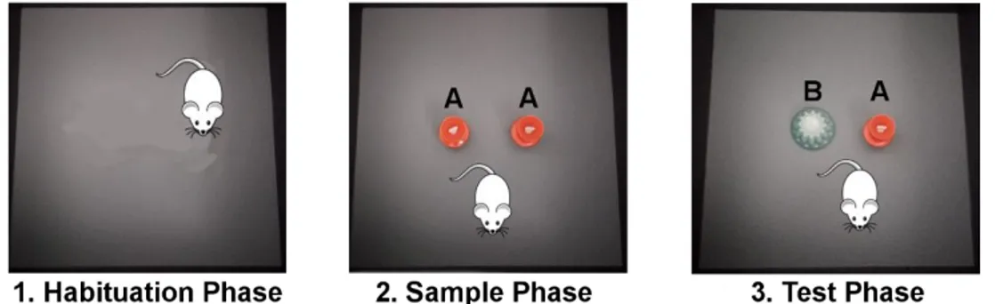
The NOR task protocol lasts 3 days (Figure 8).
Figure 8. Picture of the three phases of NOR task. In the habituation phase the animal can freely explore
the box for 10 minutes in absence of any stimulus. In the sample phase the animal can explore two copies of the same object A for 10 minutes. In the test phase, 1 or 24 hour later, the animal can explore the familiar object A or the novel object B for 5 minutes.
A total of three objects of different colours were used for the experiments. They were made of plastic or glazed ceramic and there were two copies of each object. All objects were sufficiently heavy so that they could not to be displaced by the mice. The role (familiar or novel) as well as the relative position of the two stimulus objects were counterbalanced and randomly permuted for each experimental animal. The open field arena and the stimulus objects were cleaned thoroughly between trials to ensure the absence of olfactory cues.
The test arena was a square, gray wooden box (40 × 40 × 40 cm). In the test room a camera was suspended from the ceiling directly above the center of the arena and was connected to a computer, located in an adjacent room, which used Ethovision XT (Noldus) to record the trials.
In the first day (habituation phase), all the animals were habituated to the open field arena by allowing them to freely explore it 10 min in the absence of stimulus objects. This procedure was used to reduce the possibility of context exploration interfering with object exploration
On day 2 (sample phase), mice were placed in the open field containing two identical objects and left to freely explore them for 10 min. The test phase was performed either 60 min (to analyze short-term memory or STM) or 24 h after the sample phase (to evaluate long-term memory or LTM). In the test phase one of the objects was randomly replaced with a novel object and mice were reintroduced into the open field for an additional 5-min period.
Exploration was defined as entering with the nose and/or forepaws in a defined square surrounding the stimulus object (when the animal is at a distance of less than 2cm from the object). The time spent exploring each object was scored by an observer who was blind with respect to the genotype of the animal and the object allocation during the test trial (i.e., familiar or novel). The exploration was expressed as a percentage of the total exploration time in seconds and all test trials were scored for the whole 5-min duration. Student’s t-test was used to analyze the data.
Discrimination Index (DI) was computed through the following formula where EB
and EA represent the exploration time of the novel and familiar object
respectively:
𝐷𝐼 =(𝐸𝐵− 𝐸𝐴) (𝐸𝐵+ 𝐸𝐴)
4.9 Statistical analysis
All data are presented as mean ± SEM. Origin 6 and Origin 8 (OriginLab Corporation, Northampton, Massachusetts, USA) softwares were used for statistical analysis of electrophysiological data. Statistical significances were determined by paired and unpaired t-test, one-way and two-way ANOVA, Kolmogorov-Smirnov test as indicated. P values less than 0.05 were considered significant. Levels of significance were set as *p ≤ 0.05; **p ≤ 0.01; ***p ≤ 0.001. In the legend, the number of cells and the number of animals are expressed together in the form n.cells/n.animals (i.e. 15/5 means 15 cells/5 animals).
5. RESULTS
5.1 IL-15Rα KO mice show altered inhibitory synaptic
transmission in CA1 hippocampal neurons.
To investigate the alterations in synaptic transmission associated with deletion of IL-15/IL-15 Rα pathway, I recorded glutamatergic and GABAergic transmission in CA1 pyramidal neurons from acute hippocampal slices obtained from WT and IL-15Rα-/- mice (6th-8th postnatal week). I performed in-blind experiments on
IL-15Rα+/+, IL-15Rα-/- littermates. Since IL-15Rα+/+ and WT mice showed the same
electrophysiological properties, for further experiments WT mice were used as controls (Figure 9).
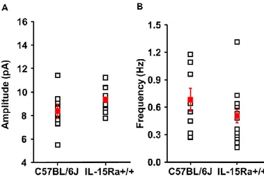
Figure 9. Post-synaptic currents recorded in CA1 pyramidal neurons obtained from C57BL/6J (WT) and IL-15Rα+/+ mice show comparable properties. The two genotypes are similar thus for some experiments C57BL/6J mice were used as controls of IL-15Rα-/- mice.
Whole-cells recordings of excitatory and inhibitory postsynaptic currents were performed at -70 mV and +0 mV respectively (with an intracellular solution of CsMetSO3), and both the frequency and the amplitude of synaptic events were
analyzed.
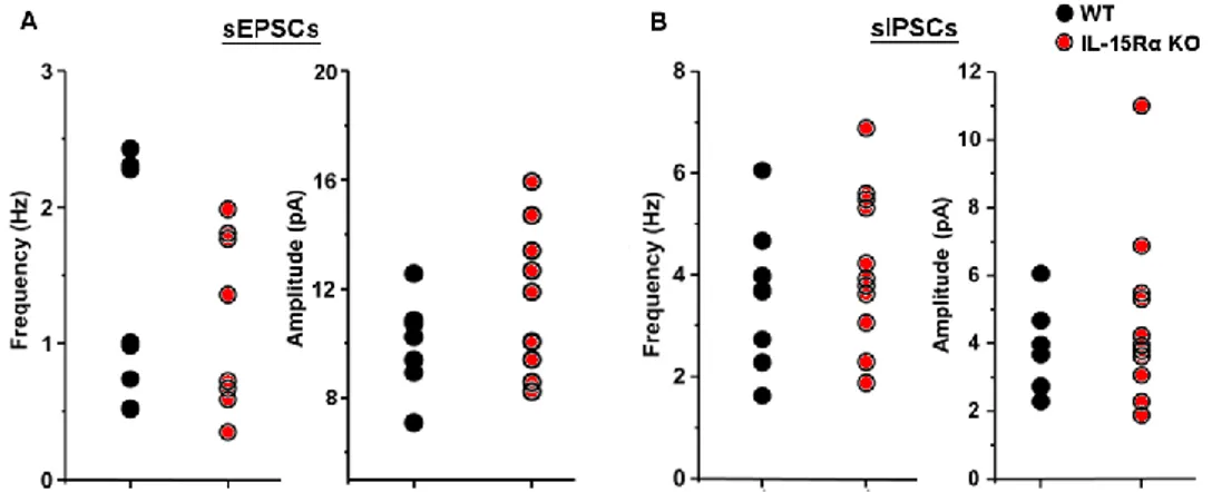
The recordings of spontaneous activity showed no differences in both spontaneous excitatory (sEPSCs) and inhibitory (sIPSCs) transmission between WT and IL-15Rα KO mice (ANOVA One-way, Figure 10). The analysis of rise and decay times showed that there were not changes in the kinetics of synaptic events (data not shown).
Figure 10. IL-15RαKO mice show normal spontaneous synaptic activity in CA1 neurons. Spontaneous
excitatory and inhibitory post-synaptic currents recorded in CA1 hippocampal neurons of IL-15Rα KO (n = 10/5) and WT (n = 8/6) mice. The mean values were computed analysing 5 minutes of stable recording at -70 mV and at 0 mV, respectively. For sIPSCs recording DNQX and AP-5 were added to bath solution to block glutamatergic transmission. A) No significant differences were found in sEPSCs between the two genotypes neither in frequency (left; WT = 1.35 ± 0.29Hz, KO = 2.41 ± 0.87Hz) or amplitude (right; WT = 10.00 ± 0.57pA; KO = 11.49 ± 0.84pA). B) Spontaneous inhibitory activity is not different in IL-15Rα KO (n = 12/4) and in WT mice (n = 9/3) regarding both frequency (left; WT = 3.63 ± 0.44Hz, KO = 4.76 ± 0.70Hz) and amplitude (right; WT = 15.73 ± 1.01pA, KO = 16.10 ± 1.44pA).
To investigate whether differences in synaptic transmission between WT and IL-15Rα KO mice were masked by the occurrence of action potential, I added TTX (a blocker of voltage gated sodium channels) to the bath solution and recorded miniature post-synaptic currents (mIPSCs and mEPSCs). In that condition I observed that the frequency of mIPCSs recorded in hippocampal slices obtained from IL-15Rα-/- mice was statistically increased compared to WT mice (p=0.034, one-way ANOVA; Figure 11D). By contrast the mIPSCs amplitude was identical between the two genotypes (p=0.77, one-way ANOVA; Figure 11E) suggesting a presynaptic alteration of the release probability of GABA at this synapse rather than modification of post-synaptic receptors, more associated with amplitude changes.
Figure 11. The frequency of mIPSCs in CA1 pyramidal neurons is significantly increased in IL-15Rα
-/-mice. A, B) Post-synaptic currents of CA1 pyramidal neurons were recorded in whole-cell patch clamp
configuration. The recordings were made from acute hippocampal slices obtained from IL-15Rα+/+ (n = 13/4)
and IL-15Rα-/- mice (n = 8/2). C) Representative mIPSCs recordings of hippocampal neurons of the two
different genotypes. D) Normalized cumulative probability curve of inter event interval (IEI) (calculated on 2 minutes recording for each cell) was significantly shifted towards the left for IL-15Rα-/- compared to
IL-15Rα+/+ (Kolmogorov-Smirnov test, p=0.049).(inset; mean mIPSCs frequency IL-15Rα-+/+ = 3.48 ± 0.37Hz,
IL-15Rα-/- = 4.68 ± 0.45Hz, p=0.034). E) Normalized cumulative probability of mIPSCs amplitudes. The
mean amplitude of mIPSCs is not affected by the different genotype (inset; IL-15Rα-+/+ = 15.60 ± 0.52pA,
IL-15Rα-/- = 15.29 ± 0.37pA, p=0.77).
As observed for the sEPSCs, no differences were found in glutamatergic mEPSCs frequency or amplitude between the two genotypes (p>0.05, one-way ANOVA;
Figure 12). These results showed that IL-15Rα deletion altered only GABAergic
transmission, possibly through a presynaptic mechanism.
Figure 12. Recordings of mEPSCs do not show any significant differences between IL-15Rα-+/+ and
IL-15Rα-/- mice. A) Representative recordings of mEPSCs of CA1 neurons from acute hippocampal slices obtained from IL-15Rα-+/+ (n = 15/4) and IL-15Rα-/- mice (n = 5/2). The means were computed on 5 minutes
recordings. B) The mean amplitude of mIPSCs is not affected by the different genotype (inset; IL-15Rα-+/+ =
9.36 ± 0.21pA, IL-15Rα-/- = 9.12 ± 0.25pA). C) The mean frequency calculated of mIPSCs was not
significantly different between IL-15Rα-+/+ and IL-15Rα-/- (IL-15Rα-+/+ = 0.50 ± 0.07Hz, IL-15Rα-/- = 0.41 ±
0.03Hz; p=0.48).
5.2 IL-15Rα KO mice show increased GABA release in CA1
area
To deep investigated alterations in the probability of GABA release at inhibitory synapses I recorded the evoked inhibitory postsynaptic currents (eIPSCs) in CA1
pyramidal neurons by using a paired-pulse protocol to stimulate inhibitory presynaptic fibers in the stratum radiatum (Figure 13).
Figure 13. Electrical stimulation of Schaffer collaterals and recordings from CA1 pyramidal neuron. A)
Anatomical location of acute hippocampal slices. B) Placement of the patch pipette in cornu ammonis 1 (CA1) and stimulating electrode on stratum radiatum; Schaeffer collaterals (SC); dentate gyrus (DG); cornu ammonis 3 (CA3). C) Paired Pulse Ratio calculation.
The paired pulse ratio (i.e. the ratio between the second and the first pulse, PPR) is related to Pr (the probability of neurotransmitter release). Synapses with low value of Pr show paired pulse facilitation (PPF) in paired stimulation protocol. This is widely held to be due to residual Ca2+ in the presynaptic terminal from the first action potential adding to the Ca2+ influx from the second pulse [70] [71]. The larger presynaptic Ca2+ leads to facilitated or increased neurotransmitter release on the second ‘pulse’ or action potential. By contrast, inhibitory synapses usually show paired pulse depression (PPD), with the first peak bigger than the second. PPD relies on the activation of presynaptic GABAB receptors and tends to
peak at 100-300 ms [72] [73]. I found that in IL-15Rα KO the paired pulse ratio was significantly lower at inter-stimulus interval (ISI) of 25, 50 and 100 ms and it came back to control values at 700ms ISI (25ms p=0.019, 50ms p=0.048, 100ms p=0.057, 700ms p=0.33; one-way ANOVA; Figure 14B). These results confirm that IL-15Rα KO mice have a higher probability of GABA release in CA1 hippocampal area.
Figure 14. Paired pulse ratio measured at different inter stimulus intervals. A) Representative recordings of
PPD at ISI = 50 ms in WT and IL-15Rα KO mice. B) At shorter ISIs there is a significant reduction of mean PPR in IL-15Rα KOmice (13/4) compared to WT (14/5) (25ms ISI: WT = 0.58 ± 0.04, KO = 0.42 ± 0.04; p=0.019, ; 50ms ISI: WT = 0.76 ± 0.04, KO = 0.55 ± 0.03; p=0.048) At ISI = 100 ms there is a reduction of PPR in IL-15Rα KO which is at the limits of significance (WT = 0.79 ± 0.05, KO = 0.66 ± 0.04; p=0.057) whereas at 700 ms PPR values were not different (WT = 0.82 ± 0.06, KO= 0.73 ± 0.06; p=0.33).
To investigate whether the increased GABA neurotransmission is due to increased inhibitory connectivity I also analysed the input output curve (I/O curve) by measuring the amplitude of eIPSCs elicited by stimuli of graded intensities. The inhibitory post-synaptic currents increased similarly in the two genotypes at increasing stimulation intensity (Figure 15), suggesting that the strength of inhibitory input onto CA1 pyramidal neurons is not affected by the lack of IL-15/IL-15Rα signalling.
Figure 15. The strength of inhibitory input onto CA1 pyramidal neurons is similar between WT and IL-15Rα KO mice. The normalized I/O curve of IPSCs recorded in CA1 neurons of WT (n = 8/3) and IL-IL-15Rα
KO mice (n = 12/5) is similar in the two genotypes.
5.3 IL-15Rα KO mice show normal CA3-CA1 glutamatergic
transmission with no alterations in short- (STP) and long-term
potentiation (LTP)
Since the IL-15Rα KO mice show memory impairments REF, I investigated the hippocampal plasticity through experiments of STP and LTP induction in
CA3-CA1 circuit. Extracellular field recordings (fEPSP) of glutamatergic transmission in hippocampal brain slices obtained from IL-15Rα KO and WT mice were performed (Figure 16A). The short-term potentiation and the long-term potentiation were similar in WT and IL-15Rα KO mice (Figure 16B).
This finding agrees with the results on sEPSCs and mEPSCs, suggesting that the effect of IL-15/15Rα signalling disruption is confined to the inhibitory system.
Figure 16. Extracellular field recording of excitatory transmission in CA3-CA1 circuit show no differences between IL-15Rα+/+ and IL-15Rα-/- mice. A) Picture of stimulation and recording electrodes position on hippocampal slice to record fEPSPs. B) LTP was induced by a high frequency stimulation protocol (HFS, 1 train of stimuli at 100 Hz of 1 s duration) in hippocampal slices obtained from IL-15Rα+/+ (n = 6/4) and
IL-15Rα-/- mice (n = 5/2). C) PPR was measured from responses to two stimuli at 50ms inter-stimulus interval
and was calculated as the ratio between the fEPSP amplitude evoked by the second stimulus over the first.
5.4 IL-15Rα KO mice are impaired in Novel Object Recognition
(NOR) task
To evaluate whether the alterations in hippocampal synaptic transmission observed in the electrophysiological recordings were correlated to a behavioural deficit, the mice have been subjected to the “novel object recognition task” (NOR). We have choosen this test because it is the best-known task to assesses recognition memory and has been shown that hippocampus, mainly the dorsal region, is essential for NOR memory formation and for object recognition [69]. Results show that IL-15Rα KO mice have a significant lower discrimination index in both the short-term memory (STM, 1h after sample phase) and long-term memory (LTM, 24h after sample phase) tests compared to controls (Figure 17). This evidence agrees with previous behavioural data [11] [10] [12] and with our findings that IL-15/IL-15Rα system affects hippocampal functionality.
Figure 17. IL-15Rα KO mice are impaired in novel object recognition test. IL-15Rα KO mice (n = 5)
showed a significant lower Discrimination Index (DI) compared to WT mice (n = 5) both 1 hour after sample phase (Short-Term Memory or STM, WT DI = 0.38 ± 0.09, KO DI = 0.15 ± 0.04, p=0.046, t-test) and 24 hours after sample phase (Long-Term Memory or LTM, WT DI = 0.22 ± 0.09, KO DI = -0.38 ± 0.21, p=0.045, t-test).
5.5 Microglia extracted from hippocampi of IL-15Rα KO mice
show an altered mRNA expression
IL15 and IL-15Rα are expressed by several cell populations in the brain ranging from immune cells to glia and neuronal cells. In order to shed light on the cellular pathways associated to IL-15-mediated regulation of neuronal function I focused on microglia since these cells are the only resident macrophages of CNS and previous studies have shown that IL-15Rα KO mice have mild microgliosis [50].At first I investigated the phenotype of microglial cells isolated from the hippocampi of IL-15Rα KO and WT mice performing RT-PCR to analyse the expression level of some inflammatory genes such as inos, TNFα, IL-1β, ym-1, arg-1, fizz-1. The results showed that microglia cells from IL-15Rα KO mice do not have different levels of expression compared to WT mice. Not surprisingly, iNOS was found to be down regulated in KO mice (p=0.049, t-test), as already described following the IL-15 signalling blockade [74]. This result suggests that the absence of IL-15/IL-15Rα signalling does not affect the state of activation of microglia (Figure 18).
Figure 18. mRNA expression levels of inflammatory genes in hippocampal microglial cells isolated from IL-15Rα KO and WT mice. Microglia isolated from IL-15Rα KO does not show a specific anti- or
![Figure 2. Several cytokines have been recognized as modulators of synaptic transmission [5]](https://thumb-eu.123doks.com/thumbv2/123dokorg/2894172.11457/13.892.190.725.308.631/figure-cytokines-recognized-modulators-synaptic-transmission.webp)