PhD course in Immunological, Hematological and Rheumatological Sciences XXXI° cycle
Curriculum: Clinical Immunology Coordinator: Prof. Angela Santoni
PhD THESIS
Immunobiology of Essential Mixed Cryoglobulinemia
Tutor: Prof. Massimo Fiorilli PhD Student: Stefania Colantuono
INDEX 1. INTRODUCTION
1.1 Cryoglobulinemia: physical properties of cryoglobulins, diagnosis and
laboratory evaluation 3
1.2 Non-HCV infectious and Essential Mixed Cryoglobulinemia 5
1.3 The complement receptor type II: CD21 8
1.4 CD21low B cells in mixed cryoglobulinemia and in other disorders 10
1.5 Anergy of CD21low B cells in HCV MC 12
2. OBJECTIVE OF THE STUDY 14
3. MATERIALS AND METHODS 3.1 Essential Mixed cryoglobulinemia patients 15
3.2 Cells and Immunophenotyping 15
3.3 PhosFlow assay for pERK 16
3.4 Apoptosis assay 17
3.5 Cell proliferation assay 18
3.6 Statistical analysis 19
4. RESULTS 4.1 Patients’ characteristics 20
4.2 Phenotypic Features of B cells in Essential mixed cryoglobulinemia 21
4.3 CD21low B cells expanded in EMC resemble CD21low B cells in HCV-MC 23
4.4 CD21low B cells of EMC patients proliferate poorly in response to TLR9 ligation 25
4.5 CD21low B cells of EMC patients display dysregulated pERK signaling and are prone to apoptosis 26
5. DISCUSSION 28
1. INTRODUCTION
1.1 Cryoglobulinemia: physical properties of cryoglobulins, diagnosis and laboratory evaluation
The term “cryoglobulinemia” refers to a clinical condition characterized by the presence of immunoglobulins in serum that precipitate, in vitro, at temperature below 37°C and redissolve after rewarming; these cold-precipitable immunoglobulins were first identified by Wintrobe and Buell in 1933 and subsequently named “cryoglobulins” by Lerner and coworkers in 1947 [1]. Interestingly, cryoglobulins are not necessarily a sign of disease since healthy people may have low concentrations of them and polyclonal cryoglobulins may be transiently detected during infection [2].
In 1974, Brouet et al [3] proposed a classification of cryoglobulinemia that is still widely used due to its good correlation with clinical features and associated diseases:
- monoclonal cryoglobulinemia (type I)
- mixed cryoglobulinemia (type II and type III)
In type I cryoglobulinemia, which account for 10-15% of the total, cryoglobulins are composed of a single monoclonal cryoprecipitable immunoglobulin (mainly IgG or IgM and rarely IgA or free immunoglobulin light chains [4] produced by the monoclonal expansion of a B cell clone that may be indolent in about 40% of patients (monoclonal gammopathy of unknown significance [MGUS]) or overtly malignant in the remaining 60% (Waldenstrom macroglobulinemia [WM], multiple myeloma [MM] or chronic lymphocytic leukemia [CLL]) [5]. In type II cryoglobulinemia, which account for almost 50-60% of cases, cryoglobulins are characterized by a polyclonal component, mainly IgG, mounting kappa or lambda chains, and a monoclonal IgM (rarely represented by IgA or IgG) endowed with rheumatoid factor (RF) activity. Most IgM react with both intact IgG and the F(ab)2 fragment and also with the Fc fragment of autologous IgGs, and these two types of molecular interaction confer greater stability to cryoprecipitating IgM-IgG immune-complexes. This type of cryoglobulinemia is usually associated with HCV infection in up to 90% of patients [6]; different causes include other infections (mainly HIV and HBV), connective tissue diseases (CTDs) and lymphoproliferative disorders. Approximately 10% of patients have no identifiable cause and in this case it is referred to as “essential mixed cryoglobulinemia” [7]. Type III cryoglobulinemia (25-30% of cases) is characterized by
polyclonal IgM with RF activity and polyclonal IgG and it’s seen in CTDs or secondary infection (mainly HCV).
However, there are unusual cryoglobulins showing a micro-heterogeneous composition, whose immunochemical structure cannot be fitted into any of the categories described above. Using sensitive methods, such as immunoblotting or two dimensional polyacrylamide gel electrophoresis, some authors described MC formed by oligoclonal IgMs and traces of polyclonal immunoglobulins and their inclusion in the Brouet classification as type II-III variant has been proposed [8,9]. The IgM micro-heterogeneity, identified in about 10% of cases, has been considered as possible intermediate state in the transition from type III to type II MC; this serological condition and the transformation from polyclonal to oligoclonal and finally monoclonal IgM-RF fraction, reflects the continuous B cell clones expansion [9].
The process of cryoprecipitation is not entirely understood and probably differs between type I and II/III. In type I, the monoclonal component undergoes crystallization and aggregation, which is dependent on temperature and concentration [10]. Although the definition of cryoglobulins is precipitation at cold temperatures, this process can occur at room temperature at high cryoglobulin concentrations [1]: this fact probably explains why distal extremities (lower temperatures) and kidneys (increase in concentration as a result of ultrafiltration) are major sites of pathology. In type II/III cryoglobulinemia, cryoprecipitation takes place in the setting of immune complex formation between polyclonal IgG and IgM with RF activity and complement fixation. Cryoprecipitation cannot be induced by the IgM or IgG components separately and requires specific antigen-avidity IgG molecules [11]: this aspect emphasizes the unique properties of cryoglobulins and explains the high incidence of HCV as a cause. Although the real mechanism of in vitro cryoprecipitation is rather obscure, non-specific Fc-Fc interactions might explain the self-aggregation of some Igs. Modifications of the primary structure of heavy and light chains are responsible, at least in part, for the different solubility of cryoglobulin and many studies on chemical analysis of cryoglobulins revealed a reduced concentration of sialic acid or a reduced amount of galactose in the Fc portion of the Ig. Furthermore, the solubility of proteins also depends on various factors such as concentration and temperature. Decreasing the temperature causes changes in the steric conformation of the molecules exposing non-polar residues resulting in solubility loss. Another factor is the pH of the solution, which may affect secondary and tertiary structures of immunoglobulins [12].
typical clinical manifestations; false negative results may occur as a consequence of improper sample handling. Samples should be transferred and centrifuged at 37°C to avoid precipitation before serum extraction. In type I, precipitation at 1 to 4 °C usually occurs within hours; samples should be stored for 7 days because precipitation can be delayed in the mixed types [13]. When cryoglobulins are detected, the measurement of the cryocrit, defined as the relative volume of the precipitate as a percentage of the total serum volume, should be reported. The correlation between the cryocrit and disease manifestation is poor [14,15], although higher cryocrit has been reported to increase the likelihood of symptomatic disease [7]; in a few studies the cryocrit was prognostic but overall, its use should be reserved for diagnosis because there is a poor correlation between the cryocrit and the response to treatment [13]. In normal human sera small amounts of cryoprecipitable immunoglobulins are frequently detected, reflecting specific molecular interactions between Ig molecules [16]. On the contrary, the cryoprecipitation process occurring in cryoglobulinemia is caused by intrinsic characteristics of both monoclonal and polyclonal IgM components and can also be caused by interaction among single components of the cryoprecipitate.
1.2 Non-HCV infectious and Essential Mixed Cryoglobulinemia
Before the discovery of HCV in 1989, mixed cryoglobulinemia (MC) vasculitis was considered an essential or idiopathic disease in most cases, but the systematic HCV screening performed in these patients revealed that most of the patients were positive for HCV infection markers.
Hepatitis B virus (HBV) is probably the cause of some cases of MC [17] and a recent study has attributed some sporadic cases to other viral, bacterial and parasitic diseases [18]. In this cohort of 18 patients affected by non HCV-infectious cryoglobulinemia, cytomegalovirus, Epstein Barr virus, parvovirus B19 and human immunodeficiency virus, pyogenic bacterial infections, parasitic infection, leprosy and candidiasis were found as causative agents.
Curiously, significant level of cryoglobulins has been found as late as six months after the onset of the symptoms of acute Chikungunya virus infection [19] and also in some patients with solid cancers [20].
Among the patients with noninfectious MC about half of cases are associated with distinctive conditions such as lymphoproliferative disorders or connective tissue diseases, while the remaining cases are classified as idiopathic or essential MC (EMC) [18, 22, 26]. In 2012, Terrier et al. analyzed data from 242 patients with non-infectious mixed cryoglobulinemia included in the French multicenter CryoVas survey [22] to describe the spectrum of clinical presentation and etiologic factors. The survey confirmed that connective tissue diseases, in particular primary Sjögren's syndrome, and hematologic disorders remain the main causes of non-infectious MC.
Other studies too, showed as autoimmune diseases are a major source of non-infectious cryoglobulinemias.
Baimpa et al. conducted a retrospective study of 536 consecutive patients with primary Sjogren syndrome to assess the prevalence of hematologic abnormalities and their associations with various disease manifestations [23] and revealed that the presence of monoclonal serum proteins, and cryoglobulinemia correlated significantly with the presence of extraglandular symptoms such as palpable purpura, lymphadenopathy, and splenomegaly. Moreover cryoglobulinemia, as neutropenia, at diagnosis were significantly associated with an increased risk of lymphoma development, similarly to the results previously observed by Brito-Zeron et al. in 2007, who identified that presence of hypocomplementaemia and/or cryoglobulins at diagnosis, together with vasculitis and severe involvement in parotid scintigraphy, were the main prognostic factors for an adverse outcome in their cohort of patients with primary SS [24] and those of Tzioufas at al. in 1996, who identified a significant correlation between the presence of mixed monoclonal cryoglobulinemia and a higher prevalence of autoantibodies to Ro/SS-A and La/SS-B, and the determination of crioglobulins being useful as a laboratory predictive factor for lymphoma development in their SS patients [25].
In 2017, The Italian Group for the Study of Cryoglobulinaemias (GISC) published the results of a prospective observational study to investigate the clinical and laboratory patterns of a large Italian cohort of HCV-unrelated MCV and the factors influencing its outcome [26].
The findings of the study confirmed that, although rare, HCV-unrelated cryoglobulinemia was not a benign disease. Similarly to HCV-associated cryoglobulinemia, it was more frequent in females aged > 60 years. Palpable purpura was observed in almost 70% of patients, together with arthralgias, fatigue and sicca syndrome as the most common
symptoms. Peripheral neuropathy (PN) was frequently part of cryoglobulinemic syndrome, and sometimes, the first sign of cryoglobulinemia. Fewer than 3% of the patients were affected by hyperviscosity syndrome. In comparison with HCV-related MC, the historical GISC series and the French HCV-negative series [22], the median cryocrit levels observed in this cohort at the time of enrollment was relatively low.
The clinical and laboratory profile of SLE-related MC, was found significantly different from those of other groups; the cessation of cryoglobulins production observed during the first year after enrollment being, probably, due to the fluctuating nature of auto-antibody production in SLE but also to SLE treatment. Cryocrit and RF concentrations tended to be higher in the patients with RA or mixed connective tissue disease than in the others.
Because of the influence of each condition on clinical severity and stability of cryoglobulins production over the time, the clinical and laboratory profiles was found very different in the different groups of patients and the outcomes of vasculitis being closely depending to the underlying condition and treatment.
Essential Mixed cryoglobulinemia (EMC) is defined as those cases with mixed cryoglobulinemia that are not associated to any known causative disease capable of inducing cryoglobulin production [21], and these account for approximately 10% of patients with cryoglobulinemia.
The pathogenesis of ECM is still unknown. Type II monoclonal mixed cryoglobulins are found in ~50% of EMC cases [18, 22, 26], suggesting an underlying benign lymphoproliferative disorder similar to that driven in HCV-MC by protracted antigenic stimulation by the virus.
In the absence of any (known) exogenous antigenic stimulus, it is likely to hypothesize that a chronic stimulation by different (auto)antigens may lead to cryoglobulins production in these cases.
1.3 The complement receptor type II: CD21
Complement receptor type II (CR2, CD21) is a 145-kDa glycosylated single polypeptide chain consisting of a long extracellular domain of 15-16 short consensus repeat sequences, a transmembrane region and a short cytoplasmic domain [27,28]; it is expressed on B cells [29], where its levels vary depending on the maturation stage of the cell, follicular dendridic cells, thymocytes and a subset of peripheral T cells [30,31]. Ligands of CD21 include the complement fragments C3d, C3dg and iC3b, that are covalently bound to the target antigens [32,33] and, in addition, it has been proposed to bind ligands such as the Epstein-Barr Virus (EBV) envelope glycoprotein gp350/220, the low affinity Fc-receptor for IgE (FceRII o CD23) and the cytokine interferon-alfa (IFN-a) [34].
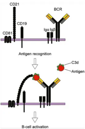
On B cell surface, CD21 forms a complex together with CD19 and CD81 (TAPA-1) [35,36], which functions as a co-receptor to the BCR. Upon simultaneous binding of complement-tagged antigens by CD21 and the BCR, the threshold for B cell activation is reduced [32,37]. In this scenario, CD21 acts as a bridge between the co-receptor complex and the BCR resulting in a co-ligation that induces the phosphorylation of the cytoplasmic tail of CD19 by BCR-associated tyrosine kinases and amplifies downstream signaling (Fig. 1) [32,38].
Figure 1. The BCR and its co-receptor
(Upper) The co-receptor complex, CD81, CD21 and CD19, and the membrane bound BCR together with its signal transduction molecules Iga and Igß. (Lower) Upon simultaneous binding of complement-tagged antigens by CD21 and the BCR, the threshold required for B cell activation is reduced. Under these circumstances, CD21 acts as a bridge between the co-receptor complex and the BCR. The opsonizing complement fragments are C3d, C3dg or iC3b [50].
CD21 plays an important role in the selection for high-affinity B cells as well as the development and maintenance of B cell memory [39]. Although the BCR co-receptor function predominates, CD21 also mediates effects independent of the BCR including the induction of the transcription factor NF-kB, the production of interleukin-6 (IL-6) and the internalization of the antigen [32,40].
CD21 also exists in a soluble form, as the receptor can be shed from the plasma membrane by proteolytic cleavage in a short consensus repeat; indeed, it has been shown that CD21 is constantly shed from peripheral blood B lymphocytes and that its levels in blood are increased after cell activation. The amounts of soluble CD21 in blood are also increased in diseases such as common variable immunodeficiency (CVID), B-CLL and malignancies associated with EBV [41], whereas the levels are decreased in autoimmune diseases [42].
Increased frequencies of a B cell subset with low or no expression of CD21 (CD21-/low B cells, below called CD21low) have been described in several diseases characterized by a chronic immune stimulation: HCV-associated MC [43], HIV infection [44], malaria [45], CVID [46], rheumatoid arthritis (RA) [47], systemic lupus erythematosus (SLE) [48] and Sjogren’s syndrome [49].
In all these different conditions CD21low B cells could appear not to be the same as they express different BCR isotypes, and these can be either mutated or unmutated; they also differ in their expression of CD27 so much so in some disorders they were selected based on being CD27+ or CD27-. Despite these differences there are several similarities between CD21low B cells expanded in the various diseases mentioned above, such as the expression of the inhibitory receptor Fc receptor like 4 (FcRL4 or FcRH4), increased levels of CD11c, and a pattern of activation and homing receptors, the latter indicating migration to inflammatory sites [50].
More than a decade ago a CD21low B cell subset was found in human tonsils and described as a novel CD27- memory B cell subset that expressed FcRL4 [51,52]; these cells were defined as memory B cells because most were switched and their BCRs contained somatic hypermutations (SHM).
Although CD21low B cells have been extensively studied in several diseases, data concerning the presence of these cells in peripheral blood from healthy individuals are relatively poor [50].
1.4 CD21low B cells in mixed cryoglobulinemia and in other disorders
As previously reported, CD21low B cells of MC patients appear to be identical to those expanded in other immunological disorders with B cell hyperactivation, such as CVID, RA, and HIV infection. These cells, as well as those found in human tonsillar tissue, show signs of previous activation and proliferation, fail to proliferate in response to the stimulation of the BCR or of Toll-like receptor 9 (TLR9), are unable to flux calcium upon BCR cross- linking and are highly prone to die by apoptosis [53,54,55]. The same behavior has been observed even in VH1-69+ IgM+IgD+CD27+CD21high MZ B cells, which therefore may be considered the precursors of their CD21low counterparts [56,57].
marker of dendritic cells. The function of CD11c in CD21low B cells is unknown but, interestingly, this receptor is also expressed in some splenic marginal zone lymphoma (SMZL) and in hairy cell leukemia [58]. Another peculiar marker of CD21low B cells is the inhibitory receptor FcRL4, first described in a subset of human tonsillar B cells with low CD21 expression [51]. Other inhibitory receptors of CD21low B cells are CD22, FcγRIIB (CD32b), CD72, CD85j, CD85k, leukocyte-associated Ig-like receptor-1 (LAIR-1) and sialic acid binding Ig-like lectin 6 (Siglec-6) [46,47,59]. The contribution of these inhibitory receptors, and particularly of FcRL4 and Siglec-6, to the dysfunction of CD21low B cells is supported by the partial recovery of the proliferative capacity and of the effector function upon silencing of these genes with siRNA [60]. A further peculiarity of CD21low B cells expanded in MC and in the other disorders is represented by the profile of trafficking receptors: indeed, it is possible to identify an increased expression of CD11c and CXCR3, which allow homing to sites of inflammation, and a reduced expression of CCR7, CD62L, CXCR4 and CXCR5, which are required for migration to lymph nodes or to germinal centers [46, 53, 59]. Human CD21low B cells resemble, under some aspects, an expanded population recently identified in aged mice. These cells, described as CD21low CD11c+ B cells and termed aged B cells (ABCs), are mostly autoreactive and develop more rapidly in female animals and in autoimmune-prone strains of mice [61,62]; interestingly, CD21low CD11c+ ABCs show the T-box expressed in T cells (T-bet) transcriptional factors which is important for the control of viral infection. Signaling through TLR7 is crucial for ABCs development; moreover, like human CD21low B cells, ABCs respond poorly to BCR or CD40 triggering but rather they robustly respond to the stimulation of TLR7 or TLR9 [63]. Recently, Tbet+CD21lowCD11c+ B cells similar to murine ABCs were found significantly expanded in patients with chronic Hepatitis C or cirrhosis and it has been demonstrated that clearance of HCV infection reduces their overall frequency indicating a dependence on infection [64]. Microarray profiling studies of CD21low B cells obtained from patients with MC [43,55] or with CVID [46,47] or from normal human tonsillar tissue [52] revealed common gene expression signatures. Among the differentially expressed transcription factors, the most strikingly up-regulated in CD21low B cells are sex determining region Y (SRY)-box 5 (SOX5), runt-related transcription factor 2 (RUNX2), early growth response 2 (EGR2) and the zinc-finger homeobox 1B (ZFHX1B) that, interestingly, is deleted in HCV-associated high-grade diffuse large B cell lymphomas (DLBCL) [65]. Down-regulated genes include T cell leukemia/lymphoma1 (TCL1), which enhances B cell survival and is over-expressed in some B cell tumors, forkhead box 1 (FOXP1), a transcriptional repressor
that is up- regulated in some DLBCL, and the IL-4 receptor. In CD21low B cells of MC patients it has been reported a striking up-regulation of Stra13, a basic helix-loop-helix transcription factor that acts as a key negative regulator of activation and cell cycle progression in B cells [66]. Stra13 was also over-expressed by VH1-69+ monoclonal B cells with high expression of CD21, while it was scarcely expressed by VH1-69+ B cells from patients with MC-associated SMZL [53]. Microarray studies [54] revealed that the CD21low B cells of MC patients display increased transcripts of genes that favor apoptosis, such as galectin-1 (LGALS1), pyrin and HIN domain family-1 (PYHIN1), death-associated protein kinase 2 (DAPK2), myeloid nuclear differentiation antigen (MNDA) and Fas (CD95). High expression of these genes correlated with increased spontaneous [55] and culture-induced [54] apoptosis of CD21low B cells.
Although the differences in gene expression profiles of CD21low and CD21high VH1-69+ B cells have not been adequately investigated, a clear-cut difference between these cell populations is represented by the lack of inhibitory receptors (e.g. FCRL4, CD22, CD72 and CD95) on CD21high B cells, whereas a common feature is the overexpression of Stra13 [53].
1.5 Anergy of CD21low B cells in HCV-MC
The ability of the adaptive immune system to provide protection against pathogens depends on a diverse repertoire of antigen receptors that allow recognition of a seemingly infinite range of foreign protein and carbohydrate antigens. Diversity is generated early in lymphocyte development by random rearrangement of immunoglobulin (Ig) genes; the random nature of this process leads inevitably to the generation of receptors that recognize self-antigens and that can be removed from the repertoire by a process of receptor editing [67] or clonal deletion [68]. However, despite these mechanisms of central tolerance silence B cells in the bone marrow, many self-reactive B cells escape to the periphery where they are silenced by anergy [69]. This last, defined as the failure to respond to BCR- mediated stimuli, is a component of normal B cell behavior [70]; it is a state of cellular lethargy resulting from the binding of the antigen by B cells (signal 1) in the absence of a significant CD4+ T cell help (signal 2).
antigen, while antigen removal results in a rapid recovery of antigen receptor signaling [71]. Two key signaling features distinguish anergic cells from other types of B lymphocytes: reduced amplitude of phosphor-protein and Ca2+ responses to antigen receptor ligation and elevated constitutive protein phosphorylation in intact cells. These signaling properties have been found both in mice and humans [70,72].
Our group and other have previously described that HCV-MC is characterized by clonal expansion of B cells showing features of anergy and exhaustion [73]. In particular, these cells fail to proliferate in response to the stimulation of Toll-like receptor 9 (TLR9), are highly prone to spontaneous apoptosis and express high constitutive levels of phosphorylated extracellular signal regulated kinase (pERK). We recently demonstrated that in HCV-MC patients, similarly to what observed in mice, anergy may be reverted by removal of chronic antigenic stimulation provided by HCV infection after treatment with direct acting antivirals (DAAs) [74]. We found that high pERK expression and accelerated apoptosis revert within 4 weeks after beginning therapy, whereas clonal B cells unresponsive to TLR9 stimulation persist for at least 24 weeks, although they may partially rescue normal CD21 expression. Thus, similar to mouse models, features of anergy in MC B cells rapidly revert after disengagement from HCV, whereas virus-specific exhaustion imparts a durable inhibitory imprint on cell function [74].
2. OBJECTIVE OF THE STUDY
B cells clonally expanded in patients with HCV-associated MC display peculiar phenotypic and functional features. In particular, these cells, chronically stimulated by HCV, display both features of anergy, induced by continual engagement of the BCR, such as high expression of the active phosphorylated form of the ERK kinase (pERK) and reduced lifespan, and of virus-specific exhaustion such as a CD21low phenotype and defective response to the stimulation of the BCR and of TLR9. Identical CD21low B cells are also expanded in patients with CVID and in HIV-infected individuals; in these cells, the high constitutive expression of pERK, together with reduced calcium flux and propensity for apoptosis, makes them closely resembling murine B cells made anergic by continual BCR engagement by antigen [71]. Gauld and coworkers have clearly showed that, in this mouse model of anergy, continuous binding of Ag and subsequent receptor signaling are essential for the maintenance of the anergic status, since many features of B cell anergy can be rapidly reversed after dissociation of self-Ag, allowing these cells to recover Ag responsiveness. Similarly, we showed that also in HCV-MC B cells recover from anergy after removal of chronic antigenic stimulation provided by HCV with DAAs [74].
Essential mixed cryoglobulinemia (EMC) has been scarcely studied so far and the pathogenic mechanisms involved in the development of the diseases are still unknown. Similar to HCV-MC, rheumatoid factor bearing IgM bind to polyclonal IgG and form cryoprecipitable immune complexes. The difference relies on the known antigenic stimulation provided by the virus in HCV-MC, whereas in EMC a possible role of chronic antigenic stimulation remains elusive.
The aim of my study was to investigate the phenotype and function of circulating B cells in EMC patients and to compare them with the B cells from patients with HCV-MC in order to assess if the former share the same features of virus-specific exhaustion and anergy induced by continual antigenic stimulation. This possibility would open the question of a possible role of a still yet unknown antigen responsible for the development of EMC.
3. MATERIALS AND METHODS
3.1 Essential Mixed Cryoglobulinemia patients
A group of 13 patients affected by essential mixed cryoglobulinemia referred to the Mixed Cryoglobulinemia Center of Lazio was studied.
The study group was compared to a group of 24 HCV-related mixed cryoglobulinemia (HCV MC) patients referred to the same Center and to 20 healthy donors (HDs).
All patients were screened, as usual, for sierological markers of infectious diseases (HCV, HBV, HIV) and autoantibodies, in order to exclude virus-associated cryoglobulinemia and to assess comorbidities.
A complete biochemical laboratory assay for all of them was performed, to evaluate cryocrit percentage, rheumatoid factor positivity, C4 values and absolute number of B cells.
3.2 Cells and immunophenotyping
Peripheral blood mononuclear cells (PBMC) were obtained by density-gradient centrifugation. Immunophenotyping was done with combinations of fluorochrome-labeled monoclonal antibodies (Becton- Dickinson Biosciences).
Flow cytometric analyses were done on a FACSCalibur instrument (Becton-Dickinson Biosciences) using the CellQuest (Becton-Dickinson Biosciences) and FlowJo (Tree Star, Ashland, OR) software.
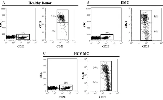
The gating strategy for enumerating CD21low B cells is depicted in Fig.2. B cells are co- stained with anti-CD20 peridinin chlorophyll (PerCP), anti-CD21 phycoerythrin (PE) and additional fluorochrome-labeled antibodies as requested. A consensus gate defining B cells with high CD21 expression (CD21high) was derived from the analysis of 20 healthy donors; setting the gate just below the cluster of CD21high B cells yielded percentages of B cells with low or no expression of CD21 (CD21low) of 6,9 ± 4,1 (mean ± SD) in healthy donors, 22,2 ± 15,5 in patients with EMC and of 54,5 ± 18 in HCV-MC patients.
CD20 SSC 10% CD20 C D 21 EMC 44% 56% SSC CD20 8% C D 21 CD20 5% Healthy Donor CD20 SSC 21% CD20 C D 21 64% 36% HCV-MC 95% A B C
Figure 2. Gating strategy for enumerating CD21low B cells.
CD20+ B cells from (A) one representative healthy donor, (B) one representative EMC patient and (C) one representative HCV-MC patient were electronically gated and percentages of CD21low B cells were calculated on the basis of a predefined consensus gate positioned at the 102 fluorescence channel.
3.3 PhosFlow assay for pERK
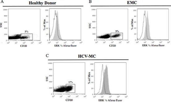
The intracellular pERK content was measured by the BD Phos-Flow system as per manufacturer’s Protocol 1 (Becton-Dickinson Biosciences). PBMCs (1.5x10⁶ cells) were split in two vials, re-suspended in 100 µL of RPMI 1640 containing 5% fetal bovine serum (complete medium), and equilibrated at 37°C for at least 20 min. An equal volume of prewarmed complete medium, either alone (unstimulated control) or containing 10 µg/mL of F(ab′)2 anti-human Ig (Jackson Immunoresearch Laboratories), was then added, and the cells were returned to 37◦C for 10 min. Cells were then fixed by the addition of 200 µL of prewarmed Phos-Flow Fix Buffer I for 10 min at 37° C, washed twice in PhosFlow Perm/Wash Buffer I, split in two vials, and stained either with anti-pERK1/2-Alexa 488 or with mouse IgG-Alexa 488 as control. Samples were simultaneously stained with fluochrome-conjugated mAbs to CD20, CD27 and IgM. The pERK-specific MFI was
calculated by subtracting the MFI values obtained with control mouse IgG from those obtained with anti- pERK antibody. A representative experiment is shown in Fig. 3.
Figure 3. Phos-Flow assay of pERK expression in unstimulated B cells: open
histograms denote staining with control antibody and gray hystograms staining with anti-pERK.
3.4 Apoptosis assay
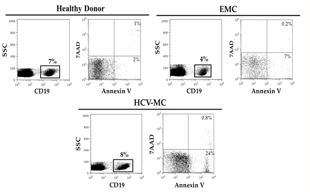
A total of 106 PBMC in 1 mL of RPMI containing 10% fetal bovine serum were incubated at 37°C for 16 hours in 96-well U-bottom plates (2x10⁵ cells/well), and then washed, resuspended in phosphate-buffered saline with 10% fetal bovine serum, and stained with Annexin V–phycoerythrin (Molecular Probes, Life Technologies), 7-amino-actinomycin (7AAD) (Sigma-Aldrich) and anti-CD20 FITC. After electronic gating of CD20+ B cells, early apoptotic B cells were identified as Annexin V+/7AAD-, and late apoptotic cells as Annexin V+/7AAD+ cells; the values reported in the Results and Discussion section refer to total apoptotic B cells (early plus late). A representative experiment is shown in Fig. 4.
SSC CD19 Annexin V 7A A D 4% 7% 0.2% EMC HCV-MC SSC 8% CD19 Annexin V 7A A D 24% 0.8% SSC CD19 7% 7A A D Annexin V 2% 1% Healthy Donor
Fig. 4. Spontaneous in vitro apoptosis of B cells.
After 16h in vitro incubation of PBMC in the absence of stimuli, cells were stained with anti-CD20, annexin V and 7AAD. Electronically gated CD20+ B cells from a healthy donor, a patient with EMC and a patient with HCV+ MC were then analyzed for annexin V and 7-AAD staining. Early apoptotic B cells are identified as annexin V+/7AAD- and late apoptotic B cells as annexin V+/7AAD+; total (early plus late) apoptotic B cells are ~3% in the healthy donor’s sample, ~7% in the EMC patient’s sample and ~25% in HCV-MC patient’s sample.
3.5 Cell proliferation assay
Cell proliferation was measured by the carboxyfluorescein diacetate succinimidyl ester (CFSE) dilution assay [81]. PBMCs were labeled with CFSE (Invitrogen, Life Technologies) and cultured at 2×10⁵ cells per well in 96-well U-bottom plates. Cells were stimulated with the TLR9 ligand CpG 2006 (2.5 µg/mL; Sigma Genosys), in the absence or presence of the BCR ligand F(ab’)2 anti-human Ig (4 µg/mL; Jackson ImmunoResearch Laboratories). Cell proliferation was measured at day 5 of culture by flow cytometry. The number of cells that started dividing (precursor cohort) was calculated using the FlowJo software. Before flow cytometric analysis, cells were permeabilized (Permeabilizing-Solution 2; Becton-Dickinson Biosciences) and counterstained with monoclonal antibodies to CD20 and IgM. Cell proliferation was measured in electronically gated whole CD20+ B cells.
Fig. 5
Strategy for the analysis of the proliferative responses of B cells from the HD, ECM and Crio-HCV is shown. At the end of a 5-day culture in the presence of stimuli, CFSE labeled peripheral blood mononuclear cells (PBMCs) were permeabilized and stained with antibodies to CD20, IgM. CD20dimIgM+ B cells were analyzed for CFSE fluorescence (histograms). Percentages in the histograms denote the percent of divided cells (number of precursor B cells that have undergone at least one division).
3.6 Statistical analysis
Continuous variables were reported as median and interquartile range or as mean and standard deviation. Categorical variables were reported as absolute and relative frequencies. Groups were compared using the Mann–Whitney test or t test for continuous variables and Fisher’s exact test for the categorical ones. Correlations were calculated by means of linear regression analysis. Significance was established at a two-sided p-value ≤0.05. Data were analyzed using Graph-Pad Prism version 7 (La Jolla, CA, USA).
4. RESULTS 4.1. Patients’ characteristics
In this study 13 patients (6 male/7 female; median age 74 y, range 59-83y) affected by EMC were enrolled. Mean cryocrit level was 3.8 ± 4.1% and the mean value of C4 was 8.3 mg/dL ± 8.6. All patients except one had a positive rheumatoid factor. Six of 13 (46%) patients showed positivity for antinuclear antibodies (ANA), 2 of 13 (15%) for SS-A and in 4 (31%) patients other autoantibodies were found: antineutrophil cytoplasmic antibodies (ANCA) (1), anti-thyroglobulin (anti-TG) and thyroid peroxidase (anti-TPO) (1), anti- parietal cell antibodies (APCA) (1), anti-gangliosides (1) and anti- CCP (1). Three patients (23%) had a concomitant diagnosis of an autoimmune disease (1 Sjogren’s syndrome, 1 Hashimoto thyroiditis and 1 Rheumatoid Arthritis).
The demographic and clinical characteristics of the patients with EMC compared to patients with HCV-MC enrolled in this study are summarized in Table 1. The two groups did not differ significantly for age and sex distribution, duration of the disease, and clinical presentation of cryoglobulinemic vasculitis at the time of enrollment with the exception of a lower prevalence of peripheral neuropathy in EMC patients. The clinical presentation of our patients did not differ from that recently described [26] in a large cohort of EMC patients, except for a lower frequency of arthralgia (38% vs 79.7%) and higher prevalence (62% vs 17.4%) of sicca syndrome in our cohort of patients.
Cryocrit values tended to be lower in EMC patients although not reaching statistical significance, and C4 levels were similar. Interestingly, the number of total circulating B- cells was significantly lower in EMC (mean: 185/mm3 ± 236) than in HCV-MC patients (mean: 529/mm3 ± 795) and was similar to the B-cell number observed in HD [76].
Table 1.
Demographic and clinical characteristics of patients with EMC and HCV-MC
4.2 Phenotypic Features of B cells in Essential mixed cryoglobulinemia
Patients with HCV-associated MC present typical features of circulating B cells [73]. Almost one third of patients is characterized by clonal expansion of marginal zone (MZ) CD27+IgM+ B cells producing a rheumatoid factor often encoded by the VH1-69 and Vk3-20 genes [77]. Usually most of the B cells have a low expression of the complement receptor CD21 and are therefore called CD21low B cells [54] that have been suggested to represent one stage of differentiation within the expanded population of activated MZ B cells in HCV associated MC. In EMC B cell phenotype has been scarcely investigated. None of the 13 patients with EMC showed a circulating B cell clone investigated by skewed kappa/lambda light chain ratio or by the expression of a VH1-69- encoded idiotype as previously described in HCV-MC [74]. It is likely that small- size VH1-69-negative clones passed undetected through kappa/lambda ratio analysis as this latter analysis is usually indicative for large (>80% of circulating B cells) B cell clones.
and 35.3 ± 10.4% and the mean percentage of memory CD27+ B cells was 41.3 ± 25.5%. Differently from HCV-MC, marginal zone IgM+CD27+ memory B cells were only slightly increased (mean 24.2 ± 15.7 % range 3-48), compared to the HDs that show percentages of ~13% [78]. I next investigated whether EMC patients had, similar to HCV-MC patients, expanded populations of circulating CD21low B-cells. EMC patients had, overall, percentages of CD21low B cells significantly increased compared to healthy donors (HD) (22.2% ± 15.5 in ECM vs 6.9 ± 4.1 in HD; p= 0.0002; Fig 6A, but significantly lower than HCV-MC patients (mean 54.5 ± 18%; p <0.0001; Fig 6A). The absolute number of circulating CD21low B-cells was also significantly lower in EMC than in HCV-MC patients (Fig. 6B). The frequency of CD21low B-cells with a CD27+ memory phenotype was around 48% in EMC and significantly higher in HCV-MC patients (68%; p=0.0079; Fig. 6C). Detailed analysis of CD27-CD21low B-cell subpopulations in EMC patients showed that switched CD27-IgM- memory B-cells represented 14.9 ± 11.2% of CD21low B-cells (Figure 6D).
In the HD the percentage of CD21low does not exceed the 10% of total B cells [50] and in our series of 13 EMC patients only 2 cases presented a percentage of CD21low B cells below 10% of total B lymphocytes (respectively 7 and 4%, Figure 6A).
Figure 6.
(A e B) CD21low B cells among circulating B cells in a cohort of 13 EMC patients compared to 24 HCV-MC patients and 20 HD. CD21low B cells in EMC patients are significantly increased compared to the HD and significantly lower than HCV-MC patients; (C) CD27+
EMC
memory B cells in CD21low B cells are significantly increate in HCV-MC (D) CD27+ memory, CD27-IgM+ naïve and CD27-IgM- switched memory B cell subpopulations as percentages of CD21low B cells in EMC patients.
4.3 CD21low B cells expanded in EMC resemble CD21low B cells in HCV-MC
CD21low B cells have a peculiar pattern of expression of different inhibitory and homing receptors [54,55,73,74]. A striking common trait is the expression of integrin αx chain (CD11c), typically a marker of dendritic cells. Another peculiar marker of CD21low B cells is Fc receptor-like 4 (FCRL4), an inhibitory receptor that was first described in a subset of human tonsillar B cells with low CD21 expression [51] and of the cell death receptor CD95 [55]. The expression of the homing receptors CCR7, CD62L, CXCR4 and CXCR5, which are required for migration to lymph nodes or to germinal centers [55], has been described to be lower in CD21low B cells.
Here I analyzed whether CD21low B cells from EMC present similar phenotypical features and investigated the expression of CD11c, CD95 and CD62L in CD21low and CD21high B cells of 4 EMC patients compared to HCV-MC patients. CD21low B cells in both disorders present similar expression pattern of these receptors with higher expression of CD11c and CD95 and lower expression of CD62L in CD21low compared to CD21high B cells (table 2).
Interestingly CD11c and CD62L are expressed at higher levels in EMC CD21low B cells compared to HCV-MC CD21low B cells. Detailed gating strategy and the phenotypical pattern of expression of CD11c, CD95 and CD62L in CD21low and high B cells in one EMC representative patient is presented in figure 7A and compared to one representative
patient with HCV-MC (7B). Since this phenotype denotes B cells with functional features of exhaustion and anergy [54,55,73,74], we investigated CD21low B-cells of EMC patients in this regard.
Figure 7.
Expression pattern of CD62L, CD11c and CD95 in CD21low and CD21high B cells in one representative EMC (A) and HCV-MC patient (B). CD19+ B cells were gated electronically among isolated PBMC by side scatter (SSC) characteristics and by CD19 expression; percentages of CD21high and CD21low B cells refer to total B cells. Dot plots (left side) and histograms show the expression of the indicated surface receptor in CD21low B cells (shadowed histograms) and CD21high B cells (open histograms).
4.4. CD21low B cells of EMC patients proliferate poorly in response to TLR9 ligation
Total B-cells of EMC and HCV-MC patients proliferated less than those of healthy donors in response to the ligation of TLR9 with CpG, and proliferation in EMC patients tended to be lesser than in HCV-MC patients although not reaching statistical significance (Fig. 8). Proliferation induced by the contemporaneous ligation of TLR9 with CpG and of the BCR with anti-Ig was similar in EMC and in healthy donors, whereas it was significantly decreased in HCV-MC (Fig. 8).
Figure 8.
(A) Proliferative responses of B cells from healthy donors (HD), EMC and HCV-MC patients to CpG and to CpG plus anti-Ig. Each symbol represents a patient studied once and bars denote the geometric means; (B) correlation of the percentage of divided cells after stimulation with CpG plus anti-Ig with the percentage of CD21low B cells in HCV-MC (empty circles) and EMC (black circles).
These different patterns of response can be explained by the fact that CpG stimulates CD27+ memory cells constitutively expressing TLR9 but not TLR9- negative naive B-cells, which however become responsive to CpG upon stimulation of the BCR [79]. Thus, the reduced response to CpG of total B-cells from EMC and HCV-MC patients is seemingly due to the fact that they contain populations of exhausted CD27+ CD21low B-cells unresponsive to TLR9 ligation. The recruitment of normal naive B-B-cells by BCR ligation with anti-Ig increases the number of cells responsive to CpG in healthy donors and in EMC patients, who have relatively small populations of CD21low B-cells, but not in HCV-MC patients who have a reduced reservoir of circulating naive B-cells because of the
presence of large populations of CD21low B-cells; this concept is supported by the observation of a negative correlation between the frequency of CD21low B-cells and the magnitude of increase of CpG-driven proliferation induced by costimulation with anti-Ig (Figure 8 B).
4.5. CD21low B cells of EMC patients display dysregulated pERK signaling and are prone to apoptosis
The CD21low B-cells of HCV-MC patients display high constitutive expression of pERK and are highly prone to spontaneous apoptosis [53,54,55]; these anomalies are similar to those found in murine B-cells reversibily made anergic by continual antigenic stimulation [71] and, indeed, they rapid revert in HCV-MC CD21low B cells after HCV infection is cleared [74]. Thus, we wondered whether also in EMC the CD21low B-cells display features of antigen-driven anergy. Unstimulated B cells of EMC patients expressed significantly higher levels of pERK than those of healthy donors and lower, although not significantly, levels than HCV-MC patients (Fig. 9 A). In EMC and HCV-MC high expression of pERK was a feature of CD21low B-cells, as revealed by the analysis of electronically gated subpopulations (not shown) and by the significant correlation between the percentage of CD21low B cells and the overall level of pERK expression in whole B cells from HCV-MC and EMC patients (Fig. 9 B).
Accelerated apoptosis, another feature of CD21low B-cells of HCV-MC patients, was also observed in EMC B-cells (Fig. 10 A). The percentages of apoptotic cells in cultured B-cells of EMC patients were intermediate between those observed in samples from healthy donors and from HCV-MC patients although, again, the difference with the latter did not reach statistical significance. Again a significant correlation between the percentage of spontaneous apoptosis of B cells and the percentage of CD21low B cells was observed (Figure 10 B).
Figure 9.
(A) Constitutive ERK phosphorylation in total B cells from healthy donors (HD), EMC and HCV-MC patients. Each symbol represents a patient studied once and bars denote the geometric means. (B) Correlation of the percentage of CD21low B cells with constitutive ERK phosphorylation in EMC (black circles) and HCV-MC patients (empty circles).
Figure 10.
(A) Spontaneous apoptosis in B cells from healthy donors (HD), EMC and HCV-MC patients. Each symbol represents a patient studied once and bars denote the geometric means; (B) correlation of percentage of spontaneous apoptotic B cells and CD21low B cells in EMC (black circles) and HCV-MC patients (empty circles).
A
B
5. DISCUSSION
The immunopathogenesis of EMC has been scarcely investigated so far and, to our knowledge, this is the first study providing a phenotypic and functional landscape of B-cells in this disorder. EMC patients display expansion of unusual CD21low B-cells that share the features of unresponsiveness, dysregulated BCR signaling and accelerated apoptosis with the CD21low B-cells expanded in other infectious or noninfectious autoimmune and lymphoproliferative disorders [46,47,48,49]. Also the expression pattern of inhibitory and homing receptors with high CD11c, CD95 and low CD62L, typically found in CD21low B cells, characterizes CD21low B cells in EMC patients.
Among the noninfectious conditions in which CD21low B-cells are expanded RA, CVID, SLE and pSS have been investigated more thoroughly [46,47,48,49]. In the healthy donor CD21low B cells account for approximately 5% of total B cells [80] and a mouse counterpart termed aged B cells (ABCs) has been described in elderly female mice [81]. Overall CD21low B cells have been identified as CD21lowCD11c+, CD21lowCD27+, CD21lowCD38- or CD21lowCD27- and there is no consensus nomenclature for this B cell population. In CVID CD21low B-cells are CD27-negative unmutated naive B-cells [46], whereas in most other diseases, the CD21low B cells, although CD27-, have been defined as memory cells based on their expression of switched and/or mutated BCRs [80]. In Sjogren’s syndrome, for instance, CD21low B cells are CD27- but carry mutated immunoglobulin genes and therefore belong to the memory B-cell compartment and, similarly in the healthy donor CD21low B cells are CD27-negative memory B cells [80]. In RA patients CD21low B cells have been described as switched memory CD27+IgG+CD11c+ [61] and CD27-IgD+IgM+ or CD27-IgD-IgM- B cells [47] but their place in the pathogenesis is not clear. In EMC CD21low B cell include all B cell subpopulations, both CD27+ memory (43.1%) CD27- naive (40.5%) and switched memory CD27- (15.5%) B cells. The heterogeneity of CD21low B cells in EMC is probably similar to RA patients. Indeed Isnardi et al. investigated the CD21low B cells after depletion of CD27+ B cells and Rubtsov focused only on CD21low CD11c+ B cells [47,61].
In EMC CD21low B cells share different functional proprieties with CD21low B cells expanded in HCV-MC and in Sjogren’s syndrome and, curiously, EMC and HCV-MC overlap with Sjogren’s syndrome in a significant proportion of cases [49], suggesting that these disorders may share pathogenetic mechanism(s) involving B-cell expansion and anergy. The CD21low B-cells of pSS patients are similar to those found in EMC and
HCV-MC with regard to dysregulated BCR signaling, lack of proliferation after BCR stimulation and accelerated apoptosis, although differ from the CD21low B-cells of the latter patients in that they do not express CD27 and proliferate, although to a somewhat reduced extent, in response to TLR9 ligation [49]. These differences might be related to distinctive pathogenetic mechanisms or to the preferential homing of FCRL4+ CD21low B- cells, which are TLR9-unresponsive [53], to the salivary gland tissue of pSS patients [82].
In our study, a striking difference between EMC and HCV-MC was the significantly lower number of circulating CD21low B-cells in the former. Interestingly, the expansions of CD21low B-cells driven by infections, such as those related to HCV [54] or to HIV [44], are on average much larger than those found in noninfectious disorders such as RA [47], pSS [49] or SLE [48]. The difference in CD21low expansion might be related to the amount of chronic antigen stimulation as shown by the fact that in HCV-associated MC, already one month after the eradication of the virus with direct acting antivirals, CD21low B cells decrease rapidly [74]. In this latter condition, as in the case of HIV infection, the antigen responsible for chronic stimulation and clonal B cell expansion can be identified in the virus but in other non-infectious diseases, such as CVID, autoimmune diseases or EMC the antigen remains almost elusive. Indirect evidence of chronic antigen stimulation, in these latter conditions, is provided by the common functional features of exhaustion and anergy characterizing CD21low B cells. Indeed CD21low B cells of HCV-MC and CVID patients display high constitutive expression of the active phosphorylated form of extracellular signal– regulated kinase (pERK); this signature, together with reduced calcium flux and proneness to apoptosis, makes them closely resemble murine B cells made anergic by continual BCR engagement by antigen [83]. In murine anergic B cells, constitutive ERK signaling and reduced lifespan are reversed by dissociation of self antigen from BCR using hapten competition, indicating the need for continual BCR occupancy for maintaining anergy [71]. This observation has many implications in regard to the pathogenic mechanisms involved in EMC, where the inciting initial trigger of disease is still obscure. Mixed cryoglobulins in EMC are formed by a rheumatoid factor (RF) antibody, almost always an IgM, binding to the Fc portion of IgGs, but it is unknown whether immune complexes, binding to RF bearing B cells, might be sufficient for providing the chronic stimulus responsible for CD21low B cell expansion observed in EMC. The better characterization of this interesting and heterogeneous B cell subpopulation might shed light in different autoimmune and immune-mediated disorders, such as EMC or CVID, with still undefined underlying pathogenic mechanisms, opening the question of a possible role of a
6. REFERENCES
[1] Lerner A.B. and Watson C.J. “Studies of cryoglobulins; unusual purpura associated with the presence of a high concentrations of cryoglobulin (cold precipitable serum immunoglobulin)”. Am J Med Sci 1947; 214 (4): 410-5.
[2] Sargur R., White P., Egner W., “Cryoglobulin evaluation: best practice?” Ann Clin Biochem 2010; 47:8-16.
[3] Brouet J.C., Clauvel J.P., Dannon F., Klein M., Seligmann M. “Biological and clinical significance of cryoglobulins: a report of 86 cases”. Am J Med 1974; 57: 775-88.
[4] Dammacco F., Sansonno D., Piccoli C., Tucci F.A., Racanelli V. “The cryoglobulins: an overview”. Eur J Clin Invest 2001;31 (7): 628-638.
[5] Harel S., Mohr M., Jahn I., et al. “Clinico-biological characteristics and treatment of type I monoclonal cryoglobulinaemia: a study of 64 cases”. Br J Haematol 2015; 168 (5): 671-678.
[6] Saadoun D., Sellam J., Ghillani-Dalbin P., Crecel R., Piette J.C., Cacoub P. “Increased risks of lymphoma and death among patients with non-hepatitis C virus-related mixed cryoglobulinemia”. Arch Intern Med 2006; 166 (19): 2101- 2108.
[7] Trejo O., Ramos-Casals M., Garcia-Carrasco M., et al. “Cryoglobulinemia: study of etiologic factors and clinical and immunologic features in 443 patients from a single center”. Medicine (Baltimore) 2001; 80 (4): 252-262.
[8] Tissot J.D., Schifferli J.A., Hochstrasser D.F., et al. “Two-dimensional polyacrylamide gel electrophoresis analysis of cryoglobulins and identification of an IgM-associated peptide”. I Immunol Meth 1994; 173: 63-75.
[9] Musset L., Diemert M.C., Taibi F., et al. “Characterization of cryoglobulins by immunoblotting”. Clin Chem 1992; 38: 798-802.
[10] Wang Y., Lomakin A., Hideshima T. et al. “Pathological crystallization of human immunoglobulins”. Proc Natl Acad Sci USA 2012; 109 (33): 13359- 13361.
[11] Sansonno D. and Dammacco F. “Hepatitis C virus, cryoglobulinemia and vasculitis: immune complex relations”. Lancet Infect Dis 2005;5 (4): 227-236.
2007; 21: 183-200.
[13] Muchtar E., Magen H., Gertz M.A. “How I treat cryoglobulinemia”. Blood 2017;29 (3): 289-298.
[14] Bryce A.H., Kyle R.A., Dispenzieri A., Gertz M.A. “Natural history and therapy of 66 patients with mixed cryoglobulinemia”. Am J Hematol 2006;81 (7): 511-518.
[15] Ferri C., Sebastiani M., Giuggioli D., et al. “Mixed cryoglobulinemia: demographic, clinical, and serologic features and survival in 231 patients”. Semin Arthritis Rheum 2004; 33 (6): 355-374.
[16] Maire M.A., Mittey M., Lambert P.H. “The presence of cryoprecipitable immunoglobulins in normal human sera may reflect specific molecular interactions”. Autoimmunity 1989; 37: 187-92.
[17] Mazzaro C, Dal Maso L, Urraro T, Mauro E, Castelnovo L, Casarin P, Monti G, Gattei V, Zignego AL, Pozzato G. “Hepatitis B virus related cryoglobulinemic vasculitis: A multicentre open label study from the Gruppo Italiano di Studio delle Crioglobulinemie – GISC.” Dig Liver Dis 2016 Jul;48 (7):780-4.
[18] Terrier B, Marie I, Lacraz A, Belenotti P, Bonnet F, Chiche L, Graffin B, Hot A, Kahn JE, Michel C, Quemeneur T, de Saint-Martin L, Hermine O, Léger JM, Mariette X, Senet P , Plaisier E, Cacoub P . “Non HCV-related infectious cryoglobulinemia vasculitis: Results from the French nationwide CryoVas survey and systematic review of the literature.” J Autoimmun. 2015 Dec;65:74-81.
[19] Oliver M, Grandadam M, Marimoutou C, Rogier C, Botelho-Nevers E, Tolou H, Moalic JL, Kraemer P, Morillon M, Morand JJ, Jeandel P, Parola P, Simon F.
“Persisting mixed cryoglobulinemia in Chikungunya infection.” Plos Negl Trop Dis 2009; 3(2): e374.
[20] Rullier P., Le Quellet A, Cognot C. “Cryoglobulins not HCV-related and solid tumors: retrospective analysis from a series of 493 patients.” Eur J Intern Med. 2009 Dec; 20 (8): e158.
[21] Ramos-Casals M., Stone J.H., Cid M.C., Bosch X. “The cryoglobulinaemias.” Lancet 2012 Jan 28; 379 (9813): 348-60.
[22] Terrier B, Krastinova E, Marie I, Launay D, Lacraz A, Belenotti P, de Saint- Martin L, Quemeneur T, Huart A, Bonnet F, Le Guenno G, Kahn JE, Hinschberger O, Rullier P, Diot
E, Lazaro E, Bridoux F, Zénone T, Carrat F, Hermine O, Léger JM, Mariette X, Senet P, Plaisier E, Cacoub P. “Management of noninfectious mixed cryoglobulinemia vasculitis: data from 242 cases included in the CryoVas survey.” Blood 2012 Jun 21; 119(25):5996-6004.
[23] Baimpa E, Dahabreh IJ, Voulgarelis M, Moutsopoulos HM. “Hematologic manifestations and predictors of lymphoma development in primary Sjö gren syndrome: clinical and pathophysiologic aspects.” Medicine (Baltimore) 2009 Sep; 88(5):284-93. [24] Brito-Zerón P, Ramos-Casals M, Bove A, Sentis J, Font J. “Predicting adverse outcomes in primary Sjogren's syndrome: identification of prognostic factors”. Rheumatology 2007 Aug; 46(8):1359-62.
[25] Tzioufas AG, Boumba DS, Skopouli FN, Moutsopoulos HM. “Mixed monoclonal cryoglobulinemia and monoclonal rheumatoid factor cross-reactive idiotypes as predictive factors for the development of lymphoma in primary Sjö gren's syndrome. ” Arthritis Rheum. 1996 May;39(5):767-72.
[26] Galli M, Oreni L, Saccardo F, Castelnovo L, Filippini D, Marson P, Mascia MT, Mazzaro C, Origgi L, Ossi E, Pietrogrande M, Pioltelli P, Quartuccio L, Scarpato S, Sollima S, Riva A, Fraticelli P, Zani R, Giuggioli D, Sebastiani M, Sarzi Puttini P, Gabrielli A, Zignego AL, Scaini P, Ferri C, De Vita S, Monti G. “HCV-unrelated cryoglobulinaemic vasculitis: the results of a prospective observational study by the Italian Group for the Study of Cryoglobulinaemias (GISC)”. Clin Exp Rheumatol. 2017 Mar-Apr;35 Suppl 103(1):67-76.
[27] Ross G.D. and Lambris J.D. “Identification of a C3bi-specific membrane complement receptor that is expressed on lymphocytes, monocytes, neutrophils and erythrocytes”. J Exp Med 1982; 155: 96-110.
[28] Ahearn J.M. and Fearon D.T. “Structure and function of the complement receptors, CR1 (CD35) and CR2 (CD21)”. Adv Immunol 1989; 46: 183-219.
[29] Iida K., Nadler L., Nussenzweig V. “Identification of the membrane receptor for the complement fragment C3d by means of a monoclonal antibody”. J Exp Med 1983; 158: 1021-1033.
[30] Fischer E., Delibrias C., Kazatchkine M.D. “Expression of CR2 (the C3dg/EBV receptor, CD21) on normal human peripheral blood T lymphocytes”. J Immunol 1991; 146: 865-869.
[31] Reynes M., Aubert J.P., Cohen J.H. et al. “Human follicular dendridic cells express CR1, CR2 and CR3 complement receptor antigens”. J Immunol 1985; 135: 2687-2694. [32] Cherukuri A., Cheng P.C., Pierce S.K. “The role of CD19/CD21 complex in B cell processing and presentation of complement tagged antigens”. J Immunol 200; 167: 163-172.
[33] Cambier J.C., Pleiman C.M., Clark M.R. “Signal transduction by the B cell antigen receptor and its coreceptors”. Annu Rev Immunol 1994; 12: 457-486.
[34] Asokan R., Hua J., Young K.A. et al. “Characterization of human complement receptor type 2 (CR2/CD21) as a receptor for IFN-alpha: a potential role in systemic lupus erythematosus”. J Immunol 2006; 177: 383-394.
[35] Fearon D.T. “The CD19-CR2-TAPA1 complex, CD45 and signaling by the antigen receptor of B lymphocytes”. Curr Opin Immunol 1993; 5: 341-348.
[36] Fearon D.T. and Carter R.H. “The CD19/CR2/TAPA1 complex of B lymphocytes: linking natural to acquired immunity”. Annu Rev Immunol 1995; 13: 127-149.
[37] Carter R.H., Tuveson D.A., Park D.J., Rhee D.J., Fearon D.T. “The CD19 complex of B lymphocytes.Activation of phospholipase C by a protein tyrosine kinase-dependent pathway that can be enhanced by the membrane IgM complex”. J Immunol 1991; 147: 3663- 3671.
[38] Matsumoto A.K., Kopicky-Burd J., Carter R.H., Tueveson D.A., Tedder T.F., Fearon D.T. “Intersection of the complement and immune systems: a signal transduction complex of the B lymphocyte-containing complement receptor type 2 and CD19”. J Exp Med 1991; 173: 55-64.
[39] Carroll M.C. “The complement system in regulation of adaptive immunity”. Nat Immunol 2004; 5:981–986.
[40] Boackle S.A., Morris M.A., Holers V.M., Karp D.R. “Complement opsonization is required for presentation of immune complexes by resting peripheral blood B cells”. J Immunol 1998; 161:6537–6543.
[41] Huemer HP, Larcher C, Prodinger WM, Petzer AL, Mitterer M, Falser N. Determination of soluble CD21 as a parameter of B cell activation. Clin Exp Immunol 1993;93:195–199.
new CD21low B cell population in the peripheral blood of patients with SLE”. Clin Immunol 2004;113:161–171.
[43] Charles E.D., Green R.M., Marukian S., Talal A.H., Lake-Bakaar G.V. et al., “Clonal expansion of immunoglobulin M+CD27+ B cells in HCV-associated mixed cryoglobulinemia”. Blood 2008; 111 (3): 1344-1356.
[44] Moir S., Malaspina A., Ogwaro K.M. et al. “HIV-1 induces phenotypic and functional perturbations of B cells in chronically infected individuals”. Proc Natl Acad Sci USA 2001;98:10362–10367.
[45] Weiss G.E., Crompton P.D., Li S. et al. “Atypical memory B cells are greatly expanded in individuals living in a malaria-endemic area”. J Immunol 2009;183:2176– 2182.
[46] Rakhmanov M., Keller B., Gutenberger S. et al. “Circulating CD21low B cells in common variable immunodeficiency resemble tissue homing, innate-like B cells”. Proc Natl Acad Sci USA 2009;106: 13451–13456.
[47] Isnardi I., Ng Y.S., Menard L., Meyers G., Saadoun D., Srdanovic I., Samuels J., Berman J., Buckner J.H., Cunningham-Rundles C., Meffre E. “Complement receptor 2/CD21- human naive B cells contain mostly autoreactive unresponsive clones”. Blood 2010; 115 (24): 5026-5036.
[48] Wehr C., Eibel H., Masilamani M. et al. “A new CD21low B cell population in the peripheral blood of patients with SLE”. Clin Immunol 2004;113:161–171.
[49] Saadoun D., Terrier B., Bannock J. et al. “Expansion of autoreactive unresponsive CD21-/low B cells in Sjogren’s syndrome-associated lymphoproliferation”. Arthritis Rheum 2013;65:1085–1096.
[50] Thorarinsdottir K., Camponeschi A., Gjertsson I., Martensson L. “CD21-/low B cells: a snapshot of a unique B cell subset in health and disease”. Scand J Immunol 2015. 82 (3): 254-261.
[51] Ehrhardt G.R., Hsu J.T., Gartland L. et al. “Expression of the immunoregulatory molecule FcRH4 defines a distinctive tissue-based population of memory B cells”. J Exp Med 2005;202:783–791.
[52] Ehrhardt G.R., Hijikata A., Kitamura H., Ohara O., Wang J.Y., Cooper M.D. “Discriminating gene expression profiles of memory B cell subpopulations”. J Exp Med
2008; 205:1807–1817.
[53] Visentini M., Cagliuso M., Conti V., Carbonari M., Cibati M., Siciliano G., Cristofoletti C., Russo G., Casato M., Fiorilli M. “Clonal B cells of HCV- associated mixed cryoglobulinemia patients contain exhausted marginal zone-like and CD21 low cells overexpressing Stra13”. Eur J Immunol 2012; 42 (6): 1468- 1476.
[54] Charles E.D., Brunetti C., Marukian S., Ritola K.D., Talal A.H. et al. “Clonal B cells in patients with Hepatitis C virus-associated mixed cryoglobulinemia contain an expanded anergic CD21low B-cell-subset”. Blood 2011; 117 (20): 5425-5437.
[55] Terrier B., Joly F., Vazquez T., Benech P., Rosenzwajg M., Carpentier W. et al. “Expansion of functionally anergic CD21-/low marginal zone-like B cell clones in hepatitis C virus infection-related autoimmunity”. J Immunol 2011; 187 (12): 6550-6563
[56] Charles E.D., Green R.M., Marukian S., Talal A.H., Lake-Bakaar G.V. et al., “Clonal expansion of immunoglobulin M+CD27+ B cells in HCV-associated mixed cryoglobulinemia”. Blood 2008; 111 (3): 1344-1356.
[57] Visentini M., Cagliuso M., Conti V., Carbonari M., Casato M., Fiorilli M. “The V(H)1- 69-expressing marginal zone B cells expanded in HCV-associated mixed cryoglobulinemia display proliferative anergy irrespective of CD21(low) phenotype”. Blood 2011; 118 (2): 3440-3441.
[58] Baseggio L., Traverse-Glehen A., Callet-Bauchu E., Morel D., Magaud J.P., Berger F., et al. “Relevance of a scoring system including CD11c expression in the identification of splenic diffuse red pulp small B-cell lymphoma (SRPL)”. Hematol Oncol 2011; 29:47-51. [59] Moir S., Ho J., Malaspina A., Wang W., Di Poto A.C., O'Shea M.A., et al. “Evidence for HIV-associated B cell exhaustion in a dysfunctional memory B cell compartment in HIV-infected viremic individuals”. J Exp Med 2008; 205:1797- 1805.
[60] Kardava L., Moir S., Wang W., Ho J., Buckner C.M., Posada J.G., O’Shea M.A. et al. “Attenuation of HIV-associated human B cell exhaustion by siRNA downregulation of inhibitory receptors”. J. Clin. Invest 2011. 121: 2614–2624.
[61] Rubtsov A.V., Rubtsova K., Fischer A., Meehan R.T., Gillis J.Z., Kappler J.W., et al. “Toll-like receptor 7 (TLR7)-driven accumulation of a novel CD11c B-cell population is important for the development of autoimmunity”. Blood 2011; 118:1305-1315.
uniquely responsive to innate stimuli accumulates in aged mice”. Blood 2011; 118:1294- 1304.
[63] Naradikian M.S., Hao Y., Cancro M.P. “Age-associated B cells: key mediators of both protective and autoreactive humoral responses”. Immunol Rev 2016; 269 (1): 118-129. [64] Chang L.Y., Li Y., Kaplan D.E. “Hepatitis C viraemia reversibly maintains subset of antigen-specific T-bet+ tissue-like memory B cells”. J Viral Hepat 2017; 24 (5): 389-396. [65] Matteucci C., Bracci M., Barba G., Carbonari M., Casato M., Visentini M. et al. “Different genomic imbalances in low and high-grade HCV-related lymphomas”. Leukemia 2008; 22: 219-222.
[66] Chan C.H., Hadlock K.G., Foung S.K., Levy S. “The V(H)1-69 gene is preferentially used both by Hepatitis C virus-associated B cell lymphomas and by normal B cells responding to the E2 viral antigen”. Blood 2001; 97 (4): 1023- 1026.
[67] Gay D., Saunders T., Camper S., Weigert M., “Receptor editing: an approach by autoreactive B cells to escape tolerance”. J. Exp. Med 1993; 177(4): 999-1008.
[68] Nemazee D.A., Bü rki K. “Clonal deletion of B lymphocytes in a transgenic mouse bearing anti-MHC class I antibody genes”. Nature 1989; 337(6207): 562- 566.
[69] Yarkoni Y., Getahun A., Cambier J.C. “Molecular underpinning of B-cell anergy”. Immunol Rev 2010; 237 (1): 249-263.
[70] Cambier J.C., Gauld S.B., Merrell K.T., Vilen B.J. “B-cell anergy: from transgenic models to naturally occurring anergic B cells?” Nature Reviews 2007; 7: 633-643.
[71] Gauld S.B., Benschop R.J., Merrell K.T., Cambier J.C. “Maintenance of B cell anergy requires constant antigen receptor occupancy and signaling”. Nat Immunol 2005; 6 (11): 1160-1167.
[72] Duty J.A., Szodoray P., Zheng N.Y., Koelsch K.A., Zhang Q., Swiatkowski M., et al. “Functional anergy in a subpopulation of naive B cells from healthy humans that express autoreactive immunoglobulin receptors”. J Exp Med 2009; 206(1):139-151.
[73] Visentini M., Cagliuso M., Conti V., Carbonari M., Cibati M., Siciliano G., et al. “Clonal B cells of HCV-associated mixed cryoglobulinemia patients contain exhausted marginal zone-like and CD21low cells overexpressing Stra13”. Eur. J. Immunol. 42 (2012) 1468-1476.