MC1R variants in childhood and adolescent melanoma:
a retrospective pooled analysis of a multicentre cohort
Cristina Pellegrini*, Francesca Botta*, Daniela Massi, Claudia Martorelli, Fabio Facchetti, Sara Gandini, Patrick Maisonneuve,
Marie-Françoise Avril, Florence Demenais, Brigitte Bressac-de Paillerets, Veronica Hoiom, Anne E Cust, Hoda Anton-Culver, Stephen B Gruber,
Richard P Gallagher, Loraine Marrett, Roberto Zanetti, Terence Dwyer, Nancy E Thomas, Colin B Begg, Marianne Berwick, Susana Puig,
Miriam Potrony, Eduardo Nagore, Paola Ghiorzo, Chiara Menin, Ausilia Maria Manganoni, Monica Rodolfo, Sonia Brugnara, Emanuela Passoni,
Lidija Kandolf Sekulovic, Federica Baldini, Gabriella Guida, Alexandros Stratigos, Fezal Ozdemir, Fabrizio Ayala, Ricardo Fernandez-de-Misa,
Pietro Quaglino, Gloria Ribas, Antonella Romanini, Emilia Migliano, Ignazio Stanganelli, Peter A Kanetsky, Maria Antonietta Pizzichetta,
Jose Carlos García-Borrón, Hongmei Nan, Maria Teresa Landi, Julian Little, Julia Newton-Bishop, Francesco Sera, Maria Concetta Fargnoli,
Sara Raimondi, for the IMI Study Group, the GEM Study Group, and the M-SKIP Study Group
Summary
Background Germline variants in the melanocortin 1 receptor gene (MC1R) might increase the risk of childhood and
adolescent melanoma, but a clear conclusion is challenging because of the low number of studies and cases. We
assessed the association of MC1R variants with childhood and adolescent melanoma in a large study comparing the
prevalence of MC1R variants in child or adolescent patients with melanoma to that in adult patients with melanoma
and in healthy adult controls.
Methods In this retrospective pooled analysis, we used the M-SKIP Project, the Italian Melanoma Intergroup, and
other European groups (with participants from Australia, Canada, France, Greece, Italy, the Netherlands, Serbia,
Spain, Sweden, Turkey, and the USA) to assemble an international multicentre cohort. We gathered phenotypic and
genetic data from children or adolescents diagnosed with sporadic single-primary cutaneous melanoma at age
20 years or younger, adult patients with sporadic single-primary cutaneous melanoma diagnosed at age 35 years or
older, and healthy adult individuals as controls. We calculated odds ratios (ORs) for childhood and adolescent
melanoma associated with MC1R variants by multivariable logistic regression. Subgroup analysis was done for
children aged 18 or younger and 14 years or younger.
Findings We analysed data from 233 young patients, 932 adult patients, and 932 healthy adult controls. Children and
adolescents had higher odds of carrying MC1R r variants than did adult patients (OR 1·54, 95% CI 1·02–2·33),
including when analysis was restricted to patients aged 18 years or younger (1·80, 1·06–3·07). All investigated
variants, except Arg160Trp, tended, to varying degrees, to have higher frequencies in young patients than in adult
patients, with significantly higher frequencies found for Val60Leu (OR 1·60, 95% CI 1·05–2·44; p=0·04) and
Asp294His (2·15, 1·05–4·40; p=0·04). Compared with those of healthy controls, young patients with melanoma had
significantly higher frequencies of any MC1R variants.
Interpretation
Our pooled analysis of MC1R genetic data of young patients with melanoma showed that
MC1R r variants were more prevalent in childhood and adolescent melanoma than in adult melanoma, especially in
patients aged 18 years or younger. Our findings support the role of MC1R in childhood and adolescent melanoma
susceptibility, with a potential clinical relevance for developing early melanoma detection and preventive strategies.
Funding SPD-Pilot/Project-Award-2015; AIRC-MFAG-11831.
Copyright © 2019 Elsevier Ltd. All rights reserved.
Introduction
Cutaneous melanoma mainly occurs in adult patients
and is rare in the paediatric population, with only 2% of
all cutaneous melanoma cases diagnosed in patients
younger than 20 years.
1–4In the child and adolescent
population, most cases of cutaneous melanoma are
diagnosed among adolescents, with only 8% occurring in
infancy and childhood.
5,6Differences exist between childhood or adolescent and
adult cutaneous melanoma regarding clinical aspects,
histo
pathological features, and disease staging.
2,7,8Cutaneous melanoma in childhood is often amelanotic,
shows broad histopathological variability, and can
present with histological uncertainty and ambiguous
atypical characteristics that do not allow a definite
malignant or benign classification.
4,9Children with
cutaneous melanoma present at a more advanced stage
of disease, with thicker lesions and higher rates of
lymph node metastasis than do their adult counterparts,
leading to a worse prognosis.
4,9However, published
studies have reported discordant data on survival
rates.
5,10Lancet Child Adolesc Health 2019 Published Online March 11, 2019 http://dx.doi.org/10.1016/ S2352-4642(19)30005-7 See Online/Comment http://dx.doi.org/10.1016/ S2352-4642(19)30026-4 *Contributed equally Department of Dermatology and Department of Biotechnological and Applied Clinical Sciences, University of L’Aquila, L’Aquila, Italy
(C Pellegrini PhD, C Martorelli PhD,
Prof M C Fargnoli MD); Division
of Epidemiology and Biostatistics (F Botta MSc,
P Maisonneuve Eng), Molecular
and Pharmaco-Epidemiology Unit, Department of Experimental Oncology
(S Gandini PhD,
S Raimondi PhD), and Division
of Melanoma, Sarcoma and Rare Cancer (F Baldini MD), European Institute of Oncology IRCCS, Milan, Italy; Department of Statistics and Quantitative Methods, University of Milano-Bicocca, Milan, Italy (F Botta); Division of Pathological Anatomy, Department of Surgery and Translational Medicine, University of Florence, Florence, Italy
(Prof D Massi MD); Pathology
Section, Department of Molecular and Translational Medicine (Prof F Facchetti MD), and Department of Dermatology
(A M Manganoni MD), Spedali
Civili di Brescia, University of Brescia, Brescia, Italy; APHP, Dermatology Department, Hôpital Cochin and Paris Descartes University, Paris, France (Prof M-F Avril MD); Genetic Variation and Human Diseases Unit (UMR-946),
Institut National de la Santé et de la Recherche Médicale (INSERM), Paris, France
(Prof F Demenais MD);
Department of Biopathology and INSERM, University of Paris-Saclay, Villejuif, France
(B Bressac-de Paillerets PhD);
Department of Oncology and Pathology, Cancer Centre, Karolinska Institutet, Stockholm, Sweden
(V Hoiom PhD); Sydney School
of Public Health and Melanoma Institute Australia, University of Sydney, Sydney, NSW, Australia (A E Cust MD); Department of Epidemiology, University of California, Irvine, CA, USA (H Anton-Culver MD); USC Norris Comprehensive Cancer Center, University of Southern California, Los Angeles, CA, USA
(S B Gruber MD); British
Columbia Cancer and Department of Dermatology and Skin Science, University of British Columbia, Vancouver, BC, Canada (R P Gallagher MD); Cancer Care Ontario, Toronto, ON, Canada
(Prof L Marrett PhD); Piedmont
Cancer Registry, Centre for Epidemiology and Prevention in Oncology in Piedmont, Turin, Italy (R Zanetti MD); George Institute for Global Health, Nuffield Department of Obstetrics and Gynaecology, University of Oxford, Oxford, UK (Prof T Dwyer MD); Lineberger Comprehensive Cancer Center, University of North Carolina, Chapel Hill, NC, USA (N E Thomas MD); Department of Epidemiology and Biostatistics, Memorial Sloan Kettering Cancer Center, New York, NY, USA
(C B Begg PhD); Department of
Internal Medicine, University of New Mexico Cancer Center, University of New Mexico, Albuquerque, NM, USA
(Prof M Berwick MD); Melanoma
Unit, Dermatology Department, Hospital Clinic Barcelona, University of Barcelona, Institut d’Investigacions Biomèdiques August Pi I Sunyer, and CIBER de Enfermedades Raras, Barcelona, Spain (S Puig MD,
M Potrony PhD); Department of Dermatology, Instituto Valenciano de Oncologia, Valencia, Spain (Prof E Nagore MD); Department of Internal Medicine and Medical
Whether adult melanoma and childhood and adolescent
melanoma share a similar pathogenesis has long been a
subject of debate. Major risk factors for paediatric
cutaneous melanoma include giant congenital melano
cytic naevi and hereditary conditions such as xeroderma
pigmentosum, immunodeficiency, and albinism.
11Other
known risk factors common to paediatric and adult
melanoma are family history of melanoma, dysplastic
naevus syndrome, elevated number of acquired melano
cytic naevi, red hair, sunsensitive phenotype, and ultra
violet radiation (UV) exposure.
12,13It is uncertain whether childhood and adolescent
cutaneous melanoma differs from the adult melanoma
regarding genetic predisposition. The paediatric form of
the disease is mostly sporadic, whereas adolescent
cutaneous melanoma is sometimes observed in
melanomaprone families. In general, there is a higher
proportion of germline mutation carriers among young
patients with cancer than among older patients,
14but
whether this tendency holds true for cutaneous
melanoma is unclear because of the rarity of this disease
occurring in children or adolescents. On the basis of the
few available studies,
12,15–21child and adolescent patients
have only rarely been found to carry germline mutations
in the two highpenetrance melanoma genes, CDKN2A
and CDK4, which are known to be significantly associated
with melanoma in a familial context alone.
The MC1R (melanocortin 1 receptor) gene is a key
determinant of human pigmentation.
22MC1R is highly
polymorphic in the general population, and specific
variants were defined as R (Asp84Glu, Arg142His,
Arg151Cys, Ile155Thr, Arg160Trp, Asp294His) or r
(Val60Leu, Val92Met, Arg163Gln) alleles, according to
their strength of association with the red hair colour
phenotype.
23Extensive in vitro and in vivo evidence
showed that both R and r alleles produce hypomorphic
proteins with compromised activity compared with
native MC1R function.
22The R alleles have been found to
have a major effect on pigmentation and UV sensitivity.
22,23By contrast, r alleles confer normal or slightly impaired
MC1R activity, resulting in a lowstrength association
with the fair skin phenotype.
23Natural MC1R variation is an established risk factor for
cutaneous melanoma across multiple populations
worldwide.
24The risk of cutaneous melanoma is higher
for carriers of an MC1R variant than for wildtype
individuals, with the strongest association among
carriers of R alleles and multiple variants.
24MC1R
variants confer a significant increased risk of cutaneous
melanoma in darkly pigmented individuals, highlighting
the effect of MC1R through nonpigmentary pathways.
25,26Moreover, MC1R variant genotypes are associated with
phenotypic characteristics of melanoma
27and melano
cytic naevi
28and seem to influence the somatic mutational
load in adult cutaneous melanoma.
29Young patients
(aged 20 years or younger)
with cutaneous melanoma
have an elevated prevalence of MC1R variants, but the
low number of available studies, coupled with the small
Research in context
Evidence before this study
The development of melanoma in children and adolescents has
been hypothesised to have a stronger genetic component than
that of melanoma in adults. We searched PubMed for studies
published up to July 31, 2018, on melanoma susceptibility in
paediatric patients, without language or date restrictions. We used
the search terms “pediatric melanoma” OR “childhood melanoma”
OR “adolescent melanoma” AND “susceptibility” OR
“predisposition” OR “genetics”. We found that genetic
predisposition for melanoma has been poorly investigated in
childhood and adolescence because of the rarity of the disease.
Most published research included few cases, mainly from
single-institution cohorts, investigating the main susceptibility genes for
melanoma CDKN2A (cyclin-dependent kinase inhibitor 2A), CDK4
(cyclin-dependent kinase 4), and MC1R (melanocortin 1 receptor).
The overall findings reported a marginal role in paediatric patients
of the two major melanoma susceptibility genes, CDKN2A and
CDK4. By contrast, a high frequency of germline variants has been
identified in the intermediate-penetrance MC1R gene, but the
very low number of paediatric cases of melanoma made any
significant conclusions impossible. Therefore, we hypothesised
that a large-scale association study could explore the importance
of the MC1R gene in paediatric melanoma predisposition.
Added value of this study
Our study assessed the effect of MC1R gene variants on
paediatric melanoma susceptibility in a large case-case study,
by comparing the prevalence of MC1R variants in child or
adolescent patients with those in adult patients and in
healthy controls. To our knowledge, our series of patients is
the largest international multicentre cohort of paediatric
patients with melanoma with available genetic data. Our
pooled analysis showed that paediatric patients had a higher
probability of carrying any MC1R variant than that of adult
patients, suggesting a major role of MC1R variants, mainly
r variants, in paediatric melanoma predisposition.
Furthermore, r variants seemed to be most strongly
associated with melanoma in patients aged 18 years
or younger.
Implications of all the available evidence
We provided evidence of genetic determinants potentially
involved in paediatric melanoma susceptibility. Our study
represents a first step to comprehend the genetic background
of paediatric melanoma and to elucidate the diversity of
paediatric and adult melanoma, with potential clinical
implications.
Specialties, University of Genoa and Ospedale Policlinico San Martino, Genoa, Italy
(Prof P Ghiorzo PhD); Diagnostic
Immunology and Molecular Oncology Unit, Veneto Institute of Oncology, IOV-IRCCS, Padua, Italy
number of cases per study, makes drawing clear
conclusions a challenge.
18–20To help elucidate the role of MC1R in childhood and
adolescent cutaneous melanoma and to better under
stand the genetic and clinical diversity of the disease
across age, with potential clinical effects in terms of
early melanoma detection and preventive strategies, we
assessed these tumours in a large multicentre cohort
pooled from the
MSKIP (melanocortin 1 receptor skin
cancer and pheno
typic characteristics) Project, the
Italian Melanoma Intergroup (IMI), and other European
groups. The aims of our study were to compare the
prevalence of MC1R variants between young patients
and healthy controls, with a casecontrol study design,
and between young patients and adult patients, with a
casecase study design.
Methods
Study design and participants
We analysed a large, multicentre cohort pooled from the
MSKIP Project, the IMI, and other European groups
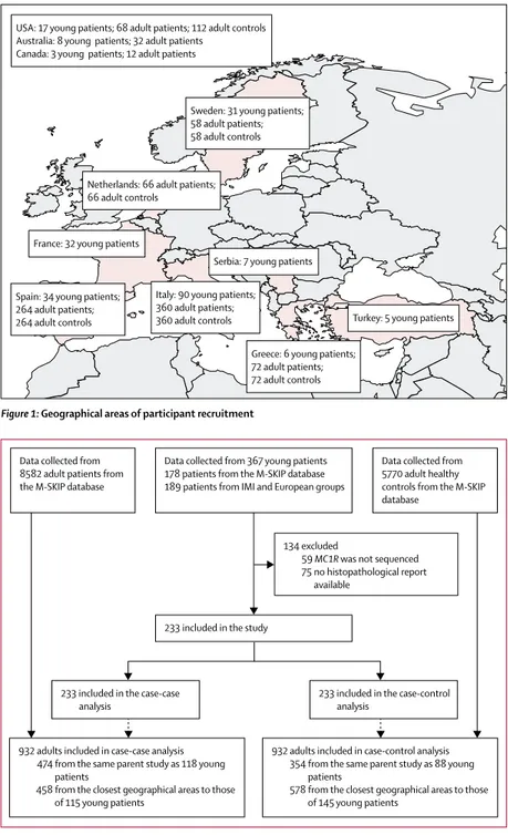
(appendix), including participants from 11 countries
(Australia, Canada, France, Greece, Italy, the Netherlands,
Serbia, Spain, Sweden, Turkey, and the USA; figure 1).
Our analysis included children and adolescents diag
nosed with sporadic singleprimary cutaneous melanoma
at age 20 years or younger, adult patients with sporadic
singleprimary cutaneous melanoma diagnosed at age
35 years or older, and healthy adult individuals as
controls. Because age is a continuous variable, and an
exact age cutoff between adolescents and adults would
not be expected, we excluded melanoma cases diagnosed
in the age range of 21–34 years to avoid a possible overlap
between categories, and thus enable comparison between
groups with distinct clinical and genetic characteristics.
Because of the known challenges in diagnosing paediatric
melanoma
30–32and to decrease misdiagnosis, participating
investigators were asked to provide the original histo
pathological reports and representative glass slides for
central review. Only patients for whom the original
histopathological report was available were eligible.
Additionally, we restricted the study to cases with
complete MC1R genotyping. We excluded familial
melanoma cases, cases with a history of cancer at any site
other than nonmelanoma skin cancer, atypical spitzoid
neoplasms or melanocytic tumors of uncertain malignant
potential, and ocular and mucosal melanomas.
Detailed information on recruitment is reported in the
appendix. Ethics committee approval was obtained at
each institution in which new blood samples were drawn.
For each young patient, four adult patients and four healthy
controls were randomly selected from the same parent
study that provided the young patient. When this was not
possible, adult patients and controls were selected from a
study that was done in the nearest geographical proximity
to the parent study of the young patient (appendix;
figure 1). Written consent was obtained from adult and
older adolescent patients and the parents of young
patients.
Procedures
For 135 young patients from the MSKIP Project, and
48 from the IMI and European groups, MC1R sequencing
had already been done in studyspecific laboratories
Figure 1: Geographical areas of participant recruitment
USA: 17 young patients; 68 adult patients; 112 adult controls Australia: 8 young patients; 32 adult patients
Canada: 3 young patients; 12 adult patients
Turkey: 5 young patients Serbia: 7 young patients
France: 32 young patients
Sweden: 31 young patients; 58 adult patients; 58 adult controls
Netherlands: 66 adult patients; 66 adult controls
Spain: 34 young patients; 264 adult patients; 264 adult controls
Greece: 6 young patients; 72 adult patients; 72 adult controls Italy: 90 young patients;
360 adult patients; 360 adult controls
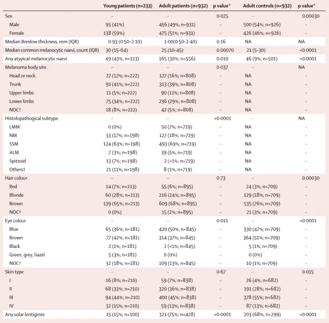
Figure 2: Flow chart of participants included in the analysis
Young patients were aged 20 years or younger, whereas adult patients and healthy controls were aged 35 years or older. M-SKIP=melanocortin 1 receptor skin cancer and phenotypic characteristics Project. IMI=Italian Melanoma Intergroup. MC1R=melanocortin 1 receptor.
233 included in the case-case
analysis 233 included in the case-control analysis 932 adults included in case-case analysis
474 from the same parent study as 118 young patients
458 from the closest geographical areas to those of 115 young patients
932 adults included in case-control analysis 354 from the same parent study as 88 young patients
578 from the closest geographical areas to those of 145 young patients
233 included in the study
134 excluded
59 MC1R was not sequenced 75 no histopathological report available
Data collected from 367 young patients 178 patients from the M-SKIP database 189 patients from IMI and European groups Data collected from
8582 adult patients from the M-SKIP database
Data collected from 5770 adult healthy controls from the M-SKIP database
(C Menin MD); Department of
Research, Fondazione IRCCS Istituto Nazionale dei Tumori, Milan, Italy (M Rodolfo MSc); Oncology Unit, Santa Chiara Hospital, Trento, Italy
(S Brugnara MD); Department
of Pathophysiology and Transplantation, University of Milan, Foundation IRCCS Ca’ Granda Ospedale Maggiore Policlinico, Milan, Italy
(E Passoni MD); Department of
Dermatology, Faculty of Medicine, Military Medical Academy, Belgrade, Serbia
(Prof L K Sekulovic MD);
Department of Basic Medical Sciences, Neurosciences and Sense Organs, University of Bari Aldo Moro, Bari, Italy
(G Guida PhD); 1st Department
of Dermatology, Andreas Sygros Hospital, Medical School, National and Kapodistrian University of Athens, Athens, Greece
(Prof A Stratigos MD);
Department of Dermatology, Faculty of Medicine, University of Ege, Izmir, Turkey
(Prof F Ozdemir MD); Melanoma
Unit, Cancer Immunotherapy and Innovative Therapies, IRCCS Istituto Nazionale dei Tumori, Fondazione G Pascale, Napoli, Italia (F Ayala MD); Dermatology Service, University Hospital Nuestra Senora de Candelaria, Santa Cruz de Tenerife, Spain
(R Fernandez-de-Misa PhD);
Dermatologic Clinic, Department of Medical Sciences, University of Torino, Turin, Italy (P Quaglino MD); Department of Medical Oncology and Haematology, Fundación Investigación Clínico de Valencia, INCLIVA Instituto de Investigación Sanitaria, Valencia, Spain
(G Ribas PhD); US Ambulatori
Melanomi, Sarcomi e Tumori Rari, UO Oncologia Medica 1, Azienda Ospedaliero-Universitaria Santa Chiara, Pisa, Italy (A Romanini MD); Plastic Surgery, San Gallicano Dermatological Institute, IRCCS, Rome, Italy
(E Migliano MD); Skin Cancer
Unit, IRCCS Scientific Institute of Romagna for the Study and Treatment of Cancer and University of Parma, Meldola, Italy (I Stanganelli MD); Department of Cancer Epidemiology, H Lee Moffitt Cancer Center and Research Institute, Tampa, FL, USA
(appendix). For the remaining 50 young patients from
IMI and European groups who provided new blood or
saliva samples, MC1R genotyping was done centrally at
the University of L’Aquila (L’Aquila, Italy) and done as
described elsewhere.
33Statistical analysis
A complete description of the statistical analysis is
available in the appendix. Briefly, we analysed the
associations between risk factors and young melanoma
by logistic regression in comparison with two reference
groups, adult patients and healthy controls, with adjust
ment for study or geographical location.
We compared the frequency of any MC1R variants
among children or adolescents with that of adult patients
and controls by logistic regression, with adjustment for
study or geographical location. These comparisons were
repeated for any MC1R R variant, for any r variant, for a
score calculated by summing across the MC1R alleles,
which gives a value of 1 to r and 2 to R variants (as proposed
elsewhere),
34and for each of the nine most prevalent MC1R
variants and any rare MC1R variants (presence or absence).
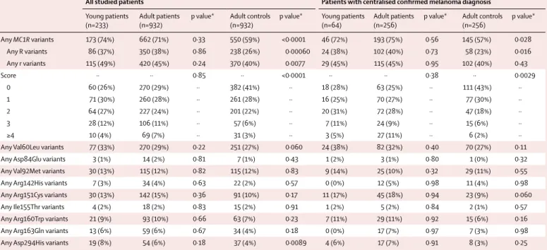
Young patients (n=233) Adult patients (n=932) p value* Adult controls (n=932) p value*
Sex ·· ·· 0·025 ·· 0·00030
Male 95 (41%) 456 (49%; n=931) ·· 500 (54%; n=926) ··
Female 138 (59%) 475 (51%; n=931) ·· 426 (46%; n=926) ··
Median Breslow thickness, mm (IQR) 0·93 (0·50–2·10) 1·00(0·50–2·40) 0·16 NA NA
Median common melanocytic naevi, count (IQR) 30 (15–64) 25 (10–45) 0·00070 21 (5–30) <0·0001 Any atypical melanocytic naevi 49 (43%; n=113) 165 (30%; n=556) 0·010 46 (9%; n=501) <0·0001
Melanoma body site ·· ·· 0·037 ·· NA
Head or neck 27 (12%; n=222) 127 (16%; n=808) ·· NA ·· Trunk 91 (41%; n=222) 313 (39%; n=808) ·· NA ·· Upper limbs 11 (5%; n=222) 90 (11%; n=808) ·· NA ·· Lower limbs 75 (34%; n=222) 236 (29%; n=808) ·· NA ·· NOC† 18 (8%; n=222) 42 (5%; n=808) ·· NA ·· Histolopathogical subtype ·· ·· <0·0001 ·· NA LMM 0 (0%) 50 (7%; n=719) ·· NA ·· NM 33 (17%; n=198) 127 (18%; n=719) ·· NA ·· SSM 124 (63%; n=198) 493 (69%; n=719) ·· NA ·· ALM 7 (3%; n=198) 39 (5%; n=719) ·· NA ·· Spitzoid 13 (7%; n=198) 2 (<1%; n=719) ·· NA ·· Others‡ 21 (11%; n=198) 8 (1%; n=719) ·· NA ·· Hair colour ·· ·· 0·73 ·· 0·00030 Red 14 (7%; n=213) 55 (6%; n=895) ·· 24 (3%; n=709) ·· Blonde 60 (28%; n=213) 216 (24%; n=895) ·· 129 (18%; n=709) ·· Brown 139 (65%; n=213) 609 (68%; n=895) ·· 535 (76%; n=709) ·· NOC† 0 (0%) 15 (2%; n=895) ·· 21 (3%; n=709) ·· Eye colour ·· ·· 0·011 ·· <0·0001 Blue 65 (36%; n=181) 420 (50%; n=845) ·· 330 (47%; n=709) ·· Brown 77 (42%; n=181) 314 (37%; n=845) ·· 364 (51%; n=709) ·· Black 2 (1%; n=181) 2 (<1%; n=845) ·· 5 (1%; n=709) ··
Green, grey, hazel 5 (3%; n=181) 0 (0%) ·· 0 (0%) ··
NOC† 32 (18%; n=181) 109 (13%; n=845) ·· 10 (1%; n=709) ·· Skin type ·· ·· 0·67 ·· 0·015 I 16 (8%; n=210) 59 (7%; n=838) ·· 26 (4%; n=682) ·· II 68 (33%; n=210) 320 (36%; n=838) ·· 191 (28%; n=682) ·· III 94 (44%; n=210) 400 (45%; n=838) ·· 378 (55%; n=682) ·· IV 32 (15%; n=210) 59 (13%; n=838) ·· 87 (13%; n=682) ··
Any solar lentigines 15 (15%; n=100) 321 (75%; n=428) <0·0001 203 (68%; n=299) <0·0001 Data are n (%), unless otherwise specified. NA=not applicable. NOC=not otherwise classifiable. LMM=lentigo maligna melanoma. NM=nodular melanoma. SSM=superficial spreading melanoma. ALM=acral lentiginous melanoma. *Logistic regression model, adjusted by matching stratum variable.†This group includes patients with doubtful or mixed information, thus not classifiable. ‡Other subtypes among children or adolescents include nevoid (n=4), epithelioid (n=3), desmoplastic (n=1), and others not specified (n=13); subtypes among adults include epithelioid (n=5), nevoid (n=1), desmoplastic (n=1), and others not specified (n=1).
We then used multivariable unconditional logistic
regression models to calculate the odds ratio (OR) for
MC1R variants after adjusting for study or geographical
location and other covariables (as available) including sex,
melanoma body site, histopathological subtype, hair
colour, and skin type. We also did a sensitivity analysis with
multivariable conditional logistic regression models.
Because of the retrospective and multicentre nature of the
study, information on covariables was not available for all
the patients. Covariables with more than 30% of missing
data were not included in the models, whereas multiple
imputation models were done for variables with less than
30% of missing data (appendix).
The primary analysis compared the entire sample of
young patients with adult controls and adult patients.
Considering the possible misdiagnosis in young patients,
we repeated the primary analysis including only the
subgroup of young patients with cutaneous melanoma
diagnosis confirmed after central slide review. We then
calculated a modified OR, applying the method proposed
by Manfred Green
35that incorporates adjustment based
on the predictive value of a positive test. We also did
sensitivity analyses on the subgroup of young and adult
patients coming from the same parental study and on the
overall sample after the exclusion of patients without
confirmed diagnosis. Subgroup analyses were done
according to age at diagnosis of young patients.
Generally, p values lower than 0·05 were considered
statistically significant. However, we also calculated
p values corrected for false discovery rate (FDR) to take
into account multiple comparisons. We used SAS
software (version 9.4) and STATA (version 15) for our
analyses.
Role of the funding source
The funder of the study had no role in study design, data
collection, data analysis, data interpretation, or writing of
the report. The corresponding author had full access to
all the data in the study and had final responsibility for
the decision to submit for publication.
Results
We retrospectively collected data up to Dec 31, 2016, of
367 young patients, 8582 adult patients, and 5770 adult
controls (figure 2). For 59 young patients, information on
MC1R was not available either because of patients’ death
(two patients) or refusal to participate in the study (n=57).
Among the remaining 308 patients, 75 had no original
histopathological report available, leaving 233 young
patients for inclusion in the statistical analysis. For the
selected 932 adult patients, 474 were from the same
parent study as the young patients and 458 came from a
geographically close study population. For the selected
932 adult controls, 354 were from the same parent study
All studied patients Patients with centralised confirmed melanoma diagnosis
Young patients
(n=233) Adult patients (n=932) p value* Adult controls (n=932) p value* Young patients (n=64) Adult patients (n=256) p value* Adult controls (n=256) p value*
Any MC1R variants 173 (74%) 662 (71%) 0·33 550 (59%) <0·0001 46 (72%) 193 (75%) 0·56 145 (57%) 0·028 Any R variants 86 (37%) 350 (38%) 0·86 238 (26%) 0·00060 24 (38%) 102 (40%) 0·73 58 (23%) 0·016 Any r variants 115 (49%) 420 (45%) 0·24 370 (40%) 0·0077 29 (45%) 115 (45%) 0·95 102 (40%) 0·43 Score ·· ·· 0·85 ·· <0·0001 ·· ·· 0·38 ·· 0·0029 0 60 (26%) 270 (29%) ·· 382 (41%) ·· 18 (28%) 63 (25%) ·· 111 (43%) ·· 1 71 (30%) 260 (28%) ·· 261 (28%) ·· 16 (25%) 70 (27%) ·· 77 (30%) ·· 2 64 (27%) 227 (24%) ·· 201 (22%) ·· 20 (31%) 72 (28%) ·· 47 (18%) ·· 3 28 (12%) 106 (11%) ·· 57 (6%) ·· 7 (11%) 24 (9%) ·· 15 (6%) ·· ≥4 10 (4%) 69 (7%) ·· 31 (3%) ·· 3 (5%) 27 (11%) ·· 6 (2%) ··
Any Val60Leu variants 77 (33%) 270 (29%) 0·22 251 (27%) 0·060 24 (38%) 82 (32%) 0·40 70 (27%) 0·11
Any Asp84Glu variants 3 (1%) 14 (2%) 0·81 7 (1%) 0·43 1 (2%) 3 (1%) 0·80 1 (0%) 0·32
Any Val92Met variants 30 (13%) 115 (12%) 0·82 115 (12%) 0·83 9 (14%) 25 (10%) 0·32 29 (11%) 0·55
Any Arg142His variants 7 (3%) 34 (4%) 0·63 22 (2%) 0·57 0 (0%) 12 (5%) 0·98 11 (4%) 0·98
Any Arg151Cys variants 30 (13%) 142 (15%) 0·36 91 (10%) 0·17 11 (17%) 45 (18%) 0·94 23 (9%) 0·060
Any Ile155Thr variants 4 (2%) 18 (2%) 0·83 15 (2%) 0·91 1 (2%) 5 (2%) 0·84 2 (1%) 0·57
Any Arg160Trp variants 21 (9%) 93 (10%) 0·66 63 (7%) 0·23 7 (11%) 29 (11%) 0·92 15 (6%) 0·16
Any Arg163Gln variants 13 (6%) 59 (6%) 0·67 34 (4%) 0·18 0 (0%) 17 (7%) 0·97 7 (3%) 0·98
Any Asp294His variants 19 (8%) 54 (6%) 0·18 37 (4%) 0·0089 4 (6%) 17 (7%) 0·91 8 (3%) 0·25
Data are n (%). For each group of children and adolescents, the adult patients or healthy controls matched for study and geographical frequency were used as comparison groups. The score was calculated by summing across the MC1R alleles, which gives a value of 1 to r and 2 to R variants.34 R variants include Asp84Glu, Arg142His, Arg151Cys, Ile155Thr, Arg160Trp, Asp294His, and other rare variants classified as R according to the algorithm proposed by Davies et al.34 The r variants include Val60Leu, Val92Met, Arg163Gln, and other rare variants classified as r according to the algorithm proposed by Davies et al.34 MC1R=melanocortin 1 receptor. *Logistic regression model, adjusted by matching stratum variable.
Table 2: Association between MC1R variants and childhood or adolescent melanoma in all study patients and in the subgroup of patients with a confirmed melanoma diagnosis after centralised slide review
(Prof P A Kanetsky PhD);
University of Trieste, Centro di Riferimento Oncologico, IRCCS, Aviano, Italy
(M A Pizzichetta MD);
Department of Biochemistry, Molecular Biology, and Immunology, University of Murcia and IMIB-Arrixaca, Murcia, Spain
(Prof J C García-Borrón PhD);
Department of Epidemiology, Richard M Fairbanks School of Public Health, Melvin & Bren Simon Cancer Center, Indiana University, Indianapolis, IN, USA (H Nan MD); Division of Cancer Epidemiology and Genetics, National Cancer Institute, National Institutes of Health, Bethesda, MD, USA
(M T Landi PhD); School of
Epidemiology and Public Health, University of Ottawa, Ottawa, ON, Canada
(Prof J Little PhD); Section of
Epidemiology and Biostatistics, Institute of Medical Research at St James’, University of Leeds, Leeds, UK
(Prof J Newton-Bishop PhD); and
Department of Public Health, Environments and Society,
London School of Hygiene & Tropical Medicine, London, UK
(F Sera MSc) Correspondence to: Dr Sara Raimondi, Division of Epidemiology and Biostatistics, European Institute of Oncology IRCCS, 20141 Milan, Italy
See Online for appendix
as the young patients and 578 came from a geographically
close study population.
Young patients had a median age
of 18 years (IQR 15–19), adult patients had a median age
of 55 years (45–67), and adult healthy controls had a
median age of 50 years (43–59). The total count of
common melanocytic naevi was higher in young patients
than in either adult patients or controls (table 1). Young
patients had a higher proportion of atypical melanocytic
naevi than those of adult patients and adult controls. We
found differences between young patients and adult
patients regarding the histopathological subtype of
melanomas and the body site where they occurred.
Children and adolescents had a lower prevalence of blue
eyes than that of adult patients or controls, and they were
less likely to have solar lentigines than adult patients or
controls (table 1).
Table 2 shows the frequencies of any MC1R variants,
any R variants, any r variants, MC1R score, and any of the
nine most prevalent MC1R variants in the young patients,
adult patients, and adult controls in our study. With a
univariable analysis, we found no significant differences
in frequency of MC1R variants between young and adult
patients. However, young patients had significantly
higher frequencies of any variants, R variants, r variants,
and MC1R score than those of healthy controls, supporting
the role of MC1R in melanoma susceptibility. We found
eight rare MC1R variants in young patients: 86insA (two
patients), Val51Ala, Thr95Met, Val122Met, Arg151His,
Ala218Thr, Phe258Leu, Lys278Glu, (one patient each).
No association was found between childhood and
adolescent melanoma and any MC1R rare variant (data
not shown).
Among the 233 young patients in our cohort,
representative histopathological slides of the tumour
were available for 85 patients and were centrally reviewed
for quality control by a dermatopathologist (DM). These
85 patients had clinicopathological characteristics similar
to those of 148 patients for whom glass slides were not
reviewed (appendix). The original diagnosis of melanoma
was confirmed in 64 (75%) of 85 patients. The samples of
the other 21 (25%) patients were deemed not repre
sentative, were difficult to interpret for technical reasons,
or were reclassified as atypical melanocytic naevi, atypical
junctional melanocytic proliferations, pagetoid melano
cytosis overlying congenital naevi, or ambiguous atypical
melanocytic prolif erations with spitzoid features. In the
reclassified cases, serial unstained slides or paraffin
blocks were not available, and thus additional immuno
histochemical or molecular analyses, which would have
clarified interpretation, were precluded. Such doubtful
cases were independently reviewed by a second dermato
pathologist (FF), but the conflicting discrepancy with the
original diagnosis remained unresolved. The median
Breslow thickness was 1·00 mm (IQR 0·50–1·90) in the
64 patients with a confirmed diagnosis and 0·45 mm
(0·10–0·75) in the 21 patients for whom the original
diagnosis was not confirmed (p=0·0005; appendix). No
other clinicopathological features differed between the
two groups (appendix).
The frequencies of MC1R variants in this subgroup of
64 children or adolescents with a confirmed diagnosis
after histopathological review and in the matched
256 adult patients, and 256 controls
were similar to those
reported for the primary analysis (table 2).
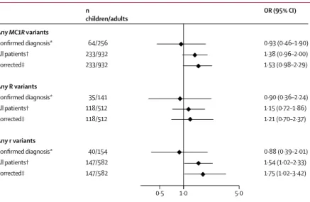
We found that children or adolescents with cutaneous
melanoma had significantly higher odds of carrying any
r variants than those of adult patients (OR 1·54, 95% CI
1·02–2·33; FDRcorrected p=0·17; figure 3). Concerning
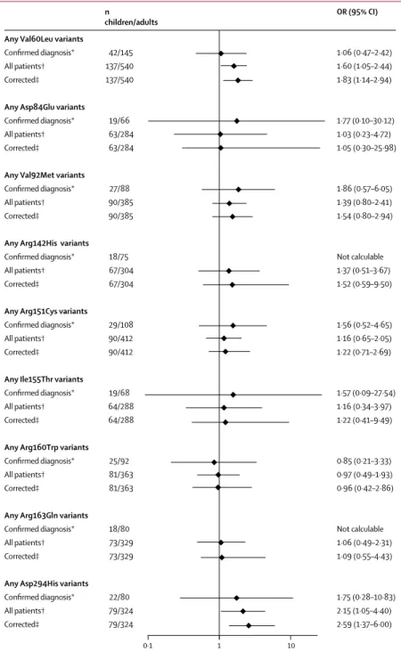
specific MC1R variants, we found a positive association
for all MC1R variants with childhood and adolescent
melanoma, except for the Arg160Trp variant (figure 4).
We found a statistically significant association for
Val60Leu (p=0·04, FDRcorrected p=0·17) and Asp294His
(p=0·04, FDRcorrected p=0·17) variants in both the
primary analysis and after correction for possible
misdiagnosis (figure 4). Similar results were obtained in
the sensitivity analysis with conditional logistic
regression models (appendix) and by excluding the
21 children and adolescents without centrally confirmed
diagnosis (appendix). Finally, when we repeated the
primary analysis on the subgroups of young patients and
adult patients from the same parental study, we obtained
stronger associations with childhood and adolescent
Figure 3: Covariable-adjusted odds ratio (OR) for the association between any MC1R variants, R variants, and r variants and childhood and adolescent melanoma compared with adult melanoma
All ORs were adjusted by sex, matching stratum variable, melanoma body site and histopathological subtype, hair colour, and skin type. For each OR, the comparison groups included child and adolescent patients matched (4:1) with adult patients by study or geographical area. The reference category for OR was MC1R wild-type (WT) individuals. Numbers of children or adolescents and adults reported here are the total numbers of patients included in each analysis, independently by MC1R status. For the analysis on any R variant versus WT, patients carrying only r variants were excluded, and for the analysis on any r variant versus WT, patients carrying only R variants were excluded. R variants include Asp84Glu, Arg142His, Arg151Cys, Ile155Thr, Arg160Trp, Asp294His, and other rare variants classified as R according to the algorithm proposed by Davies and colleagues.34 The r variants include Val60Leu, Val92Met, Arg163Gln, and other rare variants classified as r according to the algorithm proposed by Davies and colleagues.34 MC1R=melanocortin 1 receptor. *Calculated on the subgroup of patients with confirmed diagnosis of melanoma after centralised pathological review of glass slides. †Calculated on the whole sample of 233 child and adolescent patients. ‡Corrected for probability of misdiagnosis, by combining information from OR of confirmed diagnoses and OR of all patients, as suggested elsewhere.35
Any MC1R variants Confirmed diagnosis* All patients† Corrected‡ Any R variants Confirmed diagnosis* All patients† Corrected‡ Any r variants Confirmed diagnosis* All patients† Corrected‡ 64/256 233/932 233/932 35/141 118/512 118/512 40/154 147/582 147/582 n children/adults 0·93 (0·46–1·90) 1·38 (0·96–2·00) 1·53 (0·98–2·29) 0·90 (0·36–2·24) 1·15 (0·72–1·86) 1·21 (0·70–2·37) 0·88 (0·39–2·01) 1·54 (1·02–2·33) 1·75 (1·02–3·42) OR (95% CI) 0·5 1·0 5·0
melanoma than those of the primary analysis for
carriers of any MC1R variant (OR 2·04, 95% CI
1·19–3·50), r variants (2·61, 1·43–4·73), and Val60Leu
(2·67, 1·44–4·95) and Asp294His variants (3·12,
1·08–9·03; appendix).
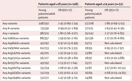
We did a subgroup analysis by age at diagnosis (patients
who were 18 years or younger and patients who were
14 years or younger at diagnosis), and we observed a
significantly higher frequency of r variants in patients
aged 18 years or younger than in adult patients (OR 1·80,
95% CI 1·06–3·07; FDRcorrected p=0·61; table 3). The
corresponding OR for patients aged 14 years or younger
was even higher, but was not significant because of the
small number of patients.
We also did a casecontrol analysis comparing young
patients with melanoma with healthy adult controls
(appendix). We found a significantly higher risk of
childhood and adolescent melanoma in carriers of any
MC1R, R, and r variants, as well as for the most common
MC1R Val60Leu, Val92Met, Arg151Cys, Arg163Gln, and
Asp294His variants compared with that in healthy controls.
Results remained significant after correction for multiple
comparison, except for the Val92Met variant (FDRcorrected
p=0·07).
Discussion
Our pooledanalysis showed that young patients had
significantly higher frequencies of any MC1R variants,
R variants, and r variants than those of healthy controls,
supporting the role of MC1R variants as genetic risk
factors for childhood and adolescent cutaneous
melanoma. We also found that the frequency of r variants
was elevated in young patients compared with that of
adult patients. The effect of r variants was supported by
analyses limited to individuals aged 18 years or younger
and was even stronger, but not significantly, for children
aged 14 years or younger, suggesting a higher prevalence
of MC1R variants in childhood melanoma. The MC1R
Val60Leu and Asp294His variants showed the most robust
association with melanoma in childhood and adolescence,
even after correction for possible misdiagnosis.
Childhood and adolescent melanoma has been report
ed to occur most commonly in white people and in
girls.
2,10,13In line with two previous studies,
12,13we found
that young patients with melanoma are characterised by
a fairer phenotype than that of healthy controls, including
traits such as red hair and skin type. By contrast,
young patients presented with more darkly pigmented
characteristics, such as brown eyes, skin type III or IV,
and a lower prevalence of freckles compared with those
of locationmatched adult patients. Consistent with
most
published studies,
2,11,36our young patients showed a high
number of melanocytic naevi, both common and atypical,
and developed melanomas mainly on the lower limbs
and the trunk. Childhood and adolescent melanoma
was more commonly diagnosed as nodular melanoma
compared with its adult counterpart. Spitzoid melanomas
Figure 4: Covariable-adjusted odds ratio (OR) for the association between the nine most prevalent MC1R variants and childhood and adolescent melanoma compared with adult melanoma
All ORs were adjusted by sex, matching stratum variable, melanoma body site and histopathological type, hair colour, and skin type. For each OR, the comparison groups included young patients matched (4:1) with adult patients by study or geographical area. The reference category for OR was MC1R wild-type (WT) individuals. Numbers of children or adolescents and adults reported here are the total numbers of patients included in each analysis, independently by MC1R status. For the analysis on each variant versus WT, patients carrying only other
MC1R variants were excluded. R variants include Asp84Glu, Arg142His, Arg151Cys, Ile155Thr, Arg160Trp,
Asp294His, and other rare variants classified as R according to the algorithm proposed by Davies and colleagues.34 The r variants include Val60Leu, Val92Met, Arg163Gln, and other rare variants classified as r according to the algorithm proposed by Davies and colleagues.34 MC1R=melanocortin 1 receptor. *Calculated on the subgroup of patients with confirmed diagnosis of melanoma after centralised pathological review of glass slides. †Calculated on the whole sample of 233 young patients. ‡Corrected for probability of misdiagnosis, by combining information from OR of confirmed diagnoses and OR of all patients, as suggested elsewhere.35
Any Val60Leu variants
Confirmed diagnosis* All patients† Corrected‡
Any Asp84Glu variants
Confirmed diagnosis* All patients† Corrected‡
Any Val92Met variants
Confirmed diagnosis* All patients† Corrected‡
Any Arg142His variants
Confirmed diagnosis* All patients† Corrected‡
Any Arg151Cys variants
Confirmed diagnosis* All patients† Corrected‡
Any Ile155Thr variants
Confirmed diagnosis* All patients† Corrected‡
Any Arg160Trp variants
Confirmed diagnosis* All patients† Corrected‡
Any Arg163Gln variants
Confirmed diagnosis* All patients† Corrected‡
Any Asp294His variants
Confirmed diagnosis* All patients† Corrected‡ 42/145 137/540 137/540 19/66 63/284 63/284 27/88 90/385 90/385 18/75 67/304 67/304 29/108 90/412 90/412 19/68 64/288 64/288 25/92 81/363 81/363 18/80 73/329 73/329 22/80 79/324 79/324 1·06 (0·47–2·42) 1·60 (1·05–2·44) 1·83 (1·14–2·94) 1·77 (0·10–30·12) 1·03 (0·23–4·72) 1·05 (0·30–25·98) 1·86 (0·57–6·05) 1·39 (0·80–2·41) 1·54 (0·80–2·94) Not calculable 1·37 (0·51–3·67) 1·52 (0·59–9·50) 1·56 (0·52–4·65) 1·16 (0·65–2·05) 1·22 (0·71–2·69) 1·57 (0·09–27·54) 1·16 (0·34–3·97) 1·22 (0·41–9·49) 0·85 (0·21–3·33) 0·97 (0·49–1·93) 0·96 (0·42–2·86) Not calculable 1·06 (0·49–2·31) 1·09 (0·55–4·43) 1·75 (0·28–10·83) 2·15 (1·05–4·40) 2·59 (1·37–6·00) OR (95% CI) 1 0·1 10 n children/adults
were more frequently identified in young patients,
whereas lentigo maligna melanomas were only seen in
adulthood.
The effect of MC1R variants in childhood and adol
escent melanoma was investigated in small series of
patients.
18–20One study
19published in 2009, identified
MC1R variants in 12 (57%) of 21 patients, with a higher
frequency of r than that of R variants. More
recently, two case series reported MC1R variants in
ten (43%) of 23 patients
18and in four (67%) of six.
20In our
pooledanalysis, MC1R variants were detected in 74% of
young patients.
Our findings showed a stronger role of MC1R r variants
in childhood and adolescent melanoma than in adult
melanoma, suggesting an involvement of biological
pathways other than pigmentation and UVsensitivity,
such as antioxidant defences, DNA repair, and cell
proliferation.
22,24,37Indeed, MC1R signalling was found to
be crucial for melanocyte key processes,
38showing that
MC1R variants, combined with HERC2/OCA2 alleles,
can determine the number of naevi bigger than 2 mm in
sunburned children.
39In our study, the MC1R variants Val60Leu and
Asp294His showed significantly higher prevalence in
childhood and adolescent melanoma than in the adult
form of the disease. The role of Val60Leu in adult
melanoma is controversial, and the magnitude of risk
varies across populations.
40A positive association of
Val60Leu with melanoma has been reported in the
Mediterranean area, where Val60Leu is the most frequent
of all variants.
40The Asp294His variant is common in
individuals with the red hair colour phenotype. The
association of Asp294His with melanoma risk shows the
heterogeneity between northern and southern European
populations, where individuals who are more darkly
pigmented are at higher risk of melanoma associated
with Asp294His than are northern, less pigmented
populations.
41To the best of our knowledge, our series of childhood
and adolescent melanoma patients is the largest
international multicentre cohort published so far with
available MC1R genetic data. The large number of young
patients with melanoma, and comparable adult patients,
provide powerful estimates of the association between
MC1R variants and childhood and adolescent melanoma
within different populations. Another strength of our
study was the centralised data quality control and
statistical analysis that provided consistency across the
numerous parent studies in defining and adjusting for
important covariates. Histopathological centralised
review of a third of the patients allowed us to calculate
association estimates in a subset of children or
adolescents with a histologically confirmed diagnosis
and was helpful for calculating corrected risk estimates
considering the issue of misdiagnosis.
Young patients with melanoma represent a hetero
geneous group, including neonates, children, and
adolescents, with various distinct presen tations.
9Child
hood melanoma might indeed differ from adol escent
melanoma, and both might differ from adult melanoma.
4To further address heterogeneity between melanomas
developed at different ages, we did a stratified analysis
for patients aged 14 years or younger and 18 years or
younger. Our nonsignificant findings from the younger
subgroup might have resulted from decreased power
related to the small sample size (59 patients) of this
subgroup, whereas a separate multivariable analysis
limited to children aged 10 years or younger was not
possible because of the low number of patients
(23 patients). In our child and adolescent sample, we
had more darkly pigmented patients from southern
European countries than from northern European
origin, which might have resulted in high frequencies of
r variants, more common in southern Europe than in
northern Europe.
42However, because young patients
were compared with adult patients and controls from
the same geographical areas, we do not believe this
affected our results. Indeed, a sensitivity analysis done
in the subgroup of young patients with adult patients
sampled from the same parent study provided similar
results. A centralised review of all melanomas would be
desirable but, unfortunately, it was not feasible because
of the retrospective nature of the study. To reduce disease
misclassification, we excluded from the analysis patients
whose histopathological reports were not available. We
also provided risk estimates corrected for our observed
Patients aged ≤18 years (n=148) Patients aged ≤14 years (n=52) Young patients/adult patients OR(95% CI) Young patients/adult patients OR (95% CI) Any variants 148/592 1·45 (0·89–2·34) 52/208 1·86 (0·69–5·03) Any R variants 73/330 0·99 (0·52–1·89) 27/115 1·63 (0·42–6·36) Any r variants 98/374 1·80 (1·06–3·07) 35/142 2·27 (0·76–6·83) Any Val60Leu variants 88/357 1·59 (0·91–2·76) 31/136 2·27 (0·76–6·80) Any Asp84Glu variants 43/192 0·97 (0·13–6·99) 15/73 Not calculated Any Val92Met variants 61/253 1·62 (0·79–3·33) 18/93 0·95 (0·11–7·97) Any Arg142His variants 45/201 1·32 (0·34–5·13) 13/80 Not calculated Any Arg151Cys variants 56/277 0·82 (0·38–1·80) 18/97 0·61 (0·10–3·88) Any Ile155Thr variants 43/192 1·13 (0·17–7·64) 15/71 Not calculated Any Arg160Trp variants 55/234 1·08 (0·45–2·58) 21/87 3·57 (0·62–20·52) Any Arg163Gln variants 51/219 1·61 (0·61–4·22) 16/84 0·68 (0·03–14·81) Any Asp294His variants 52/272 1·47 (0·58–3·70) 14/86 Not calculated Odds ratios (ORs) were adjusted by sex, matching stratum variable, melanoma body site and histological subtype, and skin type. Hair colour was not included because this category had more than 30% of missing data for these groups of patients. For each OR, the comparison group included 4:1 frequency-matched adult patients by study or geographical area. The reference category for ORs were MC1R wild-type individuals. The number of children and adults reported here are the total number of patients included in each analysis, independently by MC1R status. For the analysis on each variant versus wild type, patients carrying only other MC1R variants were excluded. R variants include Asp84Glu, Arg142His, Arg151Cys, Ile155Thr, Arg160Trp, Asp294His, and other rare variants classified as R according to the algorithm proposed by Davies et al.34 The r variants include Val60Leu, Val92Met, Arg163Gln, and other rare variants classified as r according to the algorithm proposed by Davies et al.34 MC1R=melanocortin 1 receptor.
misclassification prevalence among patients with
histopathological centralised review, a group that was
representative of the entire cohort of young patients.
Nevertheless, we should note that this correction could
not provide an exact estimate of the associations, as in a
sample with only centrally confirmed diagnosed cases,
and some imprecision of estimates could therefore not
be ruled out. Because our cohort did not include patients
with familial melanoma and the major susceptibility
genes are rarely mutated in young patients,
12,15,17,20we did
not analyse CDKN2A and CDK4 genes in our patients. It
is possible that other major melanoma predisposition
genes might influence the risk of disease in children and
adolescents, but the absence of
genetic data on these
genes, such as BAP1, prevented the analysis of possible
gene–gene interactions. Finally, although we did a high
number of statistical tests, we allowed unadjusted
p values to guide the interpretation of our results.
Because of the exploratory, rather than confirmatory,
nature of this study, we believe that our approach of
describing the tests of significance we did, as advised by
Thomas V Perneger,
43is appropriate. However, to directly
address the issue of multiple testing, we also presented
FDRcorrected p values.
In conclusion, our pooled analysis showed that natural
variations in MC1R are a genetic risk factor for childhood
and adolescent cutaneous melanoma, as well as for adult
cutaneous melanoma. MC1R variants, mainly r alleles,
were suggested to have a greater role in childhood and
adolescent melanoma than in adult melanoma, possibly
through a pigmentationindependent pathway. Add
itionally, we observed a stronger effect of r variants when
the analysis was restricted to patients with mela noma
aged 18 years or younger. Our study contributes to the
comprehension of the genetic background of paediatric
melanoma and elucidates the genetic diversity of
paediatric and adult melanoma, with potential clinical
relevance for developing early melanoma detection and
preventive strategies.
Contributors
CP and SR did the literature search. SR, SG, PM, PAK, JCGB, HN, MTL, JL, JNB, FS, and MCF contributed to study design. MFA, FD, BBdP, VH, AEC, HAC, SBG, RPG, LM, RZ, TD, NET, CBB, MB, SP, MP, EN, PG, CMe, AMM, MR, SB, EP, LKS, FBa, GG, AS, FO, FA, RFdM, PQ, GR, AR, EM, IS, PAK, MAP, and MCF contributed to data collection. DM and FF did the histophatological review. CP and CMa did the molecular analysis of MC1R for new samples. FBo and SR analysed the data. CP, DM, SG, PAK, MTL, JL, MCF, and SR contributed to data interpretation. CP, FBo, and CMa wrote the manuscript. FB did the figures.
M-SKIP Study Group
Principal investigator: Sara Raimondi (IEO, European Institute of Oncology IRCCS, Milan, Italy). Advisory Committee members: Philippe Autier (International Prevention Research Institute, Lyon, France), Maria Concetta Fargnoli (University of L’Aquila, Italy), José C GarcíaBorrón (University of Murcia, Spain), Jiali Han (Indiana University, Indianapolis, IN, USA), Peter A Kanetsky (Department of Cancer Epidemiology, H Lee Moffitt Cancer Center and Research Institute, Tampa, FL, USA), Maria Teresa Landi (National Cancer Institute, NIH, Bethesda, MD, USA), Julian Little (University of
Ottawa, Canada), Julia NewtonBishop (University of Leeds, UK), Francesco Sera (London School of Hygiene & Tropical Medicine, London, UK). Consultants: Saverio Caini (ISPO, Florence, Italy), Sara Gandini and Patrick Maisonneuve (IEO, European Institute of Oncology IRCCS, Milan, Italy). Participant investigators: Albert Hofman, Manfred Kayser, Fan Liu, Tamar Nijsten, and Andre G Uitterlinden (Erasmus MC University Medical Center, Rotterdam, Netherlands), Rajiv Kumar (German Cancer Research Center, Heidelberg, Germany), Tim Bishop and Faye Elliott (University of Leeds, Leeds, UK), Eduardo Nagore (Instituto Valenciano de Oncologia, Valencia, Spain), DeAnn Lazovich (Division of Epidemiology and Community Health, University of Minnesota, MN, USA), David Polsky (New York University School of Medicine, New York, NY, USA), Johan Hansson and Veronica Hoiom (Karolinska Institutet, Stockholm, Sweden), Paola Ghiorzo and Lorenza Pastorino (University of Genoa, Genoa, Italy), Nelleke A Gruis and
Jan Nico Bouwes Bavinck (Leiden University Medical Center, Leiden, Netherlands), Ricardo FernandezdeMisa (Hospital Universitario Nuestra Señora de Candelaria, Santa Cruz de Tenerife, Spain), Paula Aguilera, Celia Badenas, Cristina Carrera, Pol GimenezXavier, Josep Malvehy, Miriam Potrony, Susana Puig, Joan Anton PuigButille, and Gemma TellMarti (Hospital Clinic, IDIBAPS and CIBERER, Barcelona, Spain), Terence Dwyer (Murdoch Childrens Research Institute, VIC, Australia), Leigh Blizzard and Jennifer Cochrane (Menzies Institute for Medical Research, Hobart, Australia), Wojciech Branicki (Institute of Forensic Research, Krakow, Poland), Tadeusz Debniak (Pomeranian Medical University, Polabska, Poland), Niels Morling and Peter Johansen (University of Copenhagen, Denmark), Susan Mayne, Allen Bale, Brenda Cartmel, and
Leah Ferrucci (Yale School of Public Health and Medicine, New Haven, CT, USA), Ruth Pfeiffer (National Cancer Institute, NIH, Bethesda, MD, USA), Giuseppe Palmieri (Istituto di Chimica Biomolecolare, CNR, Sassari, Italy), Gloria Ribas (Fundación Investigación Clínico de Valencia Instituto de Investigación Sanitaria INCLIVA, Spain), Chiara Menin (Veneto Institute of Oncology IOVIRCCS, Padua, Italy), Alexandros Stratigos and Katerina Kypreou (University of Athens, Andreas Sygros Hospital, Athens, Greece), Anne Bowcock,
Lynn Cornelius, and M Laurin Council (Washington University School of Medicine, St Louis, MO, USA), Tomonori Motokawa (POLA Chemical Industries, Yokohama, Japan), Sumiko Anno (Shibaura Institute of Technology, Tokyo, Japan), Per Helsing and Per Arne Andresen (Oslo University Hospital, Oslo, Norway), Gabriella Guida (University of Bari, Bari, Italy), Stefania Guida (University of Modena and Reggio Emilia, Modena, Italy), Terence H Wong (University of Edinburgh, Edinburgh, UK), and the GEM Study Group.
GEM Study Group
Coordinating Center, Memorial SloanKettering Cancer Center, New York, NY, USA: Marianne Berwick (principal investigator, currently at the University of New Mexico), Colin Begg (coprincipal investigator), Irene Orlow (coinvestigator), Urvi Mujumdar (project coordinator), Amanda Hummer (biostatistician), Klaus Busam (dermatopathologist), Pampa Roy (laboratory technician), Rebecca Canchola (laboratory technician), Brian Clas (laboratory technician), Javiar Cotignola (laboratory technician), Yvette Monroe (interviewer). Study Centre, University of Sydney and The Cancer Council New South Wales, Sydney (Australia): Bruce Armstrong (principal investigator), Anne Kricker (coprincipal investigator), Melisa Litchfield (study coordinator). Study centre, Menzies Institute for Medical Research, University of Tasmania, Hobart (Australia): Terence Dwyer (principal investigator), Paul Tucker
(dermatopathologist), Nicola Stephens (study coordinator). Study centre, British Columbia Cancer Agency, Vancouver (Canada): Richard Gallagher (principal investigator), Teresa Switzer (coordinator). Study centre, Cancer Care Ontario, Toronto (Canada): Loraine Marrett (principal investigator), Beth Theis (coinvestigator), Lynn From (dermatopathologist), Noori Chowdhury (coordinator), Louise Vanasse (coordinator), Mark Purdue (research officer). David Northrup (manager for CATI). Study centre, Centro per la Prevenzione Oncologia Torino, Turin (Italy): Roberto Zanetti (principal investigator),
Stefano Rosso (data manager), Carlotta Sacerdote (coordinator). Study centre, University of California, Irvine (USA): Hoda AntonCulver (principal investigator), Nancy Leighton (coordinator), Maureen Gildea (data manager). Study centre, University of Michigan, Ann Arbor (USA): Stephen Gruber (principal investigator), Joe Bonner (data manager), Joanne Jeter (coordinator). Study centre, New Jersey Department of Health and Senior Services, Trenton (USA): Judith Klotz (principal investigator), Homer Wilcox (coprincipal investigator), Helen Weiss (coordinator). Study centre, University of North Carolina, Chapel Hill (USA): Robert Millikan (principal investigator), Nancy Thomas (coinvestigator), Dianne Mattingly (coordinator), Jon Player (laboratory technician), ChiuKit Tse (data analyst). Study centre, University of Pennsylvania, Philadelphia, PA (USA): Timothy Rebbeck (principal investigator), Peter Kanetsky (coinvestigator), Amy Walker (Laboratory technician), Saarene Panossian (laboratory technician). Consultants: Harvey Mohrenweiser, University of California, Irvine, Irvine, CA (USA); and Richard Setlow, Brookhaven National Laboratory, Upton, NY (USA).
IMI study group
Daniela Massi (University of Florence, Italy), Paola Ghiorzo and Lorenza Pastorino (University of Genoa, Italy), Chiara Menin (Veneto Institute of Oncology, IOVIRCCS, Padua, Italy), Mauro Alaibac (University of Padua, Italy), Ausilia Maria Manganoni, Fabio Facchetti (University of Brescia, Italy), Monica Rodolfo, Andrea Ferrari, Barbara Valeri (Fondazione IRCCS Istituto Nazionale dei Tumori, Milan, Italy), Maria Concetta Fargnoli, Cristina Pellegrini (University of L’Aquila, Italy), Sonia Brugnara, Mariacristina Sicher, Daniela Mangiola (S. Chiara Hospital, Trento, Italy), Emanuela Passoni, Gianluca Nazzaro (Fondazione IRCCS Ca’ Granda, Ospedale Maggiore Policlinico, Milan, Italy), Federica Baldini, Giulio Tosti, Sara Gandini, Giovanni Mazzarol (IEO, European Institute of Oncology IRCCS, Milan, Italy), Gabriella Guida, Stefania Guida, Giuseppe Giudice (Università degli Studi di Bari Aldo Moro, Bari, Italy), Fabrizio Ayala (National Cancer Institute, Fondazione G PascaleIRCCS, Naples, Italy), Pietro Quaglino, Simone Ribero, Chiara Astrua (University of Torino, Turin, Italy), Antonella Romanini (Azienda Ospedaliero Universitaria Santa Chiara, Pisa, Italy), Emilia Migliano (San Gallicano Dermatological Institute, IRCCS, Rome, Italy), Ignazio Stanganelli, Laura Mazzoni (IRCCSIRST Scientific Institute of Romagna for the Study and Treatment of Cancer, Meldola and University of Parma, Italy), and Maria Antonietta Pizzichetta (CRO Aviano National Cancer Institute, Italy).
Declaration of interests
We declare no competing interests. Acknowledgments
We have received funding for this study from the Society for Pediatric Dermatology (SPD Pilot Project Award 2015) and Italian Association for Research on Cancer (AIRC MFAG 11831). PAK has received grants for the Melanoma Susceptibility Study from the National Cancer Institute (CA75434, CA80700, CA092428). PG has received funding for the Genoa study from the Italian Association for Research on Cancer (AIRC IG 15460). JL holds a tier 1 Canada Research Chair. The study in the Melanoma Unit, Hospital Clínic (Barcelona, Spain) was supported in part by grants from Fondo de Investigaciones Sanitarias
(PI 12/00840, PI15/00956, and PI15/00716) and the CIBER de Enfermedades Raras of the Instituto de Salud Carlos III (Madrid, Spain); was cofunded by EU European Regional Development Funds and by the AGAUR 2014_SGR_603 and 2017_SGR_1134 of the Catalan Government (Spain); and was funded by a grant from Fundació La Marató de TV3 (201331–30; Catalonia, Spain), by the European Commission under the 6th Framework Programme
(LSHCCT2006–018702; GenoMEL), by the CERCA Programme– Generalitat de Catalunya, and by a research grant from the Fundación Científica de la Asociación Española Contra el Cáncer, Spain (GCB15152978SOEN). Part of the work was developed at the building Centro Esther Koplowitz, Barcelona (Spain). MFA, FD, and BBdP have received funding from the Hospital Programme for Clinical Research (France, PHRC 2007 AOM 07–195NI 07004). We thank the medical doctors who included some of the patients of this study. We wish to acknowledge the work of Gustave Roussy Biobank (BB0033–00074) in providing DNA resources.
References
1 Nikolaou V, Stratigos AJ. Emerging trends in the epidemiology of melanoma. Br J Dermatol 2014; 170: 11–19.
2 Strouse JJ, Fears TR, Tucker MA, Wayne AS. Pediatric melanoma: risk factor and survival analysis of the surveillance, epidemiology and end results database. J Clin Oncol 2005; 23: 4735–41. 3 Austin MT, Xing Y, HayesJordan AA, Lally KP, Cormier JN.
Melanoma incidence rises for children and adolescents: an epidemiologic review of pediatric melanoma in the United States. J Pediatr Surg 2013; 48: 2207–13.
4 LaChance A, Shahriari M, Kerr PE, GrantKels JM. Melanoma: kids are not just little people. Clin Dermatol 2016; 34: 742–48.
5 Lorimer PD, White RL, Walsh K, et al. Pediatric and adolescent melanoma: a national cancer data base update. Ann Surg Oncol 2016; 23: 4058–66.
6 SteliarovaFoucher E, Colombet M, Ries LAG, et al. International incidence of childhood cancer, 2001–10: a populationbased registry study. Lancet Oncol 2017; 18: 719–31.
7 Livestro DP, Kaine EM, Michaelson JS, et al. Melanoma in the young—differences and similarities with adult melanoma: a casematched controlled analysis. Cancer 2007; 110: 614–24. 8 Berg P, Lindelöf B. Differences in malignant melanoma between
children and adolescents. A 35year epidemiological study.
Arch Dermatol 1997; 133: 295–97.
9 Tracy ET, Aldrink JH. Pediatric melanoma. Semin Pediatr Surg 2016; 25: 290–98.
10 Averbook BJ, Lee SJ, Delman KA, et al. Pediatric melanoma: analysis of an international registry. Cancer 2013 119: 4012–19. 11 Pappo AS. Melanoma in children and adolescents. Eur J Cancer
2003; 39: 2651–61.
12 Youl P, Aitken J, Hayward N, et al. Melanoma in adolescents: a casecontrol study of risk factors in Queensland, Australia.
Int J Cancer 2002; 98: 92–98.
13 Whiteman DC, Valery P, McWhirter W, Green AC. Risk factors for childhood melanoma in Queensland, Australia. Int J Cancer 1997; 70: 26–31.
14 Ripperger T, Bielack SS, Borkhardt A, et al. Childhood cancer predisposition syndromes—a concise review and recommendations by the Cancer Predisposition Working Group of the Society for Pediatric Oncology and Hematology. Am J Med Genet A 2017; 173: 1017–37.
15 Berg P, Wennberg AM, Tuominen R, et al. Germline CDKN2A mutations are rare in child and adolescent cutaneous melanoma.
Melanoma Res 2004; 14: 251–55.
16 Whiteman DC, Milligan A, Welch J, Green AC, Hayward NK. Germline CDKN2A mutations in childhood melanoma.
J Natl Cancer Inst 1997; 89: 1460.
17 Hocevar M, Avbelj M, Perić B, Zgajnar J, Besić N, Battelino T. High prevalence of germline CDKN2A mutations in Slovenian cutaneous malignant melanoma families. Croat Med J 2006; 47: 851–54.
18 Lu C, Zhang J, Nagahawatte P, et al. The genomic landscape of childhood and adolescent melanoma. J Invest Dermatol 2015; 135: 816–23.
19 Daniotti M, Ferrari A, Frigerio S. Cutaneous melanoma in childhood and adolescence shows frequent loss of INK4A and gain of KIT. J Invest Dermatol 2009; 129: 1759–68.
20 Rabbie R, Rashid M, Arance AM, et al. Genomic analysis and clinical management of adolescent cutaneous melanoma.
Pigment Cell Melanoma Res 2017; 30: 307–16.
21 Wilmott JS, Johansson PA, Newell F, et al. Whole genome sequencing of melanomas in adolescent and young adults reveals distinct mutation landscapes and the potential role of germline variants in disease susceptibility. Int J Cancer 2019; 144: 1049–60.
22 Beaumont KA, Shekar SN, Newton RA, et al. Receptor function, dominant negative activity and phenotype correlations for MC1R variant alleles. Hum Mol Genet 2007; 16: 2249–60.
23 Dessinioti C, Antoniou C, Katsambas A, Stratigos AJ. Melanocortin 1 receptor variants: functional role and pigmentary associations. Photochem Photobiol 2011; 87: 978–87.
24 Kanetsky PA, Rebbeck TR, Hummer AJ, et al. Populationbased study of natural variation in the melanocortin1 receptor gene and melanoma. Cancer Res 2006; 66: 9330–37.