Contents lists available atScienceDirect
BBA - Molecular Basis of Disease
journal homepage:www.elsevier.com/locate/bbadisEmerging metabolic risk factors in hepatocellular carcinoma and their
in
fluence on the liver microenvironment
Pasquale Agosti, Carlo Sabbà, Antonio Mazzocca
⁎Interdisciplinary Department of Medicine, University of Bari School of Medicine, Piazza G. Cesare, 11, 70124 Bari, Italy
A R T I C L E I N F O
Keywords:Hepatocellular carcinoma (HCC) Non-alcoholic fatty liver disease (NAFLD) Nonalcoholic steatohepatitis (NASH) Liver microenvironment
Inflammation Insulin resistance
A B S T R A C T
Despite the reducing incidence of chronic hepatitis infections, an unexpected increasing incidence of hepato-cellular carcinoma (HCC) has being occurred. This may be explained by the increasing number of HCCs de-veloping on steatosis (NAFLD) and steatohepatitis (NASH), related to metabolic risk factors (i.e. diabetes mel-litus type II, obesity, metabolic syndrome), which are becoming emerging risk factors for HCC. This led to a growing scientific interest on the oncogenic mechanisms underlying the transition from NAFLD to HCC. However, patients with NASH receive significantly less HCC surveillance than patients with chronic hepatitis, and no specific preventive pharmacological treatments have recommended for NASH-related HCC. This review focuses on the pathogenic role of the emerging factors involved in the transition from NAFLD/NASH to HCC, including microbiota, insulin resistance, inflammation, lipid and bile acids metabolism. It will be emphasize their impact on the liver microenvironment, the implications in clinical practice and the future directions of research.
1. Introduction
Hepatocellular carcinoma (HCC) is actually the sixth cause of cancer-related death in the world and it is estimated to become the third cause in Western countries by 2030, despite the reducing in-cidence of chronic hepatitis infections [1]. The explanation for this unexpected increase of incidence may be found in the significant change in thefield of liver cancer epidemiology. In fact, an increasing number of HCCs develop on liver metabolic disorders including Non Alcoholic Fatty Liver Disease (NAFLD) and Nonalcoholic Steatohepa-titis (NASH), which are becoming the new precancerous conditions, in addition to the traditionally known virus-induced cirrhosis. Therefore, metabolic risk factors commonly associated to NAFLD or NASH, in-cluding diabetes mellitus type II, obesity and metabolic syndrome are becoming emerging risk factors for HCC. This is based upon epide-miological evidence showing the significant relationship of these con-ditions with incidence of HCC, regardless of the common risk factors such as chronic hepatitis or alcohol abuse. Therefore, it is not surprising the growing scientific interest during the last few years on the onco-genic mechanisms underlying the transition from liver metabolic
disorders to HCC involving these new metabolic risk factors. This re-view focuses on the pathogenic role of the emerging factors involved in the transition from steatosis to HCC. These factors include insulin re-sistance, inflammation, lipid and bile acids metabolism and the gut microbiota. A better understanding of the impact of these factors on the liver microenvironment may have potential benefit on the management of liver disease.
2. Emerging metabolic risk factors
Metabolic syndrome has been associated with an increased risk of HCC. In particular, each feature of this syndrome may increase cancer risk and a synergic effect has been described[2–3]. Overweight and obesity are well-recognized risk factors for HCC[4]. However, visceral adiposity shows a stronger association with HCC risk than general body weight [5]. Furthermore, obesity may influence HCC prognosis. For example, in a large study, Body Mass Index (BMI) was predictor of microvascular invasion and worsened prognosis[6], whereas visceral adiposity correlated with HCC recurrence after treatment in another study[7]. Diabetes mellitus type 2 (DM II) has been proposed as an
https://doi.org/10.1016/j.bbadis.2017.11.026
Received 23 September 2017; Received in revised form 14 November 2017; Accepted 28 November 2017
⁎Corresponding author.
E-mail address:[email protected](A. Mazzocca).
Abbreviations: ERK, extracellular signal regulated kinase; FXR, farsenoid X receptor; HCC, hepatocellular carcinoma; HSCs, hepatic stellate cells; IL1β, Interleukin 1 beta; IL18, Interleukin 18; IKKβ, inhibitor of nuclear factor kappa-B kinase subunit beta; JAK, Janus kinase; JNK, c-Jun N-terminal kinases; LPARs, lysophosphatidic acid receptors; MAPK, mitogen-activated protein kinase; MCP1, monocyte chemoattractant protein-1; MetS, metabolic syndrome; mTOR, mammalian target of rapamycin; NAFLD, non-alcoholic fatty liver disease; NASH, non-alcoholic steatohepatitis; NF-kB, nuclear factor kappa-light-chain-enhancer of activated B cells; SASP, senescence-associated secretory phenotype; STAT, signal transducer and activator of transcription; TAG, triacylglyceride; TGFβ, transforming growth factor beta; TNFα, tumor necrosis factor alpha; TLR, Toll Like Receptor
Available online 29 November 2017
0925-4439/ © 2017 Elsevier B.V. All rights reserved.
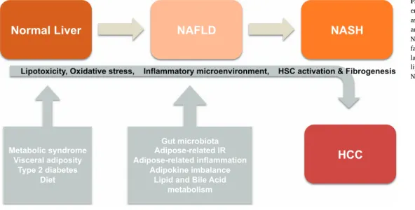
important independent predictor of HCC regardless of alcohol con-sumption [8]. Hyperlipidemia and hyperthension are two additional features of metabolic syndrome that have been studied in relation to HCC. In particular, hyperthension seems to be related to a higher risk of HCC, whereas the relation between HCC and hyperlipidemia is quite controversial[2–3]. Also, synergism between traditional and new risk factors has to be considered. For example, a strong synergic effect of alcohol abuse and DM II has been described [9]. Moreover, diabetes and obesity have been reported to enhance the risk of HCC in cohorts of chronic hepatitis patients, even more if both metabolic risk factors were present [10]. Metabolic risk factors affecting the natural history of NAFLD towards HCC are illustrated inFig. 1.
3. NAFLD and NASH as emerging precancerosis
Although NAFLD diagnosis can be made by means of imaging (i.e. ultrasound or magnetic resonance), biopsy and the consequent his-tology still remain the gold standard. Hishis-tology generally displays the accumulation of triglycerides in hepatocytes, usually in mixed macro-vesicular or micromacro-vesicular droplets, in the absence of alcohol abuse, steatogenic medication or hereditary disorders. Based on the increasing incidence of NAFLD in the general population, this condition is be-coming the most common underlying risk factor for HCC. In particular, in histology-based studies, carried out in apparently healthy candidates for liver donation, the authors reported that the prevalence of NAFLD was 12–18% in Europe and 27–38% in the United States. The pre-valence of steatosis was significantly higher in patients with metabolic risk factors, up to 94% in obese patients, 67% in overweight patients and 40–74% in diabetic patients[11]. The association between NAFLD and each component of the metabolic syndrome is well recognized and some authors have proposed hepatic steatosis itself as a feature of metabolic syndrome [12]. With regard to the prevalence of NASH, about 6–15% of cases were reported in non-selected samples, whereas prevalence was higher in patients with obesity or DM II (25–30%) and in severely obese with DM II (35%). Nevertheless, the prevalence of NASH is difficult to determine because biopsy is required, including specific criteria such as steatosis, hepatocellular injury, mainly in the form of ballooning, and lobular inflammation. Liver fibrosis may be present in noncirrhotic NASH, initially in perisinusoidal acinar zone 3 [11]. However, NASH is probably underdiagnosed because it may be misclassified with cryptogenic cirrhosis (CC), which shares the same risk factors including diabetes and obesity[13–14]. Therefore, to better estimate the prevalence of NASH, a novel NASH category including obese patients with cryptogenic cirrhosis or with unknown HCC etiology has been proposed. As a consequence, NASH resulted as the
second leading cause of HCC in patients undergoing liver transplanta-tion[15].
4. Natural history: Transition from steatosis to hepatocellular carcinoma
About 20% of NAFLD patients may have NASH, which may progress to cirrhosis in 20–45% of cases, a known precancerosis for HCC[16]. About 7% of patients with NASH-related cirrhosis may progress to HCC within 6.5 years [11]. The different long-term prognosis between NAFLD and NASH patients has been explained based on the presence (and the degree) of fibrosis, which has been described as a crucial prognostic factor. In fact,fibrosis stage strongly predicts liver disease-related and overall mortality in NAFLD patients[17]. Interestingly, a recent meta-analysis reported a rate of fibrosis progression of 0.07 stages per annum in patients with steatosis but without fibrosis at baseline, and a doubled rate for NASH patients. However, the propor-tion of patients whose liverfibrosis progressed from stage 0 to stage 3 or 4 was comparable in the two subgroups[18]. Based on the reported older age of patients with NASH compared with those with NAFLD and on the median 8-years interval between the two conditions, the higher stages offibrosis observed in NASH may reflect only a longer length of liver disease[19]. In addition, in one study, althoughfibrosis stage was significantly more advanced at stage of HCC when compared to the stage of NASH, 28% of patients showed stage 1 or stage 2fibrosis at the time of HCC diagnosis[20]. Therefore, advancedfibrosis and cirrhosis is not a necessary step for HCC development in the presence of steatosis, as shown by cases of HCC developing in non-cirrhotic livers, mainly in old male and obese patients or with criteria of metabolic syndrome [21]. In another study, by analyzing a wide healthcare claims database, the authors reported that NAFLD/NASH was the most common un-derlying risk factor for HCC (59%) and the majority of these tumors that developed on NAFLD had no International Classification of Diseases code for cirrhosis[22]. The samefindings were reported analyzing the SEER-Medicare database. The authors showed that 36% of NAFLD-re-lated HCCs developed in non-cirrhotic livers and 18% in absence of steatohepatitis. Non-cirrhotic NAFLD was the only etiology in 5.8% of HCC patients and isolated fatty liver was the only etiology in about 1% of the total cohort[23]. Since HCC may occur in NAFLD patients even in the absence of progression to NASH, steatosis itself may be con-sidered a precancerous condition. Several genetic and environmental factors influence progression of NAFLD to liver cancer and can be considered as modifiers of the natural history of liver steatosis. Patients with NAFLD or NASH have a much higher risk for developing cirrhosis and HCC in the presence of one or more metabolic syndrome features
NAFLD
NASH
HCC
Metabolic syndrome Visceral adiposity Type 2 diabetes DietNormal Liver
Gut microbiota Adipose-related IR Adipose-related inflammation Adipokine imbalance Lipid and Bile Acidmetabolism
Lipotoxicity, Oxidative stress, Inflammatory microenvironment, HSC activation & Fibrogenesis
Fig. 1. Natural history of NAFLD/NASH and emerging risk factors for HCC. Factors such as metabolic syndrome, visceral adiposity and type 2 diabetes are risk factors for NAFLD and NASH development. Additional factor including gut microbiota, adipose-re-lated inflammation, and excessive intake of lipids may accelerate the transition from NAFLD towards NASH and HCC.
[2–3]. Recent studies have shown the role of specific gene poly-morphisms as determinants of NAFLD susceptibility and progression. One of the most studied nucleotide polymorphisms in this field is rs738409 C/G resulting in an isoleucine to methionine substitution at residue 148 (I148M) in human patatinlike phospholipase domain con-taining 3 (PNPLA3) leading to alteration of TAG remodeling in lipid droplets. This variant has been associated with increased fat content, NAFLD susceptibility and progression, and with a higher risk for liver fibrosis and NAFLD-related HCC[16]. Other genetic modifiers of nat-ural history of NAFLD include glucokinase regulator (GCKR) and transmembrane 6 superfamily 2 (TM6SF2). TM6SF2 E167K and GCKR rs780094 gene variants are associated with an increased risk for fatty liver and liverfibrosis. Therefore, the combined evaluation of clinical comorbidities and specific gene variants may be useful in clinical practice and for risk stratification in NAFLD patients[11].
5. The active role of liver microenvironment in HCC development on a dysmetabolic background
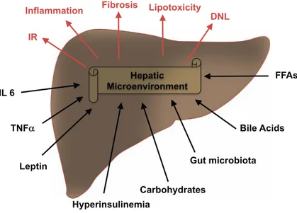
The initial“two-hit hypothesis” of NASH pathogenesis considering steatosis as thefirst step followed by a “second hit” of injury due to inflammation and oxidative stress has been gradually replaced by the “multiple-hits hypothesis”, according to which the described above events occur in parallel. Therefore, the triglycerides storage may be an early adaptive and protective stress response to hepatocytes injuries [11]. Interestingly, more recent evidence shows the key role of liver microenvironment in NAFLD progression. In fact, liver microenviron-ment is considered a proinflammatory milieu, based on the wide variety of immunologically active cells, such as Kupffer cells, T cells and several antigen presenting cells, hepatic stellate cells (HSCs) and endothelial cells. For example, HSCs express Toll like receptor 4 (TLR4), activate IKB Kinase/NF-kB and JNK pathways and release proinflammatory cytokines (i.e. IL6, TGFβ1 and MCP1) with consequent T cells recruit-ment upon activation by a liver injury [24]. Hedgehog signaling, in-volved in modulation of myofibroblast transdifferentiation by recruit-ment of NKT cells, has been related tofibrosis stage in NASH patients [25]. As a consequence of this complex proinflammatory and fibrogenic background, cell death of liver cells occur, thus amplifying these pro-cesses. Also, cellular senescence may have a relevant role in steatosis to HCC transition. In fact, the increased ability of senescent cells (i.e. HSCs) to secrete cytokines, chemokines, matrix remodeling factors and growth factors known as senescence-associated secretory phenotype (SASP) has been related tofibrosis progression[26]. In summary, the proinflammatory and fibrogenic liver microenvironment plays a de-terminant role in NAFLD progression in addition to a persistent external injury. Several pathobiological factors and mechanisms interact with the liver microenvironment in a bidirectional crosstalk creating, in the long term, potential pro-oncogenic substrates that increase the prob-ability of HCC development (Fig. 2). Among these substrates, the in-teractions with the gut microbiota, the obesity-inflammatory pheno-type, the insulin resistance, the lipid and metabolism of bile acids are the main investigatedfields and therefore hereafter reviewed. 6. Microbiota and the crosstalk with the liver microenvironment
The discovery of fatal NASH complicating jejunoileal bypass in bariatric surgery and the reversal of trend after metronidazol therapy was the basis for the scientific interest on the role of gut microbiota in NAFLD progression[27]. As reviewed in detail, a higher prevalence of small intestinal bacterial overgrowth (SIBO) has been observed in pa-tients with NASH [28]. Also, specific microflora changes may have a role in steatosis progression, particularly in obese patients. For ex-ample, NASH patients showed reduced Bacteroides and increased al-cohol-producing species [29–30]. The following main mechanisms implicated in the progression of gut microbiota-related NAFLD to NASH and HCC have been described: i) alteration of intestine permeability; ii)
persistent activation of innate immune system with consequent in-flammation; iii) changes in dietary choline and bile acid metabolism [31].
i) Patients with NAFLD show increased gut permeability with al-tered tight junctions, and these alterations may be induced by high fat diet. Therefore, dysbiosis may induce intestinal inflammation, bacterial and TLR4/TLR9 agonist traslocation to the liver, activation of in-flammatory pathways (i.e. TNFα) and eventually progression of stea-tosis[31]. ii) Innate immunity has a crucial role in modulation of the crosstalk between the gut and the liver and its persistent activation has been implicated in HCC development. In this field of investigation, many observations show a crucial role of lipopolysaccharide (LPS), a known innate immune system activator. In fact, a higher prevalence of gram-negative bacteria in microbiota known to produce LPS was in-volved in liverfibrosis progression in a mouse model[32]. The key role of LPS was also well described by Cani et al. in mouse models. These authors described increased LPS levels after high fat diet, NASH de-velopment after subcutaneous LPS infusion and beneficial metabolic effects on glucose intolerance and fat mass after antibiotics treatments [33]. Thesefindings were confirmed in human study wherein increased LPS-binding protein (LBP) levels were reported in NAFLD obese pa-tients and even more in NASH obese papa-tients, correlating with liver TNFα expression[34]. In mouse models of hepatocarcinogenesis, the gut microbiota was found to be involved in HCC progression, rather than initiation, through activation of TLR4 signaling in HSCs [35]. Another important mediator of the liver-gut interaction is Toll Like Receptor 5 (TLR5), expressed in gut mucosa. In fact, in TLR5-deficient mice, features of microbiome-mediated metabolic syndrome can be transferred to wild type mice by microflora transmittion [36]. Also, inflammasomes sensors of exogenous PAMPs and DAMPs (pathogen-and damaged-associated molecular patterns), regulating maturation of IL1β and IL18, may play a key role in the gut-liver crosstalk. In fact, changes in inflammasome-deficiency-related microbioma have been related to NAFLD progression and metabolic features, both transferred to wild-type cohabitating mice. Interestingly, antibiotics reduced NASH severity in inflammasome-deficient mice and prevented NASH pheno-type transmission[37]. All thesefindings emphasize how changes in the microbiome, in combination with loss of innate immune sensors, may induce metabolic liver disorders.
iii) Gut microbiota influences bile acid metabolisms mainly through the stimulation of the bile-acid-activated nuclear receptor, farnesoid X receptor (FXR). On the contrary, altered bile acid composition, induced by dietary fats, may result in dysbiosis[31]. Furthemore, high-fat diet has been shown to alter the gut microbiota and increase levels of the gut metabolite deoxycholic acid (DCA).
The enterohepatic circulation of DCA causes senescence-associated secretory phenotype (SASP) phenotype in HSCs, which in turn secretes inflammatory and tumor-promoting factors in the liver, thus facilitating HCC development in mice[38]. Another mechanism involved in the gut-liver crosstalk is the diet-induced microbiome changes concerning choline intake. Choline is a phospolipid component of cell membrane and has a relevant role in liver fat metabolism, very-low-lipoprotein assembly and induction of lipids transport from the liver. Therefore, choline deficiency may be involved in the development of hepatic steatosis. As previously reviewed by Aron-Wisnewsky et al., high-fat diet increases microbiota-mediated conversion of dietary choline into toxic methylamines (dimethylamine and trimethylamine). In addition, the dysfunctional liver metabolism of these toxic amines produces tri-methylamine-N-oxide, which promote liver inflammation[31]. 7. Obesity-related inflammatory phenothype and the crosstalk with liver microenvironment
Adipose tissue expansion occurring in obesity involves tissue re-modeling which is characterized by recruitment of pre-adipocytes, en-dothelial precursors, and macrophages, in a complex cell-cell
interaction. This results in adipocyte differentiation, connective tissue proliferation and angiogenesis. Also, adipose tissue remodeling induces a relevant alteration in the adipokine secretion pattern of adipocytes and recruited macrophages. Dysregulation of hormonal and in-flammatory pathways induced by visceral obesity causes a chronic low-grade inflammatory state, which is a favorable condition for HCC de-velopment [4]. The main factors involved in the obese-related in-flammation as well as in liver cancer include the release of cytokines (i.e. IL-6, TNFα) by expanded visceral adipose tissue, the adipokines imbalance and the cell senescence.
7.1. Role of TNFα and IL-6
Pro-inflammatory molecules secreted by visceral adipocytes induce activation of pro-oncogenic pathways in non-adipose tissues. In parti-cular, TNFα and IL-6 are the largest studied proinflammatory and pro-oncogenic cytokines involved in obesity-related HCC development. STAT3 was identified as the main downstream mediator of IL-6 sig-naling in inducing cell proliferation and inhibiting apoptosis. A per-sistent activation of IL-6/STAT3 pathway has been suggested to sustain the progression of liver cancer[39]. TNFα is another cytokine involved in NAFLD progression, which activates NF-κB, the mammalian target of rapamycin and the JNK signaling pathway. The involvement of the TNFα signaling pathway in HCC development, including IKKβ and the regulatory subunit NEMO/IKKγ modulators of NF-Kβ is confirmed by studies in mice[28].
7.2. Role of adipokine imbalance
Obesity is associated with fat depots, disproportion between visceral and deep subcutaneous adipose tissue, and with adverse adipokine se-cretory profile[40]. Therefore, adipokine imbalance occurs with si-multaneous increased leptin and decreased adiponectin levels resulting in a proinflammatory and pro-oncogenic pattern. Leptin has been im-plicated in NAFLD progression, liverfibrosis and eventually in the de-velopment of HCC through the activation of JAK/STAT, PI3K/Akt, and ERK pathways and the inhibition of the TGFβ1-induced apoptotic pathway[24]. Leptin exerts growth factor-like activities on hepatocytes and HCC cells, and proinflammatory, profibrogenic and proangiogenic activities on liver microenvironment. In particular, leptin induces pro-liferation, survival, expression of collagen I and TIMP 1, ROS genera-tion, and phagocytosis activities of HSCs and may be involved in the fibrogenic response to liver chronic injury by inducing expression of TGFβ[11]. Another important adipokine secreted by adipose tissue is
adiponectin, which has antinflammatory, antifibrotic, antiangiogenic, and antiproliferative activities on the liver microenvironment. Adipo-nectin exerts antifibrotic effects on HSCs through activation of the signaling AMPK axis and inhibition of TGFβ-mediated profibrogenic gene expression. In addition, adiponectin induces apoptosis of HSCs. The antinflammatory activity of adiponectin is instead mostly related to inhibition of NFkB signaling axis[11]. A direct effect of adiponectin on HCC cells has also been described. For example, adiponectin induces apoptosis and inhibits HCC cell proliferation and migration. In addition, adiponectin prevents HCC development by activation of the AMPK signaling pathway and consequent modulation of mTOR and JNK/ caspase 3 axis, resulting in growth cell inhibition and enhanced apop-tosis[4]. A number of observations support the reduced adiponectin levels observed in obese patients and in murine models. In mice, low adiponectin levels were associated to increased steatosis,fibrosis pro-gression and accelerated transition to HCC[28].
7.3. Role of cell-senescence
Interestingly, the obesity-related chronic proinflammatory state may be further enhanced by adipose cells senescence, a process of ir-reversible cell cycle arrest. In fact, as recently showed, senescent cells have an enhanced capability to secrete cytokines, chemokines, growth and matrix remodeling factors resulting in a chronic proinflammatory and pro-oncogenic pattern, known as SASP. Interestingly, SASP phe-notype in HSCs may be a relevant mechanism in the obesity-related HCC development as supported by observations in animal models[38]. For example, in a mouse model, obesity enhanced the carcinogenic effect of dimethylbenz(a)anthracene (DMBA) and increased senescence of HSCs, characterized by higher expression of SASP components in-cluding IL-6, Gro-α and CXCL9. In addition, removal of senescent HSCs or deletion of IL-1β, a SASP regulator, attenuated HCC development [38]. Based on the enhanced oncogenic pathways and the limited fi-brogenic potential of senescent HSCs, these findings may reflect the higher incidence of HCC in noncirrhotic liver, often observed as large solitary mass, in obese patients[4].
8. Insulin resistance and the crosstalk with liver microenvironment
The strong link between visceral obesity and insulin resistance (IR) is well known. However, IR may be not only adipose tissue-related. In fact, liver accumulation of fatty acid metabolites induces hepatic IR. One of the main fatty acid metabolite involved in hepatic IR is
Gut microbiota
Inflammation
Hepatic
Microenvironment
Hyperinsulinemia
FFAs
Fibrosis
DNL
Output
Input
Lipotoxicity
Leptin
Carbohydrates
Bile Acids
IR
TNF
α
IL 6
Fig. 2. Crosstalk between risk factors and the hepatic micro-environment. Input factor acting on the hepatic microenviron-ment (black) include several factors including metabolic (i.e. leptin, hyperinsulinemia and carbohydrates), gut microbiota, and bile acids. Output factors (brown) characterize the liver re-sponse causing NAFLD,fibrosis and eventually HCC.
diacylglycerol (DAG). DAG hepatocyte content observed in liver stea-tosis has been proposed as a strong predictor of hepatic IR[41]. The consequent hyperinsulinemia downregulates hepatocytes expression of IRS2 and enhances hepatic IR. In addition, although unable to suppress gluconeogenesis, insulin may chronically stimulate lipogenesis through activation of SREBP-1c, inducing further fat deposition and hepatic IR in a vicious circle[11]. Interestingly, the liver microenvironment may induce IR in nonhepatic tissues. In fact, an increase in liver fat content may be considered as the strongest predictor of skeletal muscle, hepatic and adipose tissue IR, regardless of adiposity. In other words, liver fat content may predict development of metabolic syndrome, diabetes or prediabetic conditions. For example, low levels of hepatic fats predict metabolically healthy obesity, a condition characterized by lower IR and lower cardiovascular risk[42]. The underlying mechanism may be the altered gene expression and protein synthesis and secretion ob-served in NAFLD. In fact, hepatocytes secrete a class of proteins named hepatokines, including fetuin A, fetuin B, retinol-binding protein 4 (RBP4) and selenoprotein P, which have been correlated with higher risk of IR and development of DMII[42]. It is known that an increase in insulin secretion into the blood (hyperinsulinemia) occurs to overcome IR. Hyperinsulinemia is considered a risk factor for liverfibrosis and HCC development. In fact, hyperinsulinemia may induce liverfibrosis progression by activation of HSCs, dysregulate the proliferation-apop-tosis balance in hepatic cells, and stimulate angiogenesis. The most studied pathway involved in insulin-mediated HCC development (par-ticularly in NAFLD-related HCC) is the IGF signaling axis, which exerts a growth factor-like activity on hepatocytes and a pro-angiogenic ac-tivity on the hepatic vascular system[43]. Insulin receptors (IRS) bind to insulin or IGF and share the same pro-oncogenic pathways with IGF1 receptor (IGF1R), including the activation of P13K/Akt and MAPK. A reduced expression of IGF-binding proteins (IGFBPs), along with an increased protease activity on these proteins (e.g. cathepsin D), which determines an augmented IGFs bioavailability, has been reported in mouse and human HCC cells. In addition, the overexpression of IGF1R in HCC and the increase of IGF2R circulating levels in cirrhotic patients have been observed [43]. Interestingly, IGF-1 expression in hepatic tissue adjacent to tumor was related to survival after HCC resection, suggesting an IGF-1 paracrine effect of adjacent tissue on tumor cells, which result in a more aggressive phenotype[44].
9. Lipid metabolism and the crosstalk with liver microenvironment
The consequence of the hepatic IR is an increase of free fatty acids (FFAs)flux within the liver, mainly due to dysregulation of the lipo-lysis-lipogenesis balance, to the reduced lipoprotein export and to in-hibition of lipophagy. The resulting lipotoxicity give rise to a chronic damage of hepatic tissue [40]. However, lipotoxicity is not the only result of an excess of FFAs in the liver. In fact, the alteration of lipid composition is another contributor of lipid-damaging activity. Accord-ingly, current studies are aimed at searching for specific metabolic changes as potential signatures of development of HCC in hepatic steatosis [45]. For example, a lipidomic analysis reveals that, during progression from normal liver to NAFLD or NASH, the ratio of n-6 to n-3 polyunsaturated fatty acids (PUFAs) is increased in NASH, whereas phosphatidylcholine (PC) levels are reduced in both NAFLD and NASH [46]. A choline and phospholipid reprogramming in HCC, consisting of reduced lysophosphatidylcholine (LPC) levels and increased lysopho-sphatidic acid (LPA), has been reported. Based on these observations, it has been postulated that the LPA signaling axis may act as a molecular pathogenic pathway linking steatosis to HCC[47]. Many mechanisms have been proposed to explain the role of lipotoxicity in hepatocarci-nogenesis. Lipidic-mediated ROS generation is generally caused by re-ducing equivalents from FAs mitochondrial oxidation inhibited by the effect malonyl-CoA on CTP-1. In addition, β-oxidation of LCFAs within peroxisomes and ω-oxidation in ER, both upregulated in NASH, are
implicated in ROS production[11]. The resulting oxidative stress da-mages hepatocytes by causing oxidative damage of genome and cell organelles (i.e. mitochondrial dysfunction). Furthermore, damaged hepatocytes activate pro-survival and cell stress pathways (i.e. JNK) and induce DAMPs-mediated pro-inflammatory pathways. These pa-thogenic events may be implicated in cancer development[48]. Other lipids-related mechanisms involved in hepatocarcinogenesis have been proposed. Lipotoxicity may induce ER stress by reducing the unfolded protein response with consequent accumulation of altered proteins. The protective activity of autophagy may be impaired by elevated content of saturated FAs in the liver, resulting in ER stress and enhanced IR. In addition, a pro-apoptotic effect of FFAs on hepatocytes, known as li-poapoptosis, may be involved in NAFLD progression, as indicated by the observed elevated cell death receptors (i.e. Fas and DR5) expression in NASH, mitochondrial dysfunction, and lysosomal permeabilization [11]. Saturated FAs can activate macrophages by scavenger receptors but also by TLR4 stimulation, resulting in inflammation and stimulation of the cell growth pathways (i.e. NF-kB)[49].
10. Bile acids metabolism and the crosstalk with liver 10.1. Microenvironment
The traditional concept of bile acids (BAs), as the only pathway implicated in digesting and adsorbing of dietary lipids, vitamins and steroids has been overcome. Recent observations, in fact, point out function of BAs as hormones and as regulators of cholesterol, energy and glucose homeostasis, by their binding to specific receptors. The nuclear hormone receptor farnesoid X receptor (FXR) and the protein coupled cell surface receptor (TGR5) have been the most studied. In particular, mice lacking FXR were reported to be more susceptible to develop NASH probably due to the activation of the inflammatory and fibrogenic response mediated by NF-κB signaling pathway[28]. FXR null mice develop spontaneous HCCs and this may be associated to increased levels of IL1β and activation of Wnt/βcatenin and c-myc[50]. Interestingly, the BA membrane receptor TGR5 induces gut secretion of GLP1, with consequent insulin release. This mechanism may be im-plicated in metabolic-related liver disorder. In addition, TGR5 exerts a preventive action on liver inflammation and carcinogenesis by sup-pressing macrophages NF-kB axis[28]. Besides conventional BA re-ceptors, G pregnane X receptor (PXR), vitamin D receptor (VDR) and the constitutive androstane receptor (CAR) may have a role in NAFLD progression and HCC[48].
11. Implications in clinical practice surveillance
Based on epidemiological data, it is important to identify new pa-tients at higher risk for HCC to plan personalized early screening pro-grams, considering the possibility of synergism between emerging metabolic risk factors and conventional risk factors. In patients with IR and/or metabolic risk factors (i.e. MetS, obesity, and DMII), a screening for NAFLD by liver enzymes or ultrasound evaluation should be in-cluded in the routine clinical practice[51]. In addition to imaging, several serum metabolic biomarkers have been proposed for NAFLD diagnosis including cholesterol esters, TAGs, DAGs, sphingomyelins, BAs, lactate and glutamate. In addition, reduced serum levels of lyso-phosphatidylcholine (LPC) and increased BAs were proposed as sig-natures of liver inflammation in NASH[45]. HOMA-IR, a widely ac-cepted marker of insulin resistance, correlates with NAFLD progression and may be useful to confirm uncertain diagnoses of IR-related liver disease in non-diabetic and normal body weight patients[51]. Markers of oxidative stress have been proposed in NAFLD, including decreased GSH levels and catalase activity and elevated levels of malondialdehyde (MDA), GPx and SOD activities, based on the relevant role of pro-oxi-dants-antioxidants imbalance in steatosis-associated HCC [52]. Cyto-keratin-18 fragments (CK-18), related to cell death or apoptosis, have
been proposed as markers of NAFLD progression. Three validated steatosis scores [i.e. fatty liver index (FLI), SteatoTest and the NAFLD liver fat score (NAFLD-LFS)] correlate with IR and with presence of severe steatosis. However, despite several biomarkers and imaging have been proposed, this is not sufficient to distinguish NASH from NAFLD and therefore biopsy is still necessary. In particular, proposed histolo-gical scores correlating with activity and progression of steatosis in-clude the NAFLD Activity Score (NAS), the Steatosis-Activity-Fibrosis score (SAF), and the fatty liver inhibition of progression (FLIP) algo-rithm[51].
11.1. Monitoring and prognosis
A correct and complete approach to liver steatosis in patients with risk factors is necessary to select the most appropriate time interval for disease monitoring and surveillance. It is important to identify patients in advanced stages offibrosis, candidable to a more invasive diagnostic exams and closer monitoring. Several noninvasive scoring systems evaluating liverfibrosis have been proposed, including NAFLD fibrosis score (NFS),fibrosis 4 calculator (FIB-4), Enhanced Liver Fibrosis (ELF) and FibroTest. However, these are primarily useful to exclude advanced liverfibrosis, because of the higher negative predictive values[53]. A promising imaging technique forfibrosis evaluation is elastography. A combined screening and monitoring approach (liver stiffness mea-surement by FibroScan plus serum markers) has been demonstrated to accurately diagnose or exclude severe liverfibrosis, and to reduce more than 50% biopsies[54]. Biomarkers may be useful to evaluate the re-sponse to therapy. For example, a recent study proposed a model in-cluding change in HbA1c, platelet and ALT normalization for mon-itoring improvement offibrosis after lifestyle interventions[55]. 11.2. Preventive non-pharmacological approaches
Medical history of patient on dietary habits and lifestyle (i.e. phy-sical activity) is fundamental in screening programs to understand the potentiality of non-pharmacological interventions in that patient. Weight loss in obese patients, achieved by lifestyle interventions, may reduce insulin resistance, MS and the prevalence of steatosis[56]. In one study, diet and physical activity induced improvements in NASH histological patterns, NAFLD activity score and liverfibrosis, with the best results in patients with more than 10% of weight lost [57]. In addition to total caloric intake, nutrients composition is an important component. Many observational reports showed a significant relation of specific dietary habits with lower HCC risk. One of the most studied healthy dietary pattern is Mediterranean diet, which is characterized by a greater consumption of vegetables, legumes, fruit, nuts, cereals,fish and seafood, lower consumption of dairy, meat and meat products and consequently higher monounsaturated to saturated fatty acid ratio. In one study, adherence to the traditional Mediterranean diet, assessed with the Mediterranean diet score (MDS), has been correlated with a lower risk of liver metabolic disorders and cancer [58]. In a study valuating the association of HCC incidence with two dietary indices, the Healthy Eating Index-2010 (HEI-2010) and the alternate Mediterranean Diet Score (aMED), the authors showed that higher HEI-2010 scores, which marked an higher adherence to dietary guidelines, correlated with lower HCC risk[59]. Higher intake of vegetables,fish and white meat has been associated with lower HCC risk. Observational studies suggest a potential role of coffee and tea consumption as preventive therapies for HCC development in hepatic steatosis. Also, an inverse correlation between vitamin D serum levels and risk of liver metabolic disease and HCC has been observed[60]. Despite the large number of epidemiological data, evidence stemming from human interventional studies is still weak. No beneficial effects on NASH histology were re-ported afterω-3 PUFA supplementation[61]as well as mediterranean diet showed positive effects only on NAFLD and insulin sensitivity[62]. Vitamin E, based on antioxidant properties, reduced liver enzymes
serum levels and improved histology of steatosis and lobular in-flammation but without beneficial effects on fibrosis [63]. However, long-term vitamin E supplementation has been associated to adverse outcomes, such as higher overall mortality, haemorrhagic stroke and prostate cancer [64]. Recent studies carried out in mouse models of NASH showed protective effects of green tea extracts and curcumin, likely due to their anti-inflammatory activity[65–66]. Silymarin, an-other natural compound, exerts hepatoprotection and antifibrotic ac-tivity on liver damage induced by irinotecan[67]. In mouse models of NASH, green tea extract (GTE) displays hypolipidemic, antioxidant, and anti-inflammatory properties, mainly attributed to catechin epigallo-catechin gallate (EGCG). Accordingly, several mechanisms have been proposed. For example, GTE reduce the lipogenic gene expression profile, lipid peroxidation, NFκB binding activity, protein and mRNA levels of TNFα and MCP-1, whereas it upregulate enzymatic antioxidant defenses [65]. Curcumin, a phenolic compound, has known hepato-protective properties and prevents drugs-induced liver injury. Fur-thermore, there is growing body of evidence in favor of a preventive effect of curcumin in NASH progression, based on its anti-in-flammatory, anti-oxidative and anti-carcinogenic activity. Recent evi-dence shows that curcumin attenuated NASH progression to liver cancer by several mechanisms using murine models of NASH. Curcumin attenuates hepatic expression of pro-inflammatory cytokines and che-mokines, such as interferon (IFN)γ, interleukin-1β and IFNγ-inducible protein 10. In addition, curcumin reduces nuclear translocation of NF-κB, cytoplasmic translocation of high mobility group box 1 (HMGB1), and expression of toll like receptor 4. Other described mechanisms in-clude suppression of glypican-3, VEGF, prothrombin and hepatic pro-tein expression of oxidative stress [66]. Silymarin is a natural poly-phenol compound with known antioxidant, inflammatory, anti-fibrotic and immunomodulatory activities. Silymarin reversibly inhibits P-450 cytochrome enzymes and stabilizes mitochondrial and micro-somal membranes. In addition, silymarin inhibits NF-κB axis and in-duces overexpression of immunomodulators such as 10, IFN-γ and IL-1β inhibitor (IL-1ra). Interestingly, hepatic protective activity of sily-marin has been reported not only in mouse models but also in human clinical trials, demonstrating in NAFLD patients beneficial effects on biochemical, inflammatory and ultrasonic measures of steatosis. In a recent study employing a murine model of irinotecan (IRI)-induced NASH, the authors found a preventive activity of silymarin on liver fibrogenesis via inhibition of oxidative stress and protein nitrosylation [67]. A number of studies support the use of dietary phenolic com-pounds (PC), contained in most of Mediterranean Diet nutrients, as a preventive approaches in NAFLD for their pro-lipolytic and antilipo-genic activity and for their antinflammatory and antioxidative proper-ties. For example, oleuropein, a phenolic compound with known lipid-lowering activity, reduces LDs and TG accumulation in HepG2 cells and lipid accumulation in a NAFLD mouse model. Endothelial dysfunction has a key role in NAFLD progression, being the blood–tissue interface and a regulator of the long-chain FAs metabolism, release of nitric oxide (NO) and ROS, andfibrogenesis. Interestingly, preventive effects on triglyceride accumulation and oxidative stress were shown by PC ex-tracted from olive pomace (PEOP) and tested on both rat hepatocytes and human endothelial cells[68]. Luteolin, a flavonoid present in a wide variety of vegetables, fruits and grains, has shown preventive activities on cancer, diabetes, based on anti-inflammatory, anti-oxidant and hypocholesterolemic properties displayed by this natural com-pound. In a recent study, treatment with luteolin improved NAFLD by preventing lipid accumulation induced by LXR-SREBP-1c activation in db/db mice[69]. The rationale of exercise intervention is well known, which is based on the physical activity-mediated mechanisms, in-cluding weight loss, anti-oxidative effects and improvements in adipo-kine imbalance and insulin sensitivity. A very large observational study reported physical activity in relation with a lower HCC risk in men [70]. A clinical trial showed beneficial effects of exercise on NAFLD, not only by reducing the risk of developing a new of disease but also by
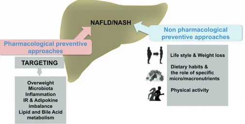
improving the existing disease [71]. Human interventional studies showed a protective effect of exercise on visceral adiposity and NAFLD regardless of weight loss[72–74]. The main non-pharmacological ap-proaches for prevention of NAFLD or NASH are shown inFig. 3. 11.3. Preventive pharmacological approaches
The main targets of the pharmacological approach for prevention of NAFLD or NASH are summarized inFig. 3.
11.3.1. Targeting overweight
Five weight loss drugs, including orlistat, lorcaserin, naltrexone-bupropion, phentermine-topiramate, and liraglutide, have been ap-proved by the US FDA as long-term treatment in obese or overweight patients in presence of one or more weight-associated comorbidities, such as DM II, hypertension or hyperlipidemia. These drugs have been proven to reduce of 5% or more weight loss at 52 weeks, and phen-termine-topiramate and liraglutide have shown greater effectiveness. However, a personalized approach is necessary due to hypoglycemic effects of liraglutide and neuropsychiatric complications related to naltrexone-bupropion in patients with chronic opiate or alcohol de-pendence[75]. In any case, to our knowledge, there is no evidence of using these drugs in liver steatosis, except for orlistat. Orlistat, an in-hibitor of gastric and pancreatic lipases, being able to induce weight loss, has been proposed in obese NASH patients. After the first pro-mising results in steatosis based on biochemical and US imaging im-provements, the treatment with orlistat in association to vitamin E and hypocaloric diet in overweight NASH patients did not induce further weight loss and improvements in insulin resistance, liver enzymes le-vels and histology in a randomized controlled trial (RCT). The two groups presented comparable results with different effects according to the amount of weight loss. In fact, reduction of more than 5% of body weight improved insulin sensitivity and steatosis, whereas improve-ment in.
NASH was shown if weight loss was more than 9%[76]. 11.3.2. Targeting microbiota
In mouse models, probiotics showed a preventive effect on IR and progression of NALFD, and could revert the pattern of NASH [28]. Human studies reported promising results. For example, In NAFLD patients, symbiotic supplementation in association to lifestyle inter-ventions, gave a significant effect than lifestyle interventions alone in reducing serum levels of ALT, AST, GGT, CRP, TNFα and NF κ-B and improvingfibrosis evaluated by transient elastography[77]. Probiotics reduced liver fat content and AST levels in NASH patients[78]. These findings show that probiotics, in particular a multistrain cocktail composed of streptococcus thermophiles, lactobacillus and
bifidobacteria, and synbiotics are a promising therapeutic support in NAFLD and NASH. However, these beneficial effects have to be con-firmed and preparations and dosage have not been fully established yet [64].
11.3.3. Targeting inflammation
Observational studies reported a correlation between aspirin use and lower risk of liverfibrosis[79]. Aspirin use has been also related to lower HCC risk, mainly in patients with daily aspirin use, for longer duration and at lower dosage[80]. Pentoxifylline, an anti-TNF agent, has been tested in several trails showing beneficial effects on transa-minases levels and hepatic steatosis. In particular, in a RCT pentox-ifylline induced a significant improvement in liver histology and fi-brosis in NASH patients [81]. Ghrelin has been reported to reduce NAFLD and NASH in mice through its antinflammatory and anti-oxi-dants activity[82]. In a clinical trial, simtuzumab, a monoclonal anti-body against lysyl oxidaselike 2, and selonsertib, an inhibitor of apoptosis signal-regulating kinase 1 modulating oxidative stress, was tested in NASH patients, alone or in combination. Selonsertib showed a reduction in liverfibrosis whereas simtuzumab was unable to induce regression of fibrosis[83]. A recently identified STAT 3 inhibitor, C 188–9, was shown to inhibit HCC tumor growth and to reduce liver steatosis, inflammation, bile ductular reactions and serum AST/ALT levels in mouse models[84].
11.3.4. Targeting insulin resistance and adipokine imbalance
The effects of antidiabetic drugs have been largely investigated in the field of liver disease. Human observational studies reported the association of the regular metformin intake and the reduced incidence of HCC in diabetic patients in a dose dependent manner[85]. However, despite the positive effects of metformin on transaminases levels, weight loss and insulin resistance documented by several trials, there is little evidence about the beneficial effects of metformin histological in NASH [56,64]. Pioglitazone, an oral antidiabetic drug belonging to thiazolidinediones (TZDs) class, has been shown to reduce liver en-zymes levels, liver steatosis and necroinflammation, with uncertain effects on liver fibrosis [56,64]. Interestingly, pioglitazone has also achieved promising results in non-diabetic NASH patients, by im-proving insulin resistance and reducing AST/ALT serum levels, steatosis and liver inflammation. However, no effects on liver fibrosis were found and the insulin sensitizing effect was not persistent at 96 weeks, as a consequence of pioglitazone-related weight gain [63]. Another pro-posed TZD is rosiglitazone, which was shown to improve steatosis and transaminases levels, without effect on fibrosis. However, side effects (i.e. weight gain, risk of congestive hearth failure, increased osteo-porosis and risk of fractures and pedal edema) and poor evidence about long-term benefits in the treatment of liver disease limit the use of TZDs Overweight
Microbiota Inflammation IR & Adipokine
imbalance Lipid and Bile Acid
metabolism TARGETING Pharmacological preventive approaches Non pharmacological preventive approaches
Life style & Weight loss Dietary habits & the role of specific micro/macronutrients Physical activity
NAFLD/NASH
Fig. 3. Potential therapeutic targets in NAFLD/NASH. The main targets for the pharmacological and non-pharmacolo-gical intervention for prevention of NAFLD or NASH are shown. The non-pharmacological intervention includes life style, physical activity and dietary habits whereas the pharmacological approach cope with different targets in-cluding overweight, inflammation, insuline resistance (IR), adipokine imbalance, and the gut microbiota.
in clinical practice. In addition, rosiglitazone has been related to a higher risk of cardiovascular events, whereas pioglitazone is contra-indicated in the presence of bladder cancer and FDA recommends caution in patients with a previous history of bladder cancer[56,64]. PPAR axis has a relevant role in nonalcoholic liver diseases, as con-firmed by the correlation between human liver PPARα gene expression and NASH severity, visceral adiposity and insulin resistance and by histological improvements following increased PPARα expression[86]. Based on this evidence, novel drugs targeting PPAR axis have been proposed. Recently, liraglutide, a GLP-1 analog, was shown to induce NASH regression in a clinical trial [87]. Elafibranor (GFT505), a PPARα/δ agonist, did not display effect in terms of disease regression compared to placebo in patients with NASH. However, in a post-hoc analysis encompassing a novel definition of NASH resolution, sig-nificant histological improvements were found in the arm treated with a higher dose of Elafibranor (GFT505)[88]. In regard with sitagliptin, a dipeptidyl peptidase 4 inhibitor, no effect on histology was reported in NASH patients [56]. Exenatide, a peptide derived from glucagonlike protein-1–receptor agonists, has been proposed as a potential effective therapy in NASH[64]. Antibodies and tyrosine kinase inhibitors turned out to be the most promising drugs targeting the IGF-I signaling axis in HCC, among several drugs tested including small molecules, antisense oligonucleotides or small interfering RNAs. The efficacy of this treat-ment may depend upon the different level of IGF-I axis deregulation in that specific subtype of tumor. Therefore, IGF-I and insulin serum le-vels, levels in tumor of IGF ligands, receptor, binding proteins and factors involved in signaling should be carefully evaluated before treatment[89].
11.3.5. Targeting lipid and bile acids metabolism
Higher duration and frequency of statins prescriptions in large co-horts of patients with DM II was associated with reduced HCC risk[90]. Interestingly, a recent study has shown a relation between statin use and lower risk of cirrhosis and HCC in HCV chronic patients, high-lighting the antifibrotic effect of fluvastatin and atorvastatin compared to other statins [91]. Statins induced NASH regression and reduced liver fibrosis in obese, diabetic mice with metabolic syndrome[92]. Based on thesefindings and on their anti-proliferative, proapoptotic, anti-angiogenic and immunomodulatory effect, statins have been pro-posed to exert a preventive action against HCC. However, only pra-vastatin and rosupra-vastatin was shown to improve histology in NASH, but there are no sufficient studies evaluating effects on histologal endpoints [56]. Despite the observed beneficial effects of ezetimibe on IR, fats deposition and liverfibrosis in obese mice, no results were reported on humans in clinical trials[64]. A promising drug targeting BAs meta-bolism is obeticholic acid (OCA), a synthetic FXR agonist. Obeticholic acid (OCA), an organic compound derived from chenodeoxycholic acid, has been shown to improve IR and to reduce markers of liver in-flammation and fibrosis in patients with DM II and steatosis[93]. Also, an improvement of histological patterns in non-cirrhotic NASH has been reported [94]. Another drug targeting BAs metabolism, urso-deoxycholic acid (UDCA), was tested based on its cytoprotective ac-tivities and capacity of preventing apoptosis and downregulating in-flammation. However, no liver histological improvements were found after treatment with UDCA [95–96]. Lysophosphatidic acid receptors (LPARs) and Autotaxin/lysophospholipase D (ATX/lysoPLD), a major LPA-producing enzyme, have been proposed as molecular targets for HCC therapy. Pharmacological approaches include small RNA inter-ference (siRNAs) to impair mRNA expression or antagonists directed against LPAR (i.e. Ki16425) and ATX inhibitors (i.e. PF-8380; S32826). In vitro studies have shown promising results in terms of reduction HCC cell proliferation, survival and invasion. In addition, in mouse models, LPA axis inhibitors reducefibrogenesis and angiogenesis[47,97]. 11.3.6. Future therapies in ongoing trials
Clinical studies are currently being conducted with the aim of
targeting multiple pathways, inflammation, oxidative stress, micro-biota, metabolism andfibrosis progression in NAFLD or NASH. Drugs tested in these trials are listed hereafter: Aramchol (a fatty acid–bile conjugate), Volixibat (a bile acid transporter inhibitor), FXR agonists (GS-9674, PX-104 and GS-9674), Polyphenols (plant-derived com-pounds with antioxidant properties), fecal microbiota transplantation and probiotics, prebiotics, and synbiotics, drugs targeting FGF-19 and FGF-21, Saroglitazar (a dual PPAR agonist), an inhibitor of phospho-diesterase-4 (ASP9831), Emricasan (a pan-caspase inhibitor), Cenicriviroc (a dual CCR2/CCR5 antagonist), anti-CD3 antibodies, Selonsertib plus acetyl-CoA carboxylase inhibitor (GS-0976), Semaglutide (a GLP 1 analog)[56].
12. Conclusions and future directions of research
NASH-related cirrhotic patients receive significantly less surveil-lance for HCC than those with HCV-related cirrhosis[98]. As a matter of fact, this is strongly in contrast with epidemiological data and re-presents a considerable problem of public health. In fact, prevention is thefirst intervention to invert the increasing incidence of HCC. How-ever, cancer prevention primarily requires the correct identification of the“new patients at higher risk for HCC”. Therefore, to early diagnose hepatic steatosis, personalized screening programs should be im-plemented in patients with diabetes mellitus, obesity and metabolic syndrome. Further studies are necessary to better disclose hepatic his-tological changes with noninvasive measures such as serum biomarkers for reducing the need of biopsies for diagnosis of NASH. In the absence of fibrosis or NASH, pharmacological interventions are not re-commended and a combined lifestyle approach, including low caloric intake with specific nutrients composition, and physical activity is still considered the most effective preventive intervention in liver steatosis. Pioglitazone and vitamin E are the only preventive treatments re-commended in patients with NASH by EASL and AASLD guidelines, despite there is no complete information on their long-term safety and efficacy[51]. Other therapies (i.e. statins, antidiabetics, etc.) may be used in NAFLD/NASH at high CVD or HCC risk, but with the main in-dication of cardiovascular prevention. Considering the strong crosstalk between the emerging metabolic factors and liver microenvironment, a wide variety of drugs targeting the underlying pathophysiological me-chanisms (i.e. inflammation, IR, adipokine imbalance, SIBO, lipid and BAs metabolism) have been proposed as cancer prevention in NASH. However, there is little evidence of any improvement or regression of liver necroinflammation and fibrosis[99]. Therefore, as recommended by US FDA, further long-duration trials are necessary to better evaluate drug efficacy in NASH patients, considering improvement of histolo-gical patterns as a primary endpoint[100].
Transparency document
The Transparency document associated with this article can be found, in online version.
Acknowledgments
The work was supported by AIRC (Italian Association for Cancer Research) Investigator Grant (IG) 2015 Id.17758 (to A. Mazzocca). References
[1] L. Rahib, B.D. Smith, R. Aizenberg, A.B. Rosenzweig, J.M. Fleshman, L.M. Matrisian, Projecting cancer incidence and deaths to 2030: the unexpected burden of thyroid, liver, and pancreas cancers in the United States, Cancer Res. 74 (11) (2014 Jun 1) 2913–2921, http://dx.doi.org/10.1158/0008-5472.CAN-14-0155.
[2] T.M. Welzel, B.I. Graubard, S. Zeuzem, H.B. El-Serag, J.A. Davila, K.A. McGlynn, Metabolic syndrome increases the risk of primary liver cancer in the United States: a study in the SEER-Medicare database, Hepatology 54 (2) (2011 Aug) 463–471,
http://dx.doi.org/10.1002/hep.24397.
[3] W. Borena, S. Strohmaier, A. Lukanova, T. Bjørge, B. Lindkvist, G. Hallmans, M. Edlinger, T. Stocks, G. Nagel, J. Manjer, A. Engeland, R. Selmer, C. Häggström, S. Tretli, H. Concin, H. Jonsson, P. Stattin, H. Ulmer, Metabolic risk factors and primary liver cancer in a prospective study of 578,700 adults, Int. J. Cancer 131 (1) (2012 Jul 1) 193–200,http://dx.doi.org/10.1002/ijc.26338.
[4] R. Karagozian, Z. Derdák, G. Baffy, Obesity-associated mechanisms of hepato-carcinogenesis, Metabolism 63 (5) (2014 May) 607–617,http://dx.doi.org/10. 1016/j.metabol.2014.01.011.
[5] S. Schlesinger, K. Aleksandrova, T. Pischon, V. Fedirko, M. Jenab, E. Trepo, P. Boffetta, C.C. Dahm, K. Overvad, A. Tjønneland, J. Halkjær, G. Fagherazzi, M.C. Boutron-Ruault, F. Carbonnel, R. Kaaks, A. Lukanova, H. Boeing, A. Trichopoulou, C. Bamia, P. Lagiou, D. Palli, S. Grioni, S. Panico, R. Tumino, P. Vineis, H.B. Bueno-de-Mesquita, S. van den Berg, P.H. Peeters, T. Braaten, E. Weiderpass, J.R. Quirós, N. Travier, M.J. Sánchez, C. Navarro, A. Barricarte, M. Dorronsoro, B. Lindkvist, S. Regner, M. Werner, M. Sund, K.T. Khaw, N. Wareham, R.C. Travis, T. Norat, P.A. Wark, E. Riboli, U. Nöthlings, Abdominal obesity, weight gain during adulthood and risk of liver and biliary tract cancer in a European cohort, Int. J. Cancer 132 (3) (2013 Feb 1) 645–657,http://dx.doi.org/ 10.1002/ijc.27645.
[6] A.B. Siegel, E.A. Lim, S. Wang, W. Brubaker, R.D. Rodriguez, A. Goyal, J.S. Jacobson, D.L. Hershman, E.C. Verna, J. Zaretsky, K. Halazun, L. Dove, R.S. Brown Jr., A.I. Neugut, T. Kato, H. Remotti, Y.J. Coppleson, J.C. Emond, Diabetes, body mass index, and outcomes in hepatocellular carcinoma patients undergoing liver transplantation, Transplantation 94 (5) (2012 Sep 15) 539–543. [7] T. Ohki, R. Tateishi, S. Shiina, E. Goto, T. Sato, H. Nakagawa, R. Masuzaki, T. Goto,
K. Hamamura, F. Kanai, H. Yoshida, T. Kawabe, M. Omata, Visceral fat accumu-lation is an independent risk factor for hepatocellular carcinoma recurrence after curative treatment in patients with suspected NASH, Gut 58 (6) (2009 Jun) 839–844,http://dx.doi.org/10.1136/gut.2008.164053.
[8] E.J. Raff, D. Kakati, J.R. Bloomer, M. Shoreibah, K. Rasheed, A.K. Singal, Diabetes mellitus predicts occurrence of cirrhosis and hepatocellular cancer in alcoholic liver and non-alcoholic fatty liver diseases, J. Clin. Transl. Hepatol. 3 (1) (2015 Mar) 9–16,http://dx.doi.org/10.14218/JCTH.2015.00001.
[9] M. Balbi, V. Donadon, M. Ghersetti, S. Grazioli, G.D. Valentina, R. Gardenal, M.D. Mas, P. Casarin, G. Zanette, C. Miranda, P. Cimarosti, Alcohol and HCV chronic infection are risk cofactors of type 2 diabetes mellitus for hepatocellular carcinoma in Italy, Int. J. Environ. Res. Public Health 7 (4) (2010 Apr) 1366–1378,
http://dx.doi.org/10.3390/ijerph7041366.
[10] C.L. Chen, H.I. Yang, W.S. Yang, C.J. Liu, P.J. Chen, S.L. You, L.Y. Wang, C.A. Sun, Lu SN, D.S. Chen, C.J. Chen, Metabolic factors and risk of hepatocellular carcinoma by chronic hepatitis B/C infection: a follow-up study in Taiwan, Gastroenterology 135 (1) (2008 Jul) 111–121,http://dx.doi.org/10.1053/j.gastro.2008.03.073. [11] T. Hardy, F. Oakley, Q.M. Anstee, C.P. Day, Nonalcoholic fatty liver disease:
pa-thogenesis and disease spectrum, Annu. Rev. Pathol. 11 (2016 May 23) 451–496,
http://dx.doi.org/10.1146/annurev-pathol-012615-044224.
[12] G. Tarantino, C. Finelli, What about non-alcoholic fatty liver disease as a new cri-terion to define metabolic syndrome? World J. Gastroenterol. 19 (22) (2013 Jun 14) 3375–3384,http://dx.doi.org/10.3748/wjg.v19.i22.3375.
[13] E. Bugianesi, N. Leone, E. Vanni, G. Marchesini, F. Brunello, P. Carucci, A. Musso, P. De Paolis, L. Capussotti, M. Salizzoni, M. Rizzetto, Expanding the natural history of nonalcoholic steatohepatitis: from cryptogenic cirrhosis to hepatocellular carci-noma, Gastroenterology 123 (1) (2002 Jul) 134–140.
[14] J.M. Regimbeau, M. Colombat, P. Mognol, F. Durand, E. Abdalla, C. Degott, F. Degos, O. Farges, J. Belghiti, Obesity and diabetes as a risk factor for hepato-cellular carcinoma, Liver Transpl. 10 (2 Suppl 1) (2004 Feb) S69–73.
[15] R.J. Wong, R. Cheung, A. Ahmed, Nonalcoholic steatohepatitis is the most rapidly growing indication for liver transplantation in patients with hepatocellular carci-noma in the U.S, Hepatology 59 (6) (2014 Jun) 2188–2195,http://dx.doi.org/10. 1002/hep.26986.
[16] L.A. Streba, C.C. Vere, I. Rogoveanu, C.T. Streba, Nonalcoholic fatty liver disease, metabolic risk factors, and hepatocellular carcinoma: an open question, World J. Gastroenterol. 21 (14) (2015 Apr 14) 4103–4110,http://dx.doi.org/10.3748/wjg. v21.i14.4103.
[17] M. Ekstedt, H. Hagström, P. Nasr, M. Fredrikson, P. Stål, S. Kechagias, R. Hultcrantz, Fibrosis stage is the strongest predictor for disease-specific mortality in NAFLD after up to 33 years of follow-up, Hepatology 61 (5) (2015 May) 1547–1554,http://dx.doi.org/10.1002/hep.27368.
[18] S. Singh, A.M. Allen, Z. Wang, L.J. Prokop, M.H. Murad, R. Loomba, Fibrosis pro-gression in nonalcoholic fatty liver vs nonalcoholic steatohepatitis: a systematic review and meta-analysis of paired-biopsy studies, Clin. Gastroenterol. Hepatol. 13 (4) (2015 Apr) (643-54.e1-9; quiz e39-40),https://doi.org/10.1016/j.cgh.2014.04. 014.
[19] S. McPherson, T. Hardy, E. Henderson, A.D. Burt, C.P. Day, Q.M. Anstee, Evidence of NAFLD progression from steatosis tofibrosing-steatohepatitis using paired biopsies: implications for prognosis and clinical management, J. Hepatol. 62 (5) (2015 May) 1148–1155,http://dx.doi.org/10.1016/j.jhep.2014.11.034. [20] K. Yasui, E. Hashimoto, K. Tokushige, K. Koike, T. Shima, Y. Kanbara, T. Saibara,
H. Uto, S. Takami, M. Kawanaka, Y. Komorizono, T. Okanoue, Japan NASH Study Group, Clinical and pathological progression of non-alcoholic steatohepatitis to hepatocellular carcinoma, Hepatol. Res. 42 (8) (2012 Aug) 767–773,http://dx.doi. org/10.1111/j.1872-034X.2012.00986.x.
[21] K. Yasui, E. Hashimoto, Y. Komorizono, K. Koike, S. Arii, Y. Imai, T. Shima, Y. Kanbara, T. Saibara, T. Mori, S. Kawata, H. Uto, S. Takami, Y. Sumida, T. Takamura, M. Kawanaka, T. Okanoue, Japan NASH Study Group, Ministry of Health, Labour, and Welfare of Japan, Characteristics of patients with nonalcoholic
steatohepatitis who develop hepatocellular carcinoma, Clin. Gastroenterol. Hepatol. 9 (5) (2011 May) 428–433 (quiz e50),https://doi.org/10.1016/j.cgh.2011.01.023. [22] A. Sanyal, A. Poklepovic, E. Moyneur, V. Barghout, Population-based risk factors
and resource utilization for HCC: US perspective, Curr. Med. Res. Opin. 26 (9) (2010 Sep) 2183–2191,http://dx.doi.org/10.1185/03007995.2010.506375. [23] R.N. Rahman, J.A. Ibdah, Nonalcoholic fatty liver disease without cirrhosis is an
emergent and independent risk factor of hepatocellular carcinoma: a population based study, Hepatology 56 (2012) 241A.
[24] K. Shetty, J. Chen, J.H. Shin, W. Jogunoori, L. Mishra, Pathogenesis of hepatocel-lular carcinoma development in non alcoholic fatty liver disease, Curr. Hepatol. Rep. 14 (2) (2015 Jun) 119–127.
[25] W.K. Syn, Y. Jung, A. Omenetti, M. Abdelmalek, C.D. Guy, L. Yang, J. Wang, R.P. Witek, C.M. Fearing, T.A. Pereira, V. Teaberry, S.S. Choi, J. Conde-Vancells, G.F. Karaca, A.M. Diehl, Hedgehog-mediated epithelial-to-mesenchymal transition andfibrogenic repair in nonalcoholic fatty liver disease, Gastroenterology 137 (4) (2009 Oct) 1478–1488.e8,http://dx.doi.org/10.1053/j.gastro.2009.06.051. [26] A. Aravinthan, C. Scarpini, P. Tachtatzis, S. Verma, S. Penrhyn-Lowe, R. Harvey,
S.E. Davies, M. Allison, N. Coleman, G. Alexander, Hepatocyte senescence predicts progression in non-alcohol-related fatty liver disease, J. Hepatol. 58 (3) (2013 Mar) 549–556,http://dx.doi.org/10.1016/j.jhep.2012.10.031.
[27] E.J. Drenick, J. Fisler, D. Johnson, Hepatic steatosis after intestinal bypass–pre-vention and reversal by metronidazole, irrespective of protein-calorie malnutrition, Gastroenterology 82 (3) (1982 Mar) 535–548.
[28] C.M. Jiang, Pu CW, Y.H. Hou, Z. Chen, M. Alanazy, L. Hebbard, Non alcoholic steatohepatitis a precursor for hepatocellular carcinoma development, World J. Gastroenterol. 20 (44) (2014 Nov 28) 16464–16473,http://dx.doi.org/10.3748/ wjg.v20.i44.16464.
[29] M. Mouzaki, E.M. Comelli, B.M. Arendt, J. Bonengel, S.K. Fung, S.E. Fischer, I.D. McGilvray, J.P. Allard, Intestinal microbiota in patients with nonalcoholic fatty liver disease, Hepatology 58 (1) (2013 Jul) 120–127,http://dx.doi.org/10.1002/ hep.26319.
[30] L. Zhu, S.S. Baker, C. Gill, W. Liu, R. Alkhouri, R.D. Baker, S.R. Gill, Characterization of gut microbiomes in nonalcoholic steatohepatitis (NASH) pa-tients: a connection between endogenous alcohol and NASH, Hepatology 57 (2) (2013 Feb) 601–609,http://dx.doi.org/10.1002/hep.26093.
[31] J. Aron-Wisnewsky, B. Gaborit, A. Dutour, K. Clement, Gut microbiota and non-alcoholic fatty liver disease: new insights, Clin. Microbiol. Infect. 19 (4) (2013 Apr) 338–348,http://dx.doi.org/10.1111/1469-0691.12140.
[32] S. De Minicis, C. Rychlicki, L. Agostinelli, S. Saccomanno, C. Candelaresi, L. Trozzi, E. Mingarelli, B. Facinelli, G. Magi, C. Palmieri, M. Marzioni, A. Benedetti, G. Svegliati-Baroni, Dysbiosis contributes tofibrogenesis in the course of chronic liver injury in mice, Hepatology 59 (5) (2014 May) 1738–1749,http://dx.doi.org/ 10.1002/hep.26695.
[33] P.D. Cani, R. Bibiloni, C. Knauf, A. Waget, A.M. Neyrinck, N.M. Delzenne, R. Burcelin, Changes in gut microbiota control metabolic endotoxemia-induced inflammation in high-fat diet-induced obesity and diabetes in mice, Diabetes 57 (6) (2008 Jun) 1470–1481,http://dx.doi.org/10.2337/db07-1403.
[34] A.G. Ruiz, F. Casafont, J. Crespo, A. Cayón, M. Mayorga, A. Estebanez, J.C. Fernadez-Escalante, F. Pons-Romero, Lipopolysaccharide-binding protein plasma levels and liver TNF-alpha gene expression in obese patients: evidence for the potential role of endotoxin in the pathogenesis of non-alcoholic steatohepatitis, Obes. Surg. 17 (10) (2007 Oct) 1374–1380.
[35] D.H. Dapito, A. Mencin, G.Y. Gwak, J.P. Pradere, M.K. Jang, I. Mederacke, J.M. Caviglia, H. Khiabanian, A. Adeyemi, R. Bataller, J.H. Lefkowitch, M. Bower, R. Friedman, R.B. Sartor, R. Rabadan, R.F. Schwabe, Promotion of hepatocellular carcinoma by the intestinal microbiota and TLR4, Cancer Cell 21 (4) (2012 Apr 17) 504–516,http://dx.doi.org/10.1016/j.ccr.2012.02.007.
[36] M. Vijay-Kumar, J.D. Aitken, F.A. Carvalho, T.C. Cullender, S. Mwangi, S. Srinivasan, S.V. Sitaraman, R. Knight, R.E. Ley, A.T. Gewirtz, Metabolic syn-drome and altered gut microbiota in mice lacking toll-like receptor 5, Science 328 (5975) (2010 Apr 9) 228–231,http://dx.doi.org/10.1126/science.1179721. [37] J. Henao-Mejia, E. Elinav, C. Jin, L. Hao, W.Z. Mehal, T. Strowig, C.A. Thaiss,
A.L. Kau, S.C. Eisenbarth, M.J. Jurczak, J.P. Camporez, G.I. Shulman, J.I. Gordon, H.M. Hoffman, R.A. Flavell, Inflammasome-mediated dysbiosis regulates progres-sion of NAFLD and obesity, Nature 482 (7384) (2012 Feb 1) 179–185,http://dx. doi.org/10.1038/nature10809.
[38] S. Yoshimoto, T.M. Loo, K. Atarashi, H. Kanda, S. Sato, S. Oyadomari, Y. Iwakura, K. Oshima, H. Morita, M. Hattori, K. Honda, Y. Ishikawa, E. Hara, N. Ohtani, Obesity-induced gut microbial metabolite promotes liver cancer through senes-cence secretome, Nature 499 (7456) (2013 Jul 4) 97–101,http://dx.doi.org/10. 1038/nature12347.
[39] E.J. Park, J.H. Lee, Yu GY, G. He, S.R. Ali, R.G. Holzer, C.H. Osterreicher, H. Takahashi, M. Karin, Dietary and genetic obesity promote liver inflammation and tumorigenesis by enhancing IL-6 and TNF expression, Cell 140 (2) (2010 Jan 22) 197–208,http://dx.doi.org/10.1016/j.cell.2009.12.052.
[40] G. Baffy, Hepatocellular carcinoma in non-alcoholic fatty liver disease: epide-miology, pathogenesis, and prevention, J. Clin. Transl. Hepatol. 1 (2) (2013 Dec) 131–137,http://dx.doi.org/10.14218/JCTH.2013.00005.
[41] N. Kumashiro, D.M. Erion, D. Zhang, M. Kahn, S.A. Beddow, X. Chu, C.D. Still, G.S. Gerhard, X. Han, J. Dziura, K.F. Petersen, V.T. Samuel, G.I. Shulman, Cellular mechanism of insulin resistance in nonalcoholic fatty liver disease, Proc. Natl. Acad. Sci. U. S. A. 108 (39) (2011 Sep 27) 16381–16385,http://dx.doi.org/10.1073/ pnas.1113359108.
[42] R.C.R. Meex, M.J. Watt, Hepatokines: linking nonalcoholic fatty liver disease and insulin resistance, Nat. Rev. Endocrinol. 13 (9) (2017 Sep) 509–520,http://dx.doi. org/10.1038/nrendo.2017.56.