published: 23 May 2017 doi: 10.3389/fphar.2017.00290
Edited by: Jianbo Xiao, University of Macau, China Reviewed by: Elena Gonzalez Burgos, Complutense University of Madrid, Spain Zhaojun Wei, Hefei University of Technology, China *Correspondence: Sengul Uysal [email protected] Andrei Mocan [email protected] Specialty section: This article was submitted to Ethnopharmacology, a section of the journal Frontiers in Pharmacology Received: 17 December 2016 Accepted: 05 May 2017 Published: 23 May 2017 Citation: Uysal S, Zengin G, Locatelli M, Bahadori MB, Mocan A, Bellagamba G, De Luca E, Mollica A and Aktumsek A (2017) Cytotoxic and Enzyme Inhibitory Potential of Two Potentilla species (P. speciosa L. and P. reptans Willd.) and Their Chemical Composition. Front. Pharmacol. 8:290. doi: 10.3389/fphar.2017.00290
Cytotoxic and Enzyme Inhibitory
Potential of Two Potentilla species
(P. speciosa L. and P. reptans Willd.)
and Their Chemical Composition
Sengul Uysal1*, Gokhan Zengin1, Marcello Locatelli2,3, Mir B. Bahadori4,
Andrei Mocan5*, Giuseppe Bellagamba2, Elisa De Luca2, Adriano Mollica2and
Abdurrahman Aktumsek1
1Department of Biology, Science Faculty, Selcuk University, Campus, Konya, Turkey,2Department of Pharmacy, University
“G. d’Annunzio” Chieti-Pescara, Chieti, Italy,3Interuniversity Consortium of Structural and Systems Biology, Rome, Italy, 4Research Center for Pharmaceutical Nanotechnology, Tabriz University of Medical Sciences, Tabriz, Iran,5Department of
Pharmaceutical Botany, “Iuliu Hatieganu” University of Medicine and Pharmacy, Cluj-Napoca, Romania
In this work, the biological and chemical fingerprints of three extracts (ethyl acetate, methanol, and water) from two Potentilla species (Potentilla reptans and P. speciosa) were investigated. Antioxidant, enzyme inhibitory, and cytotoxic activities were performed for the biological fingerprint. For the chemical characterization, total bioactive components, and individual phenolic components were determined using photometric and HPLC methods, respectively. The main identified phenolic compounds in these extracts were rutin and catechin. Methanol and water extracts contained the highest total phenolic and flavonoid content. The results of antioxidant assays showed that methanol and water extracts displayed higher antioxidant activity compared to the ethyl acetate extract. Generally, methanol and water extracts exhibited higher biological activities correlated with higher levels the bioactive components. For P. speciosa, the methanol extract exhibited the highest enzyme inhibitory activity (except BChE inhibitory activity). P. reptans exhibited also high antiproliferative activity against MCF-7 cells whilst P. speciosa had weak to moderate activity against both of A549 and MCF-7 cell lines. The results suggest that Potentilla species could be potential candidates for developing new phyto-pharmaceuticals and functional ingredients.
Keywords: Potentilla reptans, P. speciosa, antioxidant activity, enzyme inhibitory activity, cytotoxic
INTRODUCTION
Natural products and functional-food ingredients gained interest due to their valuable biological effects including antioxidant, anticancer or antimicrobial. Currently, studies on natural products and medicinal plants are one of the most important subjects in pharmaceutical area (Sut et al., 2016). In these studies, many plants or plant-derived products are suggested as potential agents for designing new pharmaceuticals or food ingredients (Raskin et al., 2002). However, there is still limited knowledge about chemical and biological profiles of many wild plant species used as folk remedies in traditional medicine.
Alzheimer’s disease (AD) and Diabetes mellitus (DM) are considered major global health problems in the 21st century (Waltenberger et al., 2016). Today, the prevalence of AD and DM is rising and is estimated to increase significantly over the next two decades. Consequently, many therapeutic strategies are developed for these health problems, and the key enzyme inhibitory theory is one of the most accepted approaches. Acetylcholinesterase (AChE) and butyrylcholinesterase (BChE) are the enzymes into synaptic cleft that terminates the cholinergic signal transfer, and which are considered targets for the treatment of AD.α-amylase and α-glucosidase are main enzymes involved in the catabolism of carbohydrates and they are of vital importance for decreasing post-prandial blood glucose level. Tyrosinase is a key enzyme in melanin biosynthesis and thus the inhibition of this enzyme is associated with the prevention of skin disorders (SD) (Schelterns and Feldman, 2003). Several synthetic and natural inhibitors (galantamine and tacrine for AD; acarbose and voglibose for DM; kojic acid for SD) were developed for the management of these diseases by drug industry. However, many researches have reported that synthetic inhibitors have unfavorable effects such as nausea and diarrhea (Nouri et al., 2014; Anantharaman
et al., 2016; Bekir et al., 2016). Due to these adverse
effects, there is an increasing search for inhibitors derived from natural products (non-toxic and effective) against key enzymes related with these diseases (Day, 1998; Qin et al., 2013).
The genus Potentilla belongs to the Rosaceae family and is represented by about 500 species around the world (Tomczyk and Latté, 2009). Also, the genus comprises about 53 species in Turkish Flora (Pesmen, 1972). Potentilla species have been used as traditional medicine for the treatment of various diseases. For example,P. fulgens Lodd. is used for the treatment of DM, cancer, stomach disorders, cough, and as wound healing (Syiem et al., 2002; Rosangkima and Prasad, 2004;
Jaitak et al., 2010; Roy et al., 2010). P. mooniana Wight. is used to treat gastric problems and mouth ulcers (Ahmed
and Borthakur, 2005; Selvam, 2008). Also, P. fruticosa L.
has several medicinal properties including strengthening the stomach and the spleen, promoting metabolism, and it is widely used as a tea (Miliauskas et al., 2004; Li et al., 2007; Liu et al., 2016). Furthermore, P. atrosanguinea Lodd. has been used for wound healing, treating diarrhea, and influenza. Owing their potential uses in different purposes, several studies focused on the biological effects and chemical profile of the genus Potentilla (Tomczyk et al., 2010;Tomovic et al., 2015; Uysal and Aktumsek, 2015). However, to the best of our knowledge, the biological and chemical fingerprints of Potentilla reptans Willd and P. speciosa L. have not yet been reported. Thus, the main purpose of present study is to evaluate biological (antioxidant capacity, enzyme inhibitory, and cytotoxic activities) and chemical (total bioactive components and individual phenolic compositions) fingerprints of P. reptans and P. speciosa. The obtained results will provide new insights on the members of this genus for potential phyto-pharmaceuticals and nutraceuticals development.
MATERIALS AND METHODS
Plant Materials
Taxonomic identification of the plant material was kindly confirmed by senior taxonomist Dr. Murad Aydın SANDA, from Department of Field Crops, Agriculture Faculty, Igdir University, Igdir, Turkey. Voucher specimens have been deposited at the Herbarium of the Department of Biology, Selcuk University, Konya, Turkey. Localities and collection periods of Potentilla species are as following:
P. reptans: Selcuk University, Alaaddin Keykubad Campus, Konya, Turkey, June 2015, (Voucher Number: GZ-1532).
P. speciosa: Nigde, Camardi, Mazmili Mountain, Turkey, July 2015 (Voucher Number: GZ-1560).
Preparation of the Extracts
Aerial parts plant materials were air-dried at room temperature. The dried plant materials were ground to a fine powder using a laboratory mill. The powdered plant samples (10 g) were extracted with 250 mL of solvent (ethyl acetate, methanol) using a Soxhlet apparatus for 6–8 h. Extracts were then filtered and concentrated under vacuum at 40◦
C by using a rotary evaporator. To obtain water extracts, powdered P. reptans and P. speciosa aerial parts (15 g) were boiled with 250 mL of distilled water for 30 min. The water extracts were then filtered and lyophilized [–80◦
C, 48 h]. Extracts were kept at 4◦
C (±1◦
C) in dark until further analysis. Abbreviation for these extracts are; Pr-EA (Potentilla reptans ethyl acetate), Pr-Met (P. reptans methanol), Pr-Wat (P. reptans water), Ps-EA (Potentilla speciosa ethyl acetate), Ps-Met (P. speciosa methanol), Ps-Wat (P. speciosa water).
Total Phenolics, Flavonoid, Saponins,
Triterpenoids, and Phenolic Composition
The total phenolic content was determined by employing the methods given in the literature (Slinkard and Singleton, 1977) with some modification. Sample solution (1 mg/mL; 0.25 mL) was mixed with diluted Folin–Ciocalteu reagent (1 mL, 1:9, v/v) and shaken vigorously. After 3 min, Na2CO3 solution
(0.75 mL, 1%) was added and the sample absorbance was read at 760 nm after a 2 h incubation at room temperature. The total phenolic content was expressed as milligrams of gallic acid equivalents (mg GAE/g extract) (Vlase et al., 2014).
The total flavonoids content was determined using AlCl3
method (Zengin et al., 2014). Briefly, sample solution (1 mg/mL; 1 mL) was mixed with the same volume of aluminum trichloride (2%) in methanol. Similarly, a blank was prepared by adding sample solution (1 mL) to methanol (1 mL) without AlCl3. The
sample and blank absorbances were read at 415 nm after a 10 min incubation at room temperature. The absorbance of the blank was subtracted from that of the sample. Rutin was used as a reference standard and the total flavonoid content was expressed as milligrams of rutin equivalents (mg RE/g extract) (Mocan et al., 2015).
The total saponins content of the extract was determined by the vanillin-sulfuric acid method (Aktumsek et al., 2013). Sample
solution (1 mg/mL; 0.25 mL) was mixed with vanillin (0.25 mL, 8%) and sulfuric acid (2 mL, 72%). The mixture was incubated for 10 min at 60◦
C. Then the mixture was cooled for another 15 min, followed by the sample absorbance measurement at 538 nm. The total saponin content was expressed as milligrams of quillaja equivalents (mg QAE/g extract).
The total triterpenoids content of the extracts was determined according to Zhang et al. (2010) method with some modifications. Briefly, sample solution (1 mg/mL; 500µL) was mixed with the vanillin–glacial acetic acid (5%, w/v, 0.5 mL) and 1 mL of perchloric acid. The mixture was incubated at 60◦
C for 10 min, cooled in an ice water bath for 15 min and then 5 mL glacial acetic acid was added and mixed well. After 6 min, the absorbance was read at 538 nm. Oleanolic acid was used as a reference standard and the content of total triterpenoids was expressed as oleanolic acid equivalents (mg OAE/g extract) through a calibration curve with oleanolic acid.
HPLC-PDA analyses were performed on a Waters liquid chromatograph equipped with a model 600 solvent pump and a 2996 photodiode array detector, and Empower v.2 Software (Waters Spa, Milford, MA, United States) was used for acquisition of data. A C18 reversed-phase packing column (Prodigy ODS (3), 4.6 × 150 mm, 5 µm; Phemomenex, Torrance, CA, United States) was used for the separation and the column was thermostated at 30 ± 1◦
C using a Jetstream2 Plus column oven. The injection volume was 20µL. The mobile phase was directly on-line degassed by using Biotech DEGASi, mod. Compact (LabService, Anzola dell’Emilia, Italy). Gradient elution was performed using the mobile phase water-acetonitrile (93:7, v/v, 3% acetic acid) (Zengin et al., 2016). The UV/Vis acquisition wavelength was set in the range of 200–500 nm. The quantitative analyses were achieved at maximum wavelength for each compound.
Biological Activities Evaluation
Antioxidant (DPPH and ABTS radical scavenging, reducing power (CUPRAC and FRAP), phosphomolybdenum, and metal chelating (ferrozine method)) and enzyme inhibitory activities [cholinesterase (ChE) Elmann’s method], tyrosinase (dopachrome method), α-amylase (iodine/potassium iodide method), andα -glucosidase (chromogenic PNPG method)) were determined using the methods previously described by Zengin et al. (2014)andDezsi et al. (2015).
For the DPPH (1,1-diphenyl-2-picrylhydrazyl) radical scavenging assay: Sample solution (1 mg/mL; 1 mL) was added to 4 mL of a 0.004% methanol solution of DPPH. The sample absorbance was read at 517 nm after a 30 min incubation at room temperature in the dark. DPPH radical scavenging activity was expressed as millimoles of trolox equivalents (mg TE/g extract).
For ABTS (2,20
-azino-bis(3-ethylbenzothiazoline) 6-sulfonic acid) radical scavenging assay: Briefly, ABTS+ was produced directly by reacting 7 mM ABTS solution with 2.45 mM potassium persulfate and allowing the mixture to stand for 12–16 in the dark at room temperature. Prior to beginning the assay, ABTS solution was diluted with methanol to an absorbance of 0.700 ± 0.02 at 734 nm. Sample solution (1 mg/mL; 1 mL) was added to ABTS solution (2 mL) and mixed. The sample
absorbance was read at 734 nm after a 30 min incubation at room temperature. The ABTS radical scavenging activity was expressed as millimoles of trolox equivalents (mmol TE/g extract) (Mocan et al., 2016a).
For CUPRAC (cupric ion reducing activity) activity assay: Sample solution (1 mg/mL; 0.5 mL) was added to premixed reaction mixture containing CuCl2(1 mL, 10 mM), neocuproine
(1 mL, 7.5 mM) and NH4Ac buffer (1 mL, 1 M, pH 7.0). Similarly,
a blank was prepared by adding sample solution (0.5 mL) to premixed reaction mixture (3 mL) without CuCl2. Then, the
sample and blank absorbances were read at 450 nm after a 30 min incubation at room temperature. The absorbance of the blank was subtracted from that of the sample. CUPRAC activity was expressed as milligrams of trolox equivalents (mg TE/g extract).
For FRAP (ferric reducing antioxidant power) activity assay: Sample solution (1 mg/mL; 0.1 mL) was added to premixed FRAP reagent (2 mL) containing acetate buffer (0.3 M, pH 3.6), 2,4,6-tris(2-pyridyl)-S-triazine (TPTZ) (10 mM) in 40 mM HCl and ferric chloride (20 mM) in a ratio of 10:1:1 (v/v/v). Then, the sample absorbance was read at 593 nm after a 30 min incubation at room temperature. FRAP activity was expressed as milligrams of trolox equivalents (mg TE/g extract).
For phosphomolybdenum method: Sample solution (1 mg/mL; 0.3 mL) was combined with 3 mL of reagent solution (0.6 M sulfuric acid, 28 mM sodium phosphate and 4 mM ammonium molybdate). The sample absorbance was read at 695 nm after a 90 min incubation at 95◦
C. The total antioxidant capacity was expressed as millimoles of trolox equivalents (mmol TE/g extract) (Mocan et al., 2016c).
For metal chelating activity assay: Briefly, sample solution (1 mg/mL; 2 mL) was added to FeCl2 solution (0.05 mL,
2 mM). The reaction was initiated by the addition of 5 mM ferrozine (0.2 mL). Similarly, a blank was prepared by adding sample solution (2 mL) to FeCl2 solution (0.05 mL, 2 mM)
and water (0.2 mL) without ferrozine. Then, the sample and blank absorbances were read at 562 nm after 10 min incubation at room temperature. The absorbance of the blank was sub-tracted from that of the sample. The metal chelating activity was expressed as milligrams of EDTA (disodium edetate) equivalents (mg EDTAE/g extract).
For ChE inhibitory activity assay: Sample solution (1 mg/mL; 50 µL) was mixed with DTNB (5,5-dithio-bis(2-nitrobenzoic) acid, Sigma, St. Louis, MO, United States) (125 µL) and AChE [acetylcholines-terase (Electric ell AChE, Type-VI-S, EC 3.1.1.7, Sigma)], or BChE [BChE (horse serum BChE, EC 3.1.1.8, Sigma)] solution (25 µL) in Tris–HCl buffer (pH 8.0) in a 96-well microplate and incubated for 15 min at 25◦
C. The reaction was then initiated with the addition of acetylthiocholine iodide (ATCI, Sigma) or butyrylthiocholine chloride (BTCl, Sigma) (25 µL). Similarly, a blank was prepared by adding sample solution to all reaction reagents without enzyme (AChE or BChE) solution. The sample and blank absorbances were read at 405 nm after 10 min incubation at 25◦
C. The absorbance of the blank was subtracted from that of the sample and the cholinesterase inhibitory activity was expressed as galanthamine equivalents (mgGALAE/g extract) (Mocan et al., 2016b).
For Tyrosinase inhibitory activity assay: Sample solution (1 mg/mL; 25 µL) was mixed with tyrosinase solution (40 µL, Sigma) and phosphate buffer (100 µL, pH 6.8) in a 96-well microplate and incubated for 15 min at 25◦
C. The reaction was then initiated with the addition of L-DOPA (40 µL, Sigma). Similarly, a blank was prepared by adding sample solution to all reaction reagents without enzyme (tyrosinase) solution. The sample and blank absorbances were read at 492 nm after a 10 min incubation at 25◦
C. The absorbance of the blank was subtracted from that of the sample and the tyrosinase inhibitory activity was expressed as kojic acid equivalents (mgKAE/g extract) (Mocan et al., 2017).
For α-amylase inhibitory activity assay: Sample solution (1 mg/mL; 25µL) was mixed with α-amylase solution (ex-porcine pancreas, EC 3.2.1.1, Sigma) (50µL) in phosphate buffer (pH 6.9 with 6 mM sodium chloride) in a 96-well microplate and incubated for 10 min at 37◦
C. After pre-incubation, the reaction was initiated with the addition of starch solution (50µL, 0.05%). Similarly, a blank was prepared by adding sample solution to all reaction reagents without enzyme (α-amylase) solution. The reaction mixture was incubated 10 min at 37◦
C. The reaction was then stopped with the addition of HCl (25 µL, 1 M). This was followed by addition of the iodine-potassium iodide solution (100µL). The sample and blank absorbances were read at 630 nm. The absorbance of the blank was subtracted from that of the sample and theα-amylase inhibitory activity was expressed as acarbose equivalents (mmol ACE/g extract) (Savran et al., 2016).
Forα-glucosidase inhibitory activity assay: Sample solution (1 mg/mL; 50 µL) was mixed with glutathione (50 µL), α-glucosidase solution (from Saccharomyces cerevisiae, EC 3.2.1.20, Sigma) (50 µL) in phosphate buffer (pH 6.8) and PNPG (4-N-trophenyl-α-D-glucopyranoside, Sigma) (50µL) in a 96-well microplate and incubated for 15 min at 37◦
C. Similarly, a blank was prepared by adding sample solution to all reaction reagents without enzyme (α-glucosidase) solution. The reaction was then stopped with the addition of sodium carbonate (50µL, 0.2 M). The sample and blank absorbances were read at 400 nm. The absorbance of the blank was subtracted from that of the sample and the α-glucosidase inhibitory activity was expressed as acarbose equivalents (mmol ACE/g extract) (Llorent-Martínez et al., 2016).
All the assays were carried out in triplicate. The results are expressed as mean values and standard deviation (SD). The differences between the different extracts were analyzed using one-way analysis of variance (ANOVA) followed by Tukey’s honestly significant differencepost hoc test withα = 0.05. This treatment was carried out using SPSS v. 14.0 program.
Cell Viability Assay
Cell viability assay was performed for the extracts of the two Potentilla species (Farimani et al., 2015). Human alveolar lung epithelial carcinoma (A549) and human breast adenocarcinoma (MCF-7) cells were cultured in 75 cm2 flasks containing RPMI 1640 medium supplemented, 10% FBS (fetal bovine serum) and antibiotics (100 mg/mL penicillin/streptomycin). Cells were grown in an atmosphere of 5% CO2at 37◦C (±1◦C) with 95%
humidity. The antiproliferative activities of Potentilla extracts were determined against A549 and MCF-7 cells using the MTT assay. Cells were seeded in 96-well plates (2 × 10⁴ cells per well) and maintained at 37◦
C (±1◦
C) with 5% CO2atmosphere
for 24 h before test extracts were added as DMSO solutions. Stock solutions were prepared by dissolvingPotentilla extracts in DMSO (100 mg/mL) to reach a final DMSO concentration of 0.1%. Equal volume of DMSO (0.1%) was added into untreated wells. After incubation (for 24, 48, and 72 h), 50 µL of MTT solution (2 mg/mL in phosphate buffer saline) was added to each well. Afterwards, the plates were incubated for additional 4 h. DMSO was used for formazan solubilization and its UV absorbance was measured at 570 nm. Doxorubicin was used as the positive control. The percentage of cytotoxicity was calculated based on the comparison with untreated cells. All of the experiments were carried out in quadruplicate and the IC50
values were expressed as average ± SD. Statistical comparisons were estimated by one-way ANOVA followed by Duncan’spost hoc test for multiple comparisons with control. Statistical analyses were performed using SPSS 16.0 software. A value ofp< 0.05 was considered to indicate statistical significance.
RESULTS AND DISCUSSION
Extraction Yield and Identification
of Phenolic Compounds
Potentilla reptans and P. speciosa were extracted using different solvents (ethyl acetate, methanol, and water) and extraction yields of samples are shown in Table 1. The solvents used for extraction play a significant role on the extraction yield. The extraction yields increased in the following order: methanol> water > ethyl acetate. The highest extraction yield was for Pr-Met (20.61%).
To understand the relationship between antioxidant capacity and phenolic components, the phenolic components of two Potentilla species were determined by using HPLC-PDA (Figure 1). As shown in Table 2, two major compounds in all samples were identified as catechin (0.52–7.30 mg/g extract) and rutin (8.09–51.51 mg/g extract). Chemical structures of all identified phenolic compounds in Potentilla extracts are presented in Figure 2. These results are supported by the findings of Wang et al. (2013) who reported that catechin, rutin, and ellagic acid were the most abundant compounds inP. parvifolia Fisch. ex Lehm. Furthermore, the aerial parts of P. fruticosa have been found to contain these phenolics (particularly elagic acid, catechins, and flavonols) (Fedoseeva, 1979). Catechin has been reported to be present in roots and rhizomes of P. erecta L., P. anserina L., P. alba L., and P. viscosa Donn ex Lehm. (Gritsenko and Smik, 1977; Zhang et al., 1988; Vennat et al., 1992;Kombal and Glasl, 1995). Furthermore, catechin has been isolated from aerial parts of some Potentilla species such as P. erecta, P. fruticosa, and P. fragarioides L. (Goncharov et al., 1989;Kombal and Glasl, 1995;Choi et al., 1998;Miliauskas et al., 2004). According to the study of Tomczyk and Latté (2009), the dominant components in aerial parts of Potentilla species were flavonoids. A number of flavonoids (such as apigenin, kaempferol, quercetin, naringenin) have been identified from
TABLE 1 | Extraction yield and total bioactive components of different solvent extracts obtained from of Potentilla reptans and P. speciosa∗
.
Assays Potentilla reptans Potentilla speciosa
Ethyl acetate Methanol Water Ethyl acetate Methanol Water
Extraction yield (%) 3.29 20.61 13.95 3.21 8.60 4.07
Total phenolics (mg GAEs/g extract)a 42.13 ± 0.36c 111.68 ± 0.65b 135.73 ± 3.94a 24.98 ± 0.50c 102.58 ± 2.27b 138.45 ± 1.54a
Total flavonoids (mg REs/g extract)b 25.10 ± 0.38c 37.95 ± 0.70a 30.56 ± 0.11b 9.34 ± 0.13c 29.83 ± 0.21a 16.30 ± 0.24b
Total saponins (mg QEs/g extract)c 506.81 ± 33.84a 459.93 ± 30.68b 268.96 ± 18.09c 347.56 ± 71.26c 928.05 ± 56.65a 575.58 ± 22.41b
Total triterpenoids (mg OAE/g extract)d 4.21 ± 0.12a 2.39 ± 0.07b 0.64 ± 0.01c 2.20 ± 0.13b 5.17 ± 0.03a 2.04 ± 0.07b
aGAEs, gallic acid equivalents;bREs, rutin equivalents;cQEs, quillaja equivalents;dOAEs, oleanolic acid equivalents.∗Values expressed are means ± SD; Data marked
with different letters within the same row indicate statistically significant differences for each sample (p< 0.05).
FIGURE 1 | HPLC-PDA analyses of the three different extracts (ethyl acetate, methanol, and water) for the two Potentilla species; chromatographic profiles are reported at 280 nm.
some Potentilla species (P. viscosa, P. multifidi L., P. discolor Bunge, and P. erecta) (Liu et al., 1984; Goncharov et al., 1989;
Xue et al., 2005; Shen et al., 2006). Biological properties of rutin have been also reported including antibacterial, antitumor, anti-inflammatory (Calabro et al., 2005), antiallergic (Zwirtes de Oliveira et al., 2006), anticarcinogenic (Webster et al., 1996), and antioxidant (Yang et al., 2008). On the basis of these considerations, rutin and catechin could play an important role in the biological effects of the investigated Potentilla extracts. The other phenolic compounds were observed in minor amounts. Ferulic and cinnamic acids were not detected in any sample. Nonetheless, tannins and triterpenoids are also known as significant biologically active components of the genusPotentilla (Xue et al., 2005;Liu et al., 2006;Tomczyk and Latté, 2009).
Determination of the Total Phenolics,
Flavonoids, Saponins, and Triterpenoids
Content
Besides identified compounds, many other compounds might be responsible for the biological effects of investigated species. In this study, the values of total phenolics, flavonoids, saponins, and triterpenoids content are shown in Table 1. Total phenolics content of Potentilla extracts varied from 24.98 to 138.45 mg GAE/g. Ps-Wat (138.45 mg GAE/g extract) had the highest
phenolics content, whereas the Ps-EA (24.98 mg GAE/g extract) had the lowest content. Contrary to our results, Sohretoglu et al. (2015) found that ethyl acetate extract of P. recta and P. astracanica Jacq. contained higher total phenolics content compared to butanol and water extracts. Tomovic et al. (2015)
reported that the total phenolics content was 116.0 mg GAE/g in water extracts ofP. reptans which is lower than for Pr-Wat (135.73 mg GAE/g), in the present study. The total phenolics content of different solvent extracts of P. atrosanguinea was reported byGupta et al. (2016)who found that the hydroalcoholic extract showed higher phenolic content (429.8 mg GAE/g dry weight of extract). Several authors also reported the total phenolics contents of Potentilla species indicating them as valuable sources of bioactive compounds (Tomczyk et al., 2010;
Wang et al., 2013;Sohretoglu et al., 2015).
The total flavonoids content ofPotentilla species ranged from 9.34 to 37.95 mg RE/g extract. The highest values were obtained from Pr-Met, Pr-Wat, and Ps-Met with 37.95, 30.56, and 29.83 mg RE/g extract, respectively. The lowest total content value was obtained from Ps-EA with 9.34 mg RE/g extract. Total flavonoids content of somePotentilla species was also reported byTomczyk et al. (2010),Sohretoglu et al. (2015), andTomovic et al. (2015). As for total saponins content, Ps-Met (928.05 mg QE/g extract) contained the highest total saponins content, followed by Pr-EA (506.81 mg QE/g) and Pr-Met (459.93 mg QE/g). According to
T ABLE 2 | Phenolic compounds of P . reptans and P . speciosa (mg/g extract) ∗. No Compounds Retention Time (min) W avelength (nm) Potentilla reptans Potentilla speciosa Ethyl acetate Methanol W ater Ethyl acetate Methanol W ater 1 Gallic acid 4. 40 ± 0. 06 271 0. 10 ± 0. 03b 0. 02 ± 0. 01c 0. 80 ± 0. 13a 0. 10 ± 0. 04b 0. 15 ± 0. 04b 1. 49 ± 0. 45a 2 Catechin 11 .9 ± 0. 27 278 4. 89 ± 0. 98a nd 0. 52 ± 0. 17b 2. 28 ± 0. 85c 5. 53 ± 1. 02b 7. 30 ± 1. 28a 3 Chlor ogenic acid 12 .7 ± 0. 23 324 0. 03 ± 0. 01b nd 0. 16 ± 0. 07a nd nd 0. 21 ± 0. 07 4 p -OH benzoic acid 13 .2 ± 0. 20 256 nd 0. 02 ± 0. 01a 0. 03 ± 0. 01a 0. 34 ± 0. 09a 0. 27 ± 0. 07b 0. 33 ± 0. 08a 5 V anillic acid 15 .7 ± 0. 19 260 0. 16 ± 0. 05b 0. 67 ± 0. 06a 0. 09 ± 0. 01c 0. 19 ± 0. 07a 0. 17 ± 0. 05a 0. 20 ± 0. 09a 6 Epicatechin 16 .4 ± 0. 23 278 0. 07 ± 0. 02b 0. 70 ± 0. 11a nd 0. 15 ± 0. 03b nd 0. 34 ± 0. 10a 7 Syringic acid 16 .7 ± 0. 17 274 nd 0. 06 ± 0. 01b 1. 90 ± 0. 23a nd nd nd 8 3-OH benzoic acid 16 .9 ± 0. 21 295 nd nd 0. 47 ± 0. 18 nd nd 0. 16 ± 0. 09 9 3-OH-4-MeO benzaldehyde 20 .4 ± 0. 18 275 nd 5. 63 ± 1. 07 nd 0. 03 ± 0. 01b 0. 83 ± 0. 15a nd 10 p -coumaric acid 22 .0 ± 0. 19 309 0. 06 ± 0. 01a 0. 01 ± 0. 005b nd 0. 15 ± 0. 07c 0. 19 ± 0. 06b 0. 29 ± 0. 11a 11 Rutin 24 .0 ± 0. 14 256 8. 09 ± 1. 01c 21 .80 ± 3. 58b 51 .51 ± 5. 19a 3. 37 ± 1. 01c 9. 21 ± 0. 99b 11 .01 ± 1. 21a 12 Sinapic acid 24 .5 ± 0. 16 324 nd 0. 09 ± 0. 02a 0. 07 ± 0. 02a 0. 06 ± 0. 01a 0. 08 ± 0. 01a 0. 05 ± 0. 01a 13 t-Ferulic acid 26 .1 ± 0. 14 315 nd nd nd nd nd nd 14 Naringin 28 .4 ± 0. 13 285 0. 13 ± 0. 04c 0. 30 ± 0. 08b 0. 46 ± 0. 10a 0. 14 ± 0. 06c 1. 39 ± 0. 47a 1. 03 ± 0. 33b 15 2.3-diMeO benzoic acid 28 .7 ± 0. 14 299 0. 46 ± 0. 11c 0. 99 ± 0. 14b 1. 97 ± 0. 24a 0. 07 ± 0. 02b 1. 66 ± 0. 55a nd 16 Benzoic acid 29 .4 ± 0. 14 275 nd 0. 65 ± 0. 13 nd 0. 03 ± 0. 01c 0. 76 ± 0. 11b 1. 24 ± 0. 41a 17 o -Coumaric acid 30 .2 ± 0. 16 276 0. 04 ± 0. 01a 0. 06 ± 0. 02a nd nd 0. 020 ± 0. 008 nd 18 Quer cetin 39 .9 ± 0. 24 367 nd 0. 30 ± 0. 09a 0. 17 ± 0. 08b 0. 31 ± 0. 12c 3. 40 ± 0. 98a 1. 03 ± 0. 34b 19 t-Cinnamic acid 42 .7 ± 0. 23 276 nd nd nd nd nd nd 20 Naringenin 46 .0 ± 0. 04 290 0. 28 ± 0. 10b 0. 64 ± 0. 12a 0. 11 ± 0. 03c 0. 04 ± 0. 01a 0. 01 ± 0. 004b nd nd: not determined; ∗V alues expressed are means ± SD; Data marked with different letters within the same row indicate statistically significant differences for each sample (p < 0.05).
FIGURE 2 | Phenolic compounds identified in Potentilla species.
the results, water extracts contained lower total saponins content than other extracts. These findings agree with the previous results obtained by Uysal and Aktumsek (2015). Nonetheless, Ps-Met
(5.17 mg OAE/g) had the highest total triterpenoids content. The lowest amount of total triterpenoids content was obtained from Pr-Wat.
TABLE 3 | Antioxidant properties of different solvent extracts obtained from of P. reptans and P. speciosa∗
.
Assays Potentilla reptans Potentilla speciosa
Ethyl acetate Methanol Water Ethyl acetate Methanol Water
DPPH scavenging (mg TEs/g extract)a 119.54 ± 4.97c 331.98 ± 1.26a 292.10 ± 6.57b 46.51 ± 6.82c 334.66 ± 0.70a 200.54 ± 9.88b
ABTS scavenging (mmol TEs/g extract)a 2.06 ± 0.12c 4.39 ± 0.06b 4.55 ± 0.19a 0.61 ± 0.07c 3.62 ± 0.10b 4.17 ± 0.19a
CUPRAC (mg TEs/g extract)a 131.03 ± 2.18b 263.39 ± 3.11a 261.41 ± 1.88a 91.85 ± 1.93b 264.62 ± 3.25a 269.18 ± 3.37a
FRAP (mg TEs/g extract)a 81.59 ± 1.76c 204.10 ± 0.39b 219.97 ± 2.06a 56.97 ± 0.82c 191.53 ± 3.47b 214.49 ± 1.84a
Phosphomolybdenum (mmol TEs/g extract)a 1.64 ± 0.06c 2.62 ± 0.02b 2.73 ± 0.15a 1.12 ± 0.03c 2.34 ± 0.02b 3.03 ± 0.08a
Metal chelating (mg EDTAEs/g extract)b 6.87 ± 0.33c 20.04 ± 0.51b 32.86 ± 0.07a 3.45 ± 0.76c 9.09 ± 1.33b 26.94 ± 1.70a
aTEs, trolox equivalents;bEDTAEs, disodium edetate equivalents;∗Values expressed are means ± SD; Data marked with different letters within the same row indicate
statistically significant differences for each sample (p< 0.05).
TABLE 4 | Enzyme inhibitory activities of different solvent extracts obtained from of P. reptans and P. speciosa∗.
Assays Potentilla reptans Potentilla speciosa
Ethyl acetate Methanol Water Ethyl acetate Methanol Water
AChE Inhibition (mg GALAE/g extract)a 3.99 ± 0.08a 3.56 ± 0.22b 1.30 ± 0.22c 3.55 ± 0.22b 3.76 ± 0.06a 0.71 ± 0.06c
BChE Inhibition (mg GALAE/g extract)a 6.15 ± 0.37a 0.09 ± 0.01c 0.52 ± 0.01b 1.43 ± 1.17c nd 2.87 ± 0.27a
α-Amylase inhibition (mmol ACE/g extract)b 1.99 ± 0.07a 1.29 ± 0.18b 0.36 ± 0.01c 1.46 ± 0.25b 2.41 ± 0.44a 0.38 ± 0.01c
α-Glucosidase inhibition (mmol ACE/g extract)b 4.94 ± 1.70c 54.19 ± 0.57a 40.99 ± 2.62b 2.80 ± 0.93c 54.57 ± 0.14a 38.59 ± 6.20b
Tyrosinase inhibition (mg KAE/g extract) 108.56 ± 6.13b 123.36 ± 4.63a 31.48 ± 4.19c 106.54 ± 9.63b 144.39 ± 1.49a 35.60 ± 4.12c
aGALAEs, galantamine equivalents;bACEs, acarbose equivalents;cKAEs, kojic acid equivalents nd: not determined.∗
Values expressed are means ± SD. Data marked with different letters within the same row indicate statistically significant differences for each sample (p< 0.05).
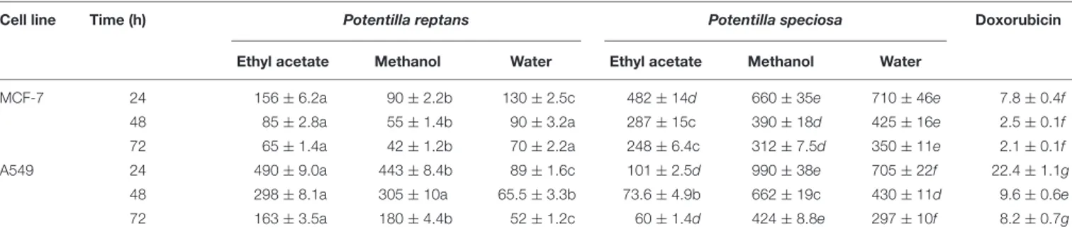
TABLE 5 | Cytotoxicity of different solvent extracts obtained from P. reptans and P. speciosa (IC50µg/ml)∗.
Cell line Time (h) Potentilla reptans Potentilla speciosa Doxorubicin
Ethyl acetate Methanol Water Ethyl acetate Methanol Water
MCF-7 24 156 ± 6.2a 90 ± 2.2b 130 ± 2.5c 482 ± 14d 660 ± 35e 710 ± 46e 7.8 ± 0.4f
48 85 ± 2.8a 55 ± 1.4b 90 ± 3.2a 287 ± 15c 390 ± 18d 425 ± 16e 2.5 ± 0.1f
72 65 ± 1.4a 42 ± 1.2b 70 ± 2.2a 248 ± 6.4c 312 ± 7.5d 350 ± 11e 2.1 ± 0.1f
A549 24 490 ± 9.0a 443 ± 8.4b 89 ± 1.6c 101 ± 2.5d 990 ± 38e 705 ± 22f 22.4 ± 1.1g
48 298 ± 8.1a 305 ± 10a 65.5 ± 3.3b 73.6 ± 4.9b 662 ± 19c 430 ± 11d 9.6 ± 0.6e
72 163 ± 3.5a 180 ± 4.4b 52 ± 1.2c 60 ± 1.4d 424 ± 8.8e 297 ± 10f 8.2 ± 0.7g
∗
Values expressed are means ± SD. Data marked with different letters indicate significant difference (p< 0.05).
Free Radical Scavenging Activity and
Total Antioxidant Capacity
The free radical scavenging activity ofP. reptans and P. speciosa was evaluated using the DPPH and ABTS radical scavenging assays. As shown in Table 3, the values of DPPH radical scavenging activity for P. speciosa and P. reptans ranged from 46.51 to 334.66 mg TE/g and from 0.61 to 4.55 mmol TE/g in ABTS radical scavenging activity, respectively. In the DPPH assay, Pr-Met and Ps-Met displayed more potent radical scavenging activity than other extracts. Additionally, in the ABTS assay, Pr-Wat (4.55 mmol TE/g extract) and Ps-Wat (4.17 mmol TE/g extract) had the highest radical scavenging activity among all samples. Tomovic et al. (2015) reported that the aerial parts and rhizome extracts of P. reptans showed DPPH radical scavenging activity with IC50 value of 12.11 and 2.57 µg/ml.
In another study, Gupta et al. (2016)found that H2O/MeOH
crude extract of P. atrosanguinea exhibited the highest DPPH radical scavenging activity (90.04 %) followed by EtOAc (88.10%) andn-BuOH (82.37%) at 200µg/ml. Besides,Liu et al. (2016)
reported that the ABTS values ofP. fruticosa varied from 303 to 1309µmol TE/g. In addition, researchers have reported that different Potentilla species have important radical scavenging activities (Choudhary et al., 2013;Rauf et al., 2013;Wang et al., 2013).
Total antioxidant capacities of Potentilla extracts were determined using phosphomolybdenum assay and the results were depicted in Table 3. Ps-Wat, Pr-Wat, and Pr-Met exhibited important total antioxidant capacities with the values of 3.03, 2.73, and 2.62 mmol TE/g extract, respectively. According also to the DPPH and ABTS results, the ethyl acetate extracts of both investigatedPotentilla species demonstrated lowest antioxidant activity in phosphomolybdenum assay.
Reducing Power and Metal Chelating
Activity
FRAP and CUPRAC assays were used to determine reducing power activity ofPotentilla species. The water extracts exhibited more pronounced activity as compared to other extracts in both CUPRAC and FRAP assays (Table 3). The lowest reducing power activity was observed for ethyl acetate extracts. The reducing power activity of the methanol extract ofP. speciosa root (FRAP: 133.35 and CUPRAC: 189.24 mg TE/g) was significantly lower than results obtained for aerial parts ofP. speciosa (FRAP: 191.53 and CUPRAC: 264.62 mg TE/g) (Zengin et al., 2016).Liu et al. (2016) reported that the FRAP values of P. fruticosa collected from eight locations ranged from 112.24 to 436.58µmol TE/g. In addition,Gupta et al. (2016)evaluated CUPRAC activities of different fractions of root extract of P. atrosanguinea in which the H2O/MeOH extract exhibited the highest reducing activity
followed byn-BuOH, EtOAc, and H2O fraction at 200µg/ml.
In the metal chelating assay, Pr-Wat (32.86 mg EDTAE/g) and Ps-Wat (26.94 mg EDTAE/g) were the most active, whereas the Pr-EA (6.87 mg EDTAE/g) and Ps-EA (3.45 mg EDTAE/g) were the least active. The ethyl acetate extracts of the studiedPotentilla extracts showed the lowest metal chelating activities and these results are supported as well by the previous findings ofUysal and Aktumsek (2015). In our previous study, we reported that metal chelating activity for the methanol extract ofP. speciosa root was 4.32 mg EDTAE/g (Zengin et al., 2016), and this value is lower than the one obtained herein for aerial part ofP. speciosa (9.09 mg EDTAE/g).
Enzyme Inhibitory Activity
The inhibitory activities of tested extracts against cholinesterases (AChE and BChE), α-amylase, α-glucosidase, and tyrosinase were tested and the results are presented in Table 4. The inhibitory activities against all enzymes ranged according to the extraction solvents. Generally, the water extract demonstrated a lower activity against all enzymes. The ethyl acetate and methanol extracts showed prominent inhibitory effects against AChE. Moreover, the highest BChE inhibitory activities were obtained from the Pr-EA and Ps-Wat with 6.15 and 2.87 mg GALAE/g. However, Ps-Met was inactive against BChE. There are several reports in literature indicating that terpenoid and phenolic compounds have promising cholinesterase inhibitory activities (Orhan et al., 2007;Stasiuk et al., 2008;Bahadori et al., 2016). Accordingly, terpenoid and phenolic richPotentilla species could be considered as promising AChE and BChE inhibitors.
The anti-diabetic activity ofPotentilla species was investigated by testing their inhibition abilities on α-amylase and α-glucosidase (Table 4). In α-glucosidase inhibitory activity, Pr-Met and Ps-Met showed the highest inhibitory activities with the values of 54.19 and 54.57 mmol ACE/g, respectively. Similarly, to our results, Kumar et al. (2013) reported that methanol extracts fromP. fulgens have strong inhibitory activity againstα-glucosidase, and ethyl acetate fraction exhibited potent α-glucosidase inhibitory activity. In addition, these authors found that isolated terpenoids from P. fulgens demonstrated significant α-glucosidase inhibitory activity. Similarly, several
triterpenoids and phenolics exhibited antidiabetic activities (Ivorra et al., 1988; Li et al., 2008;Luo et al., 2008;Etxeberria et al., 2012; Ali Asgar, 2013). Thus, the presence of these components in Potentilla species could be correlated with the observed antidiabetic activity.
The tyrosinase inhibitory activity of the studied Potentilla species ranged from 31.48 to 144.39 mg KAE/g. As shown in
Table 4, Ps-Met (144.39 mg KAE/g) and Pr-Met (123.36 mg KAE/g) displayed remarkable tyrosinase inhibitory activities. Additionally, the lowest tyrosinase inhibitory activities were observed in Pr-Wat (31.48 mg KAE/g) and Ps-Wat (35.60 mg KAE/g).
Cytotoxicity
Although some of the best anticancer drugs are from natural origin or derived, they present also negative effects on human health. Therefore, investigation of medicinal plants for discovery of potent anticancer compounds having fewer side effects is warranted. In this work, the cytotoxic activity of extracts from P. reptans and P. speciosa was determined against two human cancer cell lines (A549 and MCF-7). IC50values were expressed as
mean of quadruplicates ± SD (Table 5). The highest cytotoxicity was observed for water extract ofP. reptans (IC50< 130 µg/ml).
Compared to the positive control, doxorubicin (IC50=9.6 and
2.5µg/ml against A549 and MCF-7 cells in 48 h, respectively), P. reptans showed high antiproliferative activity against MCF-7 cells. P. speciosa exhibited weak to moderate activity against both of A549 and MCF-7 cell lines. The cytotoxicity rate of A549 and MCF-7 cells was found to be time dependent (Table 5). In general, crude extracts with IC50 values less than 1000 µg/ml
could be considered to be active. Several classes of natural compounds found in Potentilla species could be responsible for their antiproliferative activity. Previous studies revealed that triterpenoids, tannins and phenolic compounds isolated from Potentilla species exhibited cytotoxic activities against some human cancer cell lines (Li et al., 2007; Tomczyk and Latté, 2009; Choudhary et al., 2013; Rauf et al., 2015). For example two flavonoids (such as chrysin) fromP. evestita Th.Wolf showed prominent cytotoxic and antitumor promoting properties (Rauf
et al., 2015). Also, DNA topoisomerase I and II inhibitory
activity has been observed for phenolic compounds isolated from P. argentea L. (Tomczyk et al., 2008). In comparison to previous studies, P. reptans exhibited moderate to high cytotoxicity. According to our literature review, this is the first report concerning the antiproliferative activity of extracts obtained from P. reptans and P. speciosa. However, further phytochemical and pharmacological studies are needed for identification of responsible compounds and evaluation of the molecular mechanism of their anticancer action.
CONCLUSION
To sum up all, in the present work, different biological effects for twoPotentilla species were observed as well as their chemical profiles. From our results, it was apparent that the biological activities and chemical profiles were dependent on extraction
solvents and their polarity. Rutin and catechin were the major phenolic components identified in these extracts. Generally, the methanol and water extracts showed higher antioxidant activities as compared to ethyl acetate extracts. Furthermore, investigated Potentilla species revealed good inhibitory properties on tested enzymes linked to major health problems (AD and DM), and MCF-7 cells. From the present results, these twoPotentilla species could be considered as promising sources of natural-biologically active agents for pharmaceutical and food industries. However,
further experimental studies such asin vivo animal models and toxicological assays are recommended for the studiedPotentilla species.
AUTHOR CONTRIBUTIONS
SU, GZ, ML, MB, AnM, GB, and ED set up and carried out experiments. AdM and AA executed data analysis.
REFERENCES
Ahmed, A. A., and Borthakur, S. K. (2005).Ethnobotanical Wisdom of Khasis (Hynniew Treps) of Meghalaya. Dehradun: Bishen Singh Mahendra Pal Singh Publication.
Aktumsek, A., Zengin, G., Guler, G. O., Cakmak, Y. S., and Duran, A. (2013). Antioxidant potentials and anticholinesterase activities of methanolic and aqueous extracts of three endemicCentaurea L. species. Food Chem. Toxicol. 55, 290–296. doi: 10.1016/j.fct.2013.01.018
Ali Asgar, M. (2013). Anti-diabetic potential of phenolic compounds: a review.Int. J. Food Prop. 16, 91–103. doi: 10.1080/10942912.2011.595864
Anantharaman, A., Hemachandran, H., Priya, R. R., Sankari, M., Gopalakrishnan, M., Palanisami, N., et al. (2016). Inhibitory effect of apocarotenoids on the activity of tyrosinase: multi-spectroscopic and docking studies.J. Biosci. Bioeng. 121, 13–20. doi: 10.1016/j.jbiosc.2015.05.007 Bahadori, M. B., Dinparast, L., Valizadeh, H., Farimani, M. M., and Ebrahimi,
S. N. (2016). Bioactive constituents from roots of Salvia syriaca L.: acetylcholinesterase inhibitory activity and molecular docking studies.S. Afr. J. Bot. 106, 1–4. doi: 10.1016/j.sajb.2015.12.003
Bekir, J., Cazaux, S., Mars, M., and Bouajila, J. (2016). In vitro anti-cholinesterase and anti-hyperglycemic activities of flowers extracts from seven pomegranate varieties.Ind. Crops Prod. 81, 176–179. doi: 10.1016/j.indcrop.2015.11.066 Calabro, M., Tommasini, S., Donato, P., Stancanelli, R., Raneri, D., Catania, S.,
et al. (2005). The rutin/β-cyclodextrin interactions in fully aqueous solution: spectroscopic studies and biological assays. J. Pharm. Biomed. Anal. 36, 1019–1027. doi: 10.1016/j.jpba.2004.09.018
Choi, Y., Kim, M., Lee, H., Kwak, S., Yun, B., and Hu, C. (1998). Antioxidative compounds in aerial parts ofPotentilla fragarioides. Korean J. Pharmacogn. 29, 79–85.
Choudhary, A., Mittal, A. K., Radhika, M., Tripathy, D., Chatterjee, A., Banerjee, U. C., et al. (2013). Two new stereoisomeric antioxidant triterpenes from Potentilla fulgens. Fitoterapia 91, 290–297. doi: 10.1016/j.fitote.2013.09.008 Day, C. (1998). Traditional plant treatments for diabetes mellitus: pharmaceutical
foods.Br. J. Nutr. 80, 5–6. doi: 10.1017/S0007114598001718
Dezsi, ¸S., B˘ad˘ar˘au, A. S., Bischin, C., Vodnar, D. C., Silaghi-Dumitrescu, R., Gheldiu, A.-M., et al. (2015). Antimicrobial and antioxidant activities and phenolic profile ofEucalyptus globulus Labill. and Corymbia ficifolia (F. Muell.) K.D. Hill & LAS Johnson leaves. Molecules 20, 4720–4734. doi: 10.3390/ molecules20034720
Etxeberria, U., De La Garza, A. L., Campión, J., Martinez, J. A., and Milagro, F. I. (2012). Antidiabetic effects of natural plant extracts via inhibition of carbohydrate hydrolysis enzymes with emphasis on pancreatic alpha amylase.Expert Opin. Ther. Targets 16, 269–297. doi: 10.1517/14728222.2012. 664134
Farimani, M. M., Bahadori, M. B., Koulaei, S. A., Salehi, P., Ebrahimi, S. N., Khavasi, H. R., et al. (2015). New ursane triterpenoids fromSalvia urmiensis Bunge: absolute configuration and anti-proliferative activity.Fitoterapia 106, 1–6. doi: 10.1016/j.fitote.2015.07.017
Fedoseeva, G. (1979). Phenolic compounds ofPotentilla fruticosa. Chem. Nat. Comp. 15, 501–501. doi: 10.1007/BF00565058
Goncharov, N., Stupakova, E., and Komissarenko, N. (1989). Polyphenol composition of the epigeal part ofPotentilla erecta. Chem. Nat. Comp. 25, 375–376. doi: 10.1007/BF00597729
Gritsenko, O., and Smik, G. (1977). Phytochemical study ofPotentilla alba. Pharm. J. 32, 88–92.
Gupta, V. K., Kaur, R., Singla, R., and Jaitak, V. (2016). Photoprotective, antioxidant screening and new ester from dry root extracts of Potentilla atrosanguinea (Himalayan cinquefoil). S. Afr. J. Bot. 103, 49–53. doi: 10.1016/j. sajb.2015.08.007
Ivorra, M., Paya, M., and Villar, A. (1988). Hypoglycemic and insulin release effects of tormentic acid: a new hypoglycemic natural product.Planta Med. 54, 282–286. doi: 10.1055/s-2006-962433
Jaitak, V., Kaul, V. K., Kumar, N., Singh, B., Dhar, J., and Sharma, O. (2010). New hopane triterpenes and antioxidant constituents fromPotentilla fulgens. Nat. Prod. Commun. 5, 1561–1566.
Kombal, R., and Glasl, H. (1995). Flavan-3-ols and flavonoids fromPotentilla anserina. Planta Med. 61, 484–485. doi: 10.1055/s-2006-958146
Kumar, D., Ghosh, R., and Pal, B. C. (2013).α-Glucosidase inhibitory terpenoids fromPotentilla fulgens and their quantitative estimation by validated HPLC method.J. Funct. Foods 5, 1135–1141. doi: 10.1016/j.jff.2013.03.010
Li, H., Sun, H., and Hu, X. (2007). Analysis on total flavonoid in leaves ofPotentilla fruticosa in different environment and related mechanism. J. West China For. Sci. 36, 71–73.
Li, W., Chen, J., and Lu, H. (2008). Preparation ofEriobotrya japonica leaf total triterpenic acid and hypoglycemic function. U.S. Patent No. CN 1012244246. Washington, DC: U.S. Patent and Trademark Office.
Liu, P., Duan, H., Pan, Q., Zhang, Y., and Yao, Z. (2006). Triterpenes from herb of Potentilla chinensis. China J. Chin. Mater. Med. 31, 1875–1879.
Liu, Y., Su, S., and Zhu, T. (1984). Antibacterial components inPotentilla discolor. Zhongcaoyao 15, 333.
Liu, Z., Luo, Z., Jia, C., Wang, D., and Li, D. (2016). Synergistic effects ofPotentilla fruticosa L. Leaves combined with green tea polyphenols in a variety of oxidation systems.J. Food Sci. 81, C1091–C1101. doi: 10.1111/1750-3841.13292 Llorent-Martínez, E., Ortega-Barrales, P., Zengin, G., Uysal, S., Ceylan, R., Guler, G., et al. (2016).Lathyrus aureus and Lathyrus pratensis: characterization of phytochemical profiles by liquid chromatography-mass spectrometry, and evaluation of their enzyme inhibitory and antioxidant activities.RSC Adv. 6, 88996–89006. doi: 10.1039/C6RA17170B
Luo, J.-G., Ma, L., and Kong, L.-Y. (2008). New triterpenoid saponins with strong α-glucosidase inhibitory activity from the roots of Gypsophila oldhamiana. Bioorg. Med. Chem. 16, 2912–2920. doi: 10.1016/j.bmc.2007.12.053
Miliauskas, G., Van Beek, T. A., Venskutonis, P. R., Linssen, J. P., De Waard, P., and Sudhölter, E. J. (2004). Antioxidant activity ofPotentilla fruticosa. J. Sci. Food Agric. 84, 1997–2009. doi: 10.1002/jsfa.1914
Mocan, A., Schafberg, M., Cri¸san, G., and Rohn, S. (2016a). Determination of lignans and phenolic components ofSchisandra chinensis (Turcz.) Baill. using HPLC-ESI-ToF-MS and HPLC-online TEAC: contribution of individual components to overall antioxidant activity and comparison with traditional antioxidant assays. J. Funct. Foods 24, 579–594. doi: 10.1016/j.jff.2016. 05.007
Mocan, A., Vlase, L., Raita, O., Hanganu, D., Pãltinean, R., Dezsi, ¸S., et al. (2015). Comparative studies on antioxidant activity and polyphenolic content ofLycium barbarum L. and Lycium chinense Mill. leaves. Pak. J. Pharm. Sci. 28, 1511–1515.
Mocan, A., Zengin, G., Cri¸san, G., and Mollica, A. (2016b). Enzymatic assays and molecular modeling studies ofSchisandra chinensis lignans and phenolics from fruit and leaf extracts. J. Enzyme Inhib. Med. Chem. 31, 200–210. doi: 10.1080/14756366.2016.1222585
Mocan, A., Zengin, G., Uysal, A., Gunes, E., Mollica, A., Degirmenci, N. S., et al. (2016c). Biological and chemical insights ofMorina persica L.: a source
of bioactive compounds with multifunctional properties.J. Funct. Foods 25, 94–109. doi: 10.1016/j.jff.2016.05.010
Mocan, A., Zengin, G., Simirgiotis, M., Schafberg, M., Mollica, A., Vodnar, D. C., et al. (2017). Functional constituents of wild and cultivated Goji (L. barbarum L.) leaves: phytochemical characterization, biological profile, and computational studies.J. Enzyme Inhib. Med. Chem. 32, 153–168. doi: 10.1080/ 14756366.2016.1243535
Nouri, L., Mohammadi Nafchi, A., and Karim, A. A. (2014). Phytochemical, antioxidant, antibacterial, and α-amylase inhibitory properties of different extracts from betel leaves.Ind. Crops Prod. 62, 47–52. doi: 10.1016/j.indcrop. 2014.08.015
Orhan, I., Kartal, M., Tosun, F., and ¸Sener, B. (2007). Screening of various phenolic acids and flavonoid derivatives for their anticholinesterase potential. Z. Naturforsch. C 62, 829–832. doi: 10.1515/znc-2007-11-1210
Pesmen, H. (1972). “Potentilla L,” in Flora of Turkey and East Aegean Islands, ed. P. H. Davis (Edinburg, TX: Edinburg University Press), 41–68.
Qin, J., Lan, W., Liu, Z., Huang, J., Tang, H., and Wang, H. (2013). Synthesis and biological evaluation of 1, 3-dihydroxyxanthone mannich base derivatives as anticholinesterase agents.Chem. Cent. J. 7:78. doi: 10.1186/1752-153X-7-78 Raskin, I., Ribnicky, D. M., Komarnytsky, S., Ilic, N., Poulev, A., Borisjuk, N., et al.
(2002). Plants and human health in the twenty-first century.Trends Biotechnol. 20, 522–531. doi: 10.1016/S0167-7799(02)02080-2
Rauf, A., Khan, R., Khan, H., and Tokuda, H. (2015). Cytotoxic, antitumour-promoting and inhibition of protein denaturation effects of flavonoids, isolated fromPotentilla evestita Th. Wolf. Nat. Prod. Res. 29, 1775–1778. doi: 10.1080/ 14786419.2014.999336
Rauf, A., Khan, R., and Muhammad, N. (2013). Antioxidant studies of various solvent fractions and chemical constituents ofPotentilla evestita Th. Wolf. Afr. J. Pharm. Pharmacol. 7, 2710–2713. doi: 10.5897/AJPP2013.3795
Rosangkima, G., and Prasad, S. (2004). Antitumour activity of some plants from Meghalaya and Mizoram against murine ascites Dalton’s lymphoma.Indian J. Exp. Biol. 42, 981–988.
Roy, B., Swargiary, A., Syiem, D., and Tandon, V. (2010).Potentilla fulgens (Family Rosaceae), a medicinal plant of north-east India: a natural anthelmintic? J. Parasit. Dis. 34, 83–88. doi: 10.1007/s12639-010-0018-z
Savran, A., Zengin, G., Aktumsek, A., Mocan, A., Glamo´clija, J., ´Ciri´c, A., et al. (2016). Phenolic compounds and biological effects of edibleRumex scutatus andPseudosempervivum sempervivum: potential sources of natural agents with health benefits.Food Funct. 7, 3252–3262. doi: 10.1039/c6fo00695g
Schelterns, P., and Feldman, H. (2003). Treatment of Alzheimer’s disease; current status and new perspectives.Lancet Neurol. 2, 539–547. doi: 10.1016/S1474-4422(03)00502-7
Selvam, A. (2008). Inventory of vegetable crude drug samples housed in botanical survey of India. Howrah.Pharmacogn. Rev. 2, 61–94.
Shen, Y., Wang, Q., Lin, H., Shu, W., Zhou, J., and Li, Z. (2006). Study on chemical constituents ofPotentilla chinensis Ser. J. Chin. Med. Mater. 29, 237–239. Slinkard, K., and Singleton, V. L. (1977). Total phenol analysis: automation and
comparison with manual methods.Am. J. Enol. Viticult. 28, 49–55. doi: 10.3390/ molecules15128618
Sohretoglu, D., Unal, Z., and Sabuncuoglu, S. (2015). Assessment of radical scavenging activities and antiproliferative properties of two cinquefoil (Potentilla) species with their phytochemical contents. Acta Med. 4, 73–79.
Stasiuk, M., Bartosiewicz, D., and Kozubek, A. (2008). Inhibitory effect of some natural and semisynthetic phenolic lipids upon acetylcholinesterase activity. Food Chem. 108, 996–1001. doi: 10.1016/j.foodchem.2007. 12.011
Sut, S., Baldan, V., Faggian, M., Peron, G., and Dallacqua, S. (2016). Nutraceuticals, a new challenge for medicinal chemistry.Curr. Med. Chem. 23, 3198–3223. doi: 10.2174/0929867323666160615104837
Syiem, D., Syngai, G., Khup, P., Khongwir, B., Kharbuli, B., and Kayang, H. (2002). Hypoglycemic effects ofPotentilla fulgens L. in normal and alloxan-induced diabetic mice. J. Ethnopharmacol. 83, 55–61. doi: 10.1016/S0378-8741(02)00190-3
Tomczyk, M., Drozdowska, D., Bielawska, A., Bielawski, K., and Gudej, J. (2008). Human DNA topoisomerase inhibitors from Potentilla argentea and their cytotoxic effect against MCF-7.Pharmazie 63, 389–393. doi: 10.1691/ph.2008. 7810
Tomczyk, M., and Latté, K. P. (2009).Potentilla—A review of its phytochemical and pharmacological profile.J. Ethnopharmacol. 122, 184–204. doi: 10.1016/j. jep.2008.12.022
Tomczyk, M., Pleszczy´nska, M., and Wiater, A. (2010). Variation in total polyphenolics contents of aerial parts of Potentilla species and their anticariogenic activity. Molecules 15, 4639–4651. doi: 10.3390/ molecules15074639
Tomovic, M. T., Cupara, S. M., Popovic-Milenkovic, M. T., Ljujic, B. T., Kostic, M. J., and Jankovic, S. M. (2015). Antioxidant and anti-inflammatory activity of Potentilla reptans L. Acta Pol. Pharm. 72, 137–145.
Uysal, S., and Aktumsek, A. (2015). A phytochemical study onPotentilla anatolica: an endemic Turkish plant.Ind. Crops Prod. 76, 1001–1007. doi: 10.1016/j. indcrop.2015.08.017
Vennat, B., Pouget, M., and Pourrat, H. (1992). Proanthocyanidines: composition qualitative et quantitative d’un extrait de rhizomes dePotentilla tormentilla (Rosacees).J. Pharm. Belg. 47, 485–493.
Vlase, L., Mocan, A., Hanganu, D., Benedec, D., Gheldiu, A., and Crisan, G. (2014). Comparative study of polyphenolic content, antioxidant and antimicrobial activity of fourGalium species (Rubiaceae). Dig. J. Nanomater. Biostruct. 9, 1085–1094.
Waltenberger, B., Mocan, A., Šmejkal, K., Heiss, E. H., and Atanasov, A. G. (2016). Natural products to counteract the epidemic of cardiovascular and metabolic disorders.Molecules 21:807. doi: 10.3390/molecules21060807
Wang, S.-S., Wang, D.-M., Pu, W.-J., and Li, D.-W. (2013). Phytochemical profiles, antioxidant and antimicrobial activities of threePotentilla species. BMC Complement. Altern. Med. 13:321. doi: 10.1186/1472-6882-13-321 Webster, R., Gawde, M., and Bhattacharya, R. (1996). Protective effect of rutin,
a flavonol glycoside, on the carcinogen-induced DNA damage and repair enzymes in rats. Cancer Lett. 109, 185–191. doi: 10.1016/S0304-3835(96) 04443-6
Xue, P.-F., Luo, G., Zeng, W.-Z., Zhao, Y.-Y., and Liang, H. (2005). Secondary metabolites fromPotentilla multifida L.(Rosaceae). Biochem. Syst. Ecol. 33, 725–728. doi: 10.1016/j.bse.2004.12.012
Yang, J., Guo, J., and Yuan, J. (2008). In vitro antioxidant properties of rutin.LWT Food Sci. Technol. 41, 1060–1066. doi: 10.1016/j.lwt.2007.06.010
Zengin, G., Menghini, L., Malatesta, L., De Luca, E., Bellagamba, G., Uysal, S., et al. (2016). Comparative study of biological activities and multicomponent pattern of two wild Turkish species:Asphodeline anatolica and Potentilla speciosa. J. Enzyme Inhib. Med. Chem. 31, 203–208. doi: 10.1080/14756366.2016.117 8247
Zengin, G., Uysal, A., Gunes, E., and Aktumsek, A. (2014). Survey of phytochemical composition and biological effects of three extracts from a wild plant (Cotoneaster nummularia Fisch. et Mey.): a potential source for functional food ingredients and drug formulations.PLoS ONE 9:e113527. doi: 10.1371/journal. pone.0113527
Zhang, B., Nonaka, G.-I., and Nishioka, I. (1988). Potentillanin, a biflavanoid and a procyanidin glycoside fromPotentilla viscosa. Phytochemistry 27, 3277–3280. doi: 10.1016/0031-9422(88)80042-6
Zhang, L., Yang, J., Chen, X.-Q., Zan, K., Wen, X.-D., Chen, H., et al. (2010). Antidiabetic and antioxidant effects of extracts fromPotentilla discolor Bunge on diabetic rats induced by high fat diet and streptozotocin.J. Ethnopharmacol. 132, 518–524. doi: 10.1016/j.jep.2010.08.053
Zwirtes de Oliveira, I. R., Fernandes, S. C., and Vieira, I. C. (2006). Development of a biosensor based on gilo peroxidase immobilized on chitosan chemically crosslinked with epichlorohydrin for determination of rutin.J. Pharm. Biomed. Anal. 41, 366–372. doi: 10.1016/j.jpba.2005.12.019
Conflict of Interest Statement: The authors declare that the research was
conducted in the absence of any commercial or financial relationships that could be construed as a potential conflict of interest.
Copyright © 2017 Uysal, Zengin, Locatelli, Bahadori, Mocan, Bellagamba, De Luca, Mollica and Aktumsek. This is an open-access article distributed under the terms of the Creative Commons Attribution License (CC BY). The use, distribution or reproduction in other forums is permitted, provided the original author(s) or licensor are credited and that the original publication in this journal is cited, in accordance with accepted academic practice. No use, distribution or reproduction is permitted which does not comply with these terms.