Recent Patents on Inflammation & Allergy Drug Discovery 2019, 13, 1-14 1
REVIEWARTICLE
1872-213X/19 $58.00+.00 © 2019 Bentham Science Publishers
Skin Allergy to Azole Antifungal Agents for Systemic Use: A Review of the
Literature
Gianfranco Calogiuri
1,3, Lene H. Garvey
2, Eustachio Nettis
3, Paolo Romita
4, Elisabetta Di Leo
5, Riccardo
Caruso
6, Lavjay Butani
7and Caterina Foti
4,*1Department of Pneumology and Allergy Hospital Sacro Cuore - Gallipoli, Lecce, Italy; 2Allergy Clinic, Department of Dermato-Allergology, Herlev and Gentofte Hospital, University of Copenhagen, Hellerup, Denmark; 3Department of Biomedical Sciences and Human Oncology Unit of Internal Medicine "G. Baccelli", "Aldo Moro" University of Bari Medical School, Policlinico, Bari, Italy; 4Department of Biomedical Science and Human Oncology, Dermatological Clinic, University of Bari, Bari, Italy; 5Section of Allergy and Clinical Immunology, Unit of Internal Medicine-"F. Miulli" Hospital, Acquaviva delle Fonti, Bari, Italy; 6Clinica Salus - Brindisi, Brindisi, Italy; 7Section of Pediatric Nephrology, Department of Pediatrics, University of California Davis Medical Center, Sacramento, CA, USA
A R T I C L E H I S T O R Y Received: November 28, 2018 Revised: September 06, 2019 Accepted: September 06, 2019 DOI: 10.2174/1872213X13666190919162414
Abstract: Background: Antifungal azoles are the first-line agents used to treat topical and, above all,
systemic mycosis. The latter could be life-threating infections in immunocompromised patients. Che-motherapeutic antibiotics, including antifungal azoles, may induce hypersensitivity reactions; however, such immunologic adverse reactions have not been defined and carefully investigated.
Objective: The study aimed to provide an update on the evaluation and diagnosis of skin allergy to
azole antifungal agents.
Methods: This is a systematic review performed on PubMed and Google Schoolbarusing using the key
terms “allergy, hypersensitivity, anaphylaxis, immediate-type reaction, delayed-type reaction, keto-conazole, fluketo-conazole, posaketo-conazole, voriketo-conazole, itraketo-conazole, triazoles, imidazoles, antifungals, antimycotics”. The search strategy included meta-analyses, randomized controlled trials, clinical trials, observational studies, reviews and case reports.
Results: One hundred twenty-four articles matched our search terms. The most common adverse events
reported were T-cell mediated delayed-type hypersensitivity reactions, such as fixed drug eruptions, localized, generalized and exanthematous dermatitis, Steven-Johnson syndrome, toxic epidermal necro-lysis and acute generalized exhanthematous pustulosis. Rarely a drug rash, eosinophilia systemic symp-toms, has been described. Also, immediate-type reactions such as urticaria-angioedema or anaphylaxis have been reported following the administration of antifungal imidazoles, although not so frequently.
Conclusion: Despite their widespread use, triazoles seem to induce rare cutaneous hypersensitivity
reactions, but the pathomechanisms, risk factors, diagnostic and management strategies, including skin tests and challenge tests, are little known and poorly investigated.
Keywords: Allergy, antifungals, dermatitis, hypersensitivity, imidazoles, rash, triazoles. 1. INTRODUCTION
Fungal infections are not as frequent as bacterial and vi-ral infections; nevertheless, there are high incidence of these infections in humans in the last 25 years, largely as a conse-quence of the increased number of immunocompromised patients, such as patients infected by Human Immune-deficiency Virus (HIV), transplant patients and critically ill
*Address correspondence to this author at the Unit of Dermatology - De-partment of Biomedical Science and Human Oncology, University of Bari, Policlinico, Piazza Giulio Cesare, 11, Bari I-70124, Italy;
Tel: +39 080 5478107; Fax: +39 080 5478107; E-mail: [email protected]
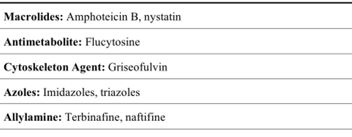
patients in High Intensive Care Units. Furthermore, in these years, the antimycotic weaponry has increased considerably, since many new antifungal molecules have been added, as shown in Table 1.
Azole antifungal agents are the largest and the most efficient class of synthetic antimycotics that can be used efficaciously to treat localized and generalized candidosis, cryptococcosis, histoplasmosis, pulmonary and systemic aspergillosis, dermatophytes, coccidioidomycosis, blasto-mycosis, penicilliosis, sporotrichosis and mucormycosis [1].
Table 1. Antimycotic Agents for Systemic Use. Macrolides: Amphoteicin B, nystatin
Antimetabolite: Flucytosine Cytoskeleton Agent: Griseofulvin Azoles: Imidazoles, triazoles Allylamine: Terbinafine, naftifine
Echinocandin: Caspofungin, micafungin, anidulafungin
The above-listed fungal infections usually affect im-mune-compromised patients such as Human Immunodefi-ciency Virus (HIV)-infected subjects, oncologic, hema-tologic and critically ill patients and last but not the least, transplant patients. Neutropenia induced by chemotherapies places these patients at risk of serious fungal infections, most commonly with Candida species.
Systemic fungal infections are more serious because they are more difficult to diagnose, more likely to become chronic and may become life-threatening conditions. Prophylactic treatment is sometimes indicated in HIV-patients and bone-marrow transplant patients despite the high risk of inducing antibiotic resistance.
Even if new antifungals are available, triazoles are still considered the first- line drugs to treat systemic fungal infec-tions and these are the most suitable and manageable drugs for patients to use at home.
Although about 20 azole antifungal chemotherapeutics are currently available in the market, most of them are mainly for topical use. They are classified into two groups: Azoles with two nitrogen atoms in the azole ring (the imida-zoles including clotrimazole, econazole, ketoconazole, mi-conazole, and tioconazole) and those with three nitrogen atoms in the azole ring (the triazoles, of which fluconazole is the most representative of the class, followed by itracona-zole, posaconaitracona-zole, voriconazole and more recent isavu-conazole). The bioisosteric triazole ring has achieved higher selectivity of fungal targets versus host [2].
There are three general mechanisms of action of antifun-gal agents: Cell membrane disruption, inhibition of cell divi-sion and inhibition of cell wall formation.
Antifungal activity stems from the presence of an aro-matic five-member heterocyclic, either an imidazole or a triazole. Ketoconazole was the first imidazole molecule, in-troduced in 1979, and is the only one used as an effective oral therapy for Candida [2]. Itraconazole, fluconazole and more recently, posaconazole, voriconazole and isavucona-zole, are the antimycotic drugs most widely used for sys-temic mycosis, while newer azoles, voriconazole and posa-conazole seem to be effective in patients with fluposa-conazole- fluconazole-resistant candida infection and to treat aspergillosis [3].
Furthermore, fluconazole and voriconazole have the best cerebrospinal fluid penetration, each resulting in concentra-tions of at least 50% of those in serum. This is important because fungal infections of the central nervous system are
associated with high morbidity and mortality rates and are difficult to treat adequately [4, 5]. Fluconazole and voricona-zole are quickly absorbed, showing a higher bioavailability through the oral route than itraconazole and posaconazole [4]. Anyway, triazoles are essential drugs in immune-compromised patients because of increased susceptibility to fungal infections [6]
All azole antifungals undergo some degree of hepatic metabolism, although for fluconazole, the drug elimination role is minimal, whereas this is not the case with itracona-zole, voriconaitracona-zole, and posaconaitracona-zole, which are highly de-pendent on the liver metabolism for their elimination. For this reason, the most frequently reported adverse events at-tributed to the triazole drugs are hepatic toxicities [7].
Hypersensitivity reactions caused by systemic antifungal azole drugs are rarely reported in clinical practice, consider-ing their widespread use, but unfortunately, when these oc-cur, they have a major influence on the therapeutic approach in critically ill patients.
The most frequently reported hypersensitivity reactions to antimycotic azoles are allergic contact dermatitis forms, due to their widespread topical use [8, 9], but systemic hy-persensitivity reactions have also been reported.
2. MATERIALS & METHODS
The English and French-language literature during a 34-year period (January 1, 1984 through July 31, 2019) was reviewed for reported immediate-type hypersensitivity reac-tions and delayed-type hypersensitivity reacreac-tions” caused by antifungal azoles.
The search was conducted using the PubMed database and Google Scholar. Search terms as “urticaria, angioedema, anaphylaxis” AND “dermatitis, fixed drug eruption, cutane-ous adverse reactions, acute generalized exanthematcutane-ous pus-tulosis, Steven-Johnson syndrome, toxic epidermal necroly-sis, drug rash with eosinophilia” AND “ketoconazole, itra-conazole, fluitra-conazole, posaitra-conazole, voriitra-conazole, isavu-conazole, triazoles, azoles, antifungal”, limited to French, and English were. Patients included were those who devel-oped skin reactions after a known antifungal azole admini-stration. Patients were excluded if they developed a toxic reaction only.
3. CLINICAL ASPECTS OF AZOLE ANTIFUNGAL HYPERSENSITIVITY
According to the World Allergy Organization (WAO), hypersensitivity reactions to drugs may be distinguished as immediate-type reactions, occurring within 1hr following drug administration, and delayed-type, appearing after 1 hour, usually within 48-72hrs, from drug intake. The timing of the appearance is related to the involvement of different immunologic pathomechanisms, the signs and symptoms of immediate reactions being directly attributable to the vasoac-tive mediators released by mast cells and basophils. The im-munological mechanisms involved in this type of reaction are usually IgE-mediated. The most common symptoms are urticaria, pruritus, flushing, angio-edema, wheezing, gastro-intestinal symptoms and even anaphylactic shock [10].
The immunological mechanism involved in the delayed-type reaction is a T cell response, also known as delayed-type IV reaction, although in fact, Ventura et al. [11] identified 4 subtypes of T cell-mediated hypersensitivity, subdividing type IV reactions into types IVa, IVb, IVc and IVd according to the subset of activated T cells involved and the inflamma-tory cytokines produced and released during the reaction, as shown in Scheme 1.
3.1. Immediate-Type-Reactions
Although ketoconazole was the first agent to be used orally, few case reports of immediate-type hypersensitivity reactions, such as urticaria-angioedema [12, 13] or anaphy-laxis [14], have been described in the literature. Diagnosis is not easy because such reactions are usually caused by food or other hidden allergens [15]. Only one case report of itra-conazole-induced anaphylaxis is present in the literature [16] and two cases of urticaria angioedema [17, 18]. Despite its widespread use, even for fluconazole, there are only a few case reports of anaphylaxis [19-21], one case of angioedema [22] and even a Kounis syndrome, of coronary ischemia oc-curring during an immediate-type hypersensitivity reaction [23]. Such reactions were presumed to be IgE-mediated [16, 19], but no specific IgE to fluconazole has been isolated
in vitro. As far as new triazoles are concerned, probably due
to their less extensive use owing to their high costs, just two episodes of angioedema [24, 25] and anaphylaxis [26] in-duced by voriconazole have been reported in the literature. A retrospective study of the French Pharmacovigilance Data-base reported 227 cases of voriconazole-induced adverse reactions, observed over a period of 4 years, from January 1, 2002 to December 31, 2005 [27]. Among them, 39 cases (17 % of a total 227) reported cutaneous involvement with erythema (38% of skin reactions), maculopapular exanthema (17%), urticaria (13%), bullous eruptions (9%), blisters (6%), and purpura (4%). Phototoxicity was the most com-mon skin adverse effect in 15 patients (43%), with erythema (43% of photosensitivity reactions), bullous eruptions (22%), eczema (5%), desquamation (10%), necrosis (5%), and cheil-itis (5%). However, in 67% of cases, voriconazole has been administered with other drugs that may potentially cause skin eruptions [20]. The study suggested that immediate-type reactions such as urticaria represent a minority of cases and no case of anaphylaxis was reported in that study [27].
No case report of immediate-type hypersensitivity in-volving posaconazole or isavuconazole has yet been re-ported, nor any close investigation with an allergy workup.
3.2. Delayed-Type Reactions
3.2.1. Fixed Drug Eruption
Fixed Drug Eruption (FDE) is a cutaneous reaction rep-resented by one or more nummular, discoid or oval erythe-matous or lilac-violet hued patches, sometimes surmounted by a blister. FDE results from systemic exposure to a drug, usually taken orally. The initial eruption is often solitary and frequently located on the lip or on the genitalia area, whereas other common locations of the initial lesion are the hip, lower back or sacrum, proximal extremities and trunk.
These lesions, which develop over a period of hours, but may persist from days to weeks, fade slowly leaving a resid-ual oval hyperpigmented area. Normally they resolve in this way after stopping the drug administration, but they may recur at the same site with re-exposure to the drug. Drugs causing FDE are usually those employed intermittently.
Several sub-types of FDE have been observed and de-scribed, based mainly on their clinical features and the dis-tribution of the patches, such as: a) a pigmenting fixed drug eruption, b) a generalized or multiple fixed drug eruption, c) a linear fixed drug eruption, d) a wandering fixed drug erup-tion, e) a non-pigmenting fixed drug eruperup-tion, f) a bullous fixed drug eruption, g) an eczematous fixed drug eruption, and g) an urticarial fixed drug eruption [28, 29].
Although FDE has been described following oral keto-conazole [30], miketo-conazole [31] and itraketo-conazole [32], it is the oral fluconazole administration which has been more frequently associated with FDE [33-51], above all in female patients who take fluconazole for vaginal candidosis. Fur-thermore, fluconazole-induced FDE may display particular morphological aspects such as lesions resembling herpes-like vesicles of the lips [52-57] or multiple bullous lesions [57-59]. The onset of herpes-like lesions on the lips in female patients taking fluconazole should be considered pathogno-monic for fluconazole hypersensitivity, although few cases have been reported in the literature.
3.2.2. Exanthema and Dermatitis
The use of systemic azole antifungals may induce a cuta-neous rash with different morphological aspects. These in-clude maculopapular exanthema with eosinophilia, which has been reported following the use of itraconazole [60-62], fluconazole [63] and ketoconazole [64, 65], but sometimes lesions may appear as purpuric dermatitis [66]. A cutaneous rash can be associated with systemic symptoms such as acute drug-induced hepatitis [67-69], which has been reported for Type of Reaction T-Cell Type Cytokines Possible effector Mechanism Clinical Symptoms
IVa Th1 IFN-g,TNF-a Monocyte / macrophage activation Contact dermatitis, bullous exanthema IVb Th2 IL-5, IL-4, IL-13, eotaxin T-Cells with eosinophilic inflammation Maculopapular and bullous exanthema IVc Cytotoxic T cells Perforin, granzyme B CD4+/CD8+ mediated T cell killing Steven Johnson and Lyell syndrome
(bullous exanthema) IVd T cells CXCL-8 GM-CSF T cell leading to recruitment and
activa-tion of neutrophils
Acute Generalized Exanthematous Pustulosis (AGEP)
fluconazole [67, 68] and voriconazole [69], or fever onset during an erythematous maculopapular rash following flu-conazole use [70,71].
Furthermore, the previous use of topical antifungal azoles may cause an under or misdiagnosed sensitization, then elic-iting a generalized cutaneous reaction following systemic administration of an antifungal azole, which may induce an allergic cross-reactivity with the topical azoles. Hidden sen-sitization can be due to a topical application of an antifungal azole, eliciting a maculopapular eruption after systemic ad-ministration [72], or alternatively to exposure to a potentially cross-reactive antifungal azole, not only to treat a cutaneous mycosis in the patient, but even due to the use of veterinary products to treat a dermatophytes infection in patients’ pets [73].
So, cross-sensitivity between topical and systemic azoles has been demonstrated for miconazole with ketoconazole [74], clotrimazole and croconazole with itraconazole [75] and clotrimazole with fluconazole [76]. Also sensitivity to imidazoles/nitroimidazoles in subjects sensitized to methyl-chloroisothiazolinone/methylisothiazolinone has been inves-tigated [77].
A particular aspect of general allergic contact dermatitis is symmetrical drug-related intertriginous and flexural exan-thema, now called the acronym SDRIFE and previously known as baboon syndrome, because it induces erythema-tous lesions of the buttocks.
Morphologically, SDRIFE is expressed as a sharply de-marcated erythema of the buttocks and perianal zone or V-shaped erythema of the inguinal area and symmetrical in-volvement of the flexures, but it is not associated with any systemic symptoms [78].
Baboon syndrome/SDRIFE has been described following the use of ketoconazole [79], itraconazole [80] and flucona-zole [81]. Yet, there is even the possibility that topical anti-fungals may cause a widespread skin reaction, going beyond the application area, and the reaction may appear as eczema-tous dermatitis in the case of isoconazole [82] or as an erythema multiforme-like eruption caused by topical tio-conazole [83], although a genuine erythema multiforme fol-lowing systemic assumption of itraconazole has also been described in the literature [84]. Systemic involvement due to topical application of azoles is more common than might be expected. Swiss dermatologists observed over a period of 4 years, from 2008 to 2011, ten patients with severe cutaneous eruptions caused by a topical formulation of associated corti-costeroid, tixocortolpivalate and an antifungal azole medica-tion, clotrimazole [85]. Patients developed widespread ec-zema, but also erythematous, maculopapular exanthema, erythema multiforme-like or blistering eruptions, that oc-curred from 7 to 21 days after beginning the topical therapy. They also evinced an intense eczematous reaction at the ap-plication sites, associated with peripheral blood eosinophilia. However, patch tests with clotrimazole resulted positive only in 4 patients [85].
3.2.3. Serious Cutaneous Adverse Reactions (SCARs) Serious Cutaneous Adverse Reactions (SCARs) to drugs consist mainly of Steven-Johnson Syndrome/Toxic
Epider-mal Necrolysis (SJS/TEN), Acute Generalized Exanthema-tousPustulosis (AGEP) and Drug Rash with Eosinophilia and Systemic Symptoms (DRESS) syndrome.
Stevens-Johnson Syndrome (SJS) and Toxic Epidermal Necrolysis (TEN) are severe mucocutaneous reactions, most commonly triggered by medications, and characterized by extensive necrosis and detachment of the epidermis with mucosal involvement in 90% of affected patients. SJS/TEN SJS is considered a disease continuum and the different forms are distinguished according to the severity of body surface involved with blisters and erosions (SJS inferior to 10%, TEN superior to 30% and overlap SJS/TEN between 10-30%). SJS/TEN is associated with fever, malaise, renal and liver impairment and skin detachment. SJS/TEN occurs more frequently in patients with an immune depressive status and related immune dysregulation like HIV infection, graft-vs-host disease, systemic lupus erythematosus, malignanci-esof mostly hematologic type and mixed essential cryoglobu-linemia, because of its immuno-mediated pathogenesis. A retrospective study by a group of Italian clinicians showed that among 35 patients with SJ/TEN, observed over 11 years in an Italian Burns Centre, 9 of them were cancer patients. Beta-lactam antibiotics and azole antimycotic fluconazole were the drugs most frequently associated to the serious skin reaction in onco-hematologic patients, while antiepileptics were more commonly the agents responsible for SJS/TEN in patients with brain tumors [86]. The first case report of SJS induced by fluconazole dates back to 1991 [87], and two years later, Spanish dermatologists described the first case of TEN triggered by fluconazole in an HIV-positive male pa-tient [88]. Since then, other cases of SJS/TEN caused by fluconazole [89-95] and voriconazole [96-98], mainly in HIV-positive patients or oncologic subjects, have been re-ported. However, SJS/TEN induced by fluconazole may af-fect even immune-competent patients [89, 93]. For that rea-son, Paszmatzi et al. have speculated that, while in HIV-positive individuals, long-term high doses of fluconazole are more likely to trigger serious cutaneous reactions, short-term low, intermittent dosages of fluconazole seem to be more
responsible for inducing SJS/TEN in
non-immunocompromised patients [93].
Sometimes SCARs may present atypical features with borderline aspects between a fixed drug eruption and SJS/TEN [99] or a photo-induced SJS/TEN, which is a par-ticular variant of that disease, in which bullae and erosions appear only in photo exposed areas [100]. In that case, itra-conazole was the culprit drug [100]. Interestingly, photoder-matitis induced by itraconazole [101] and mainly by vori-conazole [102-104], is reputed to be phototoxic reaction and not photoallergic because when photo patch tests were car-ried out [101], they resulted negative. However, because only photo patch tests can discriminate between phototoxic-ity or photoallergy and these tests are not easy to perform
in vivo routinely, the real incidence of allergic
photodermati-tis to systemic triazoles could be underestimated.
Lastly, another cutaneous hypersensitivity syndrome triggered by systemic triazoles is Acute Generalized Exan-thematous Pustulosis (AGEP). It is characterized by aseptic disseminated cutaneous pustules associated with fever, mal-aise and peripheral blood leucocytosis. Ketaconazole [105],
itraconazole [106-108] and fluconazole [109-111] have been reported to cause AGEP.
Only one recent case of atypical DRESS syndrome with no eosinophilia and agranulocytosis has been reported in the literature, following voriconazole administration in a 48-year-old Japanese woman taking the drug for pulmonary aspergillosis [112].
Suggestively, in all the aforementioned case reports of FDE, mild dermatitis and SCARs, the adverse cutaneous reaction was always associated with the oral intake of tria-zoles, including the second generation antifungal voricona-zole [97, 98], where the reaction occurred when the drug was switched from intravenous to the oral route [91]. In a single case, a patient developed a rash and hepatitis but tolerated voriconazole when it was administered through the intrave-nous route [69]. Rarely, fluconazole induced a cutaneous rash when given intravenously [70]. However, in a French post-marketing retrospective study on voriconazole adverse reactions, among 39 patients who developed cutaneous hy-persensitivity reactions, voriconazole was administered in-travenously to 14 cases [27]. Even immediate-type reactions are rarely associated with the intravenous administration of triazole molecules [16, 26].
4. ALLERGIC CROSS-REACTIVITY AMONG ANTI-FUNGAL AZOLES
Because azoles drugs include a large family of substances with an imidazole ring in their chemical structure, as shown in Table 2, it is not surprising that sometimes allergic cross-reactivity may involve compounds other than antifungals, and patients who are allergic to anti protozoic drugs may evince allergic reactions to some antimycotics, too [113]. Patch tests with specific series are required [114].
In immediate-type reactions, which are presumably sup-ported by a specific IgE to the single antimycotic molecule, no allergic cross-reactivity has been evidenced between ke-toconazole and fluconazole in skin tests [10], and challenge test evidenced no cross-sensitivity between oral itraconazole and intravenous voriconazole [16] or between itraconazole and ketoconazole or fluconazole at an oral challenge test [17]. The few case reports of immediate-type reactions
in-volving fluconazole date back to the 90’s, when no other triazole drugs such as voriconazole or posaconazole were available. In the same way, no cross-sensitivity among anti-fungal ketoconazole and proton pumps inhibitors or other azoles was revealed, when investigated [10, 115]. Lastly, isavuconazole was tolerated in a 48-year-old female patient with angioedema induced by voriconazole and administered in a graded challenge protocol [25].
On the contrary, in cases of T-cell-mediated reactions, the existence of cross-reactivity among the different imida-zole compounds has been investigated and demonstrated by different authors.
Due to their widespread use as topical medications, anti-fungal azoles may induce allergic contact dermatitis, so many researchers used patch tests to explore the potential patterns of cross-reactivity among azoles antifungal drugs.
In 1988, Motley and Reynolds firstly proposed a cross-reactive pattern involving 2-4 dichlorophenylethylimida-zoles, based on substitution of a phenyl ring close to the imi-dazoles structure [116].
Later, Baes reported a pattern of cross-reactivity among
antifungal azoles, namely
beta-substituted-1-phenylethylimodazole with an ortho-chlorine substitution, suggesting that such an ortho-chloro substitution on the phenyl or thienyl ring was the immunologic site influencing cross-reactivity among topical azoles [117]. The ortho-chloro group includes isoconazole, croconazole, tioconazole, mi-conazole and oximi-conazole and cross-reactivity between cro-conazole and itracro-conazole was evidenced in the patch tests [75], but itraconazole is the only triazole with chlorine atoms in its chemical structure, that has been substituted by fluorine atoms in the other systemic triazoles.
Moreover, Goossens et al. reviewed the literature and added their experience with 15 cases of imidazole contact dermatitis, paying particular attention to cross-reaction pat-terns. They were able to identify three common patterns of cross-reactivity: isoconazole, miconazole and econazole were linked, as were sertaconazole, miconazole and econa-zole. The third link was isoconazole and tioconazole, al-though they suggested that even cross-reactivities are unpre-dictable [9] and, in their opinion, ketoconazole seemed to be Table 2. Imidazoles Drugs.
Antifungals:
Phenethyl Imidazoles: Ketoconazole, miconazole, tioconazole, isoconazole, enilconazole, econazole, sulconazole, sertaconazole and oxiconazole Phenmethyl Imidazoles: Clotrimazole, croconazole and bifonazole
Triazoles: Fluconazole, itraconazole, posaconazole, voriconazole and isavuconazole (systemic use) and eficonazole (topical use) Antiprotozoal Agents: Metronidazole, tinidazole, secnidazole and benznidazole
Anti-Helmintic Agents: Albendazole, mebendazole and thiabendazole Antihistamine2: Cimetidine
Proton Pump Inhibitors: Lansoprazole, omeprazole, rabeprazole and esomeprazole Antiplatelet: Ticagrelor
more similar to the triazoles structure, except for the imida-zole ring, although cross-reactivity between ketoconaimida-zole and miconazole has been reported [74].
Therefore, it has been suggested that azoles belonging to phenyl ethyl imidazoles are more likely to cross-react among themselves than with phenylmethylimidazoles, which show a low degree of cross-sensitivity among themselves [118], but these are not well-established rules.
Because topical imidazoles use may be an undervalued route of sensitization to systemic triazoles, it is likely that most of the hypersensitivity reactions to triazoles are T cell-mediated, as confirmed by clinical experiences. Thus, the aforementioned classifications of imidazole cross-sensitivity for contact dermatitis could be used in systemic delayed-type hypersensitivity caused by triazoles. Nevertheless, in cases of systemic triazoles, it is possible that liver cytochrome P450 isoforms metabolic pathways may change the immu-nogenicity of triazole molecules, as shown for itraconazole, generating various metabolites such as hydroxyitraconazole, keto-itraconazole, N-desalkyl-itraconazole [119]; that could make it more difficult to foresee cross-reactive phenome-nons.
In light of the experiences described in the literature, it is difficult to establish which epitope is recognized by T cells in antifungal azoles for systemic use.
For instance, Gupta and Thami first described a cross-sensitivity between fluconazole and itraconazole, but not to ketoconazole, in a 52-year old woman with FDE induced by fluconazole. The patient underwent oral graded challenge with fluconazole, itraconazole and ketoconazole every 4 weeks. A flare-up of the lesions was observed with flucona-zole 25mg and itraconaflucona-zole 25mg, but not with ketoconaflucona-zole up to 200mg, that failed to reactivate lesions, so the authors postulated that the epitope recognized by T cells was the common azole ring. In their case report, the introduction of the third nitrogen atom in the triazole ring was sufficient to change the immunogenicity with the imidazole [39], but in many case reports, patients with FDE induced by fluconazole tolerated oral itraconazole [41, 45, 46, 49, 51, 56] in the challenge test.
On the other hand, in a 65-year-old patient with contact dermatitis to luliconazole, Tanaka et al. elicited a positive patch test to lanoconazole, but not to neticonazole. All three compounds belong to the class of vinyl-imidazoles, but only luliconazole and lanoconazole presented the same dithio-acetal moiety, which was probably recognized by T-cells [120]. Umebayashi and Ito, on the contrary, observed a pa-tient with contact dermatitis to lanoconazole who developed cross-sensitivity to netilconazole following four-months use after the first diagnosis, and attributed the imidazole cross-reactivity to their common vinyl group [121].
Yet, in another case of FDE induced by ornidazole, an anti protozoic imidazole agent in a 42-year-old woman, the patient evinced cross-reactivity to fluconazole, too, but she tolerated oral metronidazole, itraconazole and ketoconazole and topical isoconazole. On that basis, the authors suggested that since propan-2-ol is the common chemical group of both molecules, it was responsible for a cross-reaction between ornidazole and fluconazole [122].
Previously, in another FDE, it has been suggested that the propanol side chain could be the epitope causing cross-sensitivity between ornidazole and secnidazole, another anti-protozoal drug [123].
In other cases of FDE, cross-reactivity between metroni-dazole and ketaconazole and between fluconazole and tini-dazole has also been described [113, 114]. Farbre et al. ob-served the rapid onset of AGEP following the third day of therapy with fluconazole 200mg daily in a 65-year-old fe-male patient previously treated with econazole powder [111]. The authors could not perform any diagnostic patch test be-cause of the patient's compromised neurological status, but the topical application of econazole following the AGEP resolution resulted in a flare-up of pustular lesions, suggest-ing that econazole induced the hypersensitivity, and there was an immunologic cross-reactivity between econazole and fluconazole [111].
Probably, the aromatic ring in imidazoles and triazoles is an important epitope for T-cells recognition, causing cross-reactivity, but that is not the only epitope in azole molecules, and this is what makes imidazoles cross-reactivity unpredict-able.
Furthermore, a potential cross-reactivity among azoles and some preservatives commonly used in cosmetics, such as thiazolinone derivatives, i.e. Methylisothiazolinone (MI), Methylchloroisothiazolinone (MCI), Benzisothiazolinone (BIT), and Octylisothiazolinone, has been suggested [124].
These preservatives are present in cosmetics, but also in household detergents, water-based paints and other liquids for industrial purposes [124].
Thiazolinone compounds show an aromatic ring where a nitrogen atom is substituted by a sulphur atom, although such a cross-sensitivity with azoles has been considered doubtful in a few case reports [82, 125].
Recently, Stingeni et al. enrolled 149 patients (35 men and 114 women; mean age 40.0 y.o.) with a recent diagnosis of contact sensitization to MCI/MI revealed by 0.02% aque-ous patch tests [126]. Patients were investigated through patch tests with phenethyl imidazoles (econazole nitrate, fenticonazole nitrate, isoconazole nitrate, ketoconazole mi-conazole nitrate, sertami-conazole nitrate, and tiomi-conazole) and phenmethyl imidazoles (bifonazole nitrate and clotrimazole), all at 2% pet., and anti-parasite agents as nitroimidazoles, (metronidazole and tinidazole, albendazole and mebenda-zole), all at 5% pet.
They identified 9 patients (6.0%) who reacted to at least one of the patch-tested imidazoles and nitroimidazoles, par-ticularly, all nine patients reacted to imidazoles: eight to phenethyl imidazoles (5.3%) (tioconazole, 3; ketoconazole,2; isoconazole,1; miconazole,1; sertaconazole,1), and one re-acted to a phenmethyl imidazole (0.7%) (clotrimazole), al-though, interistingly some patients had never used topical azoles previously. Furthermore, authors performed a com-puterized conformational analysis to investigate the spatial electron cloud geometries of MCI, MI and the imida-zoles/nitroimidazoles that elicited positive reactions in patch tests [126].
Such a computerized conformational analysis of the dif-ferent molecular structures seemed to confirm that the elec-tronic shapes and the distributions of positive and negative ions in the chemical structures of MCI and MI were similar to those of isoconazole, miconazole, sertaconazole, and tio-conazole. It was particularly evident that isoconazole, sertaconazole and tioconazole showed a spatial electron cloud geometry similar to that of MI, whereas miconazole showed the same spatial electron cloud geometry as MCI.
For that reason, authors suggested that cross-reacting molecules are characterized not only by similar sizes or shared reactive chemical groups but also by similar spatial geometries and electron cloud distributions [126]
Due to the complexity of their chemical structure, azole antifungal drugs show a great variety of epitopes, so it is possible that T cells may recognize the whole molecule, the azole/trazole ring or the side chain. As far as chlorine or fluorine atoms are concerned, they could influence the poten-tial pattern of cross-reactivity as halogen atoms due to their electron-attraction effect, stabilizing the molecule, as evi-denced in halogenated corticosteroids [127].
The possibility that similar electronic distributions may influence the crossreactivity of azoles, not only when com-pared with MCI/MI, but among different azole molecules, contributes to make the identification of cross-reactive pat-terns more elusive.
In cases of systemic administration, the absence of cross-reactivity between fluconazole and voriconazole has been demonstrated using a graded challenge test, introducing voriconazole gradually in a patient with a previous reaction to fluconazole [68], and confirmed in another case report [128]. Although itraconazole may be tolerated in patients with fluconazole-induced FDE [45, 49, 51, 53] and with ex-anthema induced by fluconazole [63, 128], there are reports demonstrating the existence of cross-sensitivity between these two triazoles in generalized dermatitis [129, 130]. Of course, given the potential hazard, cross-reactivity was not investigated in patients with SCARs and amphotericin B was introduced as needed.
5. DIAGNOSTIC PROCEDURES AND MANAGE-MENT OF TRIAZOLE HYPERSENSITIVITY
The diagnostic accuracy of skin tests for antifungal azoles is not well established. In immediate-type reactions, skin prick tests resulted positive to ketoconazole [12], flu-conazole [19] and voriflu-conazole mg/ml, diluted 1/10 in an anaphylactic reaction [26], whereas when skin prick tests were performed for itraconazole, it resulted negative. Al-though fluconazole has been available in the market since 1990, most of the authors reported that they had not carried out skin tests with fluconazole because they did not know its negative or positive predictive value. At immediate reading, intradermal tests with fluconazole have been reported posi-tive in a single case [23] with the diluted molecules, 2mg/ml at 1/10, 1/100 and 1/1000.
In delayed-type reactions, patch tests [114] showed a high rate of false-negative results. Although many authors used petrolatum as vehicle for patch testing imidazoles, Raulin and Frosch compared petrolatum, ethanol and methyl
ethyl ketone and found more false-negative reactions with petrolatum than other vehicles [8]. However, patch test has been successfully used to confirm the AGEP diagnosis and sensitization [112]. Intradermal test with delayed reading can be useful in maculopapular exanthema [59], whereas patch test or topical open provocation test with fluconazole 10% in petrolatum applied on previous FDE lesions elicited a posi-tive response in a few cases, including a flare-up of FDE [34, 47].
Even a laboratory test like skin biopsy is a helpful tool to confirm the diagnosis, while the Lymphocyte Transforma-tion Test (LTT) seems to be a promising tool, not only to confirm the diagnosis [107], but also to investigate potential cross-sensitivities among azoles [50], although it cannot be considered an easily performed routine test. The most reli-able test at present is a challenge test with the suspected molecule, but it cannot be performed in patients with SCARs.
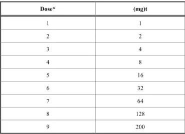
Because the polyene derivative nystatin and echino-candins are active mainly against yeasts, Candida species, azoles, triazoles and amphotericin B show a broad spectrum of activities against dermatophytes, yeasts and moulds [131], so azole antifungals remain fundamental drugs to treat sys-temic fungal infections. Drug desensitization and a graded challenge test with another triazole are the strategies fol-lowed to avoid using amphotericin B in view of its toxicity. Bittleman firstly used a desensitization protocol to itracona-zole in 1994 in a pruritic rash, as illustrated in Table 3 [59] and their protocol was replicated in a patient with fungal sinusitis [132].
Table 3. Itraconazole Desensitization Schedule Through an Oral Increasing Doses of Itraconazole and an Inter-val between Doses 30 Minutes.
Dose* (mg)t 1 1 2 2 3 4 4 8 5 16 6 32 7 64 8 128 9 200
*The itraconazole capsules were crushed; and the contents ofthe capsule were weighed, mixed in applesauce, and given to the patient.
One year later, Craig et al. performed the first desensiti-zation to fluconazole in an HIV male patient, as shown in Table 4 [129].
The same protocol was modified by Takahashi et al. who gave 4, 10, 20, 50, 100 and 200 mg daily, reaching the thera-peutic dose in 7 days instead of 15 [66]. Jariwala et al. short-ened the desensitization procedure to 5 days only, as shown
in Table 5 [71]. Apart from slow desensitization protocols for fluconazole, even rapid [21] and semi-rapid desensitiza-tion schedules as in Table 6 [130] have been developed to rapidly reach the therapeutic dose in an HIV patient with active pulmonary fungal infections.
Interestingly, treatment with fluconazole caused an im-mediate-type reaction leading to a cutaneous macular rash in
a patient, after the fifth desensitization process, suggesting a switch from an immediate-type to a delayed-type hypersensi-tivity [21].
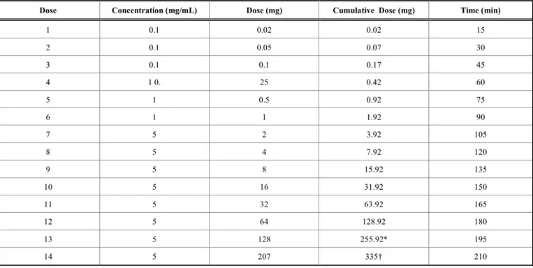
Jean and Kwuong performed a successful rapid desensiti-zation procedure to voriconazole in a 13-year old boy [26], this being the only intravenous desensitization protocol to triazoles described in literature, whereas all the others were Table 4. Slow Procedure for Fluconazole Oral Desensitization.
Day Concentration Dose Total mg
1 1mg/ml 0.2ml 0.2 2 1mg/ml 0.4ml 0.4 3 1mg/ml 0.8ml 0.8 4 1mg/ml 1.6ml 1.6 5 1mg/ml 3.2ml 3.2 6 1mg/ml 6.4ml 6.4 7 10mg/ml 1.0ml 10 8 10mg/ml 2.0ml 20 9 10mg/ml 4.0ml 40 10 50mg tablet 1 50 11 50mg tablet 2 100 12 50mg tablet 3 150 13 200mg tablet 1 200 14 100mg tablet 3 300 15 200mg tablet 2 400
Continue 400mg each day without stopping.
Table 5. Accelerated Desensitization Protocol with Oral Fluconazole.
Hour Concentration, mg/mL Volume Administered Dose Administered
0 2 1mL 2mg 6 2 1mL 2mg 12 2 2mL 4mg 18 2 4mL 8mg 24 2 8mL 16mg 30 2 15mL 30mg 36 2 30mL 60mg 42 2 60mL 120mg 48 NA NA 200mg (tablet) 60 NA NA 200mg (tablet)
A premedication with diphenhydramine, 25mg, and famotidine, 20mg, 30 minutes before the first dose. Continue the diphenhydramine, 25mg, 3 times daily and famotidine, 20mg, twice daily during the entire desensitization. Abbreviation: NA, Not Applicable).
Table 6. Fast Desensitization Procedure.
Step Concentration mg/mL Volume mL Dose Administered mg Cumulative Dose mg
1 0.02 1 0.02 0.02 2 0.02 2 0.04 0.06 3 0.02 4 0.08 0.14 4 0.2 0.8 0.16 0.30 5 0.2 1.6 0.32 0.62 6 0.2 3.2 0.64 1.26 7 2 0.75 1.50 2.76 8 2 1.5 3.00 5.76 9 2 3 6.0 11.76 10 20 0.6 12 23.76 11 20 1.2 24 47.76 12 20 2.5 50 97.76 13 20 5.0 100 197.76
The procedure is given for rapid desensitization of fluconazole, 0.02mg/mL. There was a 15-minute interval between doses. The patient was observed for 2 hours without event.
Table 7. Voriconazole Intravenous Desensitization Protocol.
Dose Concentration (mg/mL) Dose (mg) Cumulative Dose (mg) Time (min)
1 0.1 0.02 0.02 15 2 0.1 0.05 0.07 30 3 0.1 0.1 0.17 45 4 1 0. 25 0.42 60 5 1 0.5 0.92 75 6 1 1 1.92 90 7 5 2 3.92 105 8 5 4 7.92 120 9 5 8 15.92 135 10 5 16 31.92 150 11 5 32 63.92 165 12 5 64 128.92 180 13 5 128 255.92* 195 14 5 207 335† 210
*Maintenance dose is 4mg/kg every 12h, approximately 223mg. †Loading dose is 6mg/kg, approximately 335mg.
Table 7a. Graded challenge with oral voriconazole: -starting at a dose of 25mg daily on day 1, 75mg b.i.d. on day 2,
150mg b.i.d. on day 3, 300mg daily on day 4, and then 200mg b.i.d. thereafter.
given by oral route. Morales et al. preferred to desensitize a patient with angioedema due to voriconazole using isavu-conazole, probably due to the unknown pattern of triazole cross-reactivity [25]. In their experience, Oriel et al.
sug-gested that in patients with mild delayed-type cutaneous hy-persensitivity, even rapid desensitization protocols may work [133]. Graded challenge test and desensitization protocols with voriconazole are reported in Tables 7 and 8. Lastly, a
graded challenge test with isavuconazole was started intra-venously and switched to an oral formulation of isavuco-lazium (Table 9).
Although there are some differences between a graded challenge test and a desensitization procedure, because a graded challenge test involves fewer steps and may be per-formed with alternative molecules, some authors consider such methods very similar [134].
CONCLUSION
Despite their widespread use [2,6], triazoles seem to in-duce rare cutaneous hypersensitivity reactions, but the path-omechanisms, risk factors, diagnostic and management strategies, including skin tests and challenge tests, are little known and poorly investigated. Probably the severity of the diseases and the compromised clinical conditions of patients,
as for instance, in patients affected by hematologic malig-nancies, discourage medics to perform skin tests or labora-tory tests which could be seen as an unuseful diagnostic doggedness by the patient and other doctors.
CURRENT & FUTURE DEVELOPMENTS
Because of the widespread use of azole antifungal agents, also in immune-compromised patients, further, more in-depth investigations of hypersensitivity to triazoles are poorly warranted.
CONSENT FOR PUBLICATION
Not applicable.
FUNDING
None. Table 8. Oral Voriconazole IDT Protocol (117).
0.4mg/mL Oral Suspension Voriconazole
Dose Volume (mL) Dose (mg) Cumulative Dose (mg) Time (min)
1 0.25 0.1 0.1 30 2 0.5 0.2 0.3 60 3 1 0.4 0.7 90 4 1.5 0.6 1.3 120 5 2.75 1.1 2.4 150 6 4 1.6 4 180 TOTAL VOLUME = 10mL
40mg/mL Oral Suspension Voriconazole
Dose Volume (mL) Dose (mg) Cumulative Dose (mg) Time (min)
7 0.08 3.2 7.2 210 8 0.16 6.4 13.6 240 9 0.31 12.4 26 270 10 0.5 20 46 300 11 0.61 24.4 70.4 330 12 1.24 49.6 120 360 TOTAL VOLUME = 2.9mL
Table 9. Isavuconazole 1 Graded Dose Challenge.
Day Dosage, Route, and Frequency Ratio of dose Administered to Recommended Adult Dose
1 3.72mg in 250mL normal saline IV once 1:100
2 37.2mg in 250mL normal saline IV once 1:10
3 186mg PO once 1:2
4 372mg PO once 1:1
5 372mg PO every 8 hours Usual loading dose
1 After administration as isavuconazonium sulfate via 372mg vials or 186 mg capsules, the drug undergoes metabolism to the active form of isavuconazole at the equivalent of 200mg (per 372mg vial) or 100mg (per 186mg capsule).
2 To prepare, reconstitute 372mg (1 vial) with 5mL SWFI (sterile water for injection). Then add to 250mL normal saline bag (Bag A). For the final dose, take 2.5mL of Bag A and add to 250mL normal saline bag (Ba).
CONFLICT OF INTEREST
The authors declare no conflict of interest, financial or otherwise.
ACKNOWLEDGEMENTS
Declared none.
REFERENCES
[1] Verma, A.K., Arora, S.K., Arora, J.S., Rattan, A. Azole compounds as therapeutic agents for fungal infections. US6670363 (2003). [2] Thompson GR III, Cadena J, Patterson TF. Overview of antifungal
agents. Clin Chest Med 2009; 30(2): 203-15.
[http://dx.doi.org/10.1016/j.ccm.2009.02.001] [PMID: 19375628] [3] Sobel JD, Sobel R. Current treatment options for vulvovaginal
candidiasis caused by azole-resistant Candida species. Expert Opin Pharmacother 2018; 19(9): 971-7.
[http://dx.doi.org/10.1080/14656566.2018.1476490] [PMID: 29932786]
[4] Arndt CA, Walsh TJ, McCully CL, Balis FM, Pizzo PA, Poplack DG. Fluconazole penetration into cerebrospinal fluid: Implications for treating fungal infections of the central nervous system. J Infect Dis 1988; 157(1): 178-80.
[http://dx.doi.org/10.1093/infdis/157.1.178] [PMID: 2826606] [6] Ben-Ami R, Lewis RE, Kontoyiannis DP. Immunocompromised
hosts: Immunopharmacology of modern antifungals. Clin Infect Dis 2008; 47(2): 226-35.
[http://dx.doi.org/10.1086/589290] [PMID: 18540822]
[7] Dodds Ashley ES, Russell L, Lewis JS, Craig M, Andes D. Phar-macology of systemic antifungal agents. Clin Infect Dis 2006; 43: S28-39.
[http://dx.doi.org/10.1086/504492]
[8] Pérez-Mesonero R, Schneller-Pavelescu L, Ochando-Ibernón G, Vergara-Sánchez A, Sánchez-Herreros C, Martín-Alcalde E, et al. Is tioconazole contact dermatitis still a concern? Bringing allergic contact dermatitis caused by topical tioconazole back into the spot-light. Contact Dermat 2018; 80(3): 168-9.
[http://dx.doi.org/10.1111/cod.13146] [PMID: 30411354] [9] Dooms-Goossens A, Matura M, Drieghe J, Degreef H. Contact
allergy to imidazoles used as antimycotic agents. Contact Dermat 1995; 33(2): 73-7.
[http://dx.doi.org/10.1111/j.1600-0536.1995.tb00504.x] [PMID: 8549147]
[10] Johansson SG, Bieber T, Dahl R, Friedmann PS, Lanier BQ, Lockey RF, et al. Revised nomenclature for allergy for global use: Report of the Nomenclature Review Committee of the World Al-lergy Organization, October 2003. J AlAl-lergy Clin Immunol 2004; 113(5): 832-6.
[http://dx.doi.org/10.1016/j.jaci.2003.12.591] [PMID: 15131563] [11] Ventura MT, Cassano N, Romita P, Vestita M, Foti C, Vena GA.
Management of chronic spontaneous urticaria in the elderly. Drugs Aging 2015; 32(4): 271-82.
[http://dx.doi.org/10.1007/s40266-015-0249-x] [PMID: 25851216] [12] González-Delgado P, Florido-Lopez F, Saenz de San Pedro B,
Cuevas-Agusti M, Marin-Pozo JF. Hypersensitivity to ketocona-zole. Ann Allergy 1994; 73(4): 326-8.
[PMID: 7524384]
[13] Ensina LF, Tanno LK, Motta AA, Kalil J, Giavina-Bianchi P. Ke-toconazole allergy. Clinics (São Paulo) 2009; 64(4): 373-4. [http://dx.doi.org/10.1590/S1807-59322009000400018] [PMID: 19488598]
[14] van Dijke CP, Veerman FR, Haverkamp HC. Anaphylactic reac-tions to ketoconazole. Br Med J (Clin Res Ed) 1983; 287(6406): 1673-9.
[http://dx.doi.org/10.1136/bmj.287.6406.1673] [PMID: 6315129] [15] Foti C, Damiani E, Zambonin CG, Cassano N, Nettis E, Ferrannini
A, et al. Urticaria and angioedema to rubisco allergen in spinach and tomato. Ann Allergy Asthma Immunol 2012; 108(1): 60-1. [http://dx.doi.org/10.1016/j.anai.2011.09.011] [PMID: 22192968] [16] Chen J, Song X, Yang P, Wang J. Appearance of anaphylactic
shock after long-term intravenous itraconazole treatment. Ann Pharmacother 2009; 43(3): 537-41.
[http://dx.doi.org/10.1345/aph.1L343] [PMID: 19261964]
[17] Martínez-Alonso JC, Domínguez-Ortega FJ, Fuentes-Gonzalo MJ. Urticaria and angioedema due to itraconazole. Allergy 2003; 58(12): 1317-8.
[http://dx.doi.org/10.1046/j.0105-4538.2003.00316.x] [PMID: 14616112]
[18] Schmutz JL, Barbaud A, Trechot P. Urticaria and angioedema due to itraconazole. Ann Dermatol Venereol 2005; 132: 403-10. [http://dx.doi.org/10.1016/S0151-9638(05)79298-X]
[19] Neuhaus G, Pavic N, Pletscher M. Anaphylactic reaction after oral fluconazole. BMJ 1991; 302(6788): 1341-7.
[http://dx.doi.org/10.1136/bmj.302.6788.1341-b] [PMID: 2059703] [20] Koklu E, Kalay S, Koklu S, Ariguloglu EA. Fluconazole admini-stration leading to anaphylactic shock in a preterm newborn. Neo-natal Netw 2014; 33(2): 83-5.
[http://dx.doi.org/10.1891/0730-0832.33.2.83] [PMID: 24589899] [21] Randolph C, Kaplan C, Fraser B. Rapid desensitization to
flucona-zole (Diflucan). Ann Allergy Asthma Immunol 2008; 100(6): 616-7.
[http://dx.doi.org/10.1016/S1081-1206(10)60063-4] [PMID: 18592829]
[22] Abbott M, Hughes DL, Patel R, Kinghorn GR. Angio-oedema after fluconazole. Lancet 1991; 338(8767): 633.
[http://dx.doi.org/10.1016/0140-6736(91)90643-4] [PMID: 1679171] [23] Singh Mahal H. Fluconazole-induced type-1 Kounis syndrome. Am
J Ther 2016; 23(3): e961-2.
[http://dx.doi.org/10.1097/MJT.0000000000000113] [PMID: 26938747]
[24] Gençer S, Ozer S, Demirhan G, Ak O, Batirel A. Angio-oedema as an unusual tolerable side effect of voriconazole therapy. J Med Mi-crobiol 2008; 57(Pt 8): 1028-31.
[http://dx.doi.org/10.1099/jmm.0.47299-0] [PMID: 18628507] [25] Morales MK, Harris C, Shoham S. Graded isavuconazole
introduc-tion in a patient with voriconazole allergy. Transpl Infect Dis 2017; 19(6)
[http://dx.doi.org/10.1111/tid.12772] [PMID: 28851131]
[26] Jean T, Kwong K. Successful desensitization of voriconazole in an immunosuppressed pediatric patient. J Allergy Clin Immunol Pract 2015; 3(4): 637-8.
[http://dx.doi.org/10.1016/j.jaip.2015.02.008] [PMID: 25800818] [27] Eiden C, Peyrière H, Cociglio M, Djezzar S, Hansel S, Blayac JP,
et al. Adverse effects of voriconazole: Analysis of the French Pharmacovigilance Database. Ann Pharmacother 2007; 41(5): 755-63.
[http://dx.doi.org/10.1345/aph.1H671] [PMID: 17456542] [28] Ozkaya-Bayazit E, Bayazit H, Ozarmagan G. Drug related clinical
pattern in fixed drug eruption. Eur J Dermatol 2000; 10(4): 288-91. [PMID: 10846256]
[29] Mockenhaupt M. Epidemiology of cutaneous adverse drug reac-tions. Allergol Select 2017; 1(1): 96-108.
[http://dx.doi.org/10.5414/ALX01508E] [PMID: 30402608] [30] Bharija SC, Belhaj MS. Ketoconazole-induced fixed drug eruption.
Int J Dermatol 1988; 27(4): 278-9.
[http://dx.doi.org/10.1111/j.1365-4362.1988.tb03236.x] [PMID: 2968961]
[31] Kastalli S, El Aïdli S, Lakhoua G, Daghfous R. Genital fixed drug eruption induced by miconazole with positive provocation test. Tu-nis Med 2014; 92(11): 703.
[PMID: 25867162]
[32] Guliani A, Chauhan A. Fixed drug eruption due to itraconazole: A rare occurence. Postgrad Med J 2019; 95(1124): 340-1.
[http://dx.doi.org/10.1136/postgradmedj-2019-136680] [PMID: 31085618]
[33] Morgan JM, Carmichael AJ. Fixed drug eruption with fluconazole. BMJ 1994; 308(6926): 454.
[http://dx.doi.org/10.1136/bmj.308.6926.454a] [PMID: 8124179] [34] Heikkilä H, Timonen K, Stubb S. Fixed drug eruption due to
flu-conazole. J Am Acad Dermatol 2000; 42(5 Pt 2): 883-4. [http://dx.doi.org/10.1016/S0190-9622(00)90262-7] [PMID: 10767695]
[35] Ghislain PD, Ghislain E. Fixed drug eruption due to fluconazole: A third case. J Am Acad Dermatol 2002; 46(3): 467.
[http://dx.doi.org/10.1067/mjd.2002.118356] [PMID: 11862192] [36] Coondoo ABR. Fluconazole induced fixed drug eruption. Int J
[37] Lane JE, Buckthal J, Davis LS. Fixed drug eruption due to flucona-zole. Oral Surg Oral Med Oral Pathol Oral Radiol Endod 2003; 95(2): 129-30.
[http://dx.doi.org/10.1067/moe.2003.14] [PMID: 12599328] [38] Goel A, Jain C. Fluconazole induced fixed drug eruption: A rare
offender. J Dermatol 2004; 31(4): 345-6.
[http://dx.doi.org/10.1111/j.1346-8138.2004.tb00683.x] [PMID: 15187332]
[39] Gupta R, Thami GP. Fixed drug eruption caused by itraconazole: Reactivity and cross reactivity. J Am Acad Dermatol 2008; 58(3): 521-2.
[http://dx.doi.org/10.1016/j.jaad.2006.06.014] [PMID: 18280361] [40] Tavallaee M, Rad MM. Fixed drug eruption resulting from
flu-conazole use: A case report. J Med Case Reports 2009; 3: 7368-77. [http://dx.doi.org/10.4076/1752-1947-3-7368] [PMID: 19830193] [41] Walling HW, Swick BL. Cutaneous fixed drug eruption to
flucona-zole. J Drugs Dermatol 2010; 9(8): 1025-8. [PMID: 20684158]
[42] Kim CY, Kim JG, Oh CW. Fluconazole induced fixed drug erup-tion. Ann Dermatol 2011; 23(Suppl. 1): S1-3.
[http://dx.doi.org/10.5021/ad.2011.23.S1.S1] [PMID: 22028550] [43] Beecker J, Colantonio S. Fixed drug eruption due to fluconazole.
CMAJ 2012; 184(6): 675-83.
[http://dx.doi.org/10.1503/cmaj.111530] [PMID: 22231677] [44] Nakai N, Katoh N. Fixed drug eruption caused by fluconazole: A
case report and mini-review of the literature. Allergol Int 2013; 62: 139-41.
[http://dx.doi.org/10.2332/allergolint.12-LE-0464]
[45] Gaiser CA, Sabatino D. Fluconazole-induced fixed drug eruption. J Clin Aesthet Dermatol 2013; 6(3): 44-5.
[PMID: 23556037]
[46] Vora RV, Mehta MJ, Diwan NG, Gor AP, Gajjar B. Fixed drug eruption due to fluconazole: A case report. Natl J Physiol Pharm Pharmacol 2014; 4: 255-7.
[http://dx.doi.org/10.5455/njppp.2014.4.040620142]
[47] Santra R, Pramanik S, Raychaudhuri P. Fixed drug eruption due to fluconazole: Not so uncommon now-a-days. J Clin Diagn Res 2014; 8(11): HL01.
[http://dx.doi.org/10.7860/JCDR/2014/7644.5159] [PMID: 25584246]
[48] Waldman L, Reddy SB, Kassim A, Dettloff J, Reddy VB. Neutro-philic fixed drug eruption to fluconazole. Am J Dermatopathol 2015; 37(7): 574-6.
[http://dx.doi.org/10.1097/DAD.0000000000000157] [PMID: 25072682]
[49] Lai O, Hsu S. Fixed drug eruption related to fluconazole. Dermatol Online J 2016; 22: pii.
[50] Demir S, Cetin EA, Unal D, et al. Generalized fixed drug eruption induced by fluconazole without cross-reactivity to itraconazole: Lymphocyte transformation test confirms the diagnosis. Drug Saf Case Rep 2018; 5(1): 2-15.
[http://dx.doi.org/10.1007/s40800-017-0067-7] [PMID: 29294202] [51] Quint T, Wöhrl S, Kinaciyan T. Fixed drug eruption caused by
fluconazole-An underdiagnosed but recurrent problem. Contact Dermat 2019; 80(3): 172-3.
[http://dx.doi.org/10.1111/cod.13149] [PMID: 30417394] [52] Benedix F, Schilling M, Schaller M, Röcken M, Biedermann T. A
young woman with recurrent vesicles on the lower lip: Fixed drug eruption mimicking herpes simplex. Acta Derm Venereol 2008; 88(5): 491-4.
[http://dx.doi.org/10.2340/00015555-0519] [PMID: 18779889] [53] Alfonso N, Rane P, Dang A, Rataboli P, Goel H. Fluconazole
in-duced herpes labialis like lesions in an adult male. Austr Med J 2009; 14: 246-7.
[54] González-Fernández T, López-Freire S, Juangorena M, Méndez-Brea P, Vázquez-Veiga H. Herpes-like eruption due to fluconazole. J Investig Allergol Clin Immunol 2015; 25(2): 135-6.
[PMID: 25997308]
[55] Jensen ZN, Bygum A, Damkier P. Fluconazole-induced fixed drug eruption imitating herpes labialis with erythema multiforme. Eur J Dermatol 2012; 22(5): 693-4.
[http://dx.doi.org/10.1684/ejd.2012.1806] [PMID: 22858871] [56] Schneller-Pavelescu L, Ochando-Ibernón G, Vergara-de Caso E,
Silvestre-Salvador JF. Herpes simplex-like fixed drug eruption in-duced by fluconazole without cross-reactivity to itraconazole. Dermatitis 2019; 30(2): 174-5.
[http://dx.doi.org/10.1097/DER.0000000000000451] [PMID: 30829805]
[57] Sławińska M, Barańska-Rybak W, Wilkowska A, Nowicki R. Bullous fixed drug eruption due to fluconazole, imitating herpes simplex. Clin Exp Dermatol 2017; 42(5): 544-5.
[http://dx.doi.org/10.1111/ced.13098] [PMID: 28512848] [58] Nath AK, Adityan B, Thappa DM. Multifocal bullous fixed drug
eruption due to fluconazole. Indian J Dermatol 2008; 53(3): 156-7. [http://dx.doi.org/10.4103/0019-5154.43212] [PMID: 19882020] [59] Choudhury S, Loc BP, Debbarma RR, Das A. Fluconazole induced
multifocal bullous eruptions: A case report. Int J Basic Clin Phar-macol 2016; 5: 1681-3.
[http://dx.doi.org/10.18203/2319-2003.ijbcp20162492]
[60] Bittleman DB, Stapleton J, Casale TB. Report of successful desen-sitization to itraconazole. J Allergy Clin Immunol 1994; 94(2 Pt 1): 270-1.
[http://dx.doi.org/10.1053/ai.1994.v94.a55252] [PMID: 8064084] [61] Batinac T, Zamolo G, Troselj-Vukić B, Biljan D, Petranović D,
Kujundzić M. Maculopapular eruption secondary to itraconazole. Coll Antropol 2008; 32(2): 649-51.
[PMID: 18756926]
[62] Goto Y, Kono T, Teramae K, Ishii M. Itraconazole-induced drug eruption confirmed by challenge test. Acta Derm Venereol 2000; 80(1): 72-9.
[http://dx.doi.org/10.1080/000155500750012694] [PMID: 10721851] [63] Di Leo E, Nettis E, Priore MG, Ferrannini A, Vacca A. Maculo-papular rash due to fluconazole. Clin Exp Dermatol 2009; 34(3): 404-12.
[http://dx.doi.org/10.1111/j.1365-2230.2008.02895.x] [PMID: 18627382]
[64] Kahana M, Levy A, Yaron-Shiffer O, Schewach-Millet M. Drug eruption following ketoconazole therapy. Arch Dermatol 1984; 120(7): 837-42.
[http://dx.doi.org/10.1001/archderm.1984.01650430023003] [PMID: 6329105]
[65] van Ketel WG. An allergic eruption probably caused by ketocona-zole. Contact Dermat 1983; 9(4): 313-.
[http://dx.doi.org/10.1111/j.1600-0536.1983.tb04398.x] [PMID: 6311485]
[66] Kramer KE, Yaar M, Andersen W. Purpuric drug eruption secon-dary to itraconazole. J Am Acad Dermatol 1997; 37(6): 994-5. [http://dx.doi.org/10.1016/S0190-9622(97)70080-X] [PMID: 9418771]
[67] Su FW, Perumalswami P, Grammer LC. Acute hepatitis and rash to fluconazole. Allergy 2003; 58(11): 1215-6.
[http://dx.doi.org/10.1046/j.0105-4538.2003.00318.x] [PMID: 14616149]
[68] Pinto A, Chan RC. Lack of allergic cross-reactivity between flu-conazole and voriflu-conazole. Antimicrob Agents Chemother 2009; 53(4): 1715-6.
[http://dx.doi.org/10.1128/AAC.01500-08] [PMID: 19164151] [69] Alffenaar JWC, van Assen S, de Monchy JGR, Uges DRA,
Koster-ink JGW, van der Werf TS. Intravenous voriconazole after toxic oral administration. Antimicrob Agents Chemother 2010; 54(6): 2741-2.
[http://dx.doi.org/10.1128/AAC.01193-09] [PMID: 20385853] [70] Takahashi T, Hitani A, Yamada H, Nakamura T, Iwamoto A.
De-senitization to fluconazole in an AIDS patient. Ann Pharmacother 2001; 35(5): 642-3.
[http://dx.doi.org/10.1345/aph.10285] [PMID: 11346073] [71] Jariwala S, Vernon N, de Vos G. A novel method of desensitization
for fluconazole hypersensitivity in a patient with AIDS. Ann Al-lergy Asthma Immunol 2011; 106(6): 542-3.
[http://dx.doi.org/10.1016/j.anai.2011.02.020] [PMID: 21624759] [72] Fernandez L, Maquiera E, Rodriguez F, Picans I, Duque S.
Sys-temic contact dermatitis from miconazole. Contact Dermat 1996; 34(3): 217.
[http://dx.doi.org/10.1111/j.1600-0536.1996.tb02178.x] [PMID: 8833469]
[73] Rademaker M, Barker S. Contact dermatitis to a canine anti-dandruff shampoo. Australas J Dermatol 2007; 48(1): 62-3. [http://dx.doi.org/10.1111/j.1440-0960.2007.00329.x] [PMID: 17222309]
[74] Imafuku S, Nakayama J. Contact allergy to ketoconazole cross-sensitive to miconazole. Clin Exp Dermatol 2009; 34(3): 411-2.
[http://dx.doi.org/10.1111/j.1365-2230.2008.02942.x] [PMID: 19120394]
[75] Erdmann S, Hertl M, Merk HF. Contact dermatitis from clotrima-zole with positive patch-test reactions also to croconaclotrima-zole and itra-conazole. Contact Dermat 1999; 40(1): 47-8.
[http://dx.doi.org/10.1111/j.1600-0536.1999.tb05977.x] [PMID: 9928806]
[76] Nasir S, Goldsmith P. Anogenital allergic contact dermatitis caused by methylchloroisothiazolinone, methylisothiazolinone and topical clotrimazole with subsequent generalized exanthem triggered by oral fluconazole. Contact Dermat 2016; 74(5): 296-7.
[http://dx.doi.org/10.1111/cod.12513] [PMID: 27040872] [77] Stingeni L, Rigano L, Lionetti N, Bianchi L, Tramontana M, Foti
C, et al. Sensitivity to imidazoles/nitroimidazoles in subjects sensi-tized to methylchloroisothiazolinone/methylisothiazolinone: A simple coincidence? Contact Dermat 2019; 80(3): 181-3.
[http://dx.doi.org/10.1111/cod.13158] [PMID: 30468019] [78] Tan SC, Tan JW. Symmetrical drug-related intertriginous and
flexural exanthema. Curr Opin Allergy Clin Immunol 2011; 11(4): 313-8.
[http://dx.doi.org/10.1097/ACI.0b013e3283489d5f] [PMID: 21659857]
[79] Gulec AI, Uslu E, Başkan E, Yavuzcan G, Aliagaoglu C. Baboon syndrome induced by ketoconazole. Cutan Ocul Toxicol 2014; 33(4): 339-41.
[http://dx.doi.org/10.3109/15569527.2013.870187] [PMID: 24641119]
[80] Mohapatra M, Panda M, Kar BR, Raj C. Symmetric drug-related intertriginous and flexural exanthema due to Iiraconazole: An un-common side effect of a un-commonly used drug. Indian Dermatol Online J 2017; 8(6): 501-3.
[http://dx.doi.org/10.4103/idoj.IDOJ_179_17] [PMID: 29204404] [81] Nanda S. A case of SDIFE induced by fluconazole. J Am Acad
Dermatol 2016; 74(1): 89-95.
[82] Bianchi L, Hansel K, Antonelli E, Bellini V, Stingeni L. Contact allergy to isoconazole nitrate with unusual spreading over extensive regions. Contact Dermat 2017; 76(4): 243-5.
[http://dx.doi.org/10.1111/cod.12688] [PMID: 28317189] [83] Patruno C, Megna M, Bianca D, Patri A, Balato N. Erythema
mul-tiforme-like allergic contact dermatitis due to tioconazole. Clin Dermatol 2014; 2: 48-50.
[84] Park H, Knowles S, Shear NH. Serum sickness-like reaction to itraconazole. Ann Pharmacother 1998; 32(11): 1249-55.
[http://dx.doi.org/10.1345/aph.17432] [PMID: 9825096]
[85] Tang MM, Corti MAM, Stirnimann R, et al. Severe cutaneous allergic reactions following topical antifungal therapy. Contact Dermat 2013; 68(1): 56-7.
[http://dx.doi.org/10.1111/j.1600-0536.2012.02151.x] [PMID: 23227869]
[86] Gravante G, Delogu D, Marianetti M, Esposito G, Montone A. Toxic epidermal necrolysis and Steven-Johnson syndrome in on-cologic patients. Eur Rev Med Pharmacol Sci 2007; 11(4): 269-74. [PMID: 17876963]
[87] Gussenhoven MJ, Haak A, Peereboom-Wynia JD, van’t Wout JW. Stevens-Johnson syndrome after fluconazole. Lancet 1991; 338(8759): 120-6.
[http://dx.doi.org/10.1016/0140-6736(91)90112-3] [PMID: 1676446] [88] Azón-Masoliver A, Vilaplana J. Fluconazole-induced toxic epi-dermal necrolysis in a patient with human immunodeficiency virus infection. Dermatology (Basel) 1993; 187(4): 268-9.
[http://dx.doi.org/10.1159/000247261] [PMID: 8274783]
[89] Monastirli A, Pasmatzi E, Vryzaki E, Georgiou S, Tsambaos D. Fluconazole-induced Stevens-Johnson syndrome in a HIV-negative patient. Acta Derm Venereol 2008; 88(5): 521-2.
[http://dx.doi.org/10.2340/00015555-0489] [PMID: 18779900] [90] Craythorne E, Creamer D. Stevens-Johnson syndrome due to
pro-phylactic fluconazole in two patients with liver failure. Clin Exp Dermatol 2009; 34(7): e389-90.
[http://dx.doi.org/10.1111/j.1365-2230.2009.03365.x] [PMID: 19548943]
[91] Ofoma UR, Chapnick EK. Fluconazole induced toxic epidermal necrolysis: A case report. Cases J 2009; 2: 9071.
[http://dx.doi.org/10.1186/1757-1626-2-9071] [PMID: 20062708] [92] Thiyanaratnam J, Cohen PR, Powell S. Fluconazole-associated
Stevens-Johnson syndrome. J Drugs Dermatol 2010; 9(10): 1272-5. [PMID: 20941954]
[93] Pasmatzi E, Monastirli A, Georgiou S, Sgouros G, Tsambaos D. Short-term and low-dose oral fluconazole treatment can cause Ste-vens-Johnson syndrome in HIV-negative patients. J Drugs Derma-tol 2011; 10(12): 1360-7.
[PMID: 22134558]
[94] George J, Sharma A, Dixit R, Chhabra N, Sharma S. Toxic epider-mal necrolysis caused by fluconazole in a patient with human im-munodeficiency virus infection. J Pharmacol Pharmacother 2012; 3(3): 276-8.
[http://dx.doi.org/10.4103/0976-500X.99445] [PMID: 23129968] [95] Islam S, Singer M, Kulhanjian JA. Toxic epidermal necrolysis in a
neonate receiving fluconazole. J Perinatol 2014; 34(10): 792-4. [http://dx.doi.org/10.1038/jp.2014.92] [PMID: 25263725] [96] Huang DB, Wu JJ, Lahart CJ. Toxic epidermal necrolysis as a
complication of treatment with voriconazole. South Med J 2004; 97(11): 1116-7.
[http://dx.doi.org/10.1097/01.SMJ.0000144618.80128.F9] [PMID: 15586606]
[97] Curigliano G, Formica V, De Pas T, et al. Life-threatening toxic epidermal necrolysis during voriconazole therapy for invasive as-pergillosis after chemotherapy. Ann Oncol 2006; 17(7): 1174-5. [http://dx.doi.org/10.1093/annonc/mdj126] [PMID: 16410362] [98] Gomulka J, Wilson BD, Joyce JC. Toxic epidermal necrolysis due
to voriconazole: Case report and review. Dermatol Online J 2014; 20(9): 20.
[PMID: 25244163]
[99] Lester LJ, Brantley JS, Kelso RL, Kelly BC, Petitt MS, Wilkerson MG. Severe cutaneous adverse drug reaction due to fluconazole. J Drugs Dermatol 2008; 7(11): 1084-7.
[PMID: 19110744]
[100] Eloranta K, Karakorpi H, Jeskanen L, Kluger N. Photo-distributed Stevens-Johnson syndrome associated with oral itraconazole. Int J Dermatol 2016; 55(9): e508-10.
[http://dx.doi.org/10.1111/ijd.13278] [PMID: 27028785]
[101] Alvarez-Fernández JG, Castaño-Suárez E, Cornejo-Navarro P, de la Fuente EG, Ortiz de Frutos FJ, Iglesias-Diez L. Photosensitivity induced by oral itraconazole. J Eur Acad Dermatol Venereol 2000; 14(6): 501-3.
[http://dx.doi.org/10.1046/j.1468-3083.2000.00164.x] [PMID: 11444275]
[102] Willis ZI, Boyd AS, Di Pentima MC. Phototoxicity, pseudoporphy-ria, and photo-onycholysis due to voriconazole in a pediatric pa-tient with leukemia and invasive aspergillosis. J Pediatric Infect Dis Soc 2015; 4(2): e22-4.
[http://dx.doi.org/10.1093/jpids/piu065] [PMID: 26407422] [103] Haylett AK, Felton S, Denning DW, Rhodes LE.
Voriconazole-induced photosensitivity: Photobiological assessment of a case se-ries of 12 patients. Br J Dermatol 2013; 168(1): 179-85.
[http://dx.doi.org/10.1111/j.1365-2133.2012.11196.x] [PMID: 22860570]
[104] Sheu J, Hawryluk EB, Guo D, London WB, Huang JT. Voricona-zole phototoxicity in children: A retrospective review. J Am Acad Dermatol 2015; 72(2): 314-20.
[http://dx.doi.org/10.1016/j.jaad.2014.10.023] [PMID: 25481710] [105] Miteva L, Kadurina M, Schwartz RA. Childhood acute generalized
exanthematous pustulosis induced by oral ketoconazole. Acta Der-matovenerol Croat 2010; 18(4): 267-70.
[PMID: 21251445]
[106] Heymann WR, Manders SM. Itraconazole-induced acute general-ized exanthemic pustulosis. J Am Acad Dermatol 1995; 33(1): 130-1.
[http://dx.doi.org/10.1016/0190-9622(95)90038-1] [PMID: 7601931] [107] Park YM, Kim JW, Kim CW. Acute generalized exanthematous pustulosis induced by itraconazole. J Am Acad Dermatol 1997; 36(5 Pt 1): 794-6.
[http://dx.doi.org/10.1016/S0190-9622(97)80353-2] [PMID: 9146550]
[108] Cançado GGL, Fujiwara RT, Freitas PA, Correa-Oliveira R, Be-thony JM. Acute generalized exanthematous pustulosis induced by itraconazole: An immunological approach. Clin Exp Dermatol 2009; 34(8): e709-11.
[http://dx.doi.org/10.1111/j.1365-2230.2009.03440.x] [PMID: 20055840]
[109] Alsadhan A, Taher M, Krol A. Acute generalized exanthematous pustulosis induced by oral fluconazole. J Cutan Med Surg 2002; 6(2): 122-4.