UNIVERSITÀ DEGLI STUDI DI ROMA
"TOR VERGATA"
FACOLTA' DI MEDICINA E CHIRURGIA
DOTTORATO DI RICERCA IN
SCIENZE DELLO SPORT
XXI CICLO DEL CORSO DI DOTTORATO
Titolo della tesi
VIBRATION ENERGY: TRAINING METHOD AND
NEUROMUSCULAR ASSESSMENT
dottorando: ANNINO Giuseppe
Docente Guida/Tutor: Prof. Calogero FOTI
Coordinatore: Prof. Antonio LOMBARDO
Contents
ABSTRACT 3
ITRODUCTION 5
REVIEW OF THE LITERATURE 7 1. Effects of Vibration Energy on Bone Tissue 7
1.1 Bone Tissue Properties 7
1.2 Bone Tournover 8
1.3 The Biomechanical properties of bone tissue 10 1.4 Effects of Physical Exercise on Bone Tissue 12 1.5 Vibration Energy as Osteogenic Intervention 13
2. Effects of the Vibration Energy on Neuromuscular System 15 2.1 Vibratory Energy as Neuromuscular System Assessment 17
3. STUDY I
The effects of low-frequency high-magnitude whole body
vibration in physical actively osteoporotic women: a pilot study 25 4. STUDY II
Isoinertial-sEMG and Vibration sEMG test assessment
in ACL Reconstruction 42
Abstract
Purpose: A new training method based on vibration energy applied to the whole body produce changes on gravitational conditions with consequently specific effects on neuromuscular, osteo-articular and endocrine system. Aims of this study concerns, after a deep scientific review about the effects on biological system, to investigate the effects of vibration energy related to the bone tissue in a sample of osteoporotic female and the possibility of hits application in the clinical investigation of neuromuscular function as during post-surgery rehabilitation period in patients operated of Anterior Cruciate Ligament (ACL).
Methods: in the present thesis are presented two experimental study.
Study 1. This study regard the effects of vibration energy in twelve
osteoporotic females after 4 months of treatment. The experimental group improved the Amplitude-Dependent Speed of Sound (AD-SoS) QUS parameter from 1.878 ± 79,45 to 1.971, 17 ± 78,69 (p<0,002), the T-score passing from -3,50 ± 1,13 to -2, 18 ± 1,12 (p<0.002) and the Ultrasound Bone Profile Index (UPBI) improve from 0,34 ± 0,11 to 0,47 ± 0,21 (p<0,01). In the control group (low impact exercise) any of these parameters considered show significantly changes over the same period of time.
Vibrations energy stimulated an increase in bone density and strength in animals and humans. This strongly anabolic, non-invasive intervention represents, associated to good physical fitness also, early evidence of an unique non-pharmacologic treatment for osteoporosis.
Study 2, This study is direct to evaluate the vibration energy as
assessment method in rehabilitation field, specifically direct to ACL operated patients after with a follow-up from the surgery intervention about 3,5 months.
During isoinertial test the l velocity (p< 0,002), the displacement (p<0,004) was lower, while the sEMg activity of vastus lateralis and
medialis (p<0,003) was higher than the non-operated leg. The EMG activity monitored during vibration treatment was much higher in the operated compared to controlateral-one (p<0,02).
The assessment of biomechanical parameters associated to sEMG activity in isoinertial dynamic and vibration test, represented by the comparison of operated/non-operated, showed to be an effectiveness clinical method of assessment the neuromuscular behaviour of the operated limb in different exercise regimen.
Conclusion The physical exercise is the only means, capable to counteract this degeneration process is the functional mechanical stimulus. In this contest the vibration energy represent a safe and healthy training method, effectiveness for its easy application in terms of time treatment and operability. Moreover, the response of neuromuscular system to vibration, analysed trough the use of sEMG, is an important assessment method. Associated to isoinertial test integrated with sEMG, actually is the only system capable to give information on neuromuscular strategies in post-operated subject.
Key Words: Vibration – Neuromuscular Assessment – Bone – Training – sEMG.
Introduction
The responses of mammals to environmental changes inevitably involves in specific adaptations of biological systems much more stimulated. The musculo-skeletal system is a very complex biological machine that function to support and move the body. Bones, joints and muscles actively change in structure and metabolism to meet various mechanical demands placed on the system. These systems interacts and respond to patterns of environmental ipergravity, like weight lifting exercise, or ipogravity conditions, like bed rest or microgravity exposure (experienced from astronauts during space mission), with major alteration in structure and strength. These changes involves as the muscle system than the bone tissue that receive stimuli from the muscles trough tendons and ligaments. The structure and growth of bones are genetically determinates excepted for density, mass and final architecture that changes in function of daily mechanical stress (34). Then, the interaction between the human been and the gravitational mechanical stimulus, represented by the daily habitual movements, produce structural and metabolic changes direct to maintain the whole osteo-muscle-articular system in functional conditions. Besides, the human locomotion, represent the lowest mechanical stimulation which ensures the basic muscular tone. This stimulus, which is generally required to win gravitational force, it is enough to protect the bones from injuries and fracture. In fact, during locomotion, in the ground contact phase, a train of vibrations are generated and transmitted along the body, travelling in form of impact waves from the foot, to the head throughout the legs, the spine and the skull. These impact waves represents the main osteogenic stimulus during the life of human been. Sedentary life style and hypokinesia, typical characteristics of industrialised countries, are the principal causes of degenerative diseases like diabetes, osteoporosis, etc. that affects a large number of ageing people. Epidemiological studies shows the importance of an
exercise training regimen to counteract the effects of hypokinesia on muscle-skeletal system (25, 40, 41, 48, 67). Recently, a new training method based on vibration energy applied to the whole body produce changes on gravitational conditions with consequently specific effects on neuromuscular, osteo-articular and endocrine system (1, 6, 7, 8, 12, 50, 51, 72, 73) . Aims of this study concerns, after a deep scientific review about the effects on biological system, to investigate the effects of vibration energy related to the bone tissue in a sample of osteoporotic female and the possibility of hits application in the clinical investigation of neuromuscular function as during post-surgery rehabilitation period in patients operated of Anterior Cruciate Ligament (ACL).
REVIEW OF THE LITERATURE
1. Effects of Vibration Energy on Bone Tissue
Osteoporosis is currently the major cause of in older people, with most severe consequence at hip level. The prevailing view is that the major determinant of fracture risk is the bone mineral density (BMD) (18, 19). The risk of hip fracture is approximately doubled for every standard deviation reduction in BMD (33).Thus to maximize the peak bone mass and maintain bone mass or decrease bone loss would reduce the fracture risk of elderly people. Several study indicates that physical activity is effective in improving and maintain bone mass and strength and thus in preventing osteoporosis fractures (40, 41, 67). The basic form and development of skeleton is genetically determined, but hits final mass and architecture are governed by an adaptive mechanism sensitive to the mechanical gravitational environment. According Wolff’s law (1892) (74), the bone functionally respond to the habitual stress imposed upon it. Following this concept, Frost in 1987 (26) proposed his mechanostat theory to explain the architecture and mass bone adaptations to its typical mechanical environment trough different biological process within strain ranges (loading-induced deformation in bone tissue) or thresholds for bone modelling and remodelling drift, supporting the biological bases of physical exercise effects on bone tissue (48).
1.1 Bone Tissue Properties
Bone is a structural and a metabolic tissue formed of 24% organic matrix, composed primarily of proteins, glycoproteins, polysaccharides, type I collagen fibres, noncollagenous protein, and of 76% by inorganic salts conferring rigidity and consistence to be differ from other connective tissue (2). This characteristics allow it to supports the body
against gravity, to provide a lever system and mechanical function for body motion, to protects soft tissues of internal organs and bone marrow, to takes part in mineral, especially calcium, homeostasis and haematopoiesis.
The bone architecture consist for 80% of whole skeletal mass of a cortical structure, dense solid mass with only microscopic spaces and channels that cover externally the bone, whereas internally for the resting 20%, of a trabecular structure formed by a bee nest scaffolding, with plates and arches, more metabolic active but less rigid than the cortical one (2, 13, 38). In the long bones, the diaphysis is primarily compact in structure, whereas the epiphysis and metaphysis are mainly filled with trabecular bone. The interaction among organic matrix, inorganic salts, cylindrical shape of cortical bone and trabecular structure, confers to the bone structure the maximum mechanical support with minimum mass (20, 42). The bone structure and shape warrants strength in relationship to load bearing stress (20, 52).
1.2 Bone Turnover
During the individual life the bone tissue change proper structure, shape, density and strength in function of his daily activity and load borne, trough three biological mechanisms with different specific function: the growth, that influence the dimension, the modelling, that influence the shape, and the remodelling, the influence the specific adaptation (3, 26, 27).
The growth of skeletal system is due from the interaction between genetic factors and mechanical stress, and modified by nutritional and hormonal aspects. During this phase the activation of muscle and weight-bearing exercise constitute a mechanical environment that induce the cellular activity to achieve and maintain the normal architecture, shape and size of the skeleton. This sensibility of bone tissue to mechanical stimuli, in the growth phase, may have important consequences on bone mass density later in the ageing (4, 25,
46). In effects, the BMD in the elderly people depends as of peak bone mass, that in human been is achieved around at age of 20 years, than of the rate of bone loss underwent (5, 22, 40, 55).
The modelling process is defined as simultaneous removal and formation of bone at different sites operate from two mediator mechanisms related to drift cells as osteoblasts, with the formation drifts add new bone, and osteoclast, with the resorption drifts remove bone over broad surface region (26, 29). The osteoblasts also, has an important role on the balance control of inflow and outflow of calcium ad others minerals. In the modelling process Frost (29) recognised two different sub group processes distinct for specific activity. The
Micromodeling organize the cells and collagen during heir formation to
determine the kinds of tissue being formed and thicken and rearrange the individual trabeculae (42, 44) The Macromodelling that control the gross shape, size, strength, add new bone at the periosteum and endosteum, and anatomy of joints, bones, tendons and other organs. Modelling process increase the bone strength by improving geometric properties and adding mass (42).
The Remodelling process is defined as the removal of old and the formation of new bone at same site at different time and repairs micro cracks (70). In a normal adult person, activation and resorption last one month and formation another three months, thus the time scale for one complete remodelling cycle, called sigma, occurs about 4 months (28, 38) while for one complete mineralisation occur in another three to four months (38). This process can be regulated by different factors as mechanicals as hormonal (steroids, thyroids hormones) and local factors (prostaglandins, growth factors). In disuse conditions of bone structure, like those experienced in ageing, the remodelling process is associated more to resorption than formation drafts (39).
1.3 The Biomechanical properties of bone tissue
The chemical and structural composition of bones determine two important quality like strength and rigidity. The structure strength can be defined as the load magnitude at the failure points or as the ultimate load supportable and depend of its size (21, 47), whereas, the rigidity is defined as the linear deformation capacity of structure submitted to increasing external loads. The deformation of structure bone is can be measured and plotted on a load-deformation curve and the stiffness or rigidity can be determined as slope of the linear part of this curve (Fig. 1), better know as elastic modulus or Young’s modulus. The physiological significance of this aspect is due to the fact that forces applied to the bone at any point to along this curve will only deform the bone temporarily and, after removed the load, the structure returns to its original state (21).
Fig. 1 Load – Deformation Curve
The central structure of long bone is naturally designed to act the greatest stiffness in the direction of long axis and greatest impact loading resistance occurs in the transverse direction. The biomechanics property of bone tissue, on hits continuous adaptation process, depend upon two fundamental concepts that define the properties of bone without the geometric effects: stress and strain.
Stress is represented by an internal resistance in bone generated to react the external force (equal and opposite) applied per unit area and it can be classified as compressive (produced when two force are directed toward each other along the same line and as consequence the material is shortened), tensile (produced when two forces are directed along the same line but away from each other and as consequence the material is stretched) or shear (when two force are parallel to each other but not along the same line as consequence the material is bent or torque) (fig). The measure unit of stress is 1 Pascal that is equivalent to a force of 1 N (Newton) acting over an area of one square meter. In nature this three types of stress can act alone or in combination. In addition to the forces which arise during daily activity, the skeleton may be subjected to impact force trough which collision, falls and other accidents. The concept of strain is referred to ratio between stress and structure stiffness. In other words, strain regard the deformation in shape an size the bone experienced under the influence of an applied load and it is reported as the changes in length of a material divided by the its original length (fig) (34, 71).
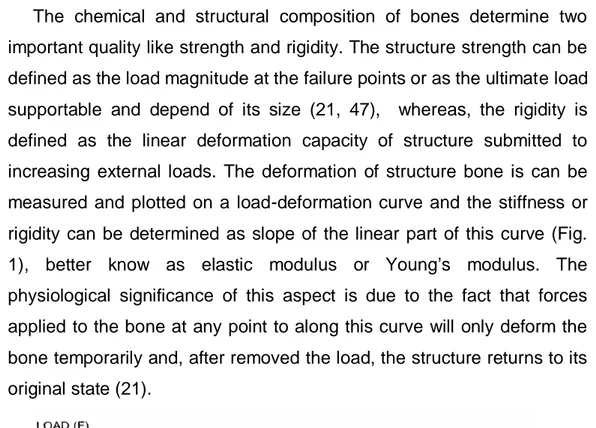
In the Frost “mechanostat theory “ the strain concept was utilised for to establish the threshold of minimum effective strain (MES) beyond which the bone tissue need to be stimulated to maintain functional homeostasis (26, 27, 29, 31). Loads inducing local strain with a magnitude comprise in a range of 1500 2500 microstrain, stimulates the modelling process with anabolic response of bone tissue, whereas mechanic stimuli under a range of 100-200 microstrain, primes the remodelling process with consequent bone resorption. (Fig. 2)(17, 29, 44).
Fig. 2 Diagram illustrating the relationship of strain and adaptive responses
1.4 Effects of Physical Exercise on Bone Tissue
The physical activity represent the principal mechanical stimulus in the regulatory process of modelling and remodelling process of bone tissue. However, the type, intensity, frequency and duration of exercise and the interrelationship that each of these variables has in terms of strain magnitude, strain frequency and strain intensity applied at bone tissue level to provide an optimal anabolic response are still largely unknown (34). In effects, the physical activity for to produce effects on the bone modeling process must be capable to generate adequate strains to bone level, not only from quantitative, but also from qualitative point of view, in terms of frequency and intensity. All these variables are integrated to each other on dynamic interactions of mechanical stimuli. Low magnitude strain to frequency level comprise between 15 – 30 Hz, typical frequency range of walking, represent a critic level for to prime an anabolic response of bone tissue. For to obtain an effective anabolic stimulus a the same frequency it need to increase the frequency (62). This aspects suggest that for a functional physical training regimen able to stimulate modelling process of bone structure must be programmed combining the variables of magnitude, intensity and frequency to obtain an enough level the mechanical stimulus.
Moreover, spatial strain distribution play a effective role in formation process of new bone, For this reason, the mechanical stimulus must be
applied with different type of movements, in relationship to a spatial strain distribution in different directions. In fact, different training regimen produce different loading characteristics of the skeleton (34). In this way, as an heavy resistance training or another type of mechanical stimulus of variable in intensity, characterised from movements performed in different directions, like in dance or in gymnastics, are able to determine specific anabolic response of bone tissue (34).
1.5 Vibration Energy as Osteogenic Intervention
Recently, in light of positive effects that vibration energy determine in muscular system, the whole body vibration has received hits scientific attention about the possible anabolic effects on bone tissue. Recent clinical studies have suggested that the whole body vibration represents a mechanical stimulus enough to improve the muscular performance (6, 7, 12, 36, 37, 50, 58). The interaction between muscle and bone responds to patterns of use or disuse with relative alterations in structure and strength (35, 66). Moreover, as show scientific evidence show that load applying loads at 30 Hz of frequency necessitate only 50 microstrains to stimulate bone formation (56). The whole body vibration has been shown to effectively counteract bone loss. The first clinical studies was conducted on animal model. Flieger in 1998 (24) showed that applying the mechanical stimulus at 50 Hz of frequency and 2 g of magnitude for 30 min per day for 12 weeks on ovariectomized rats, the bone loss was less than the other rats not exposed to vibration stimulus. Rubin et al. (63) exposed, first a mature turkey stimulated on vibrating platform, oscillating at 30Hz, 0,3 g of magnitude (g = 9,81) for 5 minutes per day for 30 days of treatment showing an increase of new bone formation in the trabecular bone of the distal tibia. Continuing on their animal studies, Rubin et al. (64) showed that applying stimulated at 30Hz for 20 minutes per day, it is obtained a 34% of increase in the density of trabecular bone in the proximal femur of adult male sheep following one year of treatment. Oxlund (51) found that an oscillating
frequency of 45 Hz was enough to increase bone formation and preserving biomechanical bone strength on ovariectomized rats. The first clinical studies on human been show a positive effects in adolescents with cerebral palsy (73) and in osteoporotic female (54). Recently other authors show the increase as in muscle strength than in bone mass after exposition to vibration stimulus in post-menopausal women. Rubin (65) showed an improvement bone mass density (BMD) of 1,5% in the spine and 2,17% in the femur, whereas the control group lost 1,6 % in the spine and 2,13% in the femur, in postmenopausal women submitted at Whole Body Vibration (WBV) treatment at 30 Hz of frequency for 20 min (2 bouts of 10 min) per day, every days for 12 months. Improvements on BMD and on muscular strength was found also after six months of WBV treatment at 35-40 Hz of frequency and 5 g of magnitude (72). These results seems to suggest that this intervention may have an anabolic effect on bone tissue. In contrast, the modest physical activity at low impact doesn’t have any effect on BMD of postmenopausal woman (68).
2. Effects of the Vibration Energy on Neuromuscular System
The vibration energy is a mechanical stimulus determinate by a sinusoidal oscillatory motion that trough its parameters of frequency (cycles per seconds measured in Hz), amplitude (peak to peak displacement in mm) and magnitude (acceleration measured in m/s2 or multiple of g, where g is the gravitational acceleration equal to 9,81 m/s2) generate the neuromuscular response. The vibration energy can be applied to the body in different ways. Locally directly on tendons or on muscle bell using vibration cable (36, 37) or vibrating dumbbell (8) and generating a typical neuromuscular response, called “tonic vibration reflex” (TVR). In the other method the vibration energy is generated by vibrating plate where the subject is positioned in standing position bent legs at suitable angle knee. In this case the vibration energy travel along the body, from feet to the head, generating the TVR in all body muscles with decreasing intensity to increase the distance from the vibrating plate. This method in scientific literature is defined “Whole Body Vibration” (WBV). In both methods the vibration provides a perturbation of gravitational field during the time-course of exposure determinate an increase of muscle strength an power (6, 7, 8). It has long been reported that mechanical vibration influence positively the normal function of muscle and tendons (32, 50) and recently has aroused interest not only in research field but also in exercise physiology, as a potentially very efficient training method for musculo-skeletal system, inducting improvements similar to those observed with heavy resistance training (6, 7, 9, 10). These effects, probably are related to the characteristics of hyper gravity imposed by vibration that are similar to those experienced from neuromuscular system during plyometrics or extraloads exercises. Besides, vibration energy change the gravitational field due to high acceleration (from 0,5 to 14 g) (16). The neuromuscular response to this hyper gravity condition characterised by fast, shorts and frequently micro contractions following the stretch-shorting cycle. This perturbation is
detected by the sensory receptors that modulate muscle stiffness trough reflex arc mediated from the activation of mechanoceptors (Muscle spindles) that leading to an enhancement of the stretch-reflex loop determinate the reflex activation of α motor neuron. This phenomena is observable from the increase in surface EMG activity during vibration exposure more than 100% than the one observed during the same voluntary muscular activity (8). This neuromuscular behaviour can be mediated not only by monosynaptic but also by polysynaptic pathway (14). Mechanical vibration is perceived not only by muscle spindle, but also by the others proprioceptors (messner, pacini, ruffini, golgi, etc.) presents in the tendons skin, ligaments and joints (57). The increase in neuromuscular performance after acute and prolonged vibration treatment is probably connected to an increase in the sensitivity of the stretch- reflex (45). Moreover, vibration stimulus seem to inhibit activation of antagonist muscles trough Ia inhibitory neurons altering the intermuscular coordination patterns leading to a decreased force in those muscles. This effects probably is the main factor determinate an increase as explosive power and maximal dynamic strength of leg extensors muscles that an increase of flexibility of the hamstrings (12).
Moreover, it need to consider the connection between the afferent signals, and their central processing unit, constituted by primary and secondary somatosensory cortex, supplementary motor area and area 4a of the brain that, all together activated by vibration energy, influences the excitatory state of peripheral and central structures, producing kinesthetic illusion and facilitating subsequent voluntary movement (49).
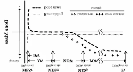
In effect, many study show the post vibration improvement of vertical jumps, dynamic strength in its force-velocity and power velocity relationship as acute effect (7) that after a prolonged treatment (1, 69) (Fig. 3)
In synthesis, the vibratory energy stimulate different sensory structures that trough the spinal loop and a sub-cortical and cortical involvement, produce a reflex muscle activation with the response of
Fig. 3 Average power (AP) and average force (AF) developed during leg press
exercise performed with progressive loads (50, 70, and 100 kg) are shown before (open symbol) andafter (filled symbol) the WBV training.
neuromuscular system more powerful and effective than voluntary activation.
2.1 Vibratory Energy as Neuromuscular System Assessment
In the last years the assessment of neuromuscular behaviour has received a powerful input from the evolution of technological systems with a consequent improvement of new instruments and devices dedicated mainly to the sport science and rehabilitation field. However, the assessment of the neuromuscular function is so far to be enough complete for understanding the complex adaptations phenomena of biological transformations consequently to the injuries or/and after surgery. Besides, there is an high percentage of patients showing a weakness or muscle impairments or dysfunctions of leg extensors muscles after a long follow-up period, probably due to proprioceptors perturbation caused from surgery intervention (23). At moment, the
350 450 550 650 750 850 950 1050 40 70 100 200 220 240 260 280 300 320 340 360 Before AF (N) After AF (N) Before AP (W) After AP (W) A P (W) AF(N ) Load (Kg)
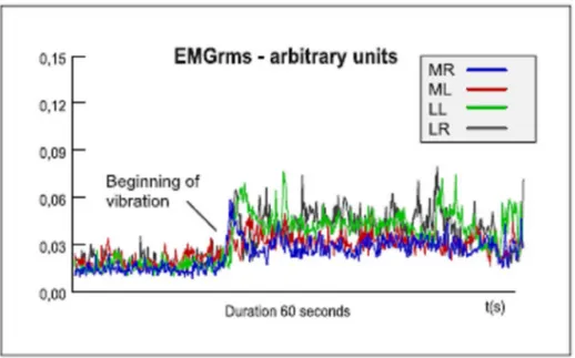
standard protocol used in rehabilitation field for evaluate the legs muscle impairment regards the assessment of muscle strength of both legs in isometric or isokinetic contraction. Both methods present evident limits regarding the unspecific muscle contraction modality, static the first and unnatural contraction type the second and in both cases the sensory structures deputed to be activated by the variation of velocity (like pacinian corpuscle) aren’t stimulated without contribute to muscle force output. These limits are exceeded by the isoinertial dynamometry that measure the biomechanics parameters of muscle contraction (force, velocity, displacement and power) in dynamic and against gravity following the natural property of neuromuscular contraction (11). Unfortunately, the isoinertial method also is inadequate and lack of specific evaluation technique that could allow the quantification and assessment of the impairment due to the proprioceptors inability to function properly consequently to the injuries or surgery intervention. In this contest of paramount importance was the first pilot investigation, conduced by the Prof. Carmelo Bosco and Rehabilitation Medical Equip of “Tor Vergata” University of Rome directed by the Prof. Foti, to analyse the possibility for detecting and quantifying the operated knee joint proprioceptors functional capacity introducing for the first time, the vibratory energy in the neuromuscular assessment field (11). This method consist on monitoring the electromyography activity of muscle during vibration for to identify altered neural strategies of motoneuron pool recruitment. In effect, an increase in EMG activity is usually observed during vibration treatment with values higher that ones observed in voluntary muscle activity (8, 59, 69) (Fig. 4).
Fig. 4 Electromyografic activity (EMGrms) recorded from leg extensor
muscles (mm. vastus lateralis : LR and LL and medialis : MR and ML of both left and right legs ), before and during whole vibration treatment.
It was suggested previously the possibility that vibration may elicit excitatory inflow trough muscle spindle – α motoneuron connections in the overall motoneuron inflow (45). It has been showed that vibration generate an enhancement of the stretch-reflex loop determinate the reflex activation at spinal level of α motoneuron without descending motor drive (59). In addition, it has been demonstrate that vibration energy-induced activation of muscle spindle receptors, not only in the muscle to which vibration was applied, but also to the neighbouring muscles (43). In light of above findings, actually the vibration energy in hits two application ways, locally on the muscle bell or to the whole body, it can retain a new assessment strategy to identify neuromusclular behaviour and possibly disfunction associated to assessment isoinertial method synchronised with EMG.
References
1. Annino G., Padua E., Di Salvo V., Menichella S., Tsarpela O., Castagna C., Di Salvo R., Manzi V., and D’Ottavio S. “Effect of whole body vibration training on lower limb performance in selected high-level ballet students” J. Strength Cond Res, 2007 Nov; 21(4):1072-6.
2. Baron R.. Anatomy and ultrastructure of bone. In Favus MJ. (ed), Primer on the metabolic bone disease and disorders of mineral metabolism. Second Ed. NY: Raven Press. 1993. 3. Baylink DJ., Finkelmann RD., Mohan S.. Growth factor to stimulate bone formation. J.
Bone Miner. Res. 1993, 8, S565-72.
4. Biewener AA., Bertram JEA.. Skeletal strain patterns in relation to exercise training during growth. J. Exp. Biol., 1993, 185, 51-69.
5. Bonjour JP., Theinz G., Law F., Slosman D., Rizzoli R.. Peak bone mass. Osteoporosis Int., 1994, 4, S7-13.
6. Bosco C., Cardinale M., Tsarpela O., Colli R., Tihanyi J., von Duvillard S., Viru A. The influence of whole body vibration on the mechanical behaviour of skeletal muscle. Biol
Sport 1998; 153: 157-164.
7. Bosco C., Colli R., Introini E., Cardinale M., Tsarpela O., Madella A., Tihanyi J., Viru A. Adaptive responses of human skeletal muscle to vibration exposure. Clin Physiol 1999; 19: 183-187.
8. Bosco C., Cardinale M., Tsarpela O. Influence of vibration on mechanical power and electromyogram activity in human arm flexor muscles. Eur J Appl Physiol 1999; 79: 306-311.
9. Bosco C., Iacovelli M., Tsarpela O., Cardinale M., Bonifazi M., Tihanyi J., Viru J., De Lorenzo A., Viru A. Hormonal responses to whole-body vibration in men. Eur J Appl
Physiol 2000; 81: 449-454.
10. Bosco C., Colli R., Bonomi R., SP. von Duvillard and Viru A. Monitoring strength training. Neuromuscular and Hormonal profile. Med. Sci. Sport Exerc. 2000; 32,1 202-08.
11. Bosco C., Tsarpela O., Foti C., Manno R, Annino G., Tarantino U. "Nuovi metodi nella valutazione delle proprietà neuro-muscolari in riabilitazione e medicina sportiva". Med Sport, 2001; 54: 195-210;
12. Bosco C., Dellisanti F., Fucci A., Tsarpela O., Annino G., Foti C., Giombini A., D'Ottavio S. "Effetto della vibrazione su forza esplosiva, resistenza alla forza veloce e flessibilità muscolare". Med Sport, 2001; 54: 287-93;
13. Buckwalter JA., Glimcher MJ., Cooper RR. Bone Biology.2: Formation, form, modeling, remodeling and regulation of cell function. J. Bone Joint Surg. (Am), 1995, 77A, 1276-89. 14. Burke JR., Schiller HH. Discharge patterns of single motor units in the tonic vibration reflex
of human triceps surae. J. Neurosurg. Psychiatry. 1976, 39 (8): 729-741.
15. Burke JR, Schutten MC., Koceja DM., Kamen G. Age-dependent effect of muscle vibration and the jendrassic manoeuvre on the patellar tendon reflex response. Arch. Phys. Med Rehabil. 1996, 77: 600-604.
16. Cardinale M. & Bosco, C. The use of vibration as an exercise intervention. Exerc Sport Sci
Rev 2003; 31: 1, 3-7.
17. Cowin SC., Moss-Saletijn, Moss ML.. Candidates for the mechanosensory system in bone. J. Biomech. Eng. 1991, 113, 191-197.
18. Cummings SR., Black DM., Nevitt MC., Browner WS., Cauley J., Ensrud K. Bone density at various sites for prediction of hip fractures. Lancet. 1993, 341, 72-75.
19. Cummings SR., Nevitt MC., Browner WS., Stones K., Fox K., Ensrud K., Cauley J., Black D., Vogt T. Risk factors for hip fracture in white woman. N. Engl. J. Med. 1995, 322: 767-773.
20. Currey J.. The mechanical adaptations of bone. Princeton NJ: Princeton Univ. Press, 1984, 3-175.
21. Einhorn TA. Bone strength: the bottom line. Calcif. Tissue Int., 1992, 51, 333-39.
22. Eisman JA., Kelly PJ., Morrison NA., Pocok NA., Yeoman R., Birminghamn J., Sambroock PN. Peak bone mass and osteoporosis prevention. Osteoporosis Int. 1983, 3, S56-60.
23. Engel A., Petschnig R., Baron R., et al. The effects of meniscectomy on the strength of the femoral quadriceps muscle after more than 3 years. Kiln. Wochenschr. 1990, 102 (22): 663-666.
24. Flieger J., Karachalios T., Khaldi L., Raptou P., Lyritis G. Mechanical stimulation in the form of vibration prevents postmenopausal bone loss in ovariectomized rats. Calc Tissue Int, 1998, 63: 510-514.
25. Forwood MR., Burr DB. Physical activity and bone mass: exercise in futility? Bone Miner. 1993, 21, 89-112.
26. Frost HM.. Bone mass and mechanostat : a proposal. Anat. Rec., 1987, 219, 1-9.
27. Frost HM. Vital biomechanics: Prposed general concepts for skeletal adaptation to mechanical usage. Calc. Tissue Int., 1988, 42, 145-156.
28. Frost HM. Some effects of basic multicellular unit-based remodelling on photon absorptiometry of trabecular bone. Bone Mineral, 1989,7, 47-65.
29. Frost HM. Skeletal structural adaptation to mechanical usage. (SATMU: 1. Redefining Wolff’s law: the bone modelling problem). Anat Rec., 1990a, 226, 403-13.
30. Frost HM. Skeletal structural adaptation to mechanical usage. (SATMU: 2. Redefining Wolff’s law: the bone modelling problem). Anat Rec., 1990b, 226, 414-22.
31. Frost HM. The role of change in mechanical usage set point in the pathogenesis of osteoporosis. J. Bone Min. Res., 1992, 7, 253-61.
32. Griffin M. Handbook of human vibration. T.J. Press (Padstow) Ltd. Padstow, Cornwall, Great Britain, 1997.
33. Hayes WC., Myers ER., Robinovitch SN., Vanderkroonenberg A., Courtney AC., McMahon TA. Etiology and prevention of age-related hip fractures. Bone, 1996, 18: S77-S66.
34. Heinonen A. Exercise as an osteogenic stimulus. Suominen H. (ed). University of Jyvaskyla, Livenstuore, 1997.
35. Huang R.P., Rubin C.T. & McLeod K.J. Changes in postural muscle dynamics as a function of age. J Gerontol A Biol Sci Med Sci 1999; 54: B352-B357.
36. Issurin VB., Liebermann DG., Tenenbaum G. Effects of vibratory stimulation training on maximal force and flexibility. J. Sport Sci., 1994, 12: 561-566.
37. Issurin VB., Tenenbaum G. Acute and residual effects of vibratory stimulation on explosive strength in elite amateur athletes. J. Sport Sci., 1999, 17: 177-182.
38. Jee WSS. The skeletal tissue. In: A Textbook of Histology. L. Weiss (ed). Urban and Schwartzenberg, Baltimore, 1988, 213-254.
39. Jee WSS.. Principles on bone physiology. J. Musculosk. Neur. Inter., 2000, 1, 11-13.
40. Johnston CC. Jr, Slemenda CW. Determinants of peak bone mass. Osteoporosis Int., 1993, 3, S54-55.
41. Kannus P., Sievanen H., Vuori I. Physical loading, exercise and bone. Bone, 1996,.18, S1-3. 42. Kimmel DB. A paradigm for skeletal strength homeostasis. J. Bone Miner. Res., 1993, .8,
S515-522.
43. Kasai T., Kawanishi I. Yabagi S. The effect of wrist muscle vibration on human voluntary elbow flexion-extension movement. Exp. Brain Res. 1992, 90: 217-220.
44. Lanyon LE. Functional strain in bone tissue as an objective and controlling stimulus for adaptive bone remodelling. J. Biomech., 1987, 20, 1083-1093.
45. Lebedev MA., Peliakov AV. Analysis of the interference electromyogram of human soleus muscle after exposure to vibration. Neirofiziologiia. 1991, 23 (1): 57-65. (article in Russian) 46. Loitz, BJ., Zernicke RF. Strenuous exercise-induce remodeling of mature bone: relationship
between in vivo strains and bone mechanics. J. Exp. Biol., 1992, 170, 1-18.
47. Martin RB., Burr DB. Structure, function and adaptation of compact bone. NY: Raven Press, 1989, 143-85.
48. Moskelide L. Osteoporosis and exercise. Bone, 1995, 17, 193-95.
49. Naito E., Kinomura S., Geyer S., Kawashima R., Roland PE. and Zilles K. Fast reaction to different sensory modalities activates common fields in the motor areas, but the anterior cingulated cortex is involved in speed reaction. J. Neurophysiol. 2000, 83: 1701-1709. 50. Nazarov V., SpivakG. Development of athlete’s strength abilities by means of
biomechanical stimulation method. Theory Pract. Physical Cult. 1987, 12: 37-39. (article in Russian).
51. Oxlund BS., Ørtoft G., Andreassen TT., Oxlund H. Low intensity, high frequency vibration appear to prevent the decrease in strength of the femur and tibia associated with ovariectomy rats. Bone, 2003, 32 (1): 69-77.
52. Oxnad CE. Bone and bones, architecture and stress fossils and osteoporosis. J. Biomech., 1993, 26, 63-79.
53. Parfitt AM. Bone remodeling and bone loss: understanding the pathophysiology of osteoporosis. Clin. Obst. Gynec., 1987, 30, 789-811.
54. Pitukcheewanont P., Safani D., Gilsanz V. & Rubin C.T. Short Term Low Level Mechanical Stimulation Increases Cancellous and Cortical Bone Density and Muscles of Females with
Osteoporosis: A Pilot Study. Endocrine Society Transactions in press. 2002 NIH Consensus Development Conference. Osteoporosis prevention, diagnosis, and therapy. NIH Consens.
Statement 2000; 17: 1-45.
55. Pocock NA., Eisman JA., Hopper JL., Yeates MG., Sambrook PN., Eberl S. Genetic determinant of bone mass in adults : a twin study. J. Clin., Invest., 1989, 80, 706-10.
56. Qin Yi-Xian, Rubin C., McLeod K. Non linear dependence of loading intensity and cycle number in the maintenance of bone mass and morphology. J. Orthop. Res. 1998, 16:482-489.
57. Ribot-Ciscar E., Vedel JP. and Roll JP. Vibration sensitivity of slowly and rapidly adapting cutaneous mechanoreceptors in the human foot and leg. Neurosci. Lett., 1989, 104: 130-135. 58. Rittweger J., Beller G., Felsenberg D. Acute physiological effects of exhaustive whole body
vibration exercise in men. Clin Physiol 2000; 20: 134-142.
59. Rothmuller C., Cafarelli E. Effects of vibration on antagonist muscle coactivation during progressive fatigue in human. J. Physiol. 1995, 485: 857-864.
60. Rubin CT., Lanyon LE. Regulation of bone mass by mechanical strain magnitude. Calc. Tissue Int., 1985, 37, 411-17.
61. Rubin CT., Lanyon LE. Osteoregulatory nature of mechanical stimuli: function as determinant for adaptive remodelling in bone. J. Orthop. Res., 1987, 5, 300-310.
62. Rubin CT., McLeod KJ. Promotion of bony ingrowth by frequency-specific, low amplitude mechanical strain. Clin. Orthop. Rel. Res. 1994, 298: 165-74
63. Rubin C., Li C., Syn Y Fritton C., McLeod K. Non-invasive stimulation of trabecular bone formation via low magnitude, high frequency strain. 41st Orthop Res Soc1995; 20: 548.
64. Rubin CT., Turner S., Bain S., Mallinckrodt C., McLeod K. Anabolism: low mechanical signal strengthen long bones. Nature 2001, 412: 603-604.
65. Rubin C., Recker R., Cullen D., Ryaby J., McCabe J. and MecLeod K. Prevention of postmenopausal bone loss by low magnitude, high-frequency mechanical stimuli: a clinical trial assessing compliance, efficacy and safety. J Bone Miner Res 2004; 19: 342-351.
66. Runge M., Rehfeld G., Resnicek E. Balance training and exercise in geriatric patients. J
Muscoloskel Neuron Interaction 2000; 1: 61-65.
67. Slemenda CW., Johnston CC. High intensity actives in young women: site specific bone mass effects among male figure skaters. Bone Miner., 1993, 20, 125-32.
68. Suominen H. Bone mineral density and long term exercise. An overview of cross-sectional athlete studies. Sport Med 1993; 16: 316-330.
69. Torvinen S., Kannus P., Sievanen H., Jarvinen TAH., Pasanen M., Kontulainen S., Jarvinen TLN., Jarvinen M., Oja P., Vuori I. Effect of four-month vertical whole body vibration on performance and balance. Med Sci Sports Exerc 2002; 34(9): 1523-1528.
70. Turner CH. Homeostatic control of bone structure.: an application of feedback theory. Bone, 1991, 12: 203-217.
71. Turner CH., Burr DB. Basic biomechanical measurements of bone: a tutorial. Bone. 1993, 14: 595-608.
72. Verschueren SM., Roelants M., Delecluse C., Swinnen S., Vanderschueren D., Boonen S. Effect of 6-month whole body vibration training on hip density, muscle strength, and postural control in postmenopausal women: a randomized controlled pilot study. J Bone Miner Res. 2004, 19(3):352-359.
73. Ward K. et al. A randomized, placebo controlled, pilot trial of low magnitude, high frequency loading treatment of children with disabling conditions who also have low bone mineral density. J Bone Min Res 2001; 16S: 1148.
STUDY I
The effects of low-frequency high-magnitude whole body vibration
in physical actively osteoporotic women: a pilot study.
Abstract
Purpose: Osteoporosis is nowadays affecting a large population. Recent studies, performed on animals and human been, have shown that low magnitude, high frequency mechanical stimuli produce anabolic effects on bone tissue, increasing both bone density and strength. Aim of this study is to verify the effects of whole body vibration on bone tissue of trained osteoporotic women underwent to high magnitude and high-frequency vibration exercise on a vibrating platform.
Method: Twenty-six osteoporotic women, trained with low impact exercise regimen, voluntarily participated in the study and were randomly divided in two groups: experimental (E) and control (C). All subjects aren’t submitted to any pharmacological therapy. The T-score, Ultrasound Bone Profile Index (UPBI) was calculated using the Amplitude-Dependent Speed of Sound (AD-SoS) measured with QUS.
Results: Thirteen osteoporotic women following four months of ten-minute treatments, three per week, of high-level of magnitude (5,0 g) and high frequency (30Hz) mechanical vibration improved the Amplitude-Dependent Speed of Sound (AD-SoS) QUS parameter from 1.878 ± 79,45 to 1.971, 17 ± 78,69 (p<0,002). The T-score in the experimental group show an inversion trend passing from -3,50 ±
1,13 to -2, 18 ± 1,12 (p<0.002) and the Ultrasound Bone Profile Index (UPBI) improve from 0,34 ± 0,11 to 0,47 ± 0,21 (p<0,01). In the control group (low impact exercise) any of these parameters considered show significantly changes over the same period of time.
Conclusion: Given that these accelerations were well tolerated by the experimental cohort, that vibrations similar to these stimulated an increase in bone density and strength in animals and humans, and that skeletal loading is an inevitable product of functional load bearing, we believe this strongly anabolic, non-invasive intervention represents, associated to good physical fitness also, early evidence of an unique non-pharmacologic treatment for osteoporosis.
Introduction
Osteoporosis is currently affecting a large population. Over 40% of women in the United States over the age of 65 are currently affected, determining a cost, which exceeds $15B per year to the health care services [1]. According to the E.S.O.P.O. study (Epidemiological Study On the Prevalence of Osteoporosis) in Italy 23% of women older than 40 years and 14% of men older than 60 years are affected by osteoporosis [2].
Many different prevention and treatment regimens have been developed to resolve the increasing problem of the osteoporosis and related fractures. Reversal of bone loss is then a critical goal for science for improving the long-term well being of the aged population. Several investigations have been conducted trying to identify an effective countermeasure to osteoporosis. However, while several pharmacological interventions have been implemented for the management of this disease [3, 4, 5, 6], it seems that sometimes the risks connected to the side-effects exceed the apparent benefits [7]. Several authors showed that the mechanical stimulus, mediated by physical activity or exercises, is the only mean which can positively influence not only the bone mass and strength but increasing muscle strength too [8, 9, 10, 11]. In addition, regular physical activity enhances health and physical fitness improving overall the quality of life in elderly population by reducing the risk of deterioration of functional capacity [12, 13]. Moreover, the osteogenic adaptation of skeleton is site-specific and related to training regimens [14, 15]. Scientific evidence shows that low impact type movement, like endurance training, has not significant results in bone gain [11]. Therefore, the impact type movement, that generates a
versatile stimulus on whole muscle-skeletal system can generate osteogenic adaptation on skeletal sites [14]. In fact, according the Wolff’s law (Wolff, 1892) [16], the bone tissue is constantly adapting to changes in its loading environment accommodating the structures of the skeleton to mechanical demands. The loading-induced deformation in bone tissue (strain) are responsible of the adaptations in bone architecture and mass [17, 18]. The mechanical strain, for determining the effects on bone remodelling, is related to the other specific factors like magnitude, frequency and application time [19, 20]. Changes of gravitational conditions can be produced also by varying magnitude and frequency of mechanical stimulus, like mechanical vibrations applied to the whole body [21]. Recent clinical studies have suggested that the whole body vibration represents a mechanical stimulus enough to improve both the muscular performance [21, 22, 23, 24]. The interaction between muscle and bone responds to patterns of use or disuse with relative alterations in structure and strength [25, 26]. The whole body vibration has been shown to effectively counteract bone loss. The first clinical studies was conducted on animal model. Flieger in 1998 [27] showed that applying the mechanical stimulus at 50 Hz of frequency and 2 g of magnitude for 30 min per day for 12 weeks on ovariectomized rats, the bone loss was less than the other rats not exposed to vibration stimulus. Rubin et al. [28] exposed, first a mature turkey stimulated on vibrating platform, oscillating at 30Hz, 0,3 g of magnitude (g = 9,81) for 5 minutes per day for 30 days of treatment showing an increase of new bone formation in the trabecular bone of the distal tibia. Continuing on their animal studies, Rubin et al. [29] showed that applying stimulated at 30Hz for 20
minutes per day, it is obtained a 34% of increase in the density of trabecular bone in the proximal femur of adult male sheep following one year of treatment. Oxlund [30] found that an oscillating frequency of 45 Hz was enough to increase bone formation and preserving biomechanical bone strength on ovariectomized rats.
The first clinical studies on human been show a positive effects in adolescents with cerebral palsy [31] and in osteoporotic female [32]. Recently other authors show the increase as in muscle strength than in bone mass after exposition to vibration stimulus in post-menopausal women. Rubin [33] showed an improvement bone mass density (BMD) of 1,5% in the spine and 2,17% in the femur, whereas the control group lost 1,6 % in the spine and 2,13% in the femur, in postmenopausal women submitted at Whole Body Vibration (WBV) treatment at 30 Hz of frequency for 20 min (2 bouts of 10 min) per day, every days for 12 months. Improvements on BMD and on muscular strength was found also after six months of WBV treatment at 35-40 Hz of frequency and 5 g of magnitude [34]. These results seems to suggest that this intervention may have an anabolic effect on bone tissue. In contrast, the modest physical activity at low impact doesn’t have any effect on BMD of postmenopausal woman [11]. Aim of this study is to verify the effects of whole body vibration associated to exercise training at low impact, on bone tissue of osteoporotic women underwent to 4 months of low-frequency vibration exercise on a vibrating platform. For ethical reasons connected not only to the experimental nature of this study but also to the short time of treatment, it was used the Quantitative Ultrasound that represent a feasible, sensitive and non-invasive method for
assessing bone tissue, over others methods that use radioactive sources or ionizing radiations [35, 36, 37, 38].
Methods
To evaluate the effects of whole body vibration on bone loss condition, twenty-six osteoporotic women (T-score –3.67 + 1.10, Age 63 + 8.6 years, Weight 66.12 + 10.7 kg, Height, 161.7 + 5.9 cm) voluntarily participated in the study and were randomly divided in two groups: experimental (E) and control (C). Table 1 presents physical characteristics of the subjects of both groups. All subjects participating at this study aren’t submitted to any pharmacological therapy.
The subjects of both groups participated at the same exercise training program (one hour three times per week) consisted in walking (15 min), flexibility and joint mobility exercises (15 min), free body exercises (15 min.), low impact step exercise (10 min.) cool down exercise (5 min.).
In addition, the subjects of experimental group performed the vibration treatment in the same day before the exercise training program.
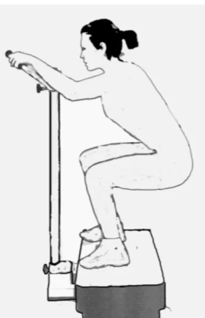
The subjects of experimental group were instructed on the outcomes and the potential benefits associated with their participation in the study. Each subject was familiarised with the experimental protocol and signed an informed participation consent, approved by the Ethical Committee of the Italian Society of Sport Science. Subjects under specific traditional treatment for osteoporosis with previous history of fractures or bone injuries were excluded from the study. They underwent to the experimental treatment consisting of whole body vertical sinusoidal vibration delivered through a specially designed vibrating plate
(Nemes LB, Ergotest Europe, Italy). The magnitude of vibration was 5 g (1g = 9.81 ms-2) and the frequency was 30Hz. The subjects exercised three times per week for a total period of three months. The treatment protocol has been previously described [39]. The total vibration exposure was ten minutes per session. The subjects were standing with both legs in semi-squat position (knees bent at 100) and were allowed to hold a standing stationary metal bar to maintain equilibrium during the exposure to vibration (Figure1). To obtain a complete whole body vibration, the mechanical waves, generated by vibrating plate, were also transmitted to the hands through the metal bar connected to it. During the time-course of the experiment, none of the subjects in the experimental group reported any discomfort from the treatment and only one subject of this group, after the first week of vibration treatment, drop-out from this experiment without appreciable reason. The Compliance of each subject participant to this study, calculated as the number of days attended divided by the 48 days in for months trial (3 days per week for 16 weeks) [40] was about 88%, without statistical difference between both groups.
Quantitative Ultrasonogrammetry (QUS)
Quantitative ultrasound (QUS) measurements were performed before and after the three months treatment period in the proximal phalange of digit II and IV of the dominant arm, using a DBM Sonic 1200 (Igea, Italy) ultrasound device. Two probes are applied to the lateral surface of the fingers, one acting as generator of signal (US frequency = 1.5 MHz) and the other as receiver. The coupling of them with the skin is mediated by a water-based gel. The velocity at which the US traverses the phalanges, in a lateral-medial direction, was
calculated by rate between the distance separating the probes, directly measured by the calliper, and the time elapsing from the emission of the US signal to its reception and expressed in m/s. The device measures the time when the electrical signal, generated by reaches an amplitude of 2 mV at the receiving probe, thus the QUS parameter calculated is the Amplitude-Dependent Speed of Sound (AD-SoS,) for each four fingers and its average value. The AD-SoS has been shown reflecting the mass and the elasticity of bone [36]. The phalanges reflect the largest variations of BMD over lifetime in women [41]. The decreasing of AD-SoS is correlated with decreasing of BMD and loss of trabecular structures, typical conditions of elderly women [42, 43].
Among the other parameters analysed by the device, in the present study, in addition to the average values of AD-SoS and the Ultrasound Bone Profile Index (UBPI) also the T-score will be considered..
The UPBI is an optimum logistic multivariate model, derived from different parameters, for fracture discrimination. It expresses the probability that the subject has a vertebral fracture at the time of QUS evaluation [44].
The T-score was calculated using the AD-SoS measurements. The individual values of QUS were then converted to a T-score according to the following formula:
T-score = (measured values – average values in young adult)/SD in young adult The device has been calibrated by manufacturer using a composite mother phantom and weekly calibrations are performed to control the ultrasound velocity in a Plexiglas phantom. All the QUS measurements were performed by the same operator. The intraoperator reproducibility was already scientifically
documented [45] and the Coefficient of Variation (SD*100/Mean of measurements) of repeated examinations was 0.15% for AD-SoS parameter, calculating on repeated measurements effectuated in the same day on the second finger of a subject 30 times. In vivo short term reproducibility was also assessed by measuring 5 times 7 subjects, randomly selected from both groups, at an interval time not exceeding 7 days; the CV% was 0.75. All the measurements effectuated in this study were performed blind, because the operator didn’t know the belonging of patients at the experimental or control group.
Statistical analysis
The data were analysed using the statistical software for the Social Science (SPSS Inc.). A paired Student’s t-test was used when comparing longitudinal data within the each group of women. The p values resulting from this calculations are two sided and the minimum level of p value to be considered as significant is 0.05. The data referred to the subject’s characteristics are expressed as mean + standard deviation.
Results
As expected, the evaluation of the control group (trained subjects only) showed mainly no changes over the QUS parameters in four months time (table 4). In detail, only five subjects showed slight improvements (table 3). On the other side, the experimental group (vibrated and trained subjects) showed remarkable improvements on the AD-SoS QUS parameter (p = 0,002), on the UPBI (p = 0,01) and on the T-score (p = 0,002) (table 4) except only one subject (table 2). Discussion
The magnitude of musculo-skeletal interactions is of paramount importance for the maintenance of bone integrity. Physical activity performed early in life has been shown to contribute to high peak bone mass [46]. The results of this study confirm the scientific evidence that some forms of exercise, in particular the ones producing high impact forces, seem to be able to reduce or reverse the age-related loss of bone [47], whereas low impact exercise regimen doesn’t have effects on remodelling bone tissue [11]. In effect, a lack of weight bearing activity could favour the likeliness of sarcopenia [48] reducing in this way signals critical to the maintenance of bone mass [26]. Vibration represents a strong stimulus for musculoskeletal structures due to the need to quickly modulate muscle stiffness to accommodate the vibratory waves [39]. Our results suggest that vibrations transmitted to the body by means of vibrating plates may be an effective alternative countermeasure to bone loss. This hypothesis is strongly supported by the effects of such treatment on human skeletal muscles. Vibration has been in fact shown to produce remarkable enhancement in strength and power production following acute [22, 23] and chronic treatments [49]. The extent of the response observed in our experiment (increase in QUS T-score by 57%) is surprising. However, it is our opinion that high magnitude (5 g), frequency (30 Hz) and time of exposure (10 min) of vibration treatment could be it assimilated to an high impact mechanical stimulus like that experienced during contact time ( 200 mms) (references) in ballistic movements (drop jump or high jump, high velocity run), enough to influence the bone tissue remodelling [21, 22, 23]. Moreover, also some influence from hormones could have determined such a remarkable adaptation to vibration
treatment considering that the total exposure time to vibration was relatively short (360 minutes). Vibration has been in fact shown to acutely increase testosterone and growth hormone levels in healthy individuals [23] following the same protocol used in the current experiment. Taking into consideration the results of these preliminary studies it would not seem far-fetched, then, to suggest that the combination of high-frequency mechanical stimuli and hormonal responses provided by vibration could represent an anabolic signal to musculo-skeletal tissues. The higher improvement obtained in these study, respect to the results present in scientific literature, could be due to different factors. One of these, associated to the overestimation of QUS measurement, following our opinion, could regards the effects of incommensurable vibration transmitted by metal bar to the hand directly, determining a local effect that could not completely representative of proper skeletal specific sites of the QUS measure. However, the present findings demonstrate, the effectiveness of high impact stimulus of vibration exercise on bone tissue and provide support for its use as a non-pharmacological intervention to prevent and/or reverse bone loss in humans.
These preliminary studies are promising, longer term, larger population scale studies must be performed in order to verify the effectiveness of vibration treatments and its combination with exercise regimen on the spine and the lower limbs for to prevent bone loss falls and related bone fractures.
FIGURES AND TABLES
Figure 1. Position assumed by subjects of Experimental Group on vibrating plate (Nemes)
Control Group Experimental Group
Variables Mean SD Mean SD
Age (years) 61.2 7.3 64.8 5.6
Height (cm) 161.6 4.4 173.1 10.6
Weight(kg) 68.6 11.2 68.0 11.9
Table 1: Descriptive data (mean ± SD) of the subjects of both groups
Exp Group T-Score AD-SoS UPBI
Subjects pre post pre post pre post
subject 1 -2,46 -2,39 1952 1957 0,39 0,38 subject 2 -2,49 -0,16 1950 2113 0,57 0,84 subject 3 -5,24 -2,04 1757 1981 0,18 0,34 subject 4 -3,04 -2,63 1911 1940 0,49 0,57 subject 5 -2,33 -1,37 1961 2028 0,36 0,45 subject 6 -3,86 -1,24 1854 2037 0,26 0,74 subject 7 -3,41 -3,54 1885 1876 0,29 0,26 subject 8 -4,53 -1,31 1807 2032 0,23 0,7 subject 9 -3,93 -3,31 1849 1892 0,27 0,24 subject 10 -2,77 -2,46 1930 1952 0,32 0,4 subject 11 -5,59 -4,11 1733 1836 0,25 0,22 subject 12 -2,41 -1,63 1955 2010 0,41 0,45
Table 2. QUS parameters for individual subjects, at the beginning and four months after the vibration treatment. The treatment was effective in all except one of the subjects of the Experimental Group.
Control Group T-Score AD-SoS UPBI
Subjects pre post pre post pre post
subject 1 -2,43 -2,74 1954 1932 0,63 0,58 subject 2 -3,63 -4,39 1870 1817 0,28 0,22 subject 3 -3,83 -3,96 1856 1847 0,21 0,22 subject 4 -3,41 -3,80 1885 1858 0,35 0,23 subject 5 -3,61 -4,11 1871 1836 0,35 0,27 subject 6 -5,23 -5,91 1758 1710 0,18 0,11 subject 7 -2,29 -2,06 1964 1980 0,38 0,39 subject 8 -4,20 -4,16 1830 1833 0,21 0,21 subject 9 -3,60 -3,51 1872 1878 0,20 0,20 subject 10 -5,64 -5,77 1729 1720 0,13 0,21 subject 11 -3,51 -3,44 1878 1883 0,26 0,31 subject 12 -3,83 -3,96 1856 1847 0,21 0,22 subject 13 -2,76 -2,20 1931 1970 0,40 0,48
Table 3. QUS parameters for individual subjects, at the beginning and four months of control group. Only five subjects showed slight benefit of exercise treatment.
Control Group Experimental Group
QUS Variables Pre Post T-test (p) = Pre-treatment Post-treatment T-test (p) =
T-Score -3,69 (0,96) -3,85 (1,15) n.s. -3,50 (1,13) -2,18 (1,12) 0,002
AD-SoS (m/s) 1865,69 (67,13) 1854,69 (80,50) n.s. 1878,67(79,45) 1971,17 (78,69) 0,002
UPBI 0,29 (0,13) 0,28 (0,13) n.s. 0,34 (0,11) 0,47 (0,21) 0,01
Table 4: Mean values ± SD of AD-SoS and UPBI before (Pre) and after (Post) three months in Experimental Group treated with Whole Body Vibration and in Control Group. Statistical differences in either groups were analysed using Student's t-test for paired observation.
References
1. NIH Consensus Development Conference. Osteoporosis prevention, diagnosis, and therapy. NIH Consens. Statement 2000; 17: 1-45.
2. Epicentro - Centro Nazionale di Epidemiologia, Sorveglianza, Promozione della Salute -ISS [Internet]: Istituto Superiore di Sanità (Italy). Available from:
http://www.epicentro.iss.it/focus/osteoporosi
3. Bjarnason N.H., Bjarnason K., Haarbo J., Rosenquist C., Christiansen C. Tibolone. Prevention of bone loss in late postmenopausal women. J Clin Endocrinol Metab 1996; 81(7): 2419-2422.
4. Hosking D., Chilvers CE., Christiansen C., Ravn P., Wasnich R., Ross P., McClung M., Balske A., Thompson D., Daley M., Yates AJ. Prevention of bone loss with alendronate in post-menopausal women under 60 years of age. N Engl J Med 1998; 338: 485-492.
5. Neer RM., Arnaud CD., Zanchetta JR., Prince R., Gaich GA., Reginster JY., Hodsman AB., Eriksen EF., Ish-Shalom S., Genant HK., Wang O., Mitlak BH. Effect of parathyroid hormone (1-34) on fractures and bone mineral density in postmenopausal women with osteoporosis. N Engl J Med 2001; 344: 1434-1441. 6. Pors Nielsen R., Barendholdt O., Hermansen F., & Munk-Jensen N. Magnitude and
pattern of skeletal response to long-term continuous and cyclic sequential oestrogen/progestin treatment. Br J Obstet Gynaecol 1994; 101: 349-324.
7. Enserink M. Women's Health: The Vanishing Promises of Hormone Replacement.
Science 2002; 297: 325-326.
8. Campbell A., Robertson M., Gardner M., Norton R., Tilyard M., Buchner D., Randomised controlled trial of general practise programme of home based exercise to prevent falls in elderly women. BMJ 1997; 315: 1965-1969.
9. Carter N., Kannus P., Khan K. Exercise in the prevention of falls in older people.
Sports Med 2001; 31: 427-438.
10. Smith R. Prevention and treatment of osteoporosis: common sense and science coincide. J Bone Joint Surg 1994; 76: 345-347.
11. Suominen H. Bone mineral density and long term exercise. An overview of cross-sectional athlete studies. Sport Med 1993; 16: 316-330.
12. Daley M., Spinks W. Exercise, mobility and aging. Sport Med 2000, 29: 1-12 13. Vuori I. Health benefit of physical activity with special reference to interaction with
diet. Public Health Nutr 2001, 4: 517-528
14. Heinonen A., Oja P., Kannus P., Sievänen H., Haapasalo H., Mänttäri A., Vuori I. Bone mineral density in female athletes representing sport with different loading characteristics of the skeleton. Bone 1995, 17: 197-203
15. Haapasalo H., Kannus P., Sievänen H., Pasanen M., Usi-Rasi K., Heinonen A., Oja P., Vuori I. Effect of starting age of physical activity on bone mineral density of female junior tennis players. J Bone Miner Res 1998, 13: 310-319.
16. Wollf J. The law of bone remodelling. Springer Verlag, Berlin, 1986.
17. Rubin CT., Lanyon LE. Regulation of bone mass by mechanical strain magnitude.
Calc. Tissue Int., 1985, 37: 411-17.
18. Frost HM. Bone mass and mechanostat : a proposal. Anat Rec, 1987, 219: 1-9. 19. Rubin CT., Lanyon LE. Regulation of bone mass by mechanical strain magnitude.
Calc. Tissue Int. 1985, 37: 411-17
20. Rubin CT., McLeod KJ. Promotion of bony ingrowth by frequency-specific, low amplitude mechanical strain. Clin. Orthop. Rel. Res. 1994, 298: 165-74
21. Bosco C., Cardinale M., Tsarpela O., Colli R., Tihanyi J., von Duvillard S., Viru A. The influence of whole body vibration on the mechanical behaviour of skeletal muscle. Biol Sport 1998; 153: 157-164.
22. Bosco C., Colli R., Introini E., Cardinale M., Tsarpela O., Madella A., Tihanyi J., Viru A. Adaptive responses of human skeletal muscle to vibration exposure. Clin
Physiol 1999; 19: 183-187.
23. Bosco C., Iacovelli M., Tsarpela O., Cardinale M., Bonifazi M., Tihanyi J., Viru J., De Lorenzo A., Viru A. Hormonal responses to whole-body vibration in men. Eur J
Appl Physiol 2000; 81: 449-454.
24. Rittweger J., Beller G., Felsenberg D. Acute physiological effects of exhaustive whole body vibration exercise in men. Clin Physiol 2000; 20: 134-142.
25. Runge M., Rehfeld G., Resnicek E. Balance training and exercise in geriatric patients. J Muscoloskel Neuron Interaction 2000; 1: 61-65.
26. Huang R.P., Rubin C.T. & McLeod K.J. Changes in postural muscle dynamics as a function of age. J Gerontol A Biol Sci Med Sci 1999; 54: B352-B357.
27. Flieger J., Karachalios T., Khaldi L., Raptou P., Lyritis G. Mechanical stimulation in the form of vibration prevents postmenopausal bone loss in ovariectomized rats.
Calc Tissue Int, 1998, 63: 510-514.
28. Rubin C., Li C., Syn Y Fritton C., McLeod K. Non-invasive stimulation of trabecular bone formation via low magnitude, high frequency strain. 41st Orthop Res Soc1995; 20: 548.
29. Rubin,C., Turner,A.S., Bain,S., Mallinckrodt,C., McLeod,K. Anabolism: Low mechanical signals strengthen long bones. Nature 2001; 412: 603-604.
30. Oxlund BS., Ørtoft G., Andreassen TT., Oxlund H. Low intensity, high frequency vibration appear to prevent the decrease in strength of the femur and tibia associated with ovariectomy rats. Bone, 2003, 32 (1): 69-77.
31. Ward K. et al. A randomized, placebo controlled, pilot trial of low magnitude, high frequency loading treatment of children with disabling conditions who also have low bone mineral density. J Bone Min Res 2001; 16S: 1148.
32. Pitukcheewanont P., Safani D., Gilsanz V. & Rubin C.T. Short Term Low Level Mechanical Stimulation Increases Cancellous and Cortical Bone Density and Muscles of Females with Osteoporosis: A Pilot Study. Endocrine Society