Alma Mater Studiorum – Università di Bologna
DOTTORATO DI RICERCA IN
SCIENZE BIOCHIMICHE E BIOTECNOLOGICHE
Ciclo XXVI
Settore Concorsuale di afferenza: 05E1 Settore Scientifico disciplinare: B10/10
TITOLO TESI
“MITOCHONDRIAL RESPIRATORY
SUPERCOMPLEX ASSOCIATION LIMITS
PRODUCTION OF REACTIVE OXYGEN
SPECIES FROM COMPLEX I”
Presentata da:
Dott.ssa. Evelina Susana Beatriz Maranzana
Coordinatore Dottorato
Relatore
i
Abstract
Evidence accumulated in the last ten years has demonstrated that a large proportion of the mitochondrial respiratory chain complexes in a variety of organisms is arranged in supramolecular assemblies called supercomplexes or respirasomes. Besides conferring a kinetic advantage (substrate channeling) and being required for the assembly and stability of Complex I, indirect considerations support the view that supercomplexes may also prevent excessive formation of reactive oxygen species (ROS) from the respiratory chain. Following this line of thought we have decided to directly investigate ROS production by Complex I under conditions in which the complex is arranged as a component of the supercomplex I1III2 or it is dissociated as an individual enzyme. The study has been
addressed both in bovine heart mitochondrial membranes and in reconstituted proteoliposomes composed of complexes I and III in which the supramolecular organization of the respiratory assemblies is impaired by: (i) treatment either of bovine heart mitochondria or liposome-reconstituted supercomplex I-III with dodecyl maltoside; (ii) reconstitution of Complexes I and III at high phospholipids to protein ratio.
The results of this investigation provide experimental evidence that the production of ROS is strongly increased in either model; supporting the view that disruption or prevention of the association between Complex I and Complex III by different means enhances the generation of superoxide from Complex I .
This is the first demonstration that dissociation of the supercomplex I1III2 in the
mitochondrial membrane is a cause of oxidative stress from Complex I. Previous work in our laboratory demonstrated that lipid peroxidation can dissociate the supramolecular assemblies; thus, here we confirm that preliminary conclusion that primary causes of oxidative stress may perpetuate reactive oxygen species (ROS) generation by a vicious circle involving supercomplex dissociation as a major determinant.
It is easy to foresee the implications of these findings in human diseases and in aging, where oxidative stress plays a major etiologic and pathogenic role.
Contributors and Funding Sources
This work was supervised by Dr. Maria Luisa Genova and Professor Giorgio Lenaz of
Dipartimento di Scienze Biomediche e Neuromotorie (Alma Mater Studiorum, Università di
Bologna, Italia). The analysis in R4B proteoliposomes were conducted in collaboration with
Giovanna Barbero from Dipartimento di Scienze Biomediche e Neuromotorie (Università di
Bologna). All other work conducted for the dissertation was completed by the student
independently.
This work was supported by MIUR (grant number PRIN2008LSHCFC_005).
Graduate study was supported by an EADIC Erasmus Mundus External Cooperation
Windows Lot 16 (UniBO-UNQ) Fellowship from the European Commission and a
dissertation research Marco Polo fellowship from Alma Mater Studiorum, Università di
iii
Contents
Abstract i
Contributors and Funding Sources ii
Contents iii
List of Figures viii
List of Tables xii
Abbreviations xiii
Chapter 1 Introduction 1
Chapter 2 Materials and Methods 73
Chapter 3 Results 113
Chapter 4 Discussion 135
Chapter 5 Conclusions 141
Contents
INTRODUCTION
1
THE MITOCHONDRIAL RESPIRATORY CHAIN 1
STRUCTURAL ORGANIZATION OF THE RESPIRATORY CHAIN 2
Solid-state organization 5
Liquid-state organization. Random collision model 8
Evidences for supramolecular organization 9
Structural evidences 9
Functional evidences 11
Pool behavior 12
Direct Transfer of Substrates (Channeling) 13
Flux Control Analysis 14
Respiratory supercomplexes in eukaryotes 15
Supercomplex I1III2 17
Supercomplexes III2IV1-2 17
Supercomplexes I1III2IV1-4 19
Respiratory strings 22
Respiratory supercomplexes in prokaryotes 24
Factors that affect supramolecular associations 26
Lipid content 26
v Scaffold
DYNAMIC ORGANIZATION: PLASTICITY MODEL 33
CoQ compartmentalization 34 COMPLEX I 35 Structure 36 Periferal arm 42 Interface domain 45 Membrane arm 46
Evolution and modular organization of Complex I 52
The N – module 53
The Q – module 53
The P – module 53
Mammalian Complex I Assembly 56
Catalytic activity of Complex I 59
NADH oxidation and intramolecular electron transfer 59
Ubiquinone reduction and coupling mechanism 60
Complex I inhibitors 61
ROS PRODUCTION IN COMPLEX I 64
Supercomplexes and ROS production in Complex I 70
HYPOTHESIS 72
MATERIALS AND METHODS
73MATERIALS 73
Reagents and solutions 73
METHODS 74
Preparation of bovine submitochondrial particles (SMP) 76
Purification of bovine Complex I-III fraction (R4B fraction) 77
Preparation of bovine Complex I-III proteoliposomes 80
Preparation of phospholipid:ubiquinone vesicles 80
Proteoliposome reconstitution 82
Determination of protein concentration 83
Ultraviolet absorbance at 280 nm (range: 0.1 – 1 mg·ml-1
) 83
Biuret Method (range: 1 – 10 mg·ml-1
) 84
Lowry Method (range: 0.01 – 0.1 mg·ml-1
) 85
Enzyme activities 87
NADH:ubiquinone oxidoreductase. 87
NADH oxidase. 87
NADH:cytochrome c reductase. 87
Measuring Reactive Oxygen Species 88
Superoxide detection. 88
Hydrogen peroxide detection. 90
Protein electrophoresis analysis 92
First dimension: Blue-native polyacrylamide gel electrophoresis
95
Second dimension: Sodium dodecyl sulfate polyacrylamide gel electrophoresis
102
Protein Immunoblotting 104
vii
Effects of DDM-treatment over respiratory mitochondrial membranes 113
Effects of lipid dilution and DDM-treatment in reconstituted supercomplex I1III2
120
Effects of chaotrope-treatment in SMP and reconstituted supercomplex I1III2
128
DISCUSSION
135Loss of enzymatic channeling between Complex I and Complex III 135
Production of reactive oxygen species from Complex I 136
Stability of Complex I 139
CONCLUSIONS
141List of Figures
Title Page
Figure 1.1 The Mitochondrial Respiratory Chain 1
Figure 1.2 Structural Organization of Mitochondrial Respiratory Complexes
3
Figure 1.3 Dynamic Organization of Mitochondrial Respiratory Complexes
4
Figure 1.4 Solid-model of Chance and Williams (1955) 7
Figure 1.5 Random collision model of Hackenbrock (1986) 8
Figure 1.6 Characterization of bovine respiratory chain supercomplexes and dimeric complex V by BN-PAGE.
10
Figure 1.7 Models of respiratory supercomplexes 18
Figure 1.8 Model for the bovine I1III2IV1 supercomplex (respirasome) 20
Figure 1.9 Cryo-EM 3D map and fitted X-ray structures of bovine I1III2IV1 supercomplex (respirasome) and Electron transfer
pathway
21
Figure 1.10 Hypothetical models for a higher organization of respiratory chain complexes (respiratory strings)
23
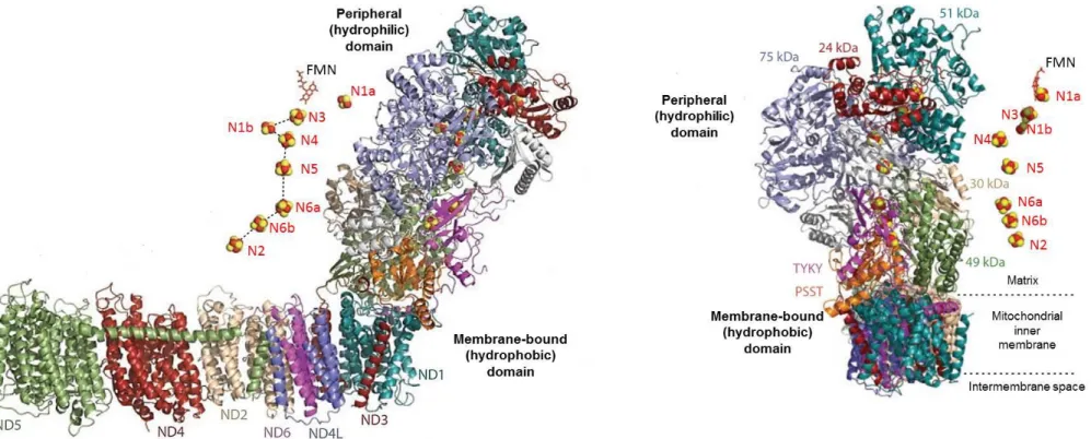
Figure 1.11 Structure of complex I from Thermus thermophilus 41
ix
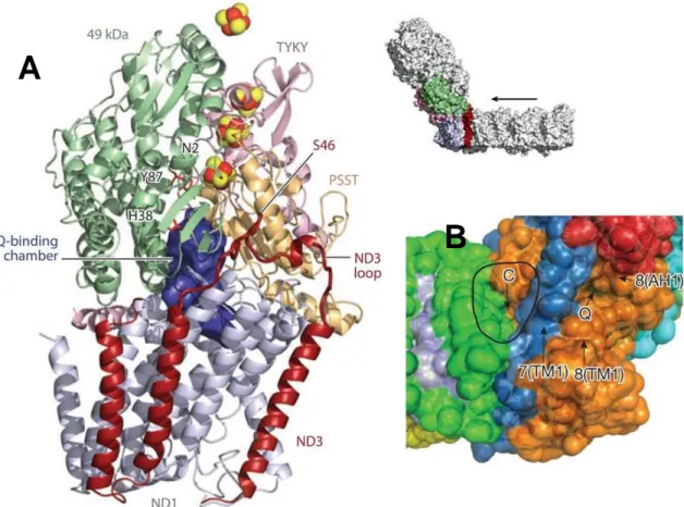
Figure 1.15 E-channel and central hydrophilic axis of Complex I 49
Figure 1.16 Putative proton-translocation channels in the antiporter-like subunits
50
Figure 1.17 Topology model of subunits in mammalian Complex I 51
Figure 1.18 Modular organization of Complex I core subunits 54
Figure 1.19 Evolutionary modules of Complex I 55
Figure 1.20 The assembly model of mammalian Complex I biogenesis 58
Figure 1.21 Proposed coupling mechanism of Complex I 62
Figure 1.22 Overview of mitochondrial ROS production 65
Figure 1.23 Modes of mitochondrial operation that lead to O2•−
production
66
Figure 1.24 Production of O2•− by Complex I 69
Figure 1.25 Production of O2•− by Complex I in I1III2 supercomplex 71
Figure 2.1 Purification of bovine Complex I-III fraction 77
Figure 2.2 Liposomes preparation 81
Figure 2.3 Electrophoresis workflow 93
Figure 2.4 Solubilization of native oxidative phosphorylation complexes 100
Figure 2.5 Separation of supramolecular assemblies of oxidative phosphorylation complexes by 1D-BN PAGE
101
Figure 2.6 Identification of individual constituents in supramolecular assemblies of oxidative phosphorylation complexes by 2D BN/SDS-PAGE
Figure 2.7 Protein blotting workflow 107
Figure 3.1 Supercomplex disassembling in bovine heart mitochondria (BHM)
116
Figure 3.2 Functional analysis of supercomplex I1III2 and complex I in
detergent-solubilized bovine heart mitochondria (BHM): NADH-ubiquinone oxidoreductase activity NADH-cytochrome c oxidoreductase activity
117
Figure 3.3 Functional analysis of supercomplex I1III2 and complex I in
detergent-solubilized bovine heart mitochondria (BHM): NADH-oxidase activity
118
Figure 3.4 Functional analysis of supercomplex I1III2 and complex I in
detergent-solubilized bovine heart mitochondria (BHM): Production of hydrogen peroxide
119
Figure 3.5 Supramolecular organization of respiratory Complex I and Complex III in R4B 1:1 and R4B 1:30 proteoliposomes. (A)
122
Figure 3.6 Functional analysis in R4B 1:1 and R4B 1:30 proteoliposomes.
123
Figure 3.7 ROS production mediated by Complex I in R4B 1:1 and R4B 1:30 proteoliposomes.
124
Figure 3.8 Disassembling of supercomplex I1III2 in R4B 1:1
proteoliposomes after detergent solubilization.
125
Figure 3.9 Functional analysis of supercomplex I1III2 and complex I in
detergent-solubilized R4B 1:1 proteoliposomes: NADH-ubiquinone oxidoreductase activity and (B) NADH-cytochrome c oxidoreductase activity
126
Figure 3.10 Functional analysis of supercomplex I1III2 and complex I in
detergent-solubilized R4B 1:1 proteoliposomes: Production of hydrogen peroxide
127
Figure 3.11 Disassembling of supercomplex I1III2 in bovine heart
submitochondrial particles (SMP) after treatment with 0.2 M
xi
Figure 3.13 Complex I activity and ROS production in bovine heart submitochondrial particles (SMP) after treatment with KSCN.
131
Figure 3.14 Complex I activity and ROS production in R4B 1:1 proteoliposomes after treatment with KSCN.
132
Figure 3.15 Production of ROS by mitochondrial Complex I in different situations where supercomplexes are maintained or dissassembled.
List of Tables
Title Page
Table 1.1 Supramolecular organization of eukaryotic respiratory complexes (mitochondrial respiratory supercomplexes)
16
Table 1.2 Supramolecular organization of prokaryotic respiratory complexes (Aerobic respiratory supercomplexes)
25
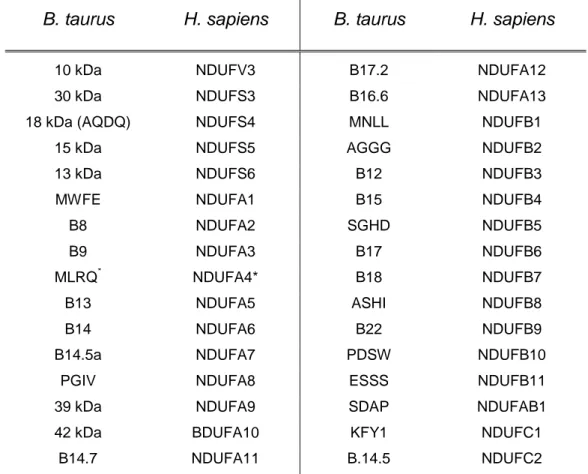
Table 1.3 Nomenclature for the 14 core subunits of Complex I and prosthetic cofactors bound by the hydrophilic subunits
39
Table 1.4 Nomenclature for the supranumerary subunits of mammalian Complex I
40
Table 1.5 Functional classification of Complex I inhibitors 63
Table 2.1 Gel buffer system formulation for BN-PAGE 98
Table 2.2 Quantity of detergent required to solubilize membrane proteins
98
Table 2.3 Gel buffer system formulation for SDS-PAGE 104
Table 3.1 Production of Reactive Oxygen Species by mitochondrial Complex I in different situations where supercomplexes are maintained or disassembled
xiii
Abbreviations
BHM Bovine heart mitochondria
DDM Dodecyl--D-maltoside ROS Reactive oxygen species
SDS Sodium dodecylsulfate
SMP Submitochondrial particles
R4B Mitochondrial fraction enriched in Complex I and Complex III
CoQ Ubiquinone, Coenzyme Q10
Cyt c Cytochrome c
Page 1
INTRODUCTION
THE MITOCHONDRIAL RESPIRATORY CHAIN
Reducing equivalents (hydrogen atoms) released from mitochondrial oxidations of the tricarboxylic acid cycle, from pyruvate oxidation, fatty acid and amino acid catabolism and other oxidative reactions, are collected by a multi enzyme system, the electron transfer chain or respiratory chain that conveys them to molecular oxygen reducing it to water. The free energy decrease of this electron transfer generates an electrochemical proton gradient (H+) by proton translocation from the mitochondrial matrix, to the space existing
between the inner and outer mitochondrial membranes. The proton gradient is then used as a source of energy to synthesize ATP from ADP and Pi by the ATPsynthase complex, or alternatively to drive other energy-linked reactions. The electron transfer chain consists of four major complexes designated as NADH:ubiquinone oxireductase (Complex I), succinate-ubiquinone oxireductase (Complex II), ubiquinol- cytochrome c oxireductase (Complex III) and cytochrome c oxidase (Complex IV) and two connecting redox-active molecules, i.e. a lipophilic quinone, designated ubiquinone (Coenzyme Q or CoQ) embedded in the membrane lipid bilayer ,and a hydrophilic hemeprotein, cytochrome c, localized on the external surface of the inner membrane (Figure 1.1).
Figure 1.1: The mitochondrial respiratory chain.Textbook description of the respiratory chain. The transmembrane protein complexes of the elec- tron transport chain generate an electrochemical gradient over the mitochondrial inner membrane. NADH is oxidized to NAD+. Electrons are transferred from NADH via Complex I and ubiquinone (Q) to Complex III. From there they pass through the peripheral electron carrier cytochrome c and complex IV to the terminal acceptor, molecular oxygen, which is reduced to water. [modified from Lehningher Principles of Biochemistry 5th edition (Nelson, D.L. and Fox, M.M)]
STRUCTURAL ORGANIZATION OF THE RESPIRATORY CHAIN
Since the early 1950s an important part of mitochondrial research has been concerned to elucidate the mechanism and structural organization of electron transport and oxidative phosphorylation. Since then, different models have been suggested for the way in which components of the electron-transfer chain interact to accomplish efficient, energy-conserving electron transfer.
When only the structural aspects of the membrane are considered, two limiting-cases which are loosely termed ‘liquid-state’ and ‘solid-state’ can be proposed [Rich, 1984. Lenaz, 1988] (Figure 1.2)
(i) In a solid-state model, the components of the respiratory chain are present as supramolecular aggregates, with the respiratory complexes I-IV arranged in an orderly sequence (Chance and Williams, 1956. Schägger and Pfeiffer, 2000).
(ii) Alternatively, in a liquid-state configuration, the respiratory multiprotein complexes, ubiquinone, and cytochrome c are randomly distributed in the membrane where they freely move by lateral diffusion. Interactions between respiratory components occur by collisional processes and the electron transport is a diffusion-coupled kinetic process (Hackenbrock et al, 1986).
The main difference between these two models concerns the mechanism of electron transfer: in a liquid random model, the diffusion of ubiquinone and cytochrome c between respiratory complexes ensure electron transfer at any effective collision with them, whereas in the solid supramolecular model, all redox reactions take place by direct electron transfer (substrate channeling) within the aggregate framework .
However, these two cases described above are clearly extreme examples and an intermediate organization is feasible. This view postulates that a dynamic equilibrium exists between complete aggregates, partial aggregates, and freely diffusing components of the respiratory chain, all active in electron transfer. The possibility of transitory functional aggregates (dynamic aggregates) among the electron transfer chain components where the efficiency of electron transfer may be increased by formation of specific associations
Page 3
Figure 1.2: Structural Organization of Mitochondrial Respiratory Complexes. A) Solid-model (Respirasomes) of the mitochondrial electron transport chain in which complexes I–IV are organized into
supramolecular aggregates (respirasomes). The substrate (cyt c or CoQ) is channeled directly from one enzyme to the next, within the respiratory complexes assembled into a huge supramolecular energy-converting machine, except for Complex II, which feeds electrons via ubiquinone to Complex III non-bound to Complex I. B) Liquid-model (Random collision model) all components of the respiratory chain diffuse
individually in the membrane, and electron transfer depends on the random, transient encounter of the four individual protein complexes and the two smaller mobile electron carriers, CoQ and cytochrome c; are assumed to diffuse laterally in the membrane as an individual entity and/or as homo-oligomers.
Black arrows show electron pathways. Complex I (I) in yellow. Complex II (II) in blue, dimeric Complex III (III2) in red, Complex IV (IV) in green. The cytochrome c; (cyt c) in pink, ubiquinol (Q), in brown. The positions of the matrix (M), the intermembrane space (IMS) and cristae or inner membrane (IM) are indicated.
A
Figure 1.3: Dynamic Organization of Mitochondrial Respiratory Complexes. Plasticity model. The supramolecular organization of the mitochondrial respiratory chain has been proposed to confer kinetic advantages on electron transfer through substrate channeling, to prevent ROS production, and to aid the assembly and stabilization of Complex I. Recently it has been reported a new functional role for the dynamic association/dissociation of mitochondrial respiratory complexes and supercomplexes, which defines dedicated CoQ and cyt c pools in order to organize electron flux to optimize the use of available substrates through the respiratory chain. These dynamic rearrangements range from all-bound to all-free respiratory complexes, and they open the possibility that different modes of organization are switched on/switched off to regulate diverse physiological functions through, i.e., ROS signaling or turnover of respiratory enzymes. I1III2IV1–4 ers to the respirasomes or supercomplexes formed by the association of one Complex I (I), a homodimer Complex III (III2), and one to four copies of Complex IV (IV1–4). Intermediate supercomplex species can be found in nature, in combination with free Complex II (II), dimers of Complex III, and Complex IV in different stoichiometries (IV1–2). Complex I requires to be associated in supercomplexes to minimize
Page 5
Solid-state organization
In [Chance and Williams, 1955], a method based on the use of beam, dual-wavelength spectrophotometer in combination with an oxygen electrode for the study of oxidative phosphorylation is published. The redox states of various respiratory-chain components were determined by spectrophotometry, simultaneously with the polarographic measurement of oxygen uptake. This method made possible the first quantitative study of the concentrations and kinetics of electron-transport enzymes not only in intact mitochondria but also in intact cells and tissues allowing depicting the respiratory chain as a solid-state assembly of flavins and cytochromes in a protein matrix. From these studies, Chance and Williams have also determined the sequence of enzymatic steps in the mitochondrial respiratory chain and hypothesized two alternative mechanisms for electron transfer from one protein carrier to another along the solid-state array [Chance and Williams, 1956] (Figure 1.4).
For the first mechanism, there are restricted rotations in the protein carriers to permit collision of the prosthetic groups. In the second, the molecules are completely fixed, and electrons then must pass through the protein moieties to the prosthetic groups. This intra-protein electron transfer mechanism through the insulating intra-protein medium today is well described as electron tunnelling transfer where the maximal distances allowing for physiological electron transfer between the interacting centres should not exceed 13–14 Å [Moser et al, 2005].
Contemporaneously, a comprehensive study of large-scale preparation of beef heart mitochondria was begun in Green’s laboratory [Crane et al, 1956]. Mitochondria from beef heart proved to possess a remarkably high degree of stability; they were capable of withstanding preparation procedure involving disruption of the tissue by relatively harsh mechanical means, and subsequent storage of the mitochondria in the frozen state for long periods of time. These preparations thus became the material of choice for future studies that aimed at a resolution and reconstitution of the respiratory chain and the phosphorylating system.
Shortly after the discovery of ubiquinone (coenzyme Q, CoQ) [Crane et al, 1957] and of its participation in electron transfer [Crane et al, 1959. Hatefi et al, 1959] the resolution of the four respiratory complexes functionally active: NADH:ubiquinone reductase (Complex I) [Hatefi et al, 1961], succinate:ubiquinone reductase (Complex II) [Ziegler and Doeg, 1961], ubiquinol:cytochrome c reductase (Complex III) [Hatefi et al, 1962a], and cytochrome c oxidase (Complex IV) [Fowler et al, 1962] from bovine heart mitochondria was possible. In 1962 Hatefi et al [Hatefi et al, 1962b] succeeded in reconstituting NADH oxidase and succinoxidase by combining complexes I, III, and IV and complexes II, III, and IV, respectively, in the presence of cytochrome c. In both cases, the reconstitution required high concentrations of the complexes and resulted in a particulate preparation which did not dissociate upon subsequent dilution.
These results gave rise to the concept that the components of the respiratory chain exist in mitochondria as a fixed assembly (“elementary particles”) [Ernster and Schatz, 1981]. Indeed, it was found that cytochrome c can form stable complexes with complex III and complex IV [Kuboyama et al, 1962] and that mitochondria contain the cytochromes in near stoichiometric amounts. Thus, it was assumed that the respiratory complexes formed a single functional unit in the mitochondria and were present in an orderly sequence which could be disrupted by appropriate reagents [Blair, 1967].
These early ideas concerning the structure of the respiratory chain where the possibility that electron-transport chain might exist as a structural unit (solid-model) has been considered also in [Lehninger, 1959] and, in the following years, Chance extended this concept to include direct communication with the ATP-synthesizing machinery [Boyer et al, 1977. Chance, 1977].
Page 7
A
B
Figure 1.4: Solid-model of Chance and Williams (1955). A) Sequence of respiratory components in solid-array determined spectrophotometrically [Chance and Williams, 1955] B) Hypotesized models of electron transfer mechanisms along the chain of fixed electron carriers in the respiratory chain. The first sequence depicts restricted rotations of protein carriers to allow electron collisions with prosthetic groups. The second sequence describes electron transport through proteins moieties towards prosthetic groups (electron tunneling). [Chance and Williams, 1956]. a3, a, c, b: cytochromes a3, a, c, and b respectively. fp: flavoprotein. DPN or DPNH: diphosphopyridine nucleotide (NADH)
Liquid-state organization. Random collision model
On the other hand, on the basis of the isolation of the functional individual respiratory complexes Green and Tzagoloff [Green and Tzagoloff, 1966] postulated that the overall respiratory activity is the result of both intracomplex electron transfer in solid-state between redox components having fixed steric relations and, in addition, of intercomplex electron transfer ensured by rapid diffusion of the mobile components acting as co-substrates (i.e., ubiquinone and cytochrome c). This proposal was supported by the kinetic analysis of Kröger and Klingenberg [Kröger and Klingenberg, 1973a. Kröger and Klingenberg, 1973b] showing that the ubiquinone behaves kinetically as a homogeneous pool in submitochondrial particles from beef heart.
Over the following years, this model was substantially confirmed by several lines of evidences leading Hackenbrock et al to the postulation of the Random Diffusion Model of Electron Transfer [Hackenbrock et al, 1986] (reviewed in [Lenaz and Genova, 2007. Lenaz and Genova, 2009a]) (Figure 1.5).
According with this model, the respiratory complexes are randomly distributed in the plane of the membrane, where they freely move by lateral diffusion. Ubiquinone and cytochrome c are also mobile electron carriers, whose diffusion rates are faster than those of the respiratory complexes. The electron-transferring reactions between all redox components and their respective redox partners occur via a long-range diffusional process, where their diffusion-coupled collision frequencies may be either higher or lower than any given reaction step within the complexes. Consequently electron transfer is rate limited by the diffusion of ubiquinone and cytochrome c.
Page 9
Evidences for supramolecular organization
Structural evidences
Despite the wide acceptance of the Random Collision Model during the following twenty years, circumstantial evidences of supramolecular organization of the respiratory complexes come from the pioneering isolation of bovine respiratory complexes where NADH:cytochrome c reductase (complex I+III) [Hatefi and Rieske, 1967] and succinate:cytochrome c reductase (complex II+III) [Tisdale, 1967] were purified, and interaction between complexes II and III in yeast [Bruel et al, 1996] was demonstrated. Stable supercomplexes of complexes III and IV were also isolated from some bacteria [Berry and Trumpower, 1985. Sone et al, 1987. Keefe and Maier, 1993. Iwasaki et al, 1995] indicating that such enzymes may be perentially associated in native membrane. Nevertheless, these reports could not challenge the prevalent view and have been overlooked.
The paradigm of how the respiratory chain is structurally organized drastically changed in 2000 when direct evidences for the existence of higher-order stoichiometric assemblies of respiratory complexes came from the development of the blue-native polyacrylamide gel electrophoresis (BN-PAGE) by Hermann Schägger and colleagues [Schägger and von Jagow, 1991. Schägger et al, 1994. Schägger, 1995].
Mitochondrial membranes solubilized with very mild non-ionic detergents like digitonin which preserve the respiratory complexes activities as well as protein interactions are used in BN-PAGE. This methodology is able to separate the largest stable and functional protein complexes that can withstand solubilization.
The complex stoichiometric composition is then determined by an orthogonal second dimension BN-PAGE (2D BN/BN-PAGE) with a relatively stronger non-ionic detergent as dodecylmaltoside or Triton X-100, to dissociate supercomplexes, or by the subunit composition with a denaturing second dimension (2D BN/SDS-PAGE). This approach allowed the separation and stoichiometric characterization of high molecular weight supramolecular aggregates of respiratory complexes first in mitochondria of bovine heart and of the yeast Saccharomyces cerevisiae [Cruciat et al, 2000. Schägger and Pfeiffer, 2000. Schägger and Pfeiffer, 2001] which remain the best characterized species (Figure 1.6)
The introduction of the BN-PAGE methodology marked the beginning of the study of the higher level of structural organization for the OXPHOS system.
After the first characterizations of supercomplexes by BN-PAGE direct structural insights in the architecture of the supercomplexes were provided more recently by the application of electron microscopy and single particle analysis [Dudkina et al, 2008. Vonck and Schäfer, 2009].
The respiratory supercomplexes are either separated by centrifugation in sucrose density gradients or electroeluted directly from preparative BN-gels, and then imaged by negative stain electron microscopy and subjected to single particle analysis. Several supercomplexes of yeast, plants and mammals have been studied.
Since then, respiratory supercomplexes have been found both in Bacteria, Achaea and in organisms belonging to different kingdoms of eukaryotes. Despite their phylogenetic distances from each other, all of them have in common that their respiratory chain share similar ultrastructure in their respiratory membranes. At this stage, one could consider that a supramolecular organization of the respiratory chain is an evolutionary-conserved trait for which selective advantages remain to be established [Chaban et al, 2013. Magalon et al, 2012].
Figure 1.6: Characterization of bovine respiratory chain supercomplexes and dimeric complex V by BN-PAGE. A) BN-PAGE of bovine heart mitochondria after solubilization by digitonin. Most Complex I and Complex III was found assembled into two major supercomplexes a and b, and two minor supercomplexes c and d. The 200 kDa mass differences indicate the presence of varying copy numbers of monomeric Complex V [Schägger and Pfeiffer; 2000] B) Supercomplexes a-d and dimeric ATP synthase (Vdim) from the BN-PAGE were dissociated by 2D BN-PAGE
A
B
Page 11
Functional evidences
When Chance and Williams have proposed their pioneering solid-state model to describe the respiratory chain organization, they also hypothesized that electron transfer between the protein components in the solid array occurs along predefined pathways [Chance and Williams, 1956].
Thus, the assumption that respirasome has a major function conferring a more efficient transfer of substrates is an inherent consequence if respiratory components are arranged in a sequentially-ordered supramacromolecular assembly.
Considering the two extreme models, the rate of electron transfer between respiratory components depends on their structural arrangement in the membrane:
For the liquid random model view, if two redox enzymes are connected by a mobile redox carrier undergoing long-range diffusion in the membrane, the overall reaction rate would be governed by the frequency of effective collisions between the mobile carrier and its two redox partners [Gupte et al, 1984. Gupte and Hackenbrock, 1988a. Gupte and Hackenbrock, 1988b]. According with this model, the mobile electron carriers components, ubiquinone and cytochrome c, constitute intermediate pools diffusing into the bulk framework of the mitochondrial inner membrane (substrate pools), then if diffusion of the quinone and quinol species is much faster than the chemical reactions of CoQ reduction and oxidation, the quinone behaves kinetically as a homogeneous pool (pool behavior).
On the other hand, if respiratory components are arranged in a solid-array configuration, the frequency of effective collisions will be determined only by the proximity between the redox components. In the case of the respiratory chain, this means direct transfer of electrons between two active sites of two enzymes that are physically adjacent by successive reduction and oxidation of the intermediate with restricted diffusion into the surrounding milieu (substrate channeling) [Ovandi, 1991].
These two models in the case of the organization of the respiratory chain are kinetically distinguishable:
Pool behavior
Kröger and Klingenberg [Kröger and Klingenberg 1973a, 1973b], based on the assumption that quinone behaves as a homogenous pool, postulated that the overall electron flux observed (Vobs) between two enzymes will follow a hyperbolic relation with the rate of
ubiquinol oxidation (vox) and the rate of ubiquinone reduction (vred):
Where CoQ is the mobile electron carrier between a first enzyme reducing ubiquinone and the second oxidizing ubiquinol. They showed this hyperbolic relation in steady-state respiration in bovine submitochondrial particles using either NADH or succinate as electron donors.
Further experimental evidences validated this pool behavior in a variety of mitochondrial systems establishing that CoQ distributes electrons randomly among the dehydrogenases and Complex III, behaving indeed as a freely diffusible intermediate. But most available data concern succinate oxidation (Complex II) in submitochondrial particles, whereas fewer data are available for NADH oxidation (Complex I).
Then, kinetics analysis changing Vred or Vox on inhibitor titration curves (e.g. titration of
Complex III by antimycin) allows to distinguish between CoQ pool behavior (random model) or CoQ channeling (supercomplexes). Pool behavior is kinetically characterized by a convex hyperbolic relationship between the integrated oxidation rate and the inhibitor concentration, whereas a linear relationship is expected by a stoichiometric association (supercomplexes) between the two enzymes.
Page 13
Direct Transfer of Substrates (Channeling)
Early evidences about substrate channeling in respiratory complexes came from [Ragan and Heron, 1978]. They demonstrated that Complex I-Complex III binary complex is formed in a 1:1 molar ratio after reconstitution of in lipid vesicles. They also showed that this binary complex contains CoQ10, and equimolar quantities of FMN and cytochrome c1.
This study also described for the first time the stoichiometric behavior for the activity of NADH:cytochrome c reductase, ascribable to the formation of a Complex I-Complex IIII supercomplex. In addition, they showed that CoQ-pool behavior could be restored if adding additional amounts of phospholipid and ubiquinone in the concentrated mixture. Under these conditions they demonstrated that Complex I and Complex III activities were independent of each other.
Heron and co-workers [Heron et al, 1978] have also reported that endogenous CoQ10
leaks out of the Complex I–III unit when extra phospholipid is present, causing a decrease in activity that could be alleviated by adding more ubiquinone.
A more direct comparison of the effect of channeling with respect to CoQ-pool behavior was performed in a simpler experimental condition in our laboratory.
A system, obtained by reconstitution of a crude mitochondrial fraction (R4B) [Hatefi et al, 1962b] enriched in Complex I and Complex III with different amounts of phospholipids and CoQ10 [Lenaz et al, 1999] was used to discriminate whether the reconstituted protein
fraction behaves as individual enzymes (CoQ-pool behavior) or as assembled supercomplexes depending on the experimental distances between the intramembrane particles.
The comparison of the experimentally determined NADH:cytochrome c reductase activity with the values expected by theoretical calculation applying the pool equation showed overlapping results at phospholipid dilutions (w:w) from 1:10 on, i.e. for theoretical distances > 50 nm. On the contrary, pool behavior was not effective and the observed rates of NADH:cytochrome c reductase activity were higher than the theoretical values [Lenaz et al, 1999. Bianchi et al, 2003. Genova et al, 2008] at a low protein:lipid dilution of 1:1 (w:w), resembling the mean nearest neighbor distance between respiratory complexes in mitochondria [Vanderkooi, 1978].
Flux Control Analysis
The first functional demonstration of the existence of supercomplexes was given by kinetic analysis of the pool function of Coenzyme Q and cytochrome c in mitochondria from Saccharomyces cerevisiae [Boumans et al, 1998]. The finding that these mitochondria did not follow pool behaviour unless treated with chaotropic agents was considered a peculiarity of this organism, because pool behaviour had been widely accepted after the kinetic studies of [Kröger and Klingenberg, 1973b].
Later on, the existence of functional supercomplexes in mammalian and plant mitochondria were confirmed flux control analysis [Bianchi et al, 2003. Bianchi et al, 2004].
In bovine heart mitochondria, Bianchi et al found that both Complex I and Complex III have flux control coefficients approaching 1, suggesting that they behave as a single enzymatic unit, so that electron transfer through Coenzyme Q is accomplished by channelling between the two complexes
In addition, flux control analysis using cyanide inhibition [Bianchi et al, 2004], showed that Complex IV appears to be randomly distributed, or in other words that a large excess of active enzyme exists in free form in the pathway from NADH to oxygen.
Page 15
Respiratory supercomplexes in eukaryotes
In regards to eukaryotes, the first experimental evidences of supramolecular organization came from the pioneering breakthroughs achieved by Hatefi and his collaborators in the 1960s. Starting from beef heart, mitochondria were isolated and purified in large amounts and subjected to systematic solubilization and fractionation. The devised scheme allowed the isolation of all five complexes from the same batch of mitochondria while at the same time was possible to isolate NADH:cytochrome c reductase (complex I+III) and succinate:cytochrome c reductase (complex II+III). However, even this evidence for a supramolecular arrangement for respiratory complexes was unseen by the subsequent studies that supported its random distribution in the inner mitochondrial membrane.
Bovine heart mitochondria have been the model for respiratory chain studies from the earliest studies, and they were also the first mammalian source where supercomplexes were detected and characterized by BN-PAGE [Schägger and Pfeiffer, 2000] and later studied by direct structural methods in the 2000’s [Schäfer et al, 2006. Schäfer et al, 2007. Althoff et al, 2011 . Dudkina et al, 2011]. (Table 1.1)
The three major complexes involved in proton translocation (complexes I, III, and IV) are found mainly assembled in supercomplexes in fungi, plant as well as mammalian mitochondria [Eubel et al, 2003. Muster et al, 2010].
The evidence for higher associations of other respiratory enzymes is negative or ambiguous. Complex II is mostly found in a free, non-associated form although Acín-Pérez et al reported Complex II associated with other respiratory complexes [Acín-Pérez et al, 2008], however this was not supported by any other structural studies [Chaban et al, 2013]. By the other side, complex V (ATP synthase) forms dimers which constitute oligomeric chains in cristae [Arnold et al, 1998. Chaban et al, 2013]. Based on their composition, respiratory supercomplexes can be classified into three main groups which relative abundance varies from organism to organism:
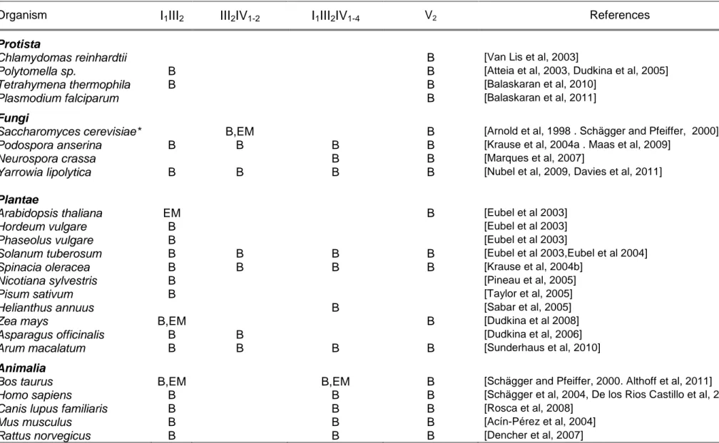
Table 1.1: Supramolecular organization of eukaryotic respiratory complexes (mitochondrial respiratory supercomplexes)
Organism I1III2 III2IV1-2 I1III2IV1-4 V2 References
Protista
Chlamydomas reinhardtii B [Van Lis et al, 2003]
Polytomella sp. B B [Atteia et al, 2003, Dudkina et al, 2005]
Tetrahymena thermophila B B [Balaskaran et al, 2010]
Plasmodium falciparum B [Balaskaran et al, 2011]
Fungi
Saccharomyces cerevisiae* B,EM B [Arnold et al, 1998 . Schägger and Pfeiffer, 2000]
Podospora anserina B B B B [Krause et al, 2004a . Maas et al, 2009]
Neurospora crassa B B [Marques et al, 2007]
Yarrowia lipolytica B B B B [Nubel et al, 2009, Davies et al, 2011]
Plantae
Arabidopsis thaliana EM B [Eubel et al 2003]
Hordeum vulgare B [Eubel et al 2003]
Phaseolus vulgare B [Eubel et al 2003]
Solanum tuberosum B B B B [Eubel et al 2003,Eubel et al 2004]
Spinacia oleracea B B B B [Krause et al, 2004b]
Nicotiana sylvestris B [Pineau et al, 2005]
Pisum sativum B [Taylor et al, 2005]
Helianthus annuus B [Sabar et al, 2005]
Zea mays B,EM B [Dudkina et al 2008]
Asparagus officinalis B B [Dudkina et al, 2006]
Arum macalatum B B B B [Sunderhaus et al, 2010]
Animalia
Page 17
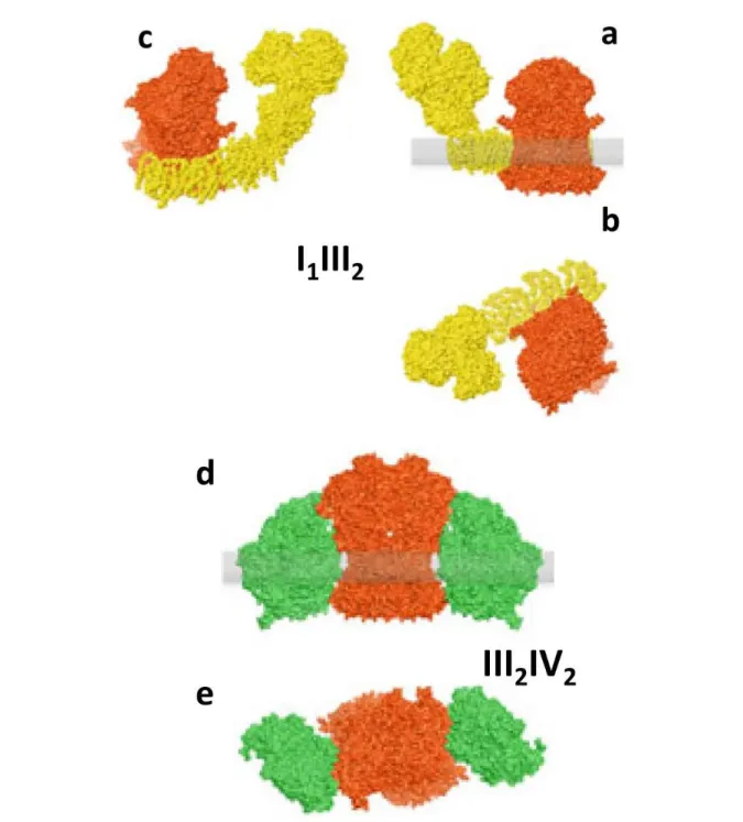
Supercomplex I
1III
2Supercomplexes consisting of one copy of Complex I associated with dimeric Complex III (supercomplex I1III2) have been found in mammals and plants [Schägger and Pfeiffer
2001. Eubel et al 2003 and, 2004. Krause 2004a] (Figure 1.7). Based on BN-PAGE, this supercomplex has an estimated molecular mass 1,500 kDa.
In plants, this is the most abundant supercomplex. Structural characterization by single-particle electron microscopy revealed that III2 is laterally attached to the membrane arm of
Complex I in its concave portion [Dudkina et al, 2005a. Dudkina et al, 2005b. Peters et al, 2008; Bultema et al 2009]. In all species studied, the complex III homodimer is associated laterally with the membrane arm of Complex I, but the interaction between the two complexes differs. In the bovine supercomplex, the interaction surface appears to be more extensive and Complex III is attached to the middle of the Complex I membrane arm, whereas in the plant Complex III attaches to the end of the membrane arm.
Supercomplexes III
2IV
1-2In some organisms, dimeric Complex III was found to associate with one or two copies of Complex IV [Heinemeyer et al, 2007. Bultema et al, 2009. Krause et al, 2004a. Dudkina et al, 2006] (Figure 1.7). The III2+IV1 (750 kDa) and III2+IV2 (1,000 kDa) supercomplexes
are the most stable in Saccharomyces cerevisiae, which in contrast to most eukaryotes, instead of Complex I, in the inner mitochondrial membrane contains three NADH dehydrogenases that are not involved in proton translocation.
Single-particle analysis at 15 Å resolution obtained by electron microscopy with docking of x-ray structures for Complex III and IV allowed to obtain a pseudo atomic model of the 3D structure which revealed that dimeric Complex III is flanked from both sides by monomeric Complexes IV, and that these monomeric complexes IV are attached to dimeric Complex III at two alternate sides with their convex sides facing the Complex III2 [Heinemeyer et al,
2007]. From the recent and more detailed 3D cryo-EM map of the III2+IV2 supercomplex,
authors also concluded that the distance between cytochrome c binding sites in complexes III and IV is about 6 nm, which supports the proposed channeling of cytochrome c between the individual complexes. The purified yeast III2IV2 supercomplex, by the other side, also
contained bound cytochrome c and catalyzed electron transfer from reduced ubiquinone (QH2) to oxygen [Mileykovskaya et al, 2012].
Figure 1.7: Models of respiratory supercomplexes. (a,b) Models for the bovine I1III2 supercomplex based on data in [Schäfer et al 2006, Schäfer et al 2007]. Complex III (red) is attached to the middlde of Complex I (yellow) membrane arm. (c) Bovine I1III2 supercomplex rotated 40° along the Complex I membrane arm to shows dimeric Complex III attached to the end of the membrane arm, as observed in plant I1III2 supercomplexes [Heinemeyer et al, 2007. Bultena et al 2009]. (d,e) Model for the yeast IIIIV supercomplex [Heinemeyer et al 2007]. Two copies of Complex IV (green)
Page 19
Supercomplexes I
1III
2IV
1-4The terms respirasome and respiratory supercomplex are synonyms for the largest stoichiometric associations of respiratory chain complexes which contain monomeric Complex I, dimeric Complex III, and up to four copies of Complex IV (Figure 1.8). The I1III2IV1 supercomplex is denominated respirasome because it is considered the minimal
unit to perform complete respiration from NADH to oxygen. The role of respirasome as functional NADH oxidase was reported by Acín-Pérez et al [Acín-Pérez et al, 2008] for a respirasome isolated from mammalian mitochondria.
They demonstrated that mouse repirasomes isolated by elution of protein bands separated by BN-PAGE contained both ubiquinone and cytochrome c. These supercomplexes showed complete NADH oxidase activity, as oxygen consumption was measured using a Clark electrode after addition of NADH. This study demonstrated that supercomplexes not only are real entities, but are competent in respiration.
Recently, three-dimensional (3D) density maps of the bovine heart I+III2+IV1 respirasome
(1,700 kDa) were determined by two different single-particle electron microscopy methods: cryoelectron tomography of digitonin-solubilized respirasomes [Dudkina et al, 2011] and cryoelectron microscopy of amphipol-solubilized respirasomes [Althoff et al, 2011] (Figure 1.9).
Docking of available crystal structures of the individual complexes [Hunte et al, 2010 . Huang et al, 2005 . Solmaz and Hunte, 2008] both 3D density maps generated very similar pseudo-atomic models.
These 3D-structures demonstrated that the Complex III2 sits in the arc of the membrane
arm of Complex I while Complex IV is present adjacent to dimeric Complex III at the distal tip of the membrane arm of Complex I. Additionally, the 3D structure reported by Althoff et al. shows distances of 13 nm between ubiquinol-binding sites of complexes I and III, and of 10-11 nm between cytochrome c binding sites of complexes III and IV (Figure 1.9). This model indicates the pathways along which ubiquinone and cytochrome c can travel to shuttle electrons between their respective protein partners.
Althoff and coworkers in addition reported the presence of significant amounts of bound phospholipids in the purified respirasome and demonstrated that cardiolipin is enriched in the surpercomplex compared with mitochondria total lipids. Moreover, analysis of lipid extracts by HPLC indicated that each respirasome contains at least one molecule of ubiquinol, and immunodetection analysis confirmed the presence of cytochrome c [Althoff et al, 2011].
Interestingly, mutual orientation of Complex IV and III in the bovine respirasome differs significantly from their orientation in the structure of the yeast supercomplex III2IV2
[Heinemeyer et al, 2007. Mileykovskaya et al, 2012]. These differences between the organization of the bovine and yeast respirasomes might be partly explained by the necessity of Complex III to interact with both Complex I and Complex IV in mammalian mitochondria. Nevertheless, considering the interaction between Complex I and Complex III homodimer there is no spatial restriction in the bovine respirasome for the dimeric Complex III and Complex IV to interact in the same way as in yeast [Chaban et al 2013]. Another possible explanation would be a string-like association between respirasomes via complexes IV [Wittig et al, 2006a].
The abundance of the supercomplexes with different compositions varies from organism to organism. Thus, I+III2 supercomplex is the most abundant in plants [Eubel et al, 2003],
III2+IV2 supercomplex in fungi [Schägger and Pfeiffer, 2000. Heinemeyer et al, 2007], and
I+III2+IV1-4 supercomplex is higher in abundance in mammals [Schägger and Pfeiffer,
2000]
Figure1.8: Model for the bovine I1III2IV1 supercomplex (respirasome) [Schäfer et al 2006,
Page 21
A
B
C
D
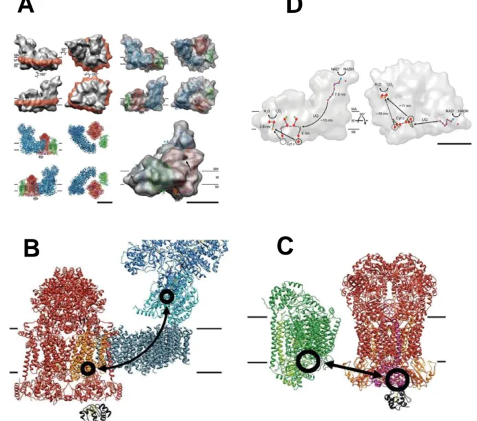
Figure 1.9: Cryo-EM 3D map and fitted X-ray structures of bovine I1III2IV1
supercomplex (respirasome) and Electron transfer pathway A). Cryo-EM 3D at 19 Å
resolution [Althoff et al 2011] with docked X-ray structure of Complex I (blue), Complex III (red), Complex IV (green), and cytochrome c (black). MA: mitochondrial matrix, M: membrane, IM: intermembrane space. Scale bars 10 nm. B) Ubiquinol binding sites are located between the 49 kDa and the PSST subunits near the first Fe-S cluster above the membrane in Complex I and the cytochrome b subunit in Complex III (orange). View from the mitochondrial membrane. C) Cytochrome c binding sites are circled and the shortest cytochrome c trajectories are marked with arrows.Dashed circles mark the unoccupied distal cytochrome c binding site. Side view. D) Electron transfer pathways in the respirasome. Outline of the supercomplex with factors active in electron transport marked in blue (FMN), purple (Fe-S clusters), green (quinol), red (hemes), and orange (copper atoms). On the left, view from the membrane.On the right, electron trajectories marked in black. The dashed circle masks the distal cytochrome c binding site, which is unoccupied in the supercomplex. Staight arrow on the left indicate the shortest distances from the cytochrome c binding side on Complex III to the site of cytochrome c oxidation in Complex IV. The shorter, proximal branch may be perred for electron transport. MA: mitochondrial matrix. M: membrane. IM intermembrane space. UQ, ubiquinol. Cyt, c, cytochrome c. Scale bar 10 nm. ,
Respiratory strings
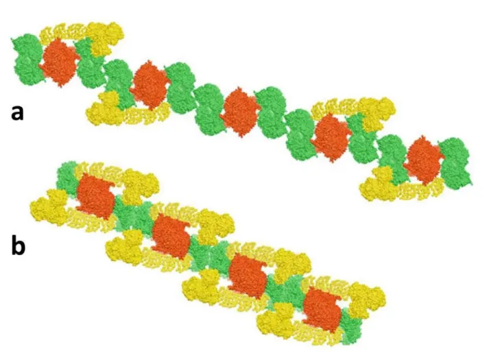
It has been also postulated that the assembly of respiratory complexes into supercomplexes is the first step in the formation of much larger supramolecular structures called respiratory strings.
The model of respiratory strings proposed by Wittig et al [Wittig et al 2006] envisions a linear aggregate of alternating Complex III homodimers and Complex IV tetramers, with Complex I bound to some of these units. This model takes into account also the occurrence of I1III2IV4, III2IV4, and IV4 supercomplexes and the ratio of respiratory
complexes I:III:IV that was determined as 1:3:6 in bovine heart mitochondria [Schägger and Pfeiffer 2001]. The existence of these assemblies was demonstrated as the isolation in large pore BN-PAGE of larger assemblies of Complex I, III, and IV in bovine hearth mitochondria with apparent masses of 35 - 45 MDa [Strecker et al 2010]. These higher assemblies are possible to split into individual Complex I, III, and IV under 2D BN/BN-PAGE with dodecylmaltoside, allowing also the identification of tetrameric Complex IV and showing the lack of oligomeric Complex I and Complex III. According these authors, this evidence suggests a specific and ordered association of these complexes as core pieces of respiratory strings and not random aggregates of respiratory supercomplexes (Figure 1.10).
By the other side, Bultema et al [Bultema et al, 2009] postulated a model based on a single-particle electron microscopy study of potato mitochondria where among others (I1III2, III2IV1, I1III2IV1) supercomplexes I2III2 consisting of two copies of Complex I bound to
both sides of a Complex III homodimer were found. In that model the string would have a basic unit of Complex III dimer with a Complex I and Complex IV bound to either side, and the contacts would be formed by Complex IV dimers (Figure 1.10).
Page 23
Figure 1.10: Hypothetical models for a higher organization of respiratory chain complexes (respiratory strings). (a)Model for bovine heart mitochondria respiratory string according to [Wittig et al 2006] and built from a linear combination of I1III2IV4 and III2IV4 supercomplexes, connected via Complex IV tetramers. (b) Model for potato mitochondrial respiratory string based on Bultema et al 2009 which consists of I2III2IV2 supercomplexes, connected via Complex IV dimers. Complex I (yellow), Complex III (red), Complex IV (green). The models are built from bovine heart Complex III (pdb code 1BGY) and Complex IV (1IOC) and Thermus thermophilus Complex I (3M9S). The figure was made using UCSF Chimera software (University of California, San Francisco)
Respiratory supercomplexes in prokaryotes
The organization of the respiratory chain in many prokaryotes is very complex. Respiratory flexibility in both electron input and output can be found at its most extreme, leading to a branched character of their respiratory chains.
Prokaryotes contain alternative terminal oxidases that, besides oxygen, enable them to use multiple alternative electron acceptors. This metabolic characteristic allow these microorganisms to colonize multiple and very diverse environments adapting their oxidative phosphorylation efficiency upon environmental changing conditions [Richardson 2000].
In regards to the exceptional diversity in respiratory complexes in Archeae or Bacteria, supramolecular organization of respiratory complexes has been reported covering only a part of their respective existing phyla in a yet restricted number of prokaryotes (Table 1.2). Interestingly, most of the reported supercomplexes of the aerobic respiratory chain in prokaryotes include the highly conserved complexes III and IV. A supramolecular organization of complexes III and IV has also been shown in eukaryotic mitochondria. Hence, ubiquinol:oxygen oxidoreductase supercomplexes were detected in all branches of the tree of life supporting the idea that this highly organized state is a general feature of living organisms [Magalon et al, 2012].
Page 25
Table 1.2: Supramolecular organization of prokaryotic respiratory complexes (Aerobic respiratory supercomplexes)
Quinol:O2 OR NADH:O2 OR H2S:O2 OR Fe(II):O2 OR References
Organism bc1-aa3 bcc-aa3 bc1-ba3 bc1-cbb3 ACIII-cbb3 CI-bc1-aa3 SQR-bc1-ba3 cyc2-cyc1-RcY-aa3
Archaea
Sulfolobus sp. X [Iwasaki et al, 1995]
Bacteria (Gram-positive)
Mycobacterium smegmatis X [Megehee et al, 2006]
Corynebacterium glutamicum X [Niebisch and Bott, 2003]
Bacillus sp. PS3 X [Sone et al 1987]
Bacillus subtilis B
Bc-caa3 supercompl exes
[Garcia Montes de Oca et al, 2012. Souza et al, 2013a]
Bacteria (Gram-negative)
Aquifex aeolicus B B B [Guiral et al, 2009. Gao et al,
2012. Prunetti et al, 2010]
Paracoccus denitrificans X B [Berry and Trumpower,
1985. Stroh et al, 2004]
Bradyrhizobium japonicum X [Keefe and Maier, 1993]
Rhodothermus marinus B [Refojo et al, 2010a. Refojo
et al, 2010b]
Acidithiobacillus ferroxidans B [Castelle et al, 2008]
Escherichia coli B
Formate-oxygen oxidoreduct
ase
[Souza et al, 2012. Souza et al, 2013b]
The enzymatic composition and overall activity of each supercomplex are indicated as well as in which microorganism it has been isolated. Small italic letters refer to the cytochromes, CI for the NADH dehydrogenase, ACIII for alternative complex III, SQR for sulfide quinone reductase, HYD for hydrogenase, SR for sulfur reductase. OR refers to oxidoreductase activity of the different supercomplex
Factors that affect supramolecular associations
In the last years, research efforts have been also placed into the search for factors responsible for gluing together the respirasome components. These aspects were extensively analyzed in a previous reviews (Lenaz and Genova, 2007; Chaban 2013) and will be briefly summarized in the following section.
Lipid content
The role of non-bilayer-forming phospholipids, cardiolipin and phosphatidylethanolamine in the formation and stability of supercomplexes is generally accepted.
These phospholipids have a relative small head group and a bulky fatty acid moiety, which results in a conical shape of the phospholipid molecule. This structural characteristic is responsible for their tendency to form hexagonal-phase structures and thus to increase the tension within a bilayer, which is important for the function of membrane proteins [Osman et al 2011].
Cardiolipin (1,3-bis(sn-3’-phosphatidyl)-sn-glycerol), an anionic phospholipid, is uniquely found in energy-transducing membranes, as the mitochondrial inner membrane. A decrease in cardiolipin content Niebisch and Bott 2003s found to be associated with decreased membrane potential, ATP synthesis, and overall mitochondrial dysfunction [Santiago et al 1973].
Phosphatidylethanolamine, by the other side, is the most abundant non-bilayer-forming phospholipid in the mitochondrial inner membrane [Bednarz-Prashad and Mize, 1978. Böttinger et al 2012]; it also binds to respiratory chain complexes [ Shinzawa-Itho et al 2007. Palsdottir and Hunte, 2004], and in vivo data indicate an important role of this phospholipid for mitochondrial functions [Birner et al 2001. Kuroda et al 2011. Joshi et al 2012].
Page 27
Cardiolipin and phosphatidylethanolamine were shown to bind to the cytochrome bc1 complex and cytochrome c oxidase, likely including the interface of both
complexes [Shinzawa-Itho et al 2007. Palsdottir and Hunte 2004 .Wenz et al 2009]. The requirement of cardiolipin for the activity of Complex I, Complex III, and Complex IV as well as for that of several mitochondrial carriers, suggests that this phospholipid plays a crucial role in the coupled electron transfer process of these enzymes [Fry and Green, 1981].
Recently, some structural evidences seem also to indicate that cardiolipin stabilizes respiratory chain supercomplexes as well as the individual complexes.
The cryo-EM maps of the yeast III2IV2 supercomplex and bovine I1III2IV1
respirasome clearly showed that the Complexes I, Complex III homodimer, and Complex IV are at some distance within the supercomplex in the lipid bilayer and these gaps between complexes appear as a lower density material in the electron microscopy maps. [Mileykovskaya et al, 2012. Althoff et al, 2011. Dudkina et al, 2011]. Consistent with these observations, about 50 cardiolipin molecules per yeast supercomplex III2IV2 were determined by mass spectrometry analysis
[Mileykovskaya et al, 2012] and about 200 cardiolipins molecules were estimated to be present in the purified bovine respirasome I1III2IV1 [Althoff et al 2011], which
is much larger due to the presence of Complex I. Due to larger distances between complexes, the bovine respirasome can potentially accommodate several times more cardiolipin molecules.
Mutations in the taz1 gene which encodes and acyltransferase involved in the metabolism of cardiolipin result in Barth syndrome, a X-linked cardiomyopathy with neutropenia and growth retardation in humans, characterized by respiratory chain dysfunction [Barth et al 1983]. Barth syndrome patient mitochondria showed a cardiolipin-dependent respirasome organization with lower cardiolipin content and polydispersity in acyl chain composition of cardiolipin molecules [Schalame and Ren, 2006]. McKenzie et al [McKenzie et al 2006] demonstrated that cardiolipin defect in Barth syndrome results in destabilization of the supercomplexes by
weakening the interactions between respiratory complexes as BN-PAGE revealed a decrease in the I1III2IV1 supercomplexes and increase of free Complex IV.
Tafazzin is found in the inner membrane in a complex including ATP synthase and the adenine nucleotide carrier; the absence of this complex due to taz1 mutations also induces altered cristae morphology [Claypool et al 2008].
Additionally, Gonzalvez and co-workers [Gonzalvez et al 2013] observed an increase in mitochondrial content, which compensates for the decrease in the level of respiratory complexes and supercomplexes. Mutants in the homologous yeast gene [Ma et al 2004] also have reduced supercomplex and complex IV contents [Brandner et al, 2005 . Li et al, 2007a].
Thus, evidences from yeast to humans actually support a role for cardiolipin in the higher organization of respiratory complexes. It has been shown that reduced formation of individual respiratory complexes and supercomplexes is also correlated with lowered cardiolipin levels due to oxidative stress and cardiolipin peroxidation in aging [Gomez and Hagen, 2012], neurodegenerative diseases [Paradies et al, 2011], and cancer [Gasparre et al, 2013].
In regards to phosphatidylethanolamine, Böttinger et al [Böttinger et al 2012], recently have demonstrated that protein transport into and across the mitochondrial inner membrane is impaired in phosphatidylethanolamine-depleted mitochondria. Though both phosphatidylethanolamine and cardiolipin are required for respiratory activity and efficient generation of by mitochondria, they play opposing roles in the stabilization of respiratory complexes. Their opposite effects on protein complex stability may be explained by the fact that cardiolipin has a negatively charged head group, whereas phosphatidylethanolamine is a zwitterionic phospholipid of neutral charge.
Page 29
Functional and Structural Consequences for Supramolecular
Association
Kinetic Advantage: Channeling
The functional consequence of supercomplex assemblies in the respiratory chain is substrate channeling between. Substrate channeling is the direct transfer of an intermediate between the active sites of two enzymes catalyzing consecutive reactions [Ovàdi, 1991]; in the case of electron transfer, this means direct transfer of electrons between two consecutive enzymes by successive reduction and reoxidation of the intermediate without its diffusion in the medium milieu. In such a case, inter-complex electron transfer becomes indistinguishable from intra-complex electron transfer, so that the so-called mobile intermediates, predicted to exhibit substrate-like behavior in the classic view of the random collision model [Hackenbrock et al 1986], would rather be buried in the interface between the two consecutive complexes.
Structural Advantage: Protein stability and Assembly Scaffold
The interdependency of supercomplexes formation and complex stability has been shown in several genetic models, in which low levels or respirasomes are detected in the absence of Complex III [Schägger et al, 2004. Acin-Perez et al, 2004], Complex IV [Diaz et al, 2006], or cytochrome c. These experimental observations lead to the proposal that the formation of respirasomes may be essential for the assembly / stability of Complex I.
Schägger and collaborators [Schägger et al, 2004], demonstrated that respirasomes is required to stabilize bacterial Complex I since mutant strains of Paracoccus denitrificans lacking Complex III or Complex IV showed complete disassembling of Complex I from supercomplexes. Reduced stability of Complex I in those mutant strains was also corroborated from an almost complete loss of NADH:ubiquinone reductase activity.
The necessity of stably assembled human Complex III for the stability of Complex I was later demonstrated using muscle biopsies and cultured patient cells with isolated deficiencies of single complexes. Human Complex I was almost completely lacking in the absence of assembled Complex III. Genetic alternations leading to a loss of Complex III prevented respirasome formation and led to secondary loss of Complex I; seen as Complex III/Complex I defects. Conversely, Complex III stability was not influenced by the absence of Complex I
[Schägger et al, 2004. Acin-Perez et al, 2004].
Animal models with mutations on Complex I proved to be useful to evaluate its effects on Complex III and Complex IV and understand their role in supercomplex assembly. Several works have demonstrated that Complex I assembly also depends on Complex IV
[Diaz et al, 2006. Li et al, 2007b. D’Aurelio et al, 2006]. While Complex I was found to be unstable in the absence of Complex III, lack of Complex IV totally abrogated assembly of Complex I [Li et al, 2007b].
Suthammark et al [Suthammarak et al, 2009] have shown that Complex I activity is dependent on the presence of Complex IV, despite no overall decrease in the intrinsic amount of Complex I. Then, Complex III defects inhibit Complex IV activity by several different mechanisms involving supercomplex destabilization. These mechanisms included also inhibition of Complex I function by weakening its interaction into the respirasome or by decreasing the amount of Complex I, or its assembly within the respirasome [Suthammarak et al, 2010].
By the other side, mutations of Complex I showed controversial results, since in some studies they did not affect the amount of other respiratory complexes [Schägger et al, 2004. Pineau et al, 2005], while in others they significantly reduced the amounts of respiratory complexes III and IV
Page 31
mutations that would be affecting (or not) subunits of Complex I involved in direct interactions with other complexes within the respirasome [Genova and Lenaz, 2013].
Because Complex I stability was found to be dependent on the assembly of supercomplexes, it was hypothesized that respirasome assembly follows the assembly of individual respiratory complexes. Several works addressing the pathway for respirasome formation suggest that these supercomplexes are assembled in stages.
Acín-Pérez et al [Acin-Perez et al, 2008] studies of pulse-chase labeling of mitochondrial translational products indicated that there is sequential incorporation of mtDNA encoded subunits into respective complexes followed by supercomplex assembly. Thus there seemed to be a temporal delay in the complete assembly of individual complexes and the following assembling into supercomplex.
However, a more extensive study showed that Complex I assembly and synthesis was very closely linked with supercomplex formation as the formation of Complex I/III supercomplex was observed to occur before Complex I was entirely formed [Marques et al, 2007].
Recently, it has been shown that Complex III and IV appear to be assembled independent of each other. In contrast, complex I is assembled in stages [Mimaki et al, 2012].

![Figure 1.4: Solid-model of Chance and Williams (1955). A) Sequence of respiratory components in solid-array determined spectrophotometrically [Chance and Williams, 1955] B) Hypotesized models of electron transfer mechanisms along the cha](https://thumb-eu.123doks.com/thumbv2/123dokorg/8165730.126795/23.892.110.693.162.351/williams-sequence-respiratory-components-determined-spectrophotometrically-hypotesized-mechanisms.webp)