Cholesterol-Lowering Drugs Inhibit Lectin-Like Oxidized
Low-Density Lipoprotein-1 Receptor Function by Membrane Raft
Disruption
□
SSara Matarazzo, Maria Chiara Quitadamo, Ruggiero Mango, Sarah Ciccone,
Giuseppe Novelli, and Silvia Biocca
Department of Systems Medicine, Laboratory of Clinical Biochemistry (S.M., S.C., S.B.), Section of Cardiology (R.M.), Department of Biomedicine and Prevention, Section of Medical Genetics (M.C.Q., G.N.), and Center of Biostatistics and Bioinformatics (S.B., G.N.), School of Medicine, University of Tor Vergata, Rome, Italy; and National Agency for the Evaluation of Universities and Research, ANVUR, St. Peter Fatebenefratelli Hospital, Rome, Italy (G.N.)
Received March 23, 2012; accepted May 8, 2012
ABSTRACT
Lectin-like oxidized low-density lipoprotein (LOX-1), the pri-mary receptor for oxidized low-density lipoprotein (ox-LDL) in endothelial cells, is up-regulated in atherosclerotic lesions. St-atins are the principal therapeutic agents for cardiovascular diseases and are known to down-regulate LOX-1 expression. Whether the effect on the LOX-1 receptor is related to statin-mediated cholesterol-lowering activity is unknown. We investi-gate the requirement of cholesterol for LOX-1-mediated lipid particle internalization, trafficking, and processing and the role of statins as inhibitors of LOX-1 function. Disruption of choles-terol-rich membrane microdomains by acute exposure of cells to methyl--cyclodextrin or chronic exposure to different st-atins (lovastatin and atorvastatin) led to a spatial disorganiza-tion of LOX-1 in plasma membranes and a marked loss of specific LOX-1 function in terms of ox-LDL binding and
inter-nalization. Subcellular fractionation and immunochemical stud-ies indicate that LOX-1 is naturally present in caveolae-en-riched lipid rafts and, by cholesterol reduction, the amount of LOX-1 in this fraction is highly decreased (ⱖ60%). In contrast, isoprenylation inhibition had no effect on the distribution and function of LOX-1 receptors. Furthermore, in primary cultures from atherosclerotic human aorta lesions, we confirm the pres-ence of LOX-1 in caveolae-enriched lipid rafts and demonstrate that lovastatin treatment led to down-regulation of LOX-1 in lipid rafts and rescue of the ox-LDL-induced apoptotic pheno-type. Taken together, our data reveal a previously unrecognized essential role of membrane cholesterol for LOX-1 receptor ac-tivity and suggest that statins protect vascular endothelium against the adverse effect of ox-LDL by disruption of mem-brane rafts and impairment of LOX-1 receptor function.
Introduction
Receptor-mediated endocytosis of oxidized low-density li-poprotein (ox-LDL) is the hallmark event in the pathogenesis of atherosclerosis. Elevated levels of ox-LDL are associated with macrophage differentiation in foam cells, apoptosis and necrosis of vascular endothelium, smooth muscle cell
migra-tion, and proliferation (Mitra et al., 2011) and are correlated to plaque instability in human coronary atherosclerotic le-sions. Most of these effects are elicited by the lectin-like oxidized low-density lipoprotein receptor-1 (LOX-1), charac-terized as the primary receptor of ox-LDL in endothelial cells (Sawamura et al., 1997; Mehta et al., 2006). LOX-1 is a scavenger receptor overexpressed in atherosclerotic lesions and up-regulated during atherogenesis in atheroma-derived cells (Mehta et al., 2006; Vohra et al., 2006). LOX-1 activa-tion triggers the oxidative stress response and has been shown to lead to plaque vulnerability and potential rupture, which is ultimately responsible for acute atherothrombotic vascular occlusion and tissue infarction. Moreover, LOX-1 is
This work was supported by FineSTRE, Finanziaria Laziale di Sviluppo, Regione Lazio (to S.B. and G.N.) and Fondazione Umberto Veronesi (to G.N.). Article, publication date, and citation information can be found at http://molpharm.aspetjournals.org.
http://dx.doi.org/10.1124/mol.112.078915.
□S The online version of this article (available at http://molpharm. aspetjournals.org) contains supplemental material.
ABBREVIATIONS: LDL, low-density lipoprotein; ox-LDL, oxidized low-density lipoprotein; LOX-1, lectin-like oxidized low-density lipoprotein receptor-1; MCD, methyl--cyclodextrin; LDL-C, low-density lipoprotein-cholesterol; EC, endothelial cell; MAb, monoclonal antibody; IR, insulin receptor subunit; HRP, horseradish peroxidase; FTI, farnesyl transferase inhibitor; ABT-100, (S)-6-[2-(4-cyanophenyl)-2-hydroxy-2-(1-methyl-1H-imidazol-5-yl)ethoxy]-4⬘-(trifluoromethoxy)-1,1⬘-biphenyl-3-carbonitrile; DMEM, Dulbecco’s modified Eagle’s medium; HEK, human embryonic kidney; E-64, trans-epoxysuccinic acid; Mes, 4-morpholineethanesulfonic acid; DiI, 1,1⬘-dioctadecyl-3,3,3⬘,3⬘-tetramethyllindocarbocyanine perchlorate; Cav-1, caveolin-1.
1521-0111/12/8202-246–254$25.00
MOLECULARPHARMACOLOGY Vol. 82, No. 2
Copyright © 2012 The American Society for Pharmacology and Experimental Therapeutics 78915/3782478
Mol Pharmacol 82:246–254, 2012
246
at Fac di FarmaciaUniv Studi di Napoli on November 13, 2012
molpharm.aspetjournals.org
Downloaded from
8915.DC1.html
up-regulated during myocardial ischemia reperfusion and appears to be associated with apoptosis, necrosis, and left ventricular functional deterioration (Li et al., 2003).
LOX-1 is encoded by a single gene, ORL1, located on hu-man chromosome 12p12.3–p13.2 (Aoyama et al., 1999). Ge-netic association studies have identified different single nu-cleotide polymorphisms within the ORL1 gene, which play a role in cardiovascular disease susceptibility. A newly identi-fied LOX-1 spliced isoform, named LOXIN, which lacks exon 5, is deficient in ox-LDL binding activity and is protective against LOX-1 induced apoptosis (Mango et al., 2005). LOXIN isoform interacts with LOX-1 receptors, inhibiting its function through the formation of nonfunctional hetero-oli-gomers (Biocca et al., 2008). LOX-1 is a type II membrane glycoprotein with an extracellular C-type lectin-like ligand-binding domain (Sawamura et al., 1997), which forms a dis-ulfide-linked heart-shaped homodimer. The C-type lectin-like ligand-binding domain possesses a basic spine structure across its ligand recognition surface known to play a role in the recognition of ox-LDL. LOX-1 dimers assemble in larger functional oligomers through noncovalent interactions (Ohki et al., 2005; Park et al., 2005; Biocca et al., 2008). More recently, other groups have reported that multimerization and cluster organization in plasma membrane are important requisites for LOX-1 activity (Cao et al., 2009; Ohki et al., 2011).
Several lines of evidence implicate cholesterol-enriched lipid microdomains, known as caveolae and lipid rafts, as essential docking sites for endocytosis of ligands, including ox-LDL, fatty acids, apoptotic cells, and also viruses. Despite the pathophysiological role of LOX-1-mediated ox-LDL endo-cytosis, details of the receptor-mediated lipid particle inter-nalization, trafficking, and processing during atherosclerotic plaque formation are not yet understood. Scavenger recep-tors, such as CD36, CD209, and CD204 are localized in lipid rafts and use lipid rafts pathways for endocytosis (Lisanti et al., 1994; Zeng et al., 2003; Cambi et al., 2004; Kiyanagi et al., 2011). Although a clathrin-independent and dynamin-2-dependent pathway has been described and thought to be involved in LOX-1 endocytosis (Kashiwakura et al., 2004; Murphy et al., 2008), these studies have not investigated the lipid raft involvement. Caveolae and lipid rafts are special-ized membrane domains, rich in cholesterol, sphingolipids, and glycerophospholipids and contain specific membrane proteins including glycosylphosphatidylinositol-anchored proteins, GTPases, and receptor-associated kinases (Parton and Simons, 2007). Cholesterol is a key component of cave-olae and raft structure and is important in modulating the fluidity of plasma membranes and regulating their function. Given the critical role of LOX-1 in atherogenesis, we wanted to investigate the requirement of cholesterol and the role of cholesterol-lowering drugs in LOX-1-mediated ox-LDL entry in human endothelial cells. To deplete cholesterol in plasma membranes, we used methyl--cyclodextrin (MCD), which specifically extracts cholesterol from the plasma mem-branes (Ilangumaran and Hoessli, 1998) and two statins, lovastatin and atorvastatin, that inhibit HMG-CoA reductase, a rate-limiting enzyme in cholesterol biosynthesis (Wang et al., 2008). For their effect on lowering circulating total and low-density lipoprotein-cholesterol (LDL-C), statins are largely used in the clinic in the treatment of patients with cardiovascular diseases. We have focused our interest on the role of statins in
reduction of the membrane cholesterol level and disruption of lipid rafts in endothelial cells and the consequences on LOX-1 expression, membrane distribution, and function.
Here we report that LOX-1 is predominantly localized in caveolae/lipid rafts in the cell plasma membranes and its function is regulated by membrane cholesterol. A decrease in plasma membrane cholesterol by statin treatment leads to down-regulation of 1 in lipid rafts, impairment of LOX-1-mediated ox-LDL internalization, and rescue of an ox-LDL-induced apoptotic phenotype in primary endothelial cells (ECs), suggesting that statins may protect vascular endothe-lium against the adverse effects of ox-LDL by disruption of LOX-1 receptor function.
Materials and Methods
DNA Constructs. For the expression in mammalian cells, human LOX-1 was subcloned into pEF/V5-His vectors (Invitrogen, Inchin-nan, Paisley, UK), as described previously (Biocca et al., 2008).
Antibodies and Reagents. Rat anti-LOX-1 (Biocca et al., 2008), MAb anti-V5 IgG (Invitrogen), mouse anti-caveolin-1 (Santa Cruz Biotechnology, Inc., Santa Cruz, CA), rabbit anti-insulin receptor subunit (IR), (BD, Franklin Lakes, NJ), and mouse anti--actin IgG (Affinity Bio Reagents, Golden, CO) were used as the primary anti-bodies. Secondary antibodies goat anti-rat IgG horseradish peroxi-dase (HRP), goat anti-mouse IgG HRP, donkey anti-rabbit IgG HRP, and Rhodamine Red X-conjugated AffiniPure donkey anti-mouse IgG were purchased from Jackson ImmunoResearch Laboratories Inc. (West Grove, PA). Filipin, MCD, and atorvastatin were purchased from Sigma-Aldrich (St. Louis, MO). Lovastatin (Enzo Life Sciences, Inc., Farmingdale, NY) was activated by NaOH addition with a lovastatin/NaOH ratio (v/v) of 2:3, at 50°C and neutralized with HCl to pH 7. The farnesyl transferase inhibitor (FTI) (S)-6-[2-(4-cyanophenyl)-2-hydroxy-2-(1-methyl-1H-imidazol-5-yl)ethoxy]-4 ⬘-(trifluoromethoxy)-1,1-biphenyl-3-carbonitrile (ABT-100) was pro-vided by Abbott Laboratories (Abbott Park, IL). For in vitro studies, ABT-100 was dissolved in dimethyl sulfoxide with dilutions made using DMEM plus 10% fetal bovine serum.
Cell Cultures and Transfection. COS and HEK-293 cells were grown in DMEM (Biowest, Miami, FL) supplemented with 10% fetal bovine serum (Gibco; Invitrogen) and 100 U/ml penicillin-streptomy-cin (Euroclone, Devon, UK). COS cells were transiently transfected with JetPEI (Polyplus Transfection, Illkirch, France), following the manufacturer’s instructions, with a DNA/transfectant reagent ratio (w/v) of 1:2. For generation of stable clones, HEK-293 cells were transfected using SuperFect (QIAGEN, Hilden, Germany) with a DNA/lipid ratio (w/v) of 1:5. At least 30 phleomycin (Zeocin)-resistant clones were isolated after 3 to 4 weeks. Of resistant clones, 20% were positive for LOX-1 expression. Details on the preparation of primary cultures of endothelial cells derived from aorta and infrarenal ab-dominal aortic aneurysm are in Supplemental Data.
Purification of Caveolae-Enriched Membrane Fractions. Caveolae-enriched membrane fractions were prepared by a deter-gent-free purification, as described previously (Song et al., 1996). Confluent 90-mm dishes of HEK-293 (clones 13 or 19) or transfected COS or primary endothelial cells were lysed in 500 mM sodium carbonate, pH 11, containing protease inhibitor cocktail set III [0.1 mM 4-(2-aminoethyl) benzenesulfonyl fluoride hydrochloride, 0.5M aprotinin, 5 mM Bestatin, 1.5M trans-epoxysuccinic acid (E-64), 10 M leupeptin, and 1 mM pepstatin A; (Calbiochem, La Jolla, CA)] and 1 M phenylmethylsulfonyl fluoride (Euroclone, Devon, UK), homogenized, and sonicated. A 5 to 45% discontinuous sucrose den-sity gradient was formed in MBS (25 mM Mes, pH 6.5, and 0.15 M NaCl) and centrifuged at 39,000 rpm for 16 to 20 h in an SW41 rotor (Beckman-Coulter, Fullerton, CA). Samples were fractionated in 1-ml aliquots from the top to the bottom. The protein concentration
at Fac di FarmaciaUniv Studi di Napoli on November 13, 2012
molpharm.aspetjournals.org
was measured in each fraction by the Bradford assay (Sigma-Al-drich). Proteins from each fraction were precipitated with 10% tri-chloroacetic acid and solubilized in SDS-polyacrylamide gel electro-phoresis sample buffer.
Western Blot Analysis. Transfected cells and human primary endothelial cells were lysed in ice-cold extraction buffer containing 10 mM Tris/HCl, pH 7.6, 100 mM NaCl, 10 mM EDTA, 0.5% Nonidet P-40, 0.5% sodium deoxycholate, protease inhibitor cocktail set III, and phenylmethylsulfonyl fluoride and centrifuged for 20 min at 4°C at 15,000g. The supernatant fraction was analyzed by SDS-poly-acrylamide gel electrophoresis in 10% SDS-poly-acrylamide gels and trans-ferred to polyvinylidene difluoride membranes (GE Healthcare, Chalfont St. Giles, Buckinghamshire, UK) for 30 min at 15 V (Semi-Dry Transfer Cell; Bio-Rad Laboratories, Hercules, CA). Immunoreactive bands were visualized by enhanced chemilumi-nescence (Sigma-Aldrich).
Immunofluorescence Analysis and Surface Labeling Quan-tification. Cell membrane immunofluorescence was performed as described previously (Cardinale et al., 2005) using MAb anti-V5 as the primary antibody and Rhodamine Red-X-conjugated AffiniPure donkey anti-mouse IgG as the secondary antibody. Samples were examined with a DMRA Leica fluorescence microscope, equipped with a charge-coupled device camera and with a confocal microscope (Nikon Instruments Spa, C1 on Eclipse TE200; EZC1 software). A cyto-enzyme-linked immunosorbent assay for quantification of mem-brane expressed proteins was performed as described previously (Biocca et al., 2008).
Ox-LDL Preparation, Labeling, and Fluorometric Assay. Human LDL was prepared from fresh healthy normolipidemic plasma of volunteers by ultracentrifugation (Sattler et al., 1992), dialyzed in phosphate-buffered saline, and filtered (0.22-m pore size). Oxidation was performed by incubating LDL (0.4 mg/ml) with 7.5M CuSO4in a CO2incubator at 37°C for 6 to 8 h, acquiring, at
the end of the incubation period, the wavelength difference spectrum using LDL (without copper) as control. Oxidation was stopped by addition of 0.3 mM EDTA, and ox-LDL was dialyzed overnight in PBS containing 0.1 mM EDTA. Agarose gel electrophoresis of native and oxidized LDL was routinely performed in 0.8% (w/v) gel pre-pared in Tris-glycine (29 mM Tris base and 192 mM glycine), pH 8.3, and subjected to 100 V constant voltage for 60 min. Gels were stained with Oil Red O or Coomassie Blue R250. Relative electrophoretic mobility was calculated as the ratio between the migration distance of ox-LDL and that of native LDL and used as measure of oxidation. Ox-LDL was labeled with 1,1 ⬘-dioctadecyl-3,3,3⬘,3⬘-tetramethyl-lindocarbocyanine perchlorate (Dil-) (Invitrogen) as described previ-ously (Stephan and Yurachek, 1993; Biocca et al., 2008). Dil-labeled ox-LDL was incubated in complete medium on ice for 1 h in the binding assay and at 37°C for different times in the uptake assay. Endothelial cells, treated or not with ox-LDL, were washed once
with DMEM without serum and incubated with DMEM supple-mented with lipid-depleted fetal calf serum (Biowest) for 1 h before the binding assay was performed. Quantitation of Dil-ox-LDL bound was assayed by Dil-extraction in isopropanol (Stephan and Yurachek, 1993), and fluorescence was determined in a spec-trofluorometer (PerkinElmer Life and Analytical Sciences, Waltham, MA) with excitation and emission wavelengths set at 520 and 578 nm, respectively.
Evaluation of Apoptosis. Apoptotic cells were visualized by staining with an annexin V assay (Bossy-Weltzel and Green, 2000) (kit from BD) and with the blue fluorescent dye Hoechst 33342 (Sigma-Aldrich). Condensed and/or fragmented nuclei were counted as apoptotic nuclei.
Statistical Data Analysis. Data are reported as mean⫾ S.D. Comparison among groups was performed using one-way analysis of variance for parameters with gaussian distributions (after confirma-tion with histograms and the Kolmogorov-Smirnov test). p⬍ 0.05 was considered statistically significant.
Results
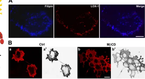
Cholesterol Is Necessary for Spatial Organization of LOX-1 Surface Receptors. To address the question of whether the membrane cholesterol level can alter the traf-ficking, distribution, and function of LOX-1 receptors, we used two compounds acting by different mechanisms. The polyene antibiotic filipin, a fluorescent drug that specifically binds to lipid rafts and nonraft membrane cholesterol (Roth-berg et al., 1990) and MCD. The latter specifically extracts cholesterol from the plasma membranes and therefore dis-rupts lipid rafts and caveolae (Ilangumaran and Hoessli, 1998). We first investigated the localization and physical interaction between LOX-1 receptors and cholesterol rich-membrane microdomains by comparing its rich-membrane distri-bution with that of filipin. Double staining of nonfixed COS cells transiently transfected with human LOX-1-V5 and fili-pin is shown in Fig. 1A. We found many colocalization sites of the two fluorescence signals. In particular, LOX-1 accumu-lates in filipin-positive dots, which suggests that LOX-1 is preferentially associated with membrane-bound cholesterol. Then, we lowered the membrane cholesterol level and dis-rupted lipid rafts by MCD and studied the trafficking and distribution of LOX-1 receptors. Whereas in the control (Fig. 1B, a and a⬘), most cells show an intense membrane fluores-cence, the plasma membrane pool of LOX-1 receptors be-comes more diffuse in MCD-treated cells (Fig. 1B, b and b⬘),
Fig. 1. Cholesterol level alters LOX-1 receptors surface
organization. A, double staining of COS cells transiently transfected with LOX-1-V5 together with filipin. After a 30-min incubation with 100 g/ml filipin, surface-ex-pressed LOX-1 receptors were visualized with MAb anti-V5 for 1 h at 4°C. B, transfected COS cells were treated with 5 mM MCD for 30 min in serum-free medium, surface stained with MAb anti-V5. and analyzed by confocal mi-croscopy. Representative images of the membrane distribu-tion of LOX-1 receptors in nontreated (a and a⬘) and in MCD-treated cells (b and b⬘) are displayed. a⬘ and b⬘ report the LOX-1 red staining converted first to black and white and then inverted to white and black, using the channel mixer of Adobe Photoshop. Scale bar, 10m.
at Fac di FarmaciaUniv Studi di Napoli on November 13, 2012
molpharm.aspetjournals.org
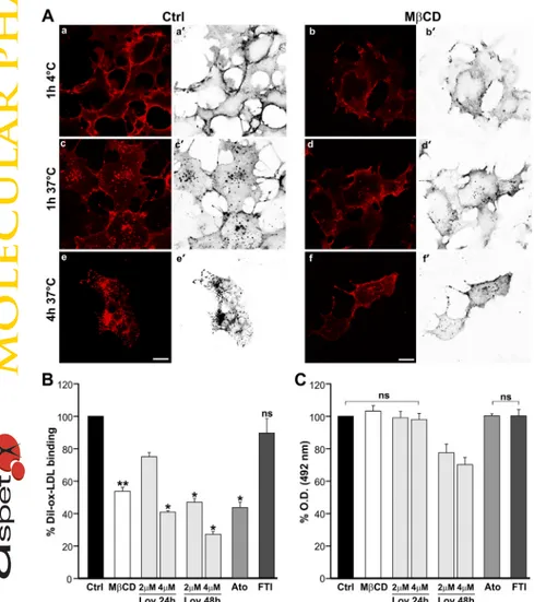
suggesting a perturbation of LOX-1 membrane localization. Taken together, these data indicate that LOX-1 is associated with cholesterol-rich domains and that plasma membrane cholesterol specifically regulates LOX-1 surface distribution. Membrane Cholesterol Depletion Inhibits LOX-1-Mediated ox-LDL Binding and Internalization. To study whether the altered membrane distribution of LOX-1 receptors is accompanied by impairment of ox-LDL binding and internalization, we transfected COS cells with LOX-1-V5 and 24 h after transfection, we treated cells with MCD and incubated them with Dil-labeled ox-LDL in serum-free me-dium. Representative confocal images of Dil-ox-LDL binding (1 h at 4°C) and uptake (1 and 4 h at 37°C) are shown in Fig. 2A. Dil-ox-LDL efficiently binds to LOX-1 receptors in control cells (Fig. 2A, a and a⬘). Of note, MCD caused a marked loss of specific ox-LDL binding, making the typical membrane fluorescence more diffuse and less intense (Fig. 2A, b and b⬘). Moreover, the intracellular dots of endocytosed ox-LDL after a 1-h incubation at 37°C (Fig. 2A, c and d) are markedly reduced both in size and number after MCD treatment, indicating strong inhibition of ox-LDL internal-ization. We also studied the intracellular uptake of ox-LDL at 37°C for 4 h (Fig. 2A, e and f). In control cells, most of the fluorescent ox-LDL is found inside the cells. In contrast, this intracellular pool is almost absent in MCD-treated COS cells, in which Dil-ox-LDL fluorescence remains at the level of the plasma membrane. Quantitation of bound Dil-ox-LDL
was obtained by its extraction from stained cells with isopro-panol and spectrofluorometric analysis. As can be seen in Fig. 2B, inhibition of ox-LDL binding is very strong, reaching 47 ⫾ 5% reduction of LOX-1 binding after treatment with MCD.
Lipid rafts are also disrupted by statins, drugs that are largely used in clinics for their activity in lowering circulat-ing cholesterol. We treated LOX-1-V5-COS transfected cells with lovastatin and atorvastatin, and quantified the fluores-cent Dil-ox-LDL binding in treated and nontreated cells (Fig. 2B). Chronic exposure of cells to statins leads to a reduction of ox-LDL binding compared with that in control cells. These data confirm that membrane cholesterol depletion in lipid rafts affects LOX-1 function. However, because statins in-hibit the synthesis of isoprenoids and prevent isoprenylation of many proteins including Ras and Rho families of GTPase, exerting cholesterol-independent or pleiotropic effects also, they may impair the function of cell surface receptors that interact with signaling molecules. To examine the role of isoprenylation on LOX-1 function, transfected COS cells were pretreated with the farnesyl transferase inhibitor ABT-100 before Dil-ox-LDL binding was measured. No effect was seen on ox-LDL binding, suggesting that cholesterol lowering rather than isoprenylation inhibition is responsible for LOX-1 function impairment.
Whether reduction of plasma membrane cholesterol causes the marked decrease in LOX-1 activity by modulating the
Fig. 2. Effect of cholesterol-lowering drugs on
LOX-1-me-diated ox-LDL binding and uptake. A, COS cells transiently transfected with LOX-1-V5 were incubated or not with 5 mM MCD in serum-free medium at 37°C for 30 min, washed, and then incubated with 10g/ml Dil-ox-LDL on ice for 1 h (binding) or 1 h or 4 h at 37°C (uptake). a⬘ to f⬘ report the Dil-ox-LDL red staining converted first to black and white and then inverted to white and black, using the channel mixer of Adobe Photoshop. Scale bar, 10m. B, cells were treated or not with MCD or with 2 or 4 M lovastatin (Lov), 2M atorvastatin (Ato), or 0.1 M FTI (ABT-100) for the times indicated. Histograms show quan-tification of Dil-ox-LDL binding measuring fluorescence by spectrofluorometer. C, surface receptors were measured by cyto-enzyme-linked immunosorbent assay by using MAb anti-V5. The data represent the average ⫾ S.D. of four separate experiments. p⬍ 0.05 was considered to be sta-tistically significant (ⴱ, p ⬍ 0.05; ⴱⴱ, p ⬍ 0.01); ns, no significant difference.
at Fac di FarmaciaUniv Studi di Napoli on November 13, 2012
molpharm.aspetjournals.org
number of exposed receptors was studied by using a surface labeling quantification assay (Biocca et al., 2008), as de-scribed under Materials and Methods. The surface appear-ance of LOX-1 in COS transfected cells, treated or not with cholesterol-lowering drugs is shown in Fig. 2C. Of interest, there is no statistically significant difference in the amount of exposed LOX-1 receptors immediately after MCD, lova-statin, and atorvastatin were washed off the cells, indicating that the effect of cholesterol reduction at the level of plasma membranes is mostly on LOX-1-mediated ox-LDL binding activity and on its distribution (as shown in Fig. 1B) and is not related to a variation in the number of exposed receptors. A decrease in surface LOX-1 receptors was only detected in cells treated for 48 h at higher concentration of lovastatin. No significant difference in surface LOX-1 receptors was found with treatment of COS transfected cells with atorvastatin and the farnesyl transferase inhibitor ABT-100.
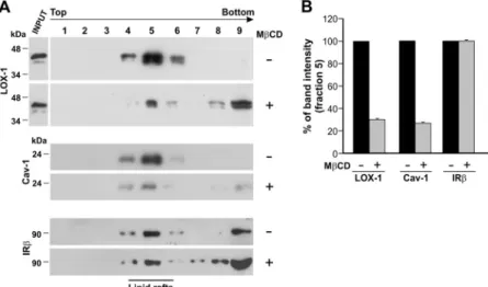
Cholesterol-Lowering Drugs Disrupt LOX-1 Distri-bution in Caveolae/Lipid Rafts. To better understand how membrane cholesterol regulates LOX-1 functional state, we studied the subcellular distribution of LOX-1 receptors in plasma membranes by fractionation of cellular membranes and purification of cholesterol-rich lipid rafts. We generated HEK-293 cell lines stably transfected with human LOX-1-V5. More than 10 stably transfected clones were analyzed and compared for LOX-1 expression by Western blot and indirect immunofluorescence, and the studies reported in this article were performed with two representative expressing clones (13 and 19). First, we quantified the total cellular LOX-1 content in HEK-293 cells incubated or not with MCD by Western blot. LOX-1 protein runs at 46 kDa in HEK-293 transfected cells and is efficiently expressed, and there is no change in the total cellular LOX-1 protein level immediately after the MCD treatment (Fig. 3A, INPUT). We then iso-lated caveolin-rich domains by a detergent-free procedure and sucrose gradient flotation centrifugation (Song et al., 1996). An aliquot of each fraction was subjected to immuno-blot analysis (Fig. 3A, fractions from 2 to 9). Of interest, LOX-1 was mostly found in fractions 5. This fraction is com-posed of lipid rafts, as confirmed by the presence of caveo-lin-1, and we have calculated that LOX-1, by using this fractionation scheme, is purified approximately 200-fold rel-ative to total cell lysate. Strikingly, MCD treatment led to a marked depletion of LOX-1 from caveolin-enriched
mem-branes, which was superimposable to that of caveolin itself, as seen in the blot visualized with anti-Cav-1 antibodies. It is worth noting that the amount of LOX-1 and caveolin-1 not clearly present in lipid rafts is now detected as associated with nonraft membrane fractions (fraction 9) and, in a lower amount, in fractions 10 to 12 (not shown). To evaluate the specificity of our data, we determined the expression of an-other plasma membrane protein, IR, which is located in raft and nonraft membranes, under the same experimental con-ditions (Winter et al., 2012). As shown in Fig. 3A, changes in cellular cholesterol did not influence the insulin receptor expression and localization in lipid rafts. The fold change in LOX-1, caveolin-1, and IR bands in fraction 5 in control and MCD-treated cells was evaluated by densitometric quanti-fication (Fig. 3B).
To verify whether chronic inhibition of cholesterol biosyn-thesis by statins also resulted in perturbation of LOX-1 dis-tribution in caveolae/lipid rafts, we used HEK-293 cells sta-bly expressing LOX-1 and treated cells with lovastatin or atorvastatin for 3 days. We then isolated lipid rafts by flota-tion centrifugaflota-tion and analyzed the presence of LOX-1, caveolin-1, and IR in each fraction of the gradient. A repre-sentative blot of the gradient derived from lovastatin-treated cells and the analysis of the band intensity in fraction 5 is shown in Fig. 4, A and B, respectively. It is worth mentioning that lovastatin, as MCD, does not change the total LOX-1 expression (Fig. 4A, INPUT). Lovastatin treatment leads to a marked decrease in LOX-1 protein (lovastatin 38⫾ 6% ver-sus control 100%) and caveolin-1 (lovastatin 50⫾ 3% versus control 100%) in lipid rafts (fraction 5). In these cells the amount of LOX-1 not present in fraction 5 is detected mostly in fraction 12. No significant difference in the intensity of the band was detected for the IR subunit. As shown in Fig. 4C, a comparable decrease in the intensity of LOX-1 band in fraction 5 was obtained by incubating cells with 2M ator-vastatin. Of importance, the farnesyl transferase inhibitor ABT-100 does not lead to a reduction in LOX-1 in lipid rafts (Fig. 4C).
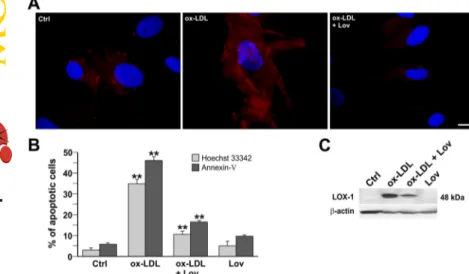
Lovastatin Acts on LOX-1 Function in Human Endo-thelial Primary Cultures from Aortic Aneurysm. It is well established that ox-LDL treatment of vascular endothe-lial cells results in apoptosis and necrosis and that most of these effects are mediated by an increase in expression and activation of LOX-1 receptors (Chen et al., 2002). To further
Fig. 3. Detergent-free purification of caveolin-rich domains
by sucrose gradient. A, distribution and localization of LOX-1 receptors on membranes derived from HEK-293 cells stably transfected with LOX-1-V5 (clone 19). Lysates of untreated cells or cells treated with 5 mM MCD at 37°C for 30 min were subjected to sucrose gradient centrifuga-tion after homogenizacentrifuga-tion in a buffer containing sodium carbonate (see under Materials and Methods) and analyzed by Western blot. Total protein extract (5%, 15g) (INPUT) was also loaded as positive internal control of electropho-retic mobility. Immunoblot analysis was carried out with anti-V5 (LOX-1), anti-caveolin 1 (Cav-1), and anti-IR an-tibodies. Fractions 4 to 6 were designated as caveolin-enriched lipid rafts indicated by the marker protein caveo-lin-1. B, histogram shows the densitometric measurements performed to compare the intensity of LOX-1, caveolin-1, and IR in fraction 5 derived from control or treated cells. The data represent the average⫾ S.D. of three separate experiments.
at Fac di FarmaciaUniv Studi di Napoli on November 13, 2012
molpharm.aspetjournals.org
explore the significance of the effect of cholesterol modulation and raft disruption by statins on LOX-1 receptor activity, we have used human endothelial primary cells isolated from atherosclerotic lesions of human aorta (infrarenal abdominal aortic aneurysm), as a model system. ECs were isolated as described in the Supplemental Data and characterized by fluorescence-activated cell sorter analysis using different markers (Supplemental Fig. 1). We incubated primary EC cells with ox-LDL or ox-LDL plus lovastatin for 24 h and analyzed the binding of fluorescent ox-LDL, as described under Materials and Methods (Fig. 5A). In control cells, bind-ing is very low because of the low endogenous LOX-1 level of expression. As expected, upon induction of LOX-1 with ox-LDL incubation, many cells show a much higher fluorescent signal (Fig. 5A, center panel). Lovastatin treatment results in a marked reduction of positive cells (right panel, ox-LDL⫹ Lov). Because most of the Dil-ox-LDL-positive cells do not thrive and many of these cells exhibit cell shrinkage, which is a feature of apoptosis, we measured the apoptotic effects after different treatments, performing annexin V membrane staining and Hoechst 33342 nuclear staining (Fig. 5B). The percentage of apoptotic cells significantly increased when ECs were exposed to ox-LDL (ox-LDL treatment 47% versus control 5%, on the basis of the annexin V assay (p⬍ 0.01). In contrast, lovastatin coincubation resulted in the rescue of the ox-LDL-induced phenotype (ox-LDL ⫹ Lov treatment 16% versus ox-LDL 47% (p ⬍ 0.01). Indeed, the percentage of apoptotic cells in this population (16⫾ 2%) approaches that of cells treated with lovastatin alone (10⫾ 3%).
Ox-LDL-dependent induction of endogenous LOX-1 recep-tors was also studied by Western blot with anti-LOX-1 poly-clonal antiserum (Fig. 5C). As can be seen, the LOX-1 band is induced in human primary endothelial cells. The 48-kDa LOX-1 band is very intense. However, its intensity markedly decreases in lysates derived from cells simultaneously incu-bated with ox-LDL and lovastatin (57⫾ 5% reduction). As expected, LOX-1 in lysates derived from nontreated cells or derived from cells incubated with lovastatin alone is under the detection threshold.
To study the intracellular distribution of endogenous LOX-1 in primary endothelial cells incubated with ox-LDL or ox-LDL plus lovastatin, we isolated lipid rafts by flotation centrifugation. We first analyzed LOX-1 distribution in tran-siently transfected COS cells incubated or not with 100g of ox-LDL for 1 h at 37°C (Fig. 6A). In the absence of ox-LDL, LOX-1 receptors are distributed in caveolae/lipid raft (frac-tions 4 and 5) and nonlipid raft membranes (frac(frac-tions 8 and 9). Of interest, upon incubation with ox-LDL, 100% of LOX-1 receptor molecules are found in fractions 4 and 5, indicating that active receptors are localized in caveolae/ lipid raft membranes.
Figure 6B shows gradients derived from primary EC cells obtained from an aortic aneurysm. In nontreated control cells, LOX-1 is distributed in raft and nonraft membranous fractions. A small, but significant, amount of LOX-1 was also found in the final fractions 11 and 12 at the bottom of the tube. As expected, caveolin-1 is almost entirely found in the caveola/lipid raft fraction (fractions 4–6). Treatment of EC
Fig. 4. Statins disrupt LOX-1 membrane distribution in
caveolin-rich lipid rafts. A, HEK-293 stably expressing LOX-1-V5 (clone 19) were treated or not with 2M lova-statin (Lov) for 3 days. Lysates of untreated and treated cells were subjected to sucrose gradient centrifugation to isolate caveolin-enriched lipid rafts and immunoblotted with anti-V5 (LOX-1), anti-caveolin (Cav-1), and anti-IR antibodies (IR). Total protein extract (5%, 15 g) (INPUT) was also loaded as a positive internal control of electropho-retic mobility. B, densitometric measurements of LOX-1, Cav-1, and IR subunit bands in fraction 5 derived from control or treated cells. The data represent the average⫾ S.D. of three separate experiments. C, Western blot anal-ysis for LOX-1 present in fraction 5 of sucrose gradients derived from HEK-293 (clone 19) treated or not with 2M lovastatin (Lov), 2M atorvastatin (Ato), or 0.1 M FTI (ABT-100) for 48 h.
Fig. 5. Lovastatin effects on human primary endothelial
cells. A, EC cells treated or not with ox-LDL (100g/ml) or ox-LDL plus 2M lovastatin (Lov) were incubated with 10 g/ml LDL for 1 h at 4°C. Fluorescence of Dil-ox-LDL was detectable only in ox-Dil-ox-LDL-treated EC cells. Scale bar, 10m. B, apoptosis assay by annexin V membrane staining and Hoechst 33342 nuclear staining performed on control EC cells, ox-LDL, ox-LDL plus lovastatin, and lov-astatin-treated cells. The data represent the average⫾ S.D. of three separate experiments.ⴱⴱ, p ⬍ 0.01. C, West-ern blot analysis for LOX-1 on EC cells treated or not with ox-LDL, ox-LDL plus lovastatin, and lovastatin alone. The gel is representative of five separate experiments. Ctrl, control.
at Fac di FarmaciaUniv Studi di Napoli on November 13, 2012
molpharm.aspetjournals.org
cells with 2M lovastatin does not change LOX-1 distribu-tion, but significantly lowers the intensity of both LOX-1 and caveolin-1 bands. We then compared (by densitometric quan-tification) the two proteins in ox-LDL- and in ox-LDL plus lovastatin-treated ECs. As observed in transfected COS cells, in the presence of the ligand ox-LDL, LOX-1 is mostly con-centrated in lipid rafts (52⫾ 3%). In primary cells, however, 38 ⫾ 6% of LOX-1 is also found in fractions 11 and 12. Because more than 45% of ox-LDL-treated cells undergo apoptosis (Fig. 5B), LOX-1 detected at the bottom of the gradient may belong to apoptotic bodies. In contrast, in the gradient derived from cells that have been coincubated with lovastatin and ox-LDL, LOX-1 is not concentrated in frac-tions 4 and 5 but has a distribution very similar to that seen in control cells and is present in all membranous fractions. As shown above, these cells show a substantial rescue of the ox-LDL-induced apoptotic phenotype.
Finally, it is worth noting that caveolin-1 is barely detect-able in lovastatin-treated endothelial cells. From a densito-metric analysis of band intensity from different experiments, gradients derived from lovastatin-treated EC cells present a reduction ofⱖ50% of the caveolin-1 band in fractions 4, 5, and 6, a value comparable to that observed in HEK-293 LOX-1-expressing cells (⫺55%) incubated with lovastatin (Fig. 4).
Discussion
Here we demonstrate that LOX-1 is distributed within lipid rafts in the plasma membranes and that its distribution in cholesterol-enriched microdomains is an absolute require-ment for its capacity to bind and internalize ox-LDL. Our observation that LOX-1 resides within lipid rafts is based on several well established raft analysis techniques. First, LOX-1 receptors colocalize with filipin, a fluorescent marker of membrane cholesterol. Second, cholesterol sequestration by MCD leads to mislocalization of LOX-1 receptors in a more diffuse distribution in the plasma membrane. Third, by biochemical fractionation of cell membranes, we show that functional LOX-1 is found almost entirely in a fraction con-taining caveolin-1 and that we define caveolae/lipid rafts in all cell types we analyzed including human primary endothe-lial cells. A more detailed morphological analysis at high resolution is necessary, however, to establish whether LOX-1 associates exclusively with caveolae. Of importance, disrup-tion of caveolae/lipid rafts by acute treatment with MCD or chronic incubation of cells with lovastatin or atorvastatin results in a marked reduction of LOX-1-mediated ox-LDL binding and uptake.
The finding that LOX-1 function is highly inhibited by treatment with cholesterol-depleting drugs highlights, for the first time, the critical importance of cholesterol in recep-tor activity and demonstrates a new role of statins as inhib-itors of LOX-1 activity. The plasma membrane cholesterol is an essential determinant of membrane fluidity by modulat-ing the structure of the phospholipid bilayer. Formation and maintenance of lipid rafts and caveolae are strictly depen-dent on cholesterol. These specialized cholesterol-rich sub-domains, highly abundant in endothelial cells, regulate var-ious signal transduction pathways and are characterized by the presence of the caveolin protein family (Li et al., 2005). Treatment with statins reduces the amount of cholesterol in these sites, increases membrane fluidity, and induces a re-distribution of caveolin-1 and other resident membrane pro-teins in the endoplasmic reticulum and plasma membrane. Many important membrane properties are affected, includ-ing cell endocytosis, permeability, and transport functions (Goonasekara et al., 2010). As shown here, LOX-1 receptors also change their distribution and lose their physiological location when cells are treated with cholesterol-depleting drugs. However, interestingly, disruption of caveolae/lipid rafts and mislocalization of LOX-1 receptors do not lead to a reduction in surface exposed LOX-1 receptors. Notwithstand-ing their presence, it appears that their affinity for ox-LDL decreases dramatically, suggesting that the assembly of LOX-1 in multimers in specific membrane microdomains is a crucial requirement for ox-LDL binding and internalization. Thus, when LOX-1 is randomly distributed in the membrane, it is unable to bind ox-LDL. Its clustered distribution is essential to enhance the interaction efficiency as well as the internalization of LOX-1-ox-LDL complexes. Several pieces of evidence from our laboratory and from others have re-cently suggested that multimerization and cluster formation are necessary for LOX-1 activity (Biocca et al., 2008; Cao et al., 2009; Ohki et al., 2011). Hetero-oligomerization with LOX-1 mutant isoforms such as LOXIN or K167N LOX-1 leads to a very severe reduction in LOX-1 function (Biocca et al., 2008, 2009). Here we indicate cholesterol-enriched
mi-Fig. 6. Effect of ox-LDL and lovastatin on LOX-1 membrane distribution
in COS and human endothelial primary cells derived from aortic aneu-rysms. A, COS cells transiently transfected with LOX-1-V5 were incu-bated or not with 100g/ml ox-LDL for 1 h at 37°C. Lysates of untreated and treated cells were subjected to sucrose gradient centrifugation and immunoblotted with MAb anti-V5. The experiment was repeated three times. B, distribution and localization of LOX-1 receptors on membranes from EC cells derived from aortic aneurysms. Lysates of untreated and treated cells with 2M lovastatin (Lov), ox-LDL (100 g/ml), and ox-LDL plus lovastatin were subjected to sucrose gradient centrifugation to iso-late caveolin-enriched lipid rafts and immunoblotted with specific anti-bodies directed against LOX-1 and caveolin-1. All collected fractions are shown, except for fraction 1, which does not contain proteins. The data are representative of five separate experiments. Ctrl, control.
at Fac di FarmaciaUniv Studi di Napoli on November 13, 2012
molpharm.aspetjournals.org
crodomains (caveolae/lipid rafts) as the sites of multimeriza-tion. Whether ox-LDL can engage multiple interactions with several LOX-1 dimers and whether these interactions strengthen the binding affinity in vivo, as was demonstrated in vitro (Ohki et al., 2011), is under study. In support of this hypothesis, it is worth mentioning that intact lipid rafts are essential for HIV-1 virus entry by scavenger receptors (Carter et al., 2009; Waheed and Freed, 2009). For DC-SIGN, a dendritic cell-specific C-type lectin receptor, a direct corre-lation between the distribution of receptors in microdomains, rather than randomly distribution, and their capacity to bind and internalize virus-sized ligand-coated particles in den-dritic cells has been described (Cambi et al., 2004). Alterna-tive nonmutually exclusive mechanisms to explain the effects of cholesterol-lowering drugs on LOX-1 activity can be pro-posed and require further studies: 1) a direct specific inter-action of cholesterol with LOX-1 receptor and 2) an indirect mechanism on membrane physical properties, which may affect other molecules involved in LOX-1-mediated ox-LDL endocytosis.
Statins, as potent cholesterol-lowering drugs, are the prin-cipal therapy for more than 25 million people at risk for cardiovascular diseases worldwide. They are largely used to lower total and LDL cholesterol and are beneficial in primary and secondary prevention of cardiovascular diseases. Lower-ing circulatLower-ing cholesterol is thought to be the principal ben-eficial effect of statins. However, they can also exert choles-terol-independent responses, because of the inhibition of the synthesis of isoprenoids, which are important lipid attach-ments for post-translational modifications of many proteins, such as Ras, Rho, and Rac, and nuclear lamina (Wang et al., 2008). Of note, treatment of cells with the farnesyl trans-ferase inhibitor ABT-100 had no effects on LOX-1 membrane distribution and its activity. We describe a different pleiotro-pic effect of statins: reduction of membrane-associated cho-lesterol, which influences the density of membrane rafts and disrupts LOX-1 cluster distribution in plasma membranes. This new mechanism of LOX-1 inhibition may explain the beneficial effects of lovastatin incubation in ox-LDL-treated primary endothelial cells derived from aortic aneurysm and suggests that statins protect vascular endothelium by inhib-iting LOX-1-mediated entry of ox-LDL. Indeed, in EC cells we show that lovastatin treatment results either in a marked reduction of ox-LDL binding, similar to that shown in statin-treated LOX-1-COS transfected cells and in a consequent substantial rescue of the ox-LDL-induced apoptotic pheno-type. Although other studies have reported the effects of statins on decreasing LOX-1 expression in animal and cellu-lar models (Li et al., 2001, 2002; Hofnagel et al., 2006; Dje N⬘Guessan et al., 2009), none of these reports have focused on the relationship between the potent membrane cholester-ol-lowering effect and LOX-1-mediated ox-LDL binding and internalization activity. It is worth mentioning, however, that in vivo and ex vivo studies have recently shown that statin-mediated membrane raft depletion and membrane cholesterol reorganization influence immune cell function, such as natural killer cell cytotoxicity and foam cell forma-tion and accumulaforma-tion in atherosclerotic lesions (Hillyard et al., 2004, 2007; Hofnagel et al., 2007; Salvary et al., 2012). In clinical use, the discrimination of the circulating LDL cho-lesterol-lowering effect from other pleiotropic effects of st-atins may be more evident in the early phase of treatment,
because only a 10% reduction in the LDL-C level is detectable after 24 h and at least 6 to 7 days are necessary to lower it significantly (Corsini et al., 2007). Of interest, in a recent trial, an early window of protection by statin without LDL-C-lowering effects has been described. A pretreatment of 12 h with 40 mg of atorvastatin before percutaneous coronary intervention improved clinical outcomes in patients with acute coronary syndromes, indicating the pleiotropic proper-ties of statins (Patti et al., 2007).
Finally, we found a marked down-regulation of caveolin-1 in lovastatin-treated human primary endothelial cells. The physiological role of caveolin-1 and caveolae in the cardiovas-cular system has been seen recently. Caveolin-1 is important for the biogenesis of caveolae and is also involved in choles-terol trafficking to and from plasma membrane. In fact, caveolin-1 directly binds cholesterol with high affinity, which can explain the high concentration of cholesterol in caveolae (Li et al., 2005). In particular, endothelial-specific overex-pression of Cav-1 enhances the progression of atherosclerosis and loss of caveolae through Cav-1 gene deletion is protective against atherosclerosis (Frank et al., 2008; Ferna´ndez-Her-nando et al., 2010). Cav-1(⫺/⫺) mice show defects in the aortic uptake of LDL particles both in vitro and in vivo. The result is that statins reduce caveolin-1 abundance in endo-thelial cells (Pelat et al., 2003; Plenz et al., 2004) and may act in synergy with the inhibitory effect on LOX-1 receptor ac-tivity. Whether caveolin-1 directly interacts with LOX-1 re-ceptors and this interaction modulates the internalization of ox-LDL remains to be determined.
The delineation of the caveola/raft-mediated pathway for endocytosis of ox-LDL through LOX-1 receptors provides the basis of future studies and supports a novel effect of statins on membrane raft function that may be relevant for other membrane receptors in cardiovascular diseases, paving the way for new therapeutic interventions.
Acknowledgments
We thank Prof. Arnaldo Ippoliti for providing surgery explants, Dr. Massimo Sanchez for his assistance with fluorescence-activated cell sorter analysis, Prof. Federica Sangiuolo and Prof. Nadia Canu for critical reading of the manuscript, and Prof. Fabrizio Loreni for helpful suggestions. We are also very grateful to Graziano Bonelli for his expert graphical assistance.
Authorship Contributions
Participated in research design: Mango, Novelli, and Biocca. Conducted experiments: Matarazzo, Quitadamo, Ciccone, and Biocca.
Contributed new reagents or analytic tools: Mango and Novelli. Performed data analysis: Matarazzo, Quitadamo, and Biocca. Wrote or contributed to the writing of the manuscript: Biocca.
References
Aoyama T, Sawamura T, Furutani Y, Matsuoka R, Yoshida MC, Fujiwara H, and Masaki T (1999) Structure and chromosomal assignment of the human lectin-like oxidized low-density-lipoprotein receptor-1 (LOX-1) gene. Biochem J 339:177–184. Biocca S, Falconi M, Filesi I, Baldini F, Vecchione L, Mango R, Romeo F, Federici G, Desideri A, and Novelli G (2009) Functional analysis and molecular dynamics simulation of LOX-1 K167N polymorphism reveal alteration of receptor activity. PLoS One 4:e4648.
Biocca S, Filesi I, Mango R, Maggiore L, Baldini F, Vecchione L, Viola A, Citro G, Federici G, Romeo F, et al. (2008) The splice variant LOXIN inhibits LOX-1 receptor function through hetero-oligomerization. J Mol Cell Cardiol 44:561–570. Bossy-Wetzel E and Green DR (2000) Detection of apoptosis by annexin V labeling.
Methods Enzymol 322:15–18.
Cambi A, de Lange F, van Maarseveen NM, Nijhuis M, Joosten B, van Dijk EM, de Bakker BI, Fransen JA, Bovee-Geurts PH, van Leeuwen FN, et al. (2004)
at Fac di FarmaciaUniv Studi di Napoli on November 13, 2012
molpharm.aspetjournals.org
mains of the C-type lectin DC-SIGN are portals for virus entry into dendritic cells. J Cell Biol 164:145–155.
Cao W, Calabro V, Root A, Yan G, Lam K, Olland S, Sanford J, Robak A, Zollner R, Lu Z, et al. (2009) Oligomerization is required for the activity of recombinant soluble LOX-1. FEBS J 276:4909 – 4920.
Cardinale A, Filesi I, Vetrugno V, Pocchiari M, Sy MS, and Biocca S (2005) Trapping prion protein in the endoplasmic reticulum impairs PrPCmaturation and prevents
PrPScaccumulation. J Biol Chem 280:685– 694.
Carter GC, Bernstone L, Sangani D, Bee JW, Harder T, and James W (2009) HIV entry in macrophages is dependent on intact lipid rafts. Virology 386:192–202. Chen M, Masaki T, and Sawamura T (2002) LOX-1, the receptor for oxidized
low-density lipoprotein identified from endothelial cells: implications in endothe-lial dysfunction and atherosclerosis. Pharmacol Ther 95:89 –100.
Corsini A, Ferri N, and Cortellaro M (2007) Are pleiotropic effects of statins real? Vasc Health Risk Manag 3:611– 613.
Dje N⬘Guessan P, Riediger F, Vardarova K, Scharf S, Eitel J, Opitz B, Slevogt H, Weichert W, Hocke AC, Schmeck B, et al. (2009) Statins control oxidized LDL-mediated histone modifications and gene expression in cultured human endothe-lial cells. Arterioscler Thromb Vasc Biol 29:380 –386.
Ferna´ndez-Hernando C, Yu J, Da´valos A, Prendergast J, and Sessa WC (2010) Endothelial-specific overexpression of caveolin-1 accelerates atherosclerosis in apolipoprotein E-deficient mice. Am J Pathol 177:998 –1003.
Frank PG, Pavlides S, Cheung MW, Daumer K, and Lisanti MP (2008) Role of caveolin-1 in the regulation of lipoprotein metabolism. Am J Physiol Cell Physiol 295:C242––C248.
Goonasekara CL, Balse E, Hatem S, Steele DF, and Fedida D (2010) Cholesterol and cardiac arrhythmias. Expert Rev Cardiovasc Ther 8:965–979.
Hillyard DZ, Jardine AG, McDonald KJ, and Cameron AJ (2004) Fluvastatin inhibits raft dependent Fc␥ receptor signalling in human monocytes. Atherosclerosis 172: 219 –228.
Hillyard DZ, Nutt CD, Thomson J, McDonald KJ, Wan RK, Cameron AJ, Mark PB, and Jardine AG (2007) Statins inhibit NK cell cytotoxicity by membrane raft depletion rather than inhibition of isoprenylation. Atherosclerosis 191:319 –325. Hofnagel O, Luechtenborg B, Eschert H, Weissen-Plenz G, Severs NJ, and Robenek
H (2006) Pravastatin inhibits expression of lectin-like oxidized low-density lipo-protein receptor-1 (LOX-1) in Watanabe heritable hyperlipidemic rabbits: a new pleiotropic effect of statins. Arterioscler Thromb Vasc Biol 26:604 – 610. Hofnagel O, Luechtenborg B, Weissen-Plenz G, and Robenek H (2007) Statins and
foam cell formation: impact on LDL oxidation and uptake of oxidized lipoproteins via scavenger receptors. Biochim Biophys Acta 1771:1117–1124.
Ilangumaran S and Hoessli DC (1998) Effects of cholesterol depletion by cyclodextrin on the sphingolipid microdomains of the plasma membrane. Biochem J 335:433– 440.
Kashiwakura Y, Watanabe M, Kusumi N, Sumiyoshi K, Nasu Y, Yamada H, Sawa-mura T, Kumon H, Takei K, and Daida H (2004) Dynamin-2 regulates oxidized low-density lipoprotein-induced apoptosis of vascular smooth muscle cell. Circu-lation 110:3329 –3334.
Kiyanagi T, Iwabuchi K, Shimada K, Hirose K, Miyazaki T, Sumiyoshi K, Iwahara C, Nakayama H, Masuda H, Mokuno H, et al. (2011) Involvement of cholesterol-enriched microdomains in class A scavenger receptor-mediated responses in hu-man macrophages. Atherosclerosis 215:60 – 69.
Li D, Chen H, Romeo F, Sawamura T, Saldeen T, and Mehta JL (2002) Statins modulate oxidized low-density lipoprotein-mediated adhesion molecule expression in human coronary artery endothelial cells: role of LOX-1. J Pharmacol Exp Ther 302:601– 605.
Li D, Williams V, Liu L, Chen H, Sawamura T, Romeo F, and Mehta JL (2003) Expression of lectin-like oxidized low-density lipoprotein receptors during isch-emia-reperfusion and its role in determination of apoptosis and left ventricular dysfunction. J Am Coll Cardiol 41:1048 –1055.
Li DY, Chen HJ, and Mehta JL (2001) Statins inhibit oxidized-LDL-mediated LOX-1 expression, uptake of oxidized-LDL and reduction in PKB phosphorylation. Car-diovasc Res 52:130 –135.
Li XA, Everson WV, and Smart EJ (2005) Caveolae, lipid rafts, and vascular disease. Trends Cardiovasc Med 15:92–96.
Lisanti MP, Scherer PE, Vidugiriene J, Tang Z, Hermanowski-Vosatka A, Tu YH, Cook RF, and Sargiacomo M (1994) Characterization of caveolin-rich membrane domains isolated from an endothelial-rich source: implications for human disease. J Cell Biol 126:111–126.
Mango R, Biocca S, del Vecchio F, Clementi F, Sangiuolo F, Amati F, Filareto A, Grelli S, Spitalieri P, Filesi I, et al. (2005) In vivo and in vitro studies support that a new splicing isoform of OLR1 gene is protective against acute myocardial infarction. Circ Res 97:152–158.
Mehta JL, Chen J, Hermonat PL, Romeo F, and Novelli G (2006) Lectin-like, oxidized low-density lipoprotein receptor-1 (LOX-1): a critical player in the devel-opment of atherosclerosis and related disorders. Cardiovasc Res 69:36 – 45. Mitra S, Deshmukh A, Sachdeva R, Lu J, and Mehta JL (2011) Oxidized low-density
lipoprotein and atherosclerosis implications in antioxidant therapy. Am J Med Sci 342:135–142.
Murphy JE, Vohra RS, Dunn S, Holloway ZG, Monaco AP, Homer-Vanniasinkam S, Walker JH, and Ponnambalam S (2008) Oxidised LDL internalisation by the LOX-1 scavenger receptor is dependent on a novel cytoplasmic motif and is regu-lated by dynamin-2. J Cell Sci 121:2136 –2147.
Ohki I, Amida H, Yamada R, Sugihara M, Ishigaki T, and Tate S (2011) Surface plasmon resonance study on functional significance of clustered organization of lectin-like oxidized LDL receptor (LOX-1). Biochim Biophys Acta 1814:345–354. Ohki I, Ishigaki T, Oyama T, Matsunaga S, Xie Q, Ohnishi-Kameyama M, Murata T,
Tsuchiya D, Machida S, Morikawa K, et al. (2005) Crystal structure of human lectin-like, oxidized low-density lipoprotein receptor 1 ligand binding domain and its ligand recognition mode to OxLDL. Structure 13:905–917.
Park H, Adsit FG, and Boyington JC (2005) The 1.4 angstrom crystal structure of the human oxidized low density lipoprotein receptor LOX-1. J Biol Chem 280:13593– 13599.
Parton RG and Simons K (2007) The multiple faces of caveolae. Nat Rev Mol Cell Biol 8:185–194.
Patti G, Pasceri V, Colonna G, Miglionico M, Fischetti D, Sardella G, Montinaro A, and Di Sciascio G (2007) Atorvastatin pretreatment improves outcomes in patients with acute coronary syndromes undergoing early percutaneous coronary interven-tion: results of the ARMYDA-ACS randomized trial. J Am Coll Cardiol 49:1272– 1278.
Pelat M, Dessy C, Massion P, Desager JP, Feron O, and Balligand JL (2003) Rosuvastatin decreases caveolin-1 and improves nitric oxide-dependent heart rate and blood pressure variability in apolipoprotein E⫺/⫺mice in vivo. Circulation 107:2480 –2486.
Plenz GA, Hofnagel O, and Robenek H (2004) Differential modulation of caveolin-1 expression in cells of the vasculature by statins. Circulation 109:e7–– e8. Rothberg KG, Ying YS, Kamen BA, and Anderson RG (1990) Cholesterol controls the
clustering of the glycophospholipid-anchored membrane receptor for 5-methyltet-rahydrofolate. J Cell Biol 111:2931–2938.
Salvary T, Gambert-Nicot S, Brindisi MC, Meneveau N, Schiele F, Se´ronde MF, Lorgis L, Zeller M, Cottin Y, Kantelip JP, et al. (2012) Pravastatin reverses the membrane cholesterol reorganization induced by myocardial infarction within lipid rafts in CD14⫹/CD16⫺circulating monocytes. Biochim Biophys Acta doi: dx.doi.org/10.1016/j.bbalip.2012.02.017.
Sattler W, Bone P, and Stocker R (1992) Isolation of Human VLDL, LDL, HDL and Two HDL Subclasses in the TL-100 Tabletop Centrifuge Using the TLA-100.4 Rotor, Technical information, Beckman Coulter, Fullerton, CA.
Sawamura T, Kume N, Aoyama T, Moriwaki H, Hoshikawa H, Aiba Y, Tanaka T, Miwa S, Katsura Y, Kita T, et al. (1997) An endothelial receptor for oxidized low-density lipoprotein. Nature 386:73–77.
Song KS, Li Shengwen, Okamoto T, Quilliam LA, Sargiacomo M, and Lisanti MP (1996) Co-purification and direct interaction of Ras with caveolin, an integral membrane protein of caveolae microdomains. Detergent-free purification of cave-olae microdomains. J Biol Chem 271:9690 –9697.
Stephan ZF and Yurachek EC (1993) Rapid fluorometric assay of LDL receptor activity by Dil-labeled LDL. J Lipid Res 34:325–330.
Vohra RS, Murphy JE, Walker JH, Ponnambalam S, and Homer-Vanniasinkam S (2006) Atherosclerosis and the lectin-like OXidized low-density lipoprotein scav-enger receptor. Trends Cardiovasc Med 16:60 – 64.
Waheed AA and Freed EO (2009) Lipids and membrane microdomains in HIV-1 replication. Virus Res 143:162–176.
Wang CY, Liu PY, and Liao JK (2008) Pleiotropic effects of statin therapy: molecular mechanisms and clinical results. Trends Mol Med 14:37– 44.
Winter PW, Van Orden AK, Roess DA, and Barisas BG (2012) Actin-dependent clustering of insulin receptors in membrane microdomains. Biochim Biophys Acta 1818:467– 473.
Zeng Y, Tao N, Chung KN, Heuser JE, and Lublin DM (2003) Endocytosis of oxidized low density lipoprotein through scavenger receptor CD36 utilizes a lipid raft pathway that does not require caveolin-1. J Biol Chem 278:45931– 45936.
Address correspondence to: Prof. Silvia Biocca, Department of Systems Medicine, University of Rome “Tor Vergata,” Via Montpellier 1, 00133, Rome, Italy. E-mail: [email protected]
at Fac di FarmaciaUniv Studi di Napoli on November 13, 2012
molpharm.aspetjournals.org