UNIVERSITY OF PISA
PhD Course in Neuroscience
Section I: Molecular, Metabolic and Functional Exploration
of the Nervous System and the Sense Organs
“MODULATING FACTORS IN THE EARLY STAGES OF COGNITIVE
DECLINE: THE ROLE OF COGNITIVE RESERVE”
Tutors:
Candidate:
Prof. Gabriele Siciliano
Dott.ssa Leda Volpi
Dott.ssa Gloria Tognoni
ACADEMIC YEAR 2012-2013
SSD MED 26
1 “God gave us memory so that we might have roses in December”
James Matthew Barrie
“You don’t remember what happened. What you remember becomes what happened”
2 To Francesco, the sweetest gift
To my grandfather Gino, who would have been very proud of me
3
TABLE OF CONTENTS
Summary …..………...4
1. Introduction ……….. 6
1.1 Toward an early detection of cognitive impairment ………...6
1.2 Non-pharmacological interventions in MCI: what can we do to slow down late-life cognitive decline? ……….16
2. Subjects and Methods ………..20
2.1 Baseline assessments ………...20 2.2 Training intervention ………...26 2.3 Statistical analysis ………....28 3. Results ………...30 3.1 Total sample ……….30 3.2 Training intervention ………...40 4. Discussion ……….49
4.1 Investigating the presence of predicting factors of cognitive decline at the very early stages ………49
4.2 Assessment of the effects of a physical and cognitive training intervention on patients with MCI ………56
4.3 Cognitive reserve: evaluation of the effect at the early stages of cognitive impairment and of the influence on the outcome of cognitive training ……….59
5. Conclusions ………63
References ……….65
4
SUMMARY
The concept of cognitive reserve (CR) is being increasingly used to explain the observation that there is often a poor correspondence between the presence of pathology at autopsy and the level of cognitive decline in life. Since dementia affects an increasing proportion of the population, imposing a large economic burden on our society, in the absence of disease-modifying treatments it is crucial to detect lifestyle factors that may play a protective role in older individuals, delaying the onset of cognitive impairment. Subjective cognitive impairment (SCI), mild cognitive impairment (MCI) and Alzheimer’s disease (AD) are part of the same spectrum of disease progression. Since memory complaints represent a very common symptom reported by the older community population, early diagnosis of cognitive decline is not always easy, which makes it extremely important to find strategies that help detect individuals who deserve specific exams in a specialist setting.
Therefore, we compared a group of individuals with SCI and a sample of patients with MCI, searching for predicting factors of initial cognitive decline. Moreover, we investigated the contribution of CR on cognitive performance in a population of subjects claiming memory difficulties and in a subgroup of patients with MCI undergoing a physical and cognitive training program. Our findings indicated that many variables, including neuroimaging, everyday functional abilities, current activities, CR and behavioral factors are differently expressed in the two populations, with medial temporal lobe atrophy, ADCS scale score and MMSE as the most predictive factors of cognitive impairment. CR resulted a powerful modulating factor of cognitive decline, which is able to support attentive and executive performances in older individuals.
The physical and cognitive training program showed a significant positive outcome in patients with MCI, with the main improvements involving memory tests, even though no changes
5
were observed in everyday functional level, nor in the use of mnemonic strategies. Reduced awareness of difficulties was a noteworthy negative predictor of responsiveness to the cognitive stimulation program. Improvement in the memory domain was detected also in subjects with low CR, meaning that neural plasticity makes the enhancement of cognitive performances possible also in late-life, therefore suggesting that cognitive stimulating activities can be useful as non-pharmacological treatments at the early stages of cognitive decline.
6
1. INTRODUCTION
1.1 Toward an early detection of cognitive impairment
Alzheimer’s disease (AD) affects an increasing proportion of individuals, imposing a large economic burden on our society. There are currently no disease-modifying treatments for AD, thus making lifestyle factors that influence dementia risk of extreme importance.
The pathophysiological process of AD is known to begin many years, if not decades, before the clinical diagnosis of dementia [Morris 2005]. This long “preclinical” phase of AD would provide a critical opportunity for therapeutic intervention. The importance of an early diagnosis of mild cognitive impairment (MCI) relies on the assumption that early detection and intervention are likely to become beneficial in the future, when course-modifying agents will be found, and which can work best in the early stages.
Recent advances in neuroimaging, cerebrospinal fluid (CSF) assays, and other biomarkers able to detect evidence of the AD pathophysiological process in vivo showed that biomarker evidence of amyloid beta (Aβ) accumulation, which is associated with functional and structural brain alterations, can be observed in clinically normal older individuals with patterns of abnormality like those seen in patients with MCI and AD dementia (Figure 1). Furthermore, clinical cohort studies suggest that there may be very subtle cognitive alterations that are detectable years before meeting the criteria for MCI, and that predict progression to AD dementia. However, early diagnosis of cognitive decline is complicated by the unfeasibility of using such invasive and/or expensive tools on the huge population of older individuals. Consequently, an important challenge of research is how to detect people with MCI in routine clinical practice.
7
A wide variety of screening tests are available for this purpose [Cullen 2007]; however, data from epidemiological studies revealed that, in general, the detection of dementia in primary care is low and for MCI is even lower [Mitchell 2011; Jessen 2011] than in a specialist setting, where the diagnosis of MCI is also considerably more predictive of future dementia. Another possible means of detecting MCI could be the use of close informant reports, but this kind of method does not seem to be easily applicable in a screening program. Therefore, a more feasible method could come from the patients themselves, reporting a cognitive change.
Subjective cognitive impairment (SCI) is a very common complaint in the older community population, with a prevalence ranging from 10-20% to 50% according to the definition of the construct and to the asked questions [Abdulrab 2008; Jonker 2000], even though it is associated with low medical help-seeking [Begum 2012]. SCI can be reported in the younger population as episodes of transient confusion, while in older people it is mainly referred as forgetfulness or needing to take notes [Ginò 2010]. Some Authors observed a five-fold greater likelihood of decline to MCI or dementia, over a 7-year mean follow-up interval, in people with subjective complaints compared with similarly aged individuals who are free of subjective complaints of impairment. [Reisberg 2005]. As an entity that can precede MCI, studies are presently noting that about 7–8% of otherwise healthy older people with SCI progress to MCI or dementia every year [Reisberg 2005; Prichep 2005].
Although a number of studies have reported associations between SCI and underlying brain changes including white matter hyperintensities [de Groot 2001; Minett 2005; Stewart 2008], smaller hippocampal volume [van Norden 2008; Stewart 2008], functional magnetic resonance imaging correlates [Rodda 2011; Haley 2011], and evidence of Aβ on brain imaging [Perrotin 2012; Merril 2012], SCI’s positive predictive value in clinical practice remains low [Palmer 2003]. Therefore, the achievement of a diagnosis of MCI relies largely
8
on chance, for example a family history of dementia, the presence of a comorbidity that alert a clinician to carry out a further investigation, a person noticing worsening memory and deciding to inform his general practitioner about it, and often also a high socioeconomic status.
From these considerations, despite the potential importance of a diagnosis of MCI, very little is known about how people fulfilling these criteria can come to medical attention. Since it is clear that the detection of predictive factors of cognitive decline would be very useful in clinical practice in the pathway leading to an early diagnosis of MCI, one aim of the present study has been to assess the presence and influence of the main modulating factors in a population of older individuals complaining of cognitive difficulties.
Figure 1: Hypothetical model of dynamic biomarkers in the pathophysiological sequence of AD including the preclinical phase [Sperling 2011].
1.1.1 Cognitive Reserve
The concept of cognitive reserve (CR) stemmed from the detection of the frequent difference between observed and expected performance for a given degree of AD pathology. In fact,
9
with the advent of autopsy studies, we know that approximately 30% of subjects with neuropathologic features of AD die without exhibiting signs of cognitive decline [Katzman 1988; Hulette 1998; Price 1999; Schmitt 2000; Morris 2001; Riley 2002; Knopman 2003]. Moreover, the use of in vivo biomarkers of AD like amyloid imaging or CSF analysis has confirmed that many elderly subjects with no clinical impairment have evidence of preclinical AD [Stern 2012; Sperling 2011]. Incidentally, after the onset of dementia, the rate of cognitive decline is much faster in patients with higher CR [Unverzagt et al., 1998; Stern et al., 1999; Andel et al., 2006; Hall et al., 2007; Bruandet et al., 2008; Roselli et al., 2009)].
These observations brought to life to the “reserve hypothesis”. There are many factors that probably contribute to CR, including genetic and environmental influences, number of neurons and synapses, the sensitivity of neurons and glia to pathology and neuroplasticity. Stern at al [2006; 2012] suggested that the brain is able to face lesions at a certain level depending on two types of reserve. The first, named Brain Reserve (BR), refers to a passive model involving intrinsic differences in the pre-existing networks as expressed by brain volume, synaptic density and neuronal number or size. Recently, the neural integrity of the locus ceruleus has been demonstrated to be associated with reduced cognitive decline independent of pathologic burden in the temporal structures linked to AD-pathology, thus suggesting that this nucleus could be part of the brain reserve structures [Wilson 2013a]. Otherwise, CR refers to an active model involving functional differences regarding the efficiency with which individuals use alternative neural networks and engage adaptive cognitive strategies in order to compensate for pathologic burden and maintain normal cognition.
Cognitively stimulating activities have been associated with reduced late-life cognitive decline, but how can we explain this association? There is no consensus in the literature about
10
the mechanism underlying this beneficial effect of cognitive activities. A possible explanation is that cognitive activity is able to delay the cognitive consequences of neuropathologic burden possibly because of an activity-dependent action on the cognitive systems in the brain (cognitive reserve hypothesis) [Draganski 2006; Woollet 2011]. Otherwise, cognitive inactivity could be a consequence of the disease rather than a risk factor (reverse causality
hypothesis) [Bosma 2002; Hultsch 1999; Jagust 2011]. Recently, many Authors have
suggested the first hypothesis as the most reliable, demonstrating that CR is able to modify the level of cognitive performance. For some Authors, cognitive stimulating activities exert a direct action on neuropathology [Landau 2012], others suggest an independent and additive effect on cognition [Wilson 2013b; Vemuri 2012; Vemuri 2011], while another hypothesis is that stimulating activities could indirectly modulate the interaction between pathology and cognitive performances [Rentz 2010; Roe 2011; Raymond 2013]. Regarding the latter hypothesis, Ewers et al [Ewers 2013] observed that high CR (measured in terms of years of education) was associated with better FDG-PET signal in temporal lobe structures in old subjects with normal Aβ CSF levels and with lower signal in the same areas in patients with preclinical AD, while both Rentz and Raymond in their works have found that CR is able to modify the correlation between Aβ deposition and neuropsychological performance in AD patients and elderly controls.
A recently proposed model for the pahophysiology of AD posits that Aβ deposition occurs early in the process but by itself does not directly cause clinical symptoms [Jack 2009; Mormino et al., 2009], while, on the other hand, impaired cognitive performance could be largely determined by neurodegeneration mediated by tau pathology. Based on this evidence, AD pathological cascade could be envisaged as a two-stage process where amyloidosis and
11
neuronal pathology (tauopathy, neuronal injury and neurodegeneration) are sequential rather than concurrent processes [Ingelsson 2004; Jack 2009; Jack 2010].
Neuroplastic changes such as axonal remodeling, dendritic branching, growth of new dendritic spines and increased myelination have been recognized in developing and mature animals exposed to enriched environments [Marham 2004; Qui 2012]. Moreover, neuroimaging studies in humans suggest that cognitive activity can lead to structural and functional changes in brain regions that might improve CR [Gaser 2003; Maguire 2000; Lee 2010]. On the basis of this evidence, cognitive stimulating activities might increase the efficiency of some cognitive systems so that higher levels of neuropathologic burden are necessary in order to exhibit cognitive decline.
An important and potential bias involving research on CR is that CR is a hypothetical construct, with no available direct measures. CR is traditionally measured by surrogate markers, such as education, occupation, other lifestyle factors like social and physical leisure activities, or premorbid QI [Valenzuela 2006a]. There is no gold standard measure of CR so Authors are using different proxies and this could contribute to discrepancy among the results of the published studies.
The rationale for using education as an indicator of CR is that schooling may increase brain reserve by promoting synaptic growth [Katzman 1993] and may promote CR by generating new cognitive strategies [Stern 2002]. Another indicator of CR is occupational status. Some Authors suggest that managerial, technical and professional occupations versus unskilled or semi-skilled work lead to a 50% reduction in the risk of dementia [Valenzuela 2006b]. An important limitation of most measures of CR is that they may exert their influence through many alternative paths. For example, education and occupation are linked to socioeconomic status differences (SES), which is considered a promoting factor of disease, as SES influences
12
the access to resources necessary to maintain good health. Moreover, education could affect exposure to environmental toxic agents, promote engagement in leisure-time physical activity or influence the ability to prevent or manage chronic conditions that accelerate cognitive aging, such as diabetes and hypertention.
Intelligence is another frequently used index of CR [Alexander 1997, Scarmeas 2003a, Scarmeas 2003b, Solé-Padullés 2009] where IQ or pre-morbid IQ are the most common proxies used to estimate CR. However, one potential limitation regarding the use of reading tests evaluating IQ as a proxy of CR in patients with AD is that they can underestimate pre-morbid verbal IQ [Alexander 1997; O’Carol 1995; Schmand 1998]. As Nucci et al. pointed out [Nucci 2012], intelligence definition and measurement are based on intellectual performance, whereas cognitive reserve is based on the concept of cognitive skills acquired during one’s lifetime [Stern 2002, Stern 2009]. Furthermore, studies that measure CR from verbal abilities or only education or leisure activities or socioeconomic status consider only one aspect of CR, which is, incidentally, the result of several contributing factors. For this reason, we have chosen to use a multiple-indicator questionnaire to measure CR.
1.1.2 Neuroimaging
Evidences from structural brain imaging have shown that brain volume decreases with age with a annual rate of decline of 0.35% in older adults, compared to 0.12% in young adults [Dennis 2008]. Physiologic aging does not alter all brain areas to the same extent; in particular, the caudate, cerebellum, hippocampi and the association cortices undergo the largest volume loss. In addition, aging brings changes in white matter integrity, preferentially
13
in the frontal and prefrontal regions, with greater changes occurring after the seventh decade [Belleville 2012].
In the last decades, increasing interest has arisen in the predictive value of MRI techniques in AD. Since autopsy studies have indicated that the medial temporal lobe structures were generally the earlier affected and most severely on AD [Braak 1991], the first quantitative MRI studies revealed a volume reduction of up to 40% in AD patients compared to controls [Seab 1988; Kesslak 1991]. In the following years several methods, including medial temporal lobe atrophy on volumetric MRI and hypometabolism on fluorodeoxyglucose-positron emission tomography have been recorded in people with MCI compared with cognitively normal individuals [DeSanti 2001; Karas 2004], with the presence of these signs taken as a high predictive factor for progression to dementia [Korf 2004; Stout 2005]. However, in clinical practice, since such sophisticated neuroimaging tools are expensive, time-consuming and not routinely available, visual rating scales of medial temporal atrophy represent a more feasible alternative. The latter, in fact, are relatively independent of protocol or scan quality and are therefore easily applicable for neurologists and radiologists.
Another important imaging feature in the assessment of cognitive decline is cerebrovascular disease. The role of cerebrovascular disease in MCI is probably under-represented, particularly in population studies in which brain imaging has not been undertaken. Both cerebrovascular disease and neurodegenerative features were shown to contribute to MCI. The importance of white-matter lesions and small lacunar infarcts is becoming increasingly apparent in vascular cognitive impairment [O’Brien 2003].
White matter hyperintensities (WMH) from T2-weighted MRI are associated with cerebrovascular pathology and thus may be preventable through treatment of vascular risk factors; however, recent evidences suggest that early WMH should be considered among the
14
factors included in the AD models. Silbert and colleagues [Silbert 2012] administered yearly MRI to 134 cognitively healthy elders who converted to MCI over a period of 19 years of follow-up and observed that increase in WMH volume accelerated an average of 10 years prior to the first clinical diagnosis of MCI. This finding, for the Authors, suggests that insidious and progressive increase of WMH may represent a marker of preclinical brain injury, indicating future cognitive decline much earlier than measures of brain atrophy. In another study, Zhuang and colleagues [Zhuang 2012] found that deficits in fractional anisotropy, a diffusion MRI-based proxy measure of white matter tract integrity, correlated better with cognitive decline (in particular, conversion from health to MCI after a 2-years follow-up) than measures of grey matter atrophy. These findings seem to point out the importance of characterizing WMH in terms of their dynamics over time rather than just the burden at a single point. Moreover, they indicate that an “acceleration” of WMH burden over time may precede a clinical diagnosis of AD, thus representing a phenotype-modulating factor. This raises the question whether WMH should be considered a marker of preclinical AD.
1.1.3 Oxidative stress
Aging is associated with a decrease in the ability to limit and repair oxidative damage and resulting
oxidative damage is thought to be a key mechanism in the aging process [Sohal 1996; Ashok 1999]. Moreover, oxidative damage of macromolecules has been implicated in many pathological processes including atherosclerosis, cancer, diabetes, and Alzheimer’s disease [Butterfield 1999; Spector 2000].
15
Under normal conditions, oxidative stress is neutralised by endogenous antioxidant compounds and enzymes within the cells. However, the brain is particularly vulnerable to oxidative damage due to the high levels of unsaturated lipids, oxygen, redox metal ions, and relatively poor antioxidant systems. Several studies have demonstrated that the oligomeric form of Aβ is highly toxic and associated with oxidative stress [Fawzi NL 2007; Walsh DM 2001; Drake J 2003]. Both AD and mild cognitive impairment (MCI) brains have significantly decreased levels of antioxidant enzymes, making the brain more vulnerable to Aβ(1–42)-induced toxic effects [Pocernich CB 2011], with marked levels of protein, lipid, DNA, and RNA oxidation, neuronal dysfunction and death [Butterfield DA 2010; Lovell MA 2007].
In a previous work, we detected increased levels of oxidized proteins and decreased values of plasma-reducing capacity in both AD and MCI patients, compared to controls [Chico 2013], and a difference in plasma-reducing capacity between AD and MCI, the former showing the lowest values. Since it is believed that oxidative damage to critical molecules occurs early in the pathogenesis of AD and precedes the phase with manifest neuropathological alterations [Baldeiras 2008; Lovell 2007; Nunomura 2001], we decided to examine oxidative stress biomarkers at the very early stages of cognitive decline; in particular, we compared blood samples from patients with MCI and SCI, and also patients with MCI undergoing the intervention training, before and after training.
16 1.2 Non-pharmacological interventions in MCI: what can we do to slow down late-life cognitive decline?
1.2.1 Cognitive Training
Participation in cognitively stimulating activities has been associated with reduced late-life cognitive decline in several studies [Bosma 2002; Hultsch 1999; Wilson 2002; Wilson 2003; Wilson 2007], even though the mechanism by which these activities exert protective effects is not yet clear. In particular, engagement in cognitively stimulating activities, such as reading, writing and playing games has been shown to contribute to delaying the onset of dementia [Wilson 2002; Verghese 2003], even though most of the studies lack correct temporal relation between cause and effect [Fratiglioni 2004].
The definition of the activities varies largely not only in the measurements used but also in the conceptual level of investigation. Some studies used simple quantitative assessment, such as number of social ties and time devoted to activities, while other studies took into account underlying dimensions and possible mechanisms by examining emotional or structural support, social integration, and social engagement. With the information available, it is often not possible to identify the effect of a specific mental or physical activity. Large variation is also present in the assessment of cognitive performance, ranging from very short global cognitive tests to large neuropsychological batteries testing multiple cognitive domains. Most of the studies examined the association between lifestyle assessed at baseline and cognitive performance at follow-up; only six studies related lifestyle to cognitive decline. Moreover, numerous cognitive training interventions have been carried out in small-scale samples of subjects.
In normal elderly individuals, there are evidences that cognitive training helps to do better on the specific task for which they were trained than untrained individuals [Churchill 2002].
17
Although some studies showed that this improvement can be retained for months [Derwinger 2003], it is not yet clear if the improvement in any of the domains can be transferred to real-life situations. A large randomized clinical trial (ACTIVE) carried out in the USA in 1998, showed a clear and durable beneficial effect from cognitive training on the targeted cognitive abilities, but no effects on everyday function [Ball 2002].
When we consider patients with MCI, things get even more confusing, since results from cognitive stimulating trainings involve heterogeneous groups in which some individuals will develop dementia, while others will stay as they are or even improve, making it difficult to really assess the benefits due to the intervention. Therefore, the impact of non-pharmacologic interventions in elderly with MCI has yet to be elucidated. Some Authors suggest that cognitive training can optimize cognitive functions in such patients, slowing down the rate of cognitive and functional decline [Belleville 2008], while others observed modest cognitive
enhancement following training but with individuals with MCI
exhibiting cognitive performance typical of individuals without cognitive impairment, this suggesting cognitive plasticity [Olchik 2008]. Therefore, the entity of the effects of cognitive training on functional abilities and the modulating factors playing a role on the intervention’s outcome in older adults with MCI remain unknown.
1.2.2 Physical training
The possible beneficial effect on cognition from physical training, in normal elderly, has been assessed in several small, randomized controlled studies [Churchill 2002]. However, the results of short-term trials of physical exercise are equivocal, suggesting that, while high levels of fitness are achieved after years, brief periods of exercise cannot have beneficial
18
effects on a wide array of cognitive process, even though they can be effective in a subset of cognitive domains that are more sensitive to age-related decrements. This hypothesis is essentially derived from two studies. In a randomized controlled intervention, Kramer and colleagues [Kramer 1999] observed substantial improvements on cognitive tasks requiring executive control in people who received aerobic training, compared with anaerobically trained people. This finding was confirmed in a subsequent meta-analysis [Colcombe 2003].
So what is the contribution of physical exercise on dementia? A recent meta-analysis of studies including patients with neurodegenerative diseases found that higher physical activity seems to be associated with a 28% reduction in incident dementia [Hamer 2009]. Frequent exercisers (>3/week) have been reported to have a stable or improved cognitive pattern over 5 years [Middleton 2008]. Moreover, higher muscle mass is associated with 43% decreased risk of AD [Boyle 2009]. However, when we consider people with MCI, the efficacy of such interventions becomes less clear because of the lack of a certain diagnosis of degenerative disease in the population studied, as well as because of the inclusion of mixed samples in some reviews [Ahlskog JE 2011; Heyn 2004; Van Uffelen JGZ 2008]. In a recent meta-analysis of random controlled-trials of exercise effects on cognitive outcome in people with MCI, Gates and colleagues [Gates 2013] observed that 69% of the trials examined reported significant benefits for at least one outcome, in particular regarding verbal fluency [Lam 2011; Molloy 1988; Maki 2012], global cognition [Muscari 2010; Lam 2011; Lautenschlager 2008], executive functions [Baker 2010; Nagamastu 2012] and memory [Boyle 2009; Busse 2008; Nagamastu 2012]. In particular, aerobic training was found to improve global cognitive function in three trials and also verbal fluency, isolated resistance training seemed to promote memory in two trials, while multi-modal training strategies gave negligible results.
19
In their review on the effect of lifestyle activities on cognitive decline, Fratiglioni and colleagues concluded that social, mental and physical components of lifestyle share all the same pathways, which involve the cognitive reserve hypothesis [Fratiglioni 2004]. Therefore, the aims of the present study have been:
1) To investigate the presence of predicting factors of cognitive decline at the very early
stages in a population of elderly individuals;
2) To assess the effect of physical and cognitive training on cognitive and functional performances in a group of older adults with MCI;
3) To evaluate if cognitive reserve is able to influence the outcome of cognitive training; 4) To search for other possible modulating factors acting on cognitive intervention
20
2. SUBJECTS AND METHODS
2.1 Baseline assessments
Subjects aged from 65 to 89 years, in good health with no significant cerebral vascular disease, psychoses, depression, mental retardation or severe chronic internal diseases, were recruited from January 2010 to March 2013 from the general practitioners of Pisa and sent to the Memory Unit of the Neurological Clinic, University of Pisa. Patients with evidence of cognitive disease other than MCI, including toxic, metabolic, traumatic, demyelinating, neoplastic, or focal cerebrovascular disease, were excluded. All participants signed a free and informed consent previously approved by the institution’s ethics committee that oversaw the study.
A neuropsychological and clinical evaluation was performed. The investigation was performed using a two-phase scheme, consisting of a screening phase aimed to identify subjects with possible cognitive impairment, and a subsequent complete clinical and neuropsychological evaluation aimed at recognising individuals with a diagnosis of Mild Cognitive Impairment.
The screening evaluation included: Mini Mental State Examination (MMSE) [Folstein 1975, Magni 1996], Clock Drawing Test (CDT) [Sunderland 1989], Clinical Dementia Rating Scale (CDR) [Hughes 1982]; Geriatric Depression Scale (GDS) [Yesavage 1991]. MMSE was used to screen the cognitive status and to estimate the severity of the same. The CDT was chosen as a valid screening method for MCI. CDR, a structured interview administered to the patient and an informant, is a numeric scale used to quantify the severity of symptoms of dementia. The GDS is a self-report assessment used to identify depression in the elderly.
21
To be eligible for phase II, all participants had to meet the following operational criteria:
1) a global Clinical Dementia Rating of 0.5, with at least a 0.5 in the memory domain with MMSE score greater than or equal to 24 and CDT greater than 8;
2) a CDT score lower or equal than 8 even if the CDR score is equal to 0 and the MMSE score greater than or equal to 24;
3) Geriatric Depression Scale (GDS) score lower than 9.
The protocol investigation of phase II consisted of: (i) a neuropsychological assessment of all cognitive functions in order to identify MCI and its subtypes, (ii) a clinical evaluation, including the collection of medical history and a complete neurological examination.
A comprehensive battery of neuropsychological tasks was chosen to assess all cognitive domains. Short-term memory was evaluated with digit span forward [Orsini 1987], Corsi blocks task [Orsini 1987] and the Rey-Osterrieth Complex Figure Test – ROCF [Carlesimo 2002] immediate (1 minute after copying). Retrospective memory was assessed using the Rey Auditory Verbal Learning Task – RAVLT [Carlesimo 1996], Babcock short story recall [Carlesimo 2002] and the Rey-Osterrieth Complex Figure Test – ROCF [Carlesimo 2002], delayed recall (20 minutes after copying). Visuo-spatial abilities and non-verbal intelligence were measured with the Raven Coloured Progressive Matrices – CPM-47 [Carlesimo 1996]. Executive functions and attention were assessed using the phonemic verbal fluency [Carlesimo 1996], semantic verbal fluency [Novelli 1986], the Trail Making Test – TMT A, B, B-A [Giovagnoli 1996] and attentional matrices [Spinnler 1987]. Constructional praxis was measured with the copy of Rey-Osterrieth Complex Figure Test, with the copy of freehand drawings and the copy of drawings with programming elements [Carlesimo 1996].
22 • ADCS MCI-ADL Scale [Galasko 1997]: a modification of the ADCS-ADL scale designed to increase sensitivity to impairments in instrumental activities that may occur in MCI; the impairment in activities of daily living is determined by a clinical interview with the patient and informant. We have added three more sub-items obtained with an arbitrary score: “memory” (obtained from the sum of the scores of items 1, 8, 12 and the second and third question of items 13 and 14), “planning” (which resulted from the sum of the scores regarding “housekeeping”, “handling money”, “shopping”, “cooking”, “using household appliances”) and “initiative” (obtained from the sum of the scores of questions 2, 6, 18 and the first question of items 13 and 14) (see Appendix).
- A questionnaire concerning demographic, occupational and social data, education, medical history, pharmacological drug use and lifestyle habits (tobacco smoking, alcohol consumption, recreational, social and physical activities);
- the Cognitive Reserve Index questionnaire (CRIq) [Nucci 2011], an instrument for a standardized measure of the cognitive reserve accumulated by individuals through their lifespan, tested in an Italian-speaking population. The CRIq includes demographic data and items grouped into three sections: education, working activity and leisure time, each of which returns a subscore.
A clinical diagnosis was then made, based on the results of both the neuropsychological assessment and the clinical evaluation.
The diagnostic criteria for MCI proposed by the European Consortium on Alzheimer’s Disease Working Group on MCI [Winblad 2004; Portet 2006] were applied: (1) Cognitive complaints corroborated by an informant; (2) The reporting of a decline in cognitive functioning relative to previous abilities during the past year by the patient or informant; (3)
23
Cognitive disorders as evidenced by clinical evaluation (impairment in memory or in another cognitive domain); (4) no or minimal impairment in activities of daily living as determined by the ADCS MCI-ADL Scale and (5) not sufficiently impaired, cognitively and functionally, to meet the National Institute of Neurological and Communicative Disorders and Stroke/Alzheimer’s Disease and Related Disorders Association criteria for AD (these patients were already excluded in the screening phase).
Subjects who complained of a memory problem but who did not show any deficit during the neuropsychological assessment were classified as subjective cognitive impairment (SCI), while the diagnosis of "probable" Alzheimer's disease was made according to current research criteria [McKhann 1984].
All patients who gave consent to proceed in the study and a subgroup of subjects with SCI underwent then a venous blood sample and a structural MRI .
Blood exams: laboratory exams, including blood count, protein electrophoresis, glucose,
creatinine, transaminases, electrolytes, thyroid function, lipid profile, vitamin B12, folic acid, homocysteine, uric acid, BUN, GGT, ESR, CRP, and urine test were performed in order to detect any treatable causes of cognitive decline.
Evaluation of oxidative stress biomarkers: in order to assess the oxidative stress status, we
decided to measure blood levels of advanced oxidation protein products (AOPP) and of two components of the antioxidant defense system: ferric reducing antioxidant power (FRAP) and thiols.
In particular AOPP, a marker of oxidative damage to proteins, was assessed according to Witko-Sarsat et al. [Witko-Sarsat 1996]. Briefly, plasma was mixed with PBS, acetic acid and potassium iodide. The absorbance was read spectrophotometrically at 340 nm and compared
24
with that of a solution of chloramine T dissolved in the same buffer. A calibration curve was established by substituting the sample with chloramine T. The data were expressed as nanomoles per millilitre of chloramines T equivalents [Witko-Sarsat 1996]. In order to measure non-enzymatic antioxidant properties, iron-reducing ability of plasma (FRAP) was assessed according to Benzie and Strain [Benzie and Strain 1996]: the FRAP reagent (sodium-acetate, tripyridyltriazine in hydrochloric acid, and ferric chloride) pre-warmed to 37 ° C was mixed with plasma; the absorbance was read after 4 min at 620 nm. A calibration curve was established by substituting the sample with a solution of iron sulphate in hydrochloric acid; the data were expressed in millimoles per litre. AOPP and FRAP levels were correlated with plasma concentration of total proteins, assessed spettrophotometrically at 600 nm.
The content of plasmatic total thiols was estimated by evaluating the sulphydryl groups (-SH) present in the molecules, following the protocol described by Miao-Lin Hu [Hu ML 1994]. At the time of determination, to 50 µl of plasma was added 150 µl of a buffer consisting of Tris - EDTA and 10 µl of 2,2-ditiobisnitrobenzoico acid (DTNB) and 800 µl of absolute methanol. This was made following incubation at room temperature for 15 minutes, at the end of which, the sample was centrifuged at 3000 g for 10 minutes. The absorbance of the supernatant was assessed at a wavelength of 412 nm and subtracting the value of a blank consisting of DTNB. The values of plasmatic total thiols are expressed in µmol/l.
MR acquisition design: MRI data were acquired using a GE HDxt 1.5 T Signa
Neuro-optimized System (General Electric Medical Systems) fitted with 40 mT/m high-speed gradients. The MR protocol consisted of different anatomical studies. In particular, the following sequences were acquired: axial T2-weighted FSE acquisition (TR/TE=3000/102 ms, NEX=2, scan time = 2'36''); sagittal T2-weighted FSE acquisition (TR/TE=6420/125 ms,
25
NEX=2, scan time = 4'50''); axial FLAIR (TR/TI=10000/2500 ms, NEX=1, scan time = 3'30''); axial T2*-weighted GRE (TR/TE=1000/30 ms, NEX=2, scan time = 4' 52''); T1-weighted 3D FSPGR (TR/TE=12650/5300 ms, prep time=700 ms, NEX=1, isotropic voxel= 1x1x1 mm3, scan time = 10' 08'').
The degree of white matter changes was rated on a 4-point scale according to Wahlund et al [Wahlund 2001]. Ratings were made on the MRI images on the computer screen with both T2 and FLAIR images. Five different regions were rated in the right and left hemispheres separately: the frontal area, the parieto-occipital area, the temporal area, the infratentorial area and the basal ganglia. White matter changes were defined as ill-defined hyperintensities ≥5 mm on both T2 and PD/FLAIR images. Lacunes were defined as well-defined areas of >2 mm with signal characteristics the same as cerebrospinal fluid. If lesions with these characteristics were ≤2 mm, they were considered as perivascular spaces, except around the anterior commissure, where perivascular spaces can be large. Changes in the basal ganglia were rated in the same way and considered as white matter lesions even if located in the grey matter nuclei, which contains a small amount of white matter. The definitions of rating scores (0 –3) are shown in Table 1.
The reduction in medial temporal lobe and hippocampal volume was assessed using a visual scale according to Scheltens et al [Scheltens 1992]. Briefly, the characteristics of each patient were defined in terms of height of the hippocampal formation (hippocampus and parahippocampal gyrus) and enlargement of the surrounding cerebrospinal fluid spaces, resulting in five different scores (0-4), (table 2).
26
Table 1. The white matter hyperintensities rating scale for MRI and CT [Wahlund 2001].
Table 2. Visual rating of medial temporal lobe atrophy [Scheltens 1992].
2.2 Training intervention
Patients diagnosed as MCI were selected for the training protocol and underwent a psycho-behavioral evaluation which included the following scales:
- the Alzheimer's Disease Assessment Scale-cognitive (ADAS-Cog) [Fioravanti 1994]; - Neuropsychiatric Inventory (NPI) [Cummings 1997];
- Anosognosia Questionnaire Dementia Short version [Migliorelli 1995] was
27
disease: a discrepancy of 4 or more points between the answers was considered meaningful.
- Memory Functioning Questionnaire [Pedone 2005]: the section “mnemonics usage” is
a questionnaire of 18 items with a score ranging from 1 to 7 for each item, the lowest score indicating the higher use of mnemonics strategies.
The complete neuropsychological evaluation was performed at baseline (T0) and at the end of the training period (7 months, T7); the ADAS-Cog was performed at T0, 3 months after the beginning of the training (T3) and at T7. The results obtained from neuropsychological tests were compared to those of a control group of patients with a diagnosis of MCI who were not attending the training protocol.
2.2.1 Cognitive training protocol
The training sessions lasted two hours each, and were run three times weekly over a seven-month period.
The adopted cognitive training program was based on a mixed strategy which provides a free alternation between single-mode training sessions and sessions of recreational and leisure cognitive activities. Participants Single-mode training activities are designed to specifically stimulate cognitive functions such as memory, attention, verbal fluency, executive functions, and others. The recreational and leisure activities are intended to promote a context of increased socialization and interpersonal exchange, in order to arouse curiosity and participation and to reduce the anxiety level linked to an excessive repetition of unimodal activities. The program also consisted of lessons aimed to implement compensatory strategies for cognitive processes such as memory, attention, metacognition and thinking.
28
2.2.2 Physical training protocol
The program provided for each subject sessions of 60 minutes each, three times a week for seven months.
In broad terms, each session is structured with a first session of bodyweight workouts, a second session of aerobic exercise on a cyclette or treadmill and a third session of cool-down.
2.3 Statistical analysis
The Mann-Whitney U-test and the Kruskal–Wallis one-way analysis of variance by ranks were used to compare baseline parameters of the SCI and MCI groups, while the ANCOVA test was chosen to investigate the effects of other continuous variables that are not of primary interest, known as covariates. In particular, the Mann-Whitney U-test was chosen to compare data from the Wahlund score, current activities, thiols dosage, NPI and ADCS scores, which are variables with non-normal distribution; Kruskal-Wallis test is a non-parametric method used to compare variables with normal distribution in groups of unequal size, such as CRI scores, the atrophy score, AOPP and FRAP values.
Logistic regression was used to measure the relationship between the diagnosis (SCI/MCI) and the principal baseline variables, by means of probability scores as the predicted values of the dependent variables. Spearman's rank correlation coefficient was used to assess the statistical dependence between the principal baseline variables. Student’s t test for paired samples was used to compare pre and post test performance for the training group, while Student’s t-test for unpaired samples was used to compare socio-demographic data of subjects with a different diagnosis and also the results regarding neuropsychological evaluation and scales/questionnaires between the training and the control group.
29
The data were analyzed using the SPSS statistical program for Windows version 12.0 and the level of significance adopted for the statistical tests was 5%.
30
3. RESULTS
3.1 Total sample
In the period ranging from January 2010 to March 2013, 272 subjects were recruited for the study. The neuropsychological evaluation revealed that 161 were SCI, 94 were MCI and 17 were affected by a mild form of dementia. In the latter group, 13 patients had a diagnosis of probable AD, 2 were affected by vascular dementia, one had frontotemporal dementia, while another one had Parkinson’s disease dementia.
3.1.1 Comparison of the groups (MCI and SCI)
In terms of medications used by study participants, all groups were on similar drug regimens. The most commonly used medication was an antihypertensive therapy in both groups. Treatment for diabetes, dyslipidemia, osteoporosis, arthrosis, gastritis, mild anxiety and/or mood disorders and insomnia was reported less frequently.
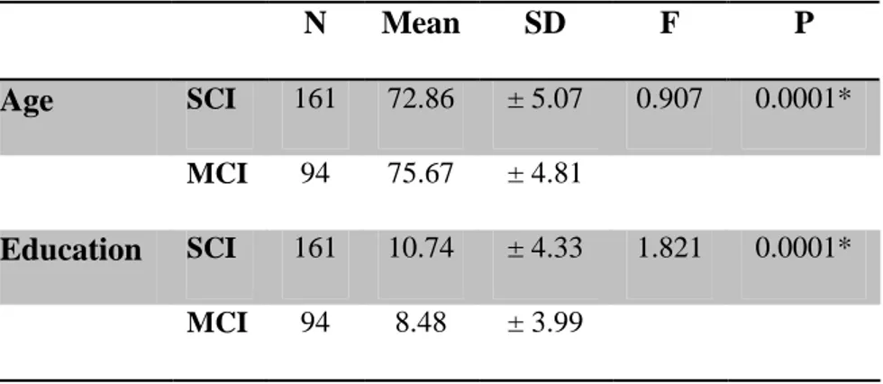
Regarding the socio-demographic data, Table 3 shows means, standard deviations and p-values for the variables age and education.
Table 3. Means, standard deviations and p-values for socio-demographic data performed with t-Student test. SD=standard deviation.
N Mean SD F P
Age SCI 161 72.86 ± 5.07 0.907 0.0001*
MCI 94 75.67 ± 4.81
Education SCI 161 10.74 ± 4.33 1.821 0.0001*
31 Psychobehavioral evaluation. From the comparison between SCI and MCI patients, a
significant difference was observed with regard to the extent of behavioral problems derived from the NPI scale (p<0.0001), everyday functional abilities expressed by the ADCS score (p<0.001), the CRI-total score and its sub-scores – CRI-school and CRI-work (table 4). The ANCOVA test showed that the difference between SCI and MCI subjects regarding CR was not mediated by the difference of age but was influenced by the different level of education (table 5).
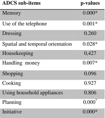
Comparing the sub-items of the ADCS scale, we found that patients with MCI exhibited a significant impairment regarding the following items: memory, use of the telephone, orientation, handling money, planning and initiative, compared to the SCI group (table 6). Regarding the NPI items, symptoms of mood disturbances, anxiety, apathy, irritability and sleep difficulties were significantly more present in the MCI than in the SCI group (table 7), while delusions, hallucinations, agitation/aggression, elation/euphoria, disinhibition were not found in any patient.
No statistically significant differences came to light for physical, recreational and social activities, even though the mean frequency of social and recreational activities was higher in the SCI group compared to the MCI.
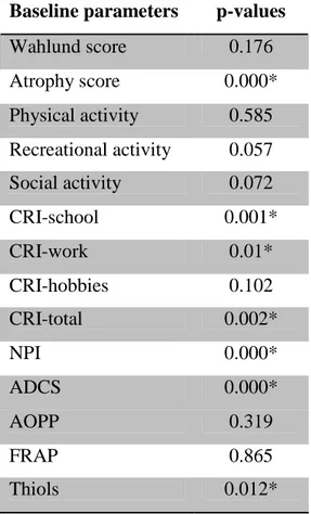
Neuroimaging. The difference between subjects with MCI and with SCI was remarkable with
regard to the degree of atrophy (p<0.0001), which was significantly more relevant in the former; patients with MCI also exhibited a higher mean degree of white matter abnormalities, even though the difference did not reach a statistical significance (Table 4).
32
Table 4. Comparison of the baseline variables between the SCI and MCI groups. Baseline parameters p-values
Wahlund score 0.176 Atrophy score 0.000* Physical activity 0.585 Recreational activity 0.057 Social activity 0.072 CRI-school 0.001* CRI-work 0.01* CRI-hobbies 0.102 CRI-total 0.002* NPI 0.000* ADCS 0.000* AOPP 0.319 FRAP 0.865 Thiols 0.012*
Table 5. Results from the ANCOVA test used to evaluate the influence of age and education on the difference in CRI-total values between the MCI and SCI groups. df= degree of freedom.
Covariate df F P
Age 1 1.258 0.265
Education 1 139.851 0.000*
Oxidative stress. Compared to subjects with SCI, patients with MCI had lower blood levels of
thiols (p<0.012) and FRAP (n.s.) and higher levels of AOPP (n.s.), even if these last two comparisons did not reach statistical significance (Table 4).
33
Table 6. Comparison of the ADCS scale sub-items between the SCI and MCI groups obtained with the Mann-Whitney U-test.
ADCS sub-items p-values
Memory 0.000*
Use of the telephone 0.001*
Dressing 0.260
Spatial and temporal orientation 0.028*
Housekeeping 0,427
Handling money 0.007*
Shopping 0.096
Cooking 0.927
Using household appliances 0.806
Planning 0.000*
Initiative 0.000*
Table 7. Results from the comparison between the SCI and MCI groups regarding neuropsychiatric symptoms, assessed with the Mann-Whitney U-test.
NPI items p-values
Depression/Dysphoria 0.006*
Anxiety 0.019*
Apathy/Indifference 0.000*
Irritability 0.037*
Aberrant motor behaviour 0.145
Sleep and Nighttime
Behavior Disorders
0.027*
Appetite and Eating
Disorders
34 3.1.2 Logistic regression
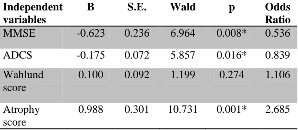
Gathering together all the principal parameters available, we have tried to highlight which factors are most predictive in discriminating between the diagnosis of SCI and MCI. For this purpose, direct logistic regression was performed to assess the impact of a number of factors on the likelihood that subjects would have a diagnosis of MCI rather than just a subjective memory complaint. The model containing the four independent variables MMSE, ADCS score, Wahlund score, atrophy score, as a whole, explained between 42.4% (Cox and Snell R square) and 58.8% (Negelkerke R squared) of variance in the diagnosis and correctly classified 77.1% of cases. As shown in table 8, only three of the independent variables made a unique statistically significant contribution to the model (MMSE, ADCS score and atrophy score). The strongest predictor of having a diagnosis of MCI was the atrophy score, recording an odds ratio of 2.685. This indicated that subjects complaining of memory difficulties who had a moderate-to-severe level of medial temporal atrophy were over 2 times more likely to have a diagnosis of MCI after a neuropsychological evaluation than those who had a normal temporal volume, controlling for all the factors in the model.
Table 8. Logistic regression predicting likelihood for a subject complaining of cognitive difficulties in reporting a diagnosis of MCI.
Independent variables
B S.E. Wald p Odds
Ratio MMSE -0.623 0.236 6.964 0.008* 0.536 ADCS -0.175 0.072 5.857 0.016* 0.839 Wahlund score 0.100 0.092 1.199 0.274 1.106 Atrophy score 0.988 0.301 10.731 0.001* 2.685
35 3.1.3 Correlations between variables
The relationships between the principal baseline variables were assessed using the Spearman's rank correlation coefficient, taking into account the total sample and then examining separately the MCI and the SCI group.
Considering the total sample:
Neuroimaging. As shown in table 9, the entity of white matter abnormalities positively
correlated with age (ρs = 0.424, p <0.001), atrophy (ρs = 0.354, p =0.001) and hypertension (ρs = 0.446, p =0.049), while negatively correlated with CRI-total (ρs = -0.221, p = 0.045), CRI-school (ρs = -0.233, p =0.034), CRI-work (ρs = -0.359, p =0.001).
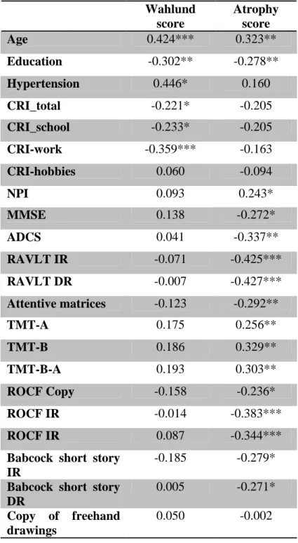
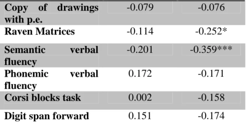
Temporal lobe and hippocampal atrophy correlated with age (ρs = 0.323, p = 0.002), education (ρs = -0.278, p = 0.010), NPI (ρs = 0.243, p = 0.029), MMSE (ρs = -0.272, p = 0.012), ADCS (ρs = -0.337, p = 0.002); considering the neuropsychological tests, a significant correlation was observed between atrophy and a low performance at RAVLT Immediate and Delayed recall (ρs = -0.425, p <0.001, ρs = -0.427, p <0.001, respectively), attentive matrices (ρs = -0.292, p = 0.007), TMT-A, TMT-B, TMT-B-A (ρs = 0.256, p =.019, ρs = 0.329, p =.003, ρs = 0.303, p = 0.007, respectively), ROCF Copy, Immediate and Delayed recall (ρs = -0.236, p = 0.030, ρs = -0.383, p <0.001, ρs = -.0344, p = 0.001, respectively), Babcock short story Immediate and Delayed recall (ρs = -0.279, p = 0.011, ρs = -0.271, p = 0.013, respectively), Raven Matrices (ρs = -0.252, p = 0.022) and semantic verbal fluency (ρs = -0.359, p = 0.001);
Cognitive Reserve and functional scales. As shown in table 10, there was a remarkable correlation between CRI-total and education (ρs = 0.804, p <0.001) Wahlund score (ρs =
-36
0.221, p = 0.045), physical and recreational activities (ρs = .0366, p <0.001, ρs = 0.232, p = 0.015, respectively) and the resort to mnemonic strategies (ρs = -0.273, p = 0.037); among the neuropsychological tests, there was a correlation with best performances at RAVLT Immediate recall (ρs = .228, p = 0.016), Babcock short story Immediate recall (ρs = 0.220, p = 0.021), attentional matrices (ρs = 0.237, p = 0.012), TMT-A, TMT-B, TMT-B-A (ρs = -0.232, p = 0.014, ρs = -0.348, p <0.001, ρs = -0.325, p = 0.001), ROCF-copy (ρs = 0.205, p = 0.030), copy of drawings (ρs = 0.193, p = 0.047) and semantic verbal fluency (ρs = 0.216, p = 0.024). Regarding CRI subscores: CRI-school was the one which behaved in the most similar way to CRI-total, with significant correlations with education (ρs = 0.852, p <0.001), Wahlund score (ρs = -.233, p = 0.034), physical and recreational activities (ρs = 0.232, p =.015, ρs = 0.240, p = 0.012, respectively), RAVLT Immediate and Delayed recall (ρs = 0.322, p =0.001, ρs = 0.230, p =.014), attentive matrices (ρs = .238, p =.012), TMT-A, TMT-B, TMT B-A (ρs = -0.315, p = 0.001, ρs = -0.364, p <0.001, ρs = -0.287, p = 0.003, respectively), Babcock short story Immediate recall (ρs = 0.231, p = 0.015), Raven matrices (ρs = 0.232, p = 0.014). CRI-work correlated with education (ρs = 0.646, p <0.001) and with best performance at TMT-B (ρs = -0.223, p = 0.023) and TMT B-A (ρs = -0.215, p = 0.028), while CRI-hobbies was linked to education (ρs = 0.318, p <0.001), current physical and social activities (ρs = 0.346, p <0.001, ρs = 0.270, p = 0.005, respectively) and good performances at attentive matrices (ρs = 0.241, p = 0.011), TMT B-A (ρs = -0.195, p = 0.048) and phonemic verbal fluency (ρs = 0.258, p = 0.006).
The use of mnemonic strategies was correlated with education (ρs = -0.242, p = 0.034) and CRI-total (see above). The behavioral symptoms (obtained from the NPI scale) were
37
associated to lower ADCS scores (ρs = -0.305, p = 0.001), lower MMSE (ρs = -0.294, p = 0.001) and fewer social activities (ρs = -0.205, p = 0.025).
Table 9. Correlation relationships between neuroimaging indexes, functional scales and neuropsychological tests in the total sample.
IR=Immediate recall; DR=Delayed recall; p.e.= programming elements. Wahlund score Atrophy score Age 0.424*** 0.323** Education -0.302** -0.278** Hypertension 0.446* 0.160 CRI_total -0.221* -0.205 CRI_school -0.233* -0.205 CRI-work -0.359*** -0.163 CRI-hobbies 0.060 -0.094 NPI 0.093 0.243* MMSE 0.138 -0.272* ADCS 0.041 -0.337** RAVLT IR -0.071 -0.425*** RAVLT DR -0.007 -0.427*** Attentive matrices -0.123 -0.292** TMT-A 0.175 0.256** TMT-B 0.186 0.329** TMT-B-A 0.193 0.303** ROCF Copy -0.158 -0.236* ROCF IR -0.014 -0.383*** ROCF IR 0.087 -0.344***
Babcock short story IR
-0.185 -0.279*
Babcock short story DR
0.005 -0.271*
Copy of freehand drawings
38 Copy of drawings with p.e. -0.079 -0.076 Raven Matrices -0.114 -0.252* Semantic verbal fluency -0.201 -0.359*** Phonemic verbal fluency 0.172 -0.171
Corsi blocks task 0.002 -0.158
Digit span forward 0.151 -0.174
Table 10. Correlation relationships between CRI indexes, functional scales and neuropsychological tests in the total sample.
CRI school CRI work
CRI hobbies CRI total
Education 0.852*** 0.646*** 0.318*** 0.804*** Physical activity 0.232* 0.172 0.346*** 0.366*** Recreational activities 0.240* 0.091 0.159 0.232* Social activities 0.082 0.013 0.270** 0.147 Mnemonic strategies -0.253 -0.170 -0.201 -0.273* NPI -0.104 -0.134 0.000 -0.117 MMSE 0.024 -0.044 -0.058 -0.042 ADCS 0.089 -0.051 0.098 0.065 RAVLT IR 0.322*** 0.172 0.084 0.228* RAVLT DR 0.230* 0.072 -0.009 0.111 Attentive matrices 0.238* 0.069 0.241* 0.237* TMT-A -0.315*** -0.159 -0.098 -0.232* TMT-B -0.364*** -0.223* -0.189 -0.348*** TMT-B-A -0.287** -0.215* -0.195* -0.325*** ROCF Copy 0.073 0.175 0.168 0.205* ROCF IR 0.093 0.010 0.009 0.038 ROCF DR 0.054 -0.021 0.023 0.005 Babcock short story IR 0.231* 0.132 0.136 0.220* Babcock short story DR 0.123 0.092 0.072 0.122
39 Copy of freehand drawings 0.150 0.135 0.133 0.193* Copy of drawings with p.e. 0.079 0.025 0.020 0.044 Raven Matrices 0.232* 0.003 0.031 0.116 Semantic verbal fluency 0.179 0.231* 0.056 0.216* Phonemic verbal fluency 0.182 -0.125 0.258** 0.117
Corsi blocks task -0.006 0.066 -0.083 0.002
Digit span
forward
0.110 0.059 0.100 0.128
Considering only the MCI sample:
Neuroimaging. Regarding the neuroimaging indexes, the entity of white matter changes was
correlated with age (ρs = 0.441, p = 0.001), temporal atrophy (ρs = 0.281, p = 0.038), Clock Drawing test (ρs = -0.317, p = 0.019), attentive matrices (ρs = -0.300, p = 0.026) TMT-B and TMT B-A (ρs = 0.294, p =. 0029, ρs = 0.281, p =. 0048, respectively). The atrophy score was not correlated with any variable, except for the Wahlund score (see above).
Cognitive Reserve and functional scales. A significant correlation persisted between
CRI-total and the attentional sphere assessed with TMT B and TMT B-A (ρs = -0.483, p <0.001,
ρs = -0.506, p <0.001, respectively), as with ROCF-copy (ρs = 0.271, p = 0.029) and physical and recreational activities (ρs = 0.248, p = 0.050, ρs = 0.258, p = 0.041, respectively) . A noteworthy correlation with the attentional domain (TMT B and TMT B-A) was observed also with the sub-indexes CRI-school (ρs = -0.395, p = 0.002, ρs = -0.356, p = 0.006) and CRI-work (ρs = -0.475, p <0.001, ρs = -0.517, p <0.001). CRI-work was also associated with better visuo-spatial performances at ROCF-copy (ρs = .0295, p = 0.017) and copy of drawings (ρs = 0.251, p = 0.044).
40
The use of mnemonic strategies was correlated with CRI-total (ρs = -0.273, p = 0.037). NPI score was linked to functional abilities expressed by ADCS (ρs = -0.255, p = 0.035);
Regarding the SCI group:
Neuroimaging. The entity of temporal atrophy was correlated with age (ρs = -0.609, p <0.001) and to higher white matter abnormalities (ρs = 0.418, p = 0.019). As for MCI, the severity of the white matter changes correlated only with age (ρs = -0.385, p = 0.032).
Cognitive Reserve and functional scales. Education was correlated to physical and
recreational activities (ρs = 0.390, p = 0.008, ρs = 0.402, p = 0.006, respectively) and with all CRI indexes (CRI-total: ρs = 0.774, p <0.001, CRI-school: ρs = 0.913, p<0.001, CRI-work: ρs = 0.680, p<0.001, CRI-hobbies: ρs = 0.314, p = 0.025). Both the CRI-total and all the sub-indexes correlated with both education and physical activity (CRI-total: ρs = 0.438, p = 0.002, CRI-school: ρs = 0.381, p = 0.009, CRI-work: ρs = 0.307, p = 0.038, CRI-hobbies:
ρs = 0.407, p = 0.005); CRI-school was also linked to recreational activities (ρs = 0.309, p = 0.036) and to better performances at RAVLT Immediate recall (ρs = 0.349, p = 0.016) and semantic verbal fluency (ρs = 0.323, p = 0.035).
The NPI score was negatively correlated with MMSE (ρs = -0.324, p = 0.028);
3.2 Training intervention
Thirty-five patients (18 F, 17 M; mean age 74.63 ± 4.29 years; mean years of education 9.29 ± 4.09) completed the physical and cognitive training program. All patients habitually
41
followed a dietary regimen of Mediterranean type. Thirteen subjects had at least one relative with cognitive decline in old age (even though no clear diagnosis was made). Hypertension was the most frequent co-morbidity factor, affecting 14 patients, followed by hypercholesterolemia (8 patients), diabetes (7), heart disease (mainly arrhythmias, 5) and previous thyroid disease (4). All diseases were treated with the appropriate therapies.
The control group included sixteen patients (7 M and 9 F, mean age 72.88 ± 3.48 years of education 8.06 ± 4.1). No significant differences were detected between the intervention and the control group regarding age, nor education (table 11).
Table 11. Means, standard deviations and p-values for socio-demographic data performed with Student t-test. SD=standard deviation.
N Mean SD P Age Training group 35 74.63 ± 4.29 0,159 Control group 16 72.88 ± 3.48 Education Training group 35 9.29 ± 4.09 0,327 Control group 16 8.06 ± 4.1 3.2.1 Baseline assessment
Neuropsychological evaluation. At T0, in the training group, 22 patients were amnestic-MCI
MCI), one was non-amnestic-MCI (na-MCI) and 12 were amnestic-multidomain MCI (a-md-MCI). The ADAS-cog mean total score was 14.01 ± 4.73. Regarding the control group, ten patients were a-MCI, two were na-MCI, three were a-md-MCI and one was na-md-MCI.
Neuroimaging: None of the patients had brain lesions compatible with the exclusion criteria
42
white matter abnormalities, while the mean atrophy score was 3.43 ± 1.56, with 17 patients exhibiting mild or no reduction of medial temporal lobe and hippocampal volume (table 12).
Cognitive Reserve and functional scales. Seventeen patients performed at least three hours of
physical activity per week, while the others were rather sedentary; the medium weekly frequency was 15.73 ± 11.75 hours spent on leisure activities and 6.20 ± 7.93 hours on social activities.
Regarding the CRIq, the mean CRI total score was 106.02 ± 16.71, with sub-scores of 102.15 ± 14.83 (CRI-school), 101.60 ± 17.11 (CRI-work) and 110.09 ± 21.84 (CRI-hobbies). NPI total mean score at T0 was 6.7 ± 5.61, with anxiety (18), irritability (17), depression (12) and apathy (10) being the most frequent type of symptoms. The mean ADCS total score was 45 ± 5.3.
3.2.2 T3 assessment
At T3, subjects performed the ADAS-cog, with a mean total score of 13.05 ± 5.74.
3.2.3 T7 assessments
Neuropsychological assessment: After the training program, 25 subjects maintained the
diagnosis of MCI, one patient evolved into dementia and 9 returned to a normal cognitive profile. The neuropsychological evaluation revealed that, of the MCI patients, 11 were a-MCI, 3 na-MCI and 11 a-md-MCI. The comparison of the neuropsychological assessment between T0 and T7 revealed a significant improvement at RAVLT Immediate and Delayed recall (p<0.05, p<0.01, respectively), Babcock short story Delayed recall (p<0.001) and phonemic verbal fluency (p<0.006).
43
We also investigated the progression of neuropsychological scores from T0 to T7 separating the trained patients into two groups on the basis of their CR level (low CR= CRI-total ≤100; high CR = CRI-total >100). Both groups confirmed the memory improvement; subjects with low CR were particularly improved in RAVLT Immediate and Delayed recall (p<0.05, p<0.02, respectively) and the high CR group exhibited the most significant improvement involving Babcock short story Delayed recall (p<0.002).
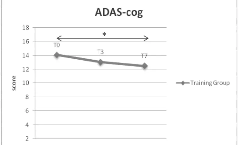
Regarding ADAS-cog, a statistically significant improvement was observed between T0 and T7 regarding total ADAS-cog score (p<0.03) (Figure 2) and the sub-score word recall (p<0.04). When we examined the results in the samples separated by CR, we observed that the improvement was more remarkable in the low-CR group compared to the high-CR sample, regarding both the total score (p<0.003 vs p<0.05) and the sub-score word recall (p<0.01 vs n.s.).
Functional scales. NPI: in the training group, total mean score at T0 was 6.7 ± 5.61, with
anxiety (18), irritability (17), depression (12) and apathy (10) being the most frequent type of symptoms. At T7, total score was 5.19 ± 3.89, with a reduction of the intensity of symptoms regarding anxiety, irritability, depression, apathy and sleep disturbances, even though none were statistically significant (Table 11). ADCS: no significant differences were observed at T7 regarding functional everyday abilities.
Anosognosia questionnaire: comparing patients’ and caregiver’s answers, unawareness has
been observed in 7 patients, while 6 subjects overestimated their difficulties. The “unaware” subjects had lower CRI scores (even though the difference did not reach statistical significance) and higher scores at mnemonic strategies usage questionnaire than the other individuals (p<0.03; Table 12; Figure 3). We also compared the effects of the training intervention on the neuropsychological profile of the two groups and we observed that
![Figure 1: Hypothetical model of dynamic biomarkers in the pathophysiological sequence of AD including the preclinical phase [Sperling 2011]](https://thumb-eu.123doks.com/thumbv2/123dokorg/7940491.119830/9.892.127.618.599.894/figure-hypothetical-biomarkers-pathophysiological-sequence-including-preclinical-sperling.webp)