GCN5-Dependent Acetylation of HIV-1
Integrase Enhances Viral Integration
Mariaelena Terreni
Ph.D Thesis in Molecular Biology
Supervisor: Dott.ssa Anna Cereseto
Scuola Normale Superiore
C
ONTENTS
AIM OF THE THESIS ...9
INTRODUCTION ... 13
HIV-1 and AIDS ... 15
The structure of HIV-1... 15
The morphologic structure of the viral particle... 15
The organization of the viral genome ... 16
The HIV-1 replication cycle: an overview... 17
HIV-1 integration... 22
IN structural overview ... 22
The mechanism of integration ... 25
Integration site selection by HIV-1... 26
HIV-1 integration and the host cell ... 31
Cellular cofactors of HIV-1 integration... 31
INI1... 31
BAF, Lap2α and Emerin ... 32
HMGA1 ... 33
LEDGF/p75... 33
TRN-SR2 ... 36
p300 ... 37
Post-translational modifications of IN ... 38
Restriction of infection at the integration step: KAP1 ... 40
IN inhibitors to treat HIV-1 infection... 40
Acetylation... 43
The diversity of HATs ... 43
HDACs... 44
Histone acetylation in the regulation of gene expression ... 45
Bromodomain structure and function ... 46
Acetylation of non-histone proteins ... 49
Acetylation of trancription factors... 50
Acetylation of HMG proteins ... 51
Acetylation of nuclear import factors ... 52
Acetylation of non-nuclear proteins: α-tubulin and cortactin acetylation ... 53
Acetylation of viral proteins ... 54
Acetylation of non-histone substrates and the protein modification code.... 57
GCN5... 58
Metazoan GCN5-containing complexes... 62
GCN5 and PCAF exhibit distinct, yet partially overlapping functions... 67
MATERIALS AND METHODS... 69
Plasmids... 71
Recombinant proteins production and proteolytic processing ... 72
In vitro acetylation assay... 73
In vitro binding assay... 74
In vitro integration assays ... 74
Cell culture and transfection ... 75
Immunoprecipitation and Western blotting ... 75
Antibodies ... 75
Virus and viral vector production ... 76
Transient and stable knockdown of GCN5 expression... 77
HIV-1 infectivity assays ... 77
RT-Q-PCR analysis... 78
Statystical Analysis... 79
RESULTS ... 81
HIV-1 IN is acetylated by GCN5 acetyltransferase ... 83
IN is acetylated by GCN5 in vitro: identification of the acetylation sites ... 83
IN is acetylated by GCN5 in vivo: detection of IN acetylation by a novel anti-acetylated IN antibody ... 86
IN interacts with GCN5... 89
IN interacts with GCN5 in vitro: mapping of the regions involved in the
interaction ... 89
Acetylation by GCN5 enhances IN catalytic activity in vitro ... 92
Production of constitutively acetylated recombinant IN by tethered catalysis. 92 In vitro integration assays with constitutively acetylated recombinant IN... 93
HIV-1 infectivity is reduced in GCN5 knockdown cells ... 94
Mutations at IN acetylation sites reduce HIV-1 integration efficiency and replication capacity ... 99
Reduced integration efficiency of HIV-1 viruses mutated at IN acetylation sites in single round infections... 99
HIV-1 clones mutated at IN acetylation sites exhibit delayed peaks of replication and reduced virus production in multiple round infections ... 100
DISCUSSION ... 103
Acetylation of the IN CTD by cellular HATs... 105
IN acetylated lysines are located within the HAT- interacting region... 106
Comparative analysis of GCN5 and p300 function in HIV-1 integration ... 109
The reduced integration efficiency of HIV-1 clones mutated at IN acetylated lysines is due to a lack of acetylation... 110
A recent debate: the role of IN acetylation during the replication cycle of HIV-1 ... 111
C
HAPTER
1
Integration of the reverse transcribed viral cDNA into the host cellular genome is an essential event during the replication cycle of HIV-1. This step is catalyzed by the viral integrase (IN) protein, which has been identified as a substrate for acetylation by the cellular histone acetyltransferase (HAT) p300 (Cereseto et al., 2005; Topper et al., 2007). The modified residues have been mapped to the IN C-terminal domain (CTD), corresponding to lysines 264, 266 and 273 (Cereseto et al., 2005; Topper et al., 2007). While IN acetylation by p300 was shown to increase both IN binding affinity to DNA and its strand transfer activity in vitro (Cereseto et al., 2005), conflicting results were obtained regarding the relevance of this post-translational modification (PTM) of IN during HIV-1 replication, with one study reporting a severe replication impairment for a mutant viral clone in which lysine-to-arginine substitutions were introduced at the IN sites targeted by p300-mediated acetylation (Cereseto et al., 2005), and a subsequent report claiming that the virus expressing the acetylation-defective mutant IN was able to replicate with kinetics similar to the wild type virus (Topper et al., 2007).
Based on these premises, the aim of the present thesis was that of investigating the role of IN acetylation by cellular HATs during HIV-1 integration and how this PTM influences the viral replication cycle. Since proteins modified by acetylation are often substrates for multiple HATs, we set out to explore whether IN might be acetylated by enzymes other than p300 and indeed found the existence of a physical and functional interaction between IN and the member of the GNAT family of HATs GCN5, resulting in the acetylation of four lysines in the IN CTD, at positions 258, 264, 266 and 273. Given that IN lysines 264, 266 and 273 proved to be common substrates of GCN5 and p300, we decided to perform a comparative analysis of the roles played by these enzymes during HIV-1 integration and replication. With the present study we obtained results supporting the notion that HIV-1 integration is modulated by an acetylation balance that controls the activity of the IN enzyme and involves at least two different cellular HATs, GCN5 and p300. Finally, the identification of a novel pathway of IN regulation unveils a new
mechanism that can be exploited for future therapeutic development.
C
HAPTER
2
HIV-1 and AIDS
The Human Immunodeficiency Virus (HIV) is a member of the genus Lentivirus in the Retroviridae family. Retroviruses are so called because, upon entry into the host cell, their RNA genome is reverse transcribed into a DNA copy that is stably inserted into the cellular chromosomal DNA. The integrated form of the viral DNA, the provirus, then serves as template for the formation of viral RNAs and proteins that constitute the progeny virions. The reverse flow of genetic information from RNA to DNA and the establishment of the viral DNA in an integrated form in the host cell genome are the defining hallmarks of retroviruses.
Since its initial description in 1983, HIV type 1 (HIV-1) was identified as the etiological agent of the acquired immunodeficiency syndrome (AIDS) (Barre-Sinoussi et al., 1983; Broder and Gallo, 1984; Gallo et al., 1984), a clinical condition characterized by profound immunosuppression with associated opportunistic infections and malignant tumors, wasting, and central nervous system (CNS) degeneration.
Even if the AIDS epidemic was first identified only in the 1980s, the degree of morbidity and mortality caused by HIV and the global impact of HIV infection on health care resources and economics are already enormous. Although HIV incidence has been steadily declining since the late 1990s, levels of new infections are still high, and, with significant reductions in mortality due to the scale up of antiretroviral therapy over the past few years, the number of people living with HIV worldwide has increased: UNAIDS estimated that there were about 33 million people living with HIV infection and AIDS worldwide at the end of 2009 (UNAIDS, 2010).
The structure of HIV-1
The morphologic structure of the viral particle
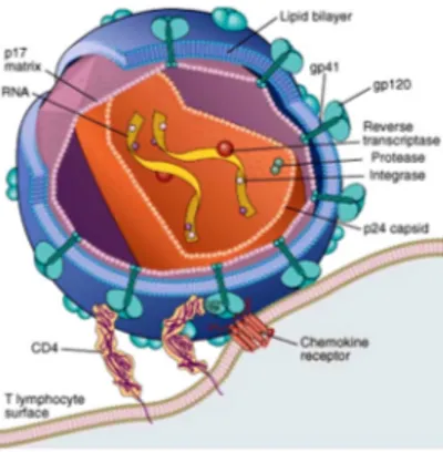
HIV-1 particles are around 120 nm in diameter and roughly spherical. The viral genome consists of two copies of a single stranded RNA about 9500 nucleotides (nt) long and is tightly associated with the nucleocapsid protein p7. This ribonucleoprotein complex is enclosed by a conical capsid made up of the viral protein p24. The capsid environment also contains other viral proteins, such as
reverse transcriptase and integrase. The capsid is surrounded by a layer of matrix protein, p17, which is in turn anchored to the inside of the viral lipoprotein membrane, the envelope. This is formed when the virus buds from the infected cell, taking some of the host plasma membrane with it. The major HIV-1 proteins associated with the envelope are gp120 and gp41. The gp41 transmembrane (TM) subunit traverses the envelope, while the gp120 surface (SU) subunit is present on the outer surface and is non-covalently linked to gp41, thus forming a complex that mediates the attachment to target cells. Also enclosed within the virion are the viral protease and the accessory proteins Vif, Vpr and Nef. The structure of a HIV-1 mature virion is schematically represented in Figure 1.
Figure 1. Schematic representation of a mature HIV-1 virion. (Abbas, 2010).
The organization of the viral genome
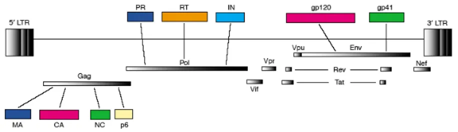
The HIV-1 genome is comprised of nine open reading frames (ORFs). The gag (group-specific antigen), pol (polymerase) and env (envelope) ORFs encode the classical structural and enzymatic precursor polyproteins, which are subsequently cleaved into the individual components common to all retroviruses (Figure 2). The four Gag proteins, p17 matrix (MA), p24 capsid (CA), p7 nucleocapsid (NC) and p6, together with the two Env proteins, gp120 and gp41, are the structural components that make up the core of the virion and the outer membrane envelope. The three pol-encoded proteins, protease (PR), reverse transcriptase (RT) and integrase (IN), provide essential enzymatic functions and are also included within the viral particle. In addition to the products of the gag, pol and env genes, HIV-1
genome encodes six additional proteins (Figure 2), often called accessory proteins: two expression enhancing factors, Tat (transactivator of transcription) and Rev (regulator of expression of the virion), as well as four proteins that modulate cell systems and functions for the advantage of the virus, Vif (viral infectivity factor), Vpu (viral protein U), Nef (negative factor), and Vpr (viral protein R).
Long terminal repeats (LTRs) at each end of the viral genome are generated during the process of reverse transcription and, therefore, exist only as repeats in the viral DNA. In the context of the provirus, the major function of the LTRs consists in the regulation of viral RNA synthesis. The 5’ LTR normally acts as the HIV-1 transcriptional promoter, whose function is to recruit the RNA polymerase II (RNA pol II) holoenzyme to the start site of viral RNA synthesis, while the 3' LTR acts in transcription termination and polyadenylation.
Figure 2. Schematic representation of the proteins encoded by HIV-1 proviral DNA. (Freed, 2004).
The HIV-1 replication cycle: an overview
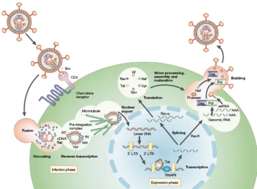
The replication cycle of HIV-1 is a complex multistep process that depends on both viral and host cell factors (Figure 3) (Fields et al., 2007). The first step in HIV-1 infection involves the interaction between the virion SU gp120 protein and the primary receptor CD4 expressed on T-lymphocytes, T-cell precursors within the bone-marrow and thymus, monocytes and macrophages, eosinophils, dendritic cells and microglial cells of the CNS (Wyatt and Sodroski, 1998). CD4 binding induces conformational changes in gp120, resulting in the exposure of the coreceptor-binding determinants (Sullivan et al., 1998). The two major coreceptors required for entry of HIV-1 are the chemokine receptor molecules CCR5 and CXCR4 (Alkhatib et al., 1996; Choe et al., 1996; Deng et al., 1996; Feng et al.,
1996), which are used by monocyte/macrophage (M)-tropic strains (R5 isolates) and T-cell (T)-tropic viruses (X4 isolates), respectively (Berger et al., 1998). The final step for viral entry requires the gp41-mediated fusion of the viral envelope components with the host cell membrane. The current model to explain membrane fusion assumes that, after the formation of a ternary gp120-CD4-coreceptor complex, conformational changes occurr in gp41 that lead to the exposure of its N-terminal part. This part, also known as the fusion-peptide, is then inserted into the target lipid bilayer (Eckert and Kim, 2001), triggering the fusion of the HIV-1 envelope with the host cell membrane and virus internalization.
Following entry of the HIV-1 core into the cytoplasm of the target cell, reverse transcription of the viral genomic RNA into a linear double-stranded cDNA copy takes place within a large macromolecular complex, known as the reverse transcription complex (RTC), which comprises proteins of both cellular and viral origin (Fassati and Goff, 2001; Karageorgos et al., 1993). The enzyme that catalyzes the reaction is RT. RT possesses three catalytic activities essential for replication of the virus: RNA-dependent DNA polymerase (i.e. reverse transcriptase), RNase H (i.e. for degradation of the genomic RNA in RNA/DNA hybrids during DNA synthesis) and DNA-dependent DNA polymerase activity (i.e. for synthesis of the second strand of the viral cDNA) (Fields et al., 2007).
The next step in the replication cycle is the translocation of the viral double-stranded cDNA into the nucleus as a component of a large nucleoprotein complex called the pre-integration complex (PIC), which is composed of both viral and cellular proteins (Bukrinsky et al., 1993b; Farnet and Haseltine, 1991; Miller et al., 1997). To cross the intact nuclear envelope and enter the nucleus, the PIC must be actively transported through the nuclear pore complex (NPC). Many attempts have been made to determine the viral and cellular factors mediating nuclear import of the HIV-1 PIC. Various viral karyophilic proteins, such as MA, Vpr and IN have been suggested to actively translocate the PIC into the host cell nucleus (Bukrinsky et al., 1993b; Depienne et al., 2000; Farnet and Haseltine, 1991; Haffar et al., 2000; Jenkins et al., 1998; Miller et al., 1997; Sherman and Greene, 2002; Zennou et al., 2000). The cellular protein lens epithelium-derived growth factor p75 (LEDGF/p75), along with a short section of triple-stranded DNA present within the
viral cDNA and known as the DNA flap, have also been implicated in promoting translocation of the PIC into the nuclei of infected cells (Cherepanov et al., 2003; Llano et al., 2004b; Zennou et al., 2000). In addition, CA recently emerged to play a key role in mediating HIV-1 nuclear import (Yamashita and Emerman, 2004) (Yamashita et al., 2007). With regard to cellular cofactors, several nuclear-import receptors, such as the importin α/β heterodimer (Ao et al., 2005; Armon-Omer et al., 2004; Gallay et al., 1997; Hearps and Jans, 2006; Levin et al., 2009), importin 7 (Ao et al., 2007; Zaitseva et al., 2009) and transportin-SR2 (TRN-SR2, TNPO3, transportin 3) (Brass et al., 2008; Christ et al., 2008; Konig et al., 2008), as well as several components of the nuclear pore complex, among which Nup 98, Nup153 and RanBP2 (Nup358) (Brass et al., 2008; Ebina et al., 2004; Konig et al., 2008; Woodward et al., 2009; Zhang et al.), were all proposed to be required for efficient translocation of the HIV-1 PIC into the host cell nucleus. However, at present, the exact mechanism governing nuclear import of the HIV-1 PIC still awaits further clarification.
Following the PIC arrival in the nucleus, the viral IN protein catalyzes the integration of HIV-1 cDNA into the host cellular genome. The integration reaction and the host factors participating in the process will be discussed in full detail in the next section.
The integrated viral DNA is then transcribed, thus generating full-length progeny viral RNA and a number of spliced mRNA transcripts to be translated in the cytoplasm (Coffin et al., 1997). Transcription of HIV-1 proviral DNA is broadly similar to host gene transcription and involves a large array of cellular transcriptional activator and repressor proteins, as well as the entire RNA Pol II machinery. An exception is represented by the enhancement of expression from the HIV-1 LTR promoter, which is mediated by the viral regulatory protein Tat. Tat is an atypical transcriptional activator, since it does not bind to DNA, but to a RNA stem-loop structure, the transactivation response region (TAR), which forms at the 5′-end of nascent HIV-1 transcripts (Berkhout et al., 1989; Dingwall et al., 1989). Through its interaction with TAR, Tat activates transcription from the HIV-1 LTR by at least two different molecular mechanisms. The first mechanism consists in the recruitment of the cellular cofactor P-TEFb (positive transcription elongation factor
b), composed of the subunits cyclin T1 and CDK9 kinase. Tat interaction with cyclin T1 (Wei et al., 1998) increases its binding affinity to the TAR element, thus resulting in the formation of a ternary Tat/P-TEFb/TAR complex, which brings the kinase activity of P-TEFb in the proximity of the HIV-1 promoter. The Cdk9 subunit of P-TEFb then phosphorylates the carboxyl-terminal domain (CTD) of the host cell RNA pol II, which stimulates the elongation process and thereby the overall transcriptional efficiency (Bieniasz et al., 1999; Chen et al., 1999; Fujinaga et al., 1998; Garber et al., 1998; Ivanov et al., 1999). The second mechanism of Tat-induced transcriptional activation involves the recruitment of chromatin-modifying complexes to the HIV-1 promoter (Benkirane et al., 1998; Col et al., 2001; Hottiger and Nabel, 1998; Marzio et al., 1998)((Agbottah et al., 2006; Mahmoudi et al., 2006), which results in an increased DNA accessibilty to transcription factors and activators, thus promoting transcription initiation and efficient elongation.
The nuclear export of unspliced or partially spliced HIV-1 messages to the cytoplasm requires the action of the viral regulatory protein Rev. In the absence of Rev function, incompletely spliced HIV-1 mRNAs, primarily encoding viral structural proteins, are retained in the nucleus by cellular mRNA-processing factors, known as splicing commitment factors (Chang and Sharp, 1989; Emerman et al., 1989; Malim et al., 1989). Although these commitment factors can prevent incompletely spliced mRNAs from accessing the canonical cellular mRNA-export pathway, they cannot prevent the nuclear export of HIV-1 mRNAs bound by Rev. Rev contains an RNA binding motif that directly interacts with a stem-loop secondary structure, termed the Rev responsive element (RRE), which is present in all incompletely spliced viral mRNAs. The RRE can accommodate the binding of at least 8 Rev molecules and, at a certain threshold concentration of Rev protein in the nucleus, functional Rev/RRE complexes are formed, which greatly stimulate the export of unspliced and singly spliced mRNAs to the cytoplasm. The nuclear export of Rev with its RNA cargo is mediated by the cellular nucleocytoplasmic transport factor Crm1 (chromosome region maintenance 1), which interacts with the Rev nuclear export signal (NES) sequence (Fornerod et al., 1997; Fukuda et al., 1997; Stade et al., 1997). Once in the cytoplasm, the complex disassembles (Gorlich and Mattaj, 1996; Nigg, 1997) and Rev is released along with its unspliced or partially spliced
HIV-1 RNA cargo, allowing the latter to be incorporated into progeny virions or translated into viral proteins. The nuclear import of Rev is then achieved through the direct interaction of its nuclear localization signal (NLS) motif with the cellular cofactor importin β (Henderson and Percipalle, 1997; Szebeni et al., 1997), which results in the translocation of the complex through the nuclear pore.
HIV-1 assembly takes place in lipid rafts, plasma membrane microdomains enriched in cholesterol and glycosphingolipid (Ono and Freed, 2005), and is directed by Gag. The MA domain of Gag appears to be responsible for both directing the association of the polyprotein with the lipid bilayer and determining the specificity of membrane targeting (Facke et al., 1993) (Freed et al., 1994; Ono et al., 2000), while the CA domain seems to play a key role in driving the viral particle assembly (Ehrlich et al., 1992; Gross et al., 1997; von Schwedler et al., 1998). The third major domain synthesized as part of the Gag precursor is NC, whose main function in assembly involves the encapsidation of full-length, unspliced HIV-1 genomic RNA into virions through specific interaction with a cis-acting target known as packaging signal or ψ-site (Berkowitz et al., 1996).
Virus particle production is completed upon budding of the nascent virion from the plasma membrane (Adamson and Freed, 2007). To allow virus release from the infected cell, the p6 domain of Gag uses its highly conserved Pro-Thr-Ala-Pro (PTAP) motif (Huang et al., 1995), also known as “late domain” (since point mutations in this motif block the virus release at a late stage), to recruit components of the cellular endosomal sorting machinery, which normally function to promote the budding of vesicles into late endosome to form multivesicular bodies (MVBs) (Bieniasz, 2009; Demirov and Freed, 2004; Fujii et al., 2007; Morita and Sundquist, 2004). Concomitant with virus release, PR cleaves the Gag and Gag-Pol precursors into their respective protein domains (Coffin et al., 1997), leading to virion maturation, a reassembly event that results in the formation of the characteristic cone-shaped core and is essential for the acquisition of infectivity (Adamson and Freed, 2007; Ganser-Pornillos et al., 2008). Only after maturation is the virus ready for another round of infection.
Figure 3. Schematic representation of the HIV-1 replication cycle. (Peterlin and Trono, 2003).
HIV-1 integration
A distinguishing feature of retroviral replication is the insertion of a DNA copy of the genomic RNA into a chromosome of the host cell following reverse transcription. The provirus then serves as the template for the synthesis of viral RNAs and is maintained as part of the host cell genome for the lifetime of the infected cell. Retroviral mutants deficient in the ability to integrate generally fail to establish a productive infection.
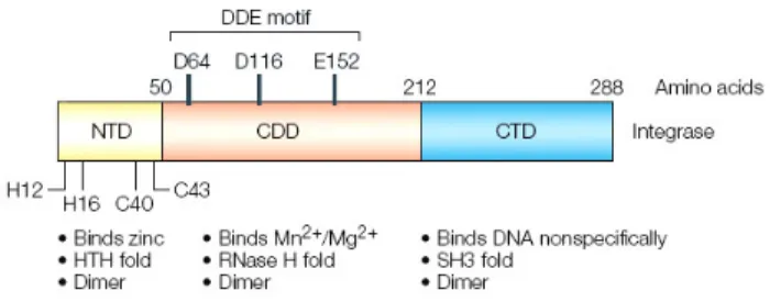
The integration reaction is catalyzed by the viral IN enzyme, a 32 kDa protein of 288 aminoacids generated by PR-mediated cleavage of the C-terminal portion of the HIV-1 Gag-Pol polyprotein.
IN structural overview
HIV-1 IN is made up of three structurally and functionally distinct domains: the N-terminal domain (NTD) spanning residues 1 to 50, the protease-resistant central catalytic core domain (CCD) from amino acids 50 to 212, and the C-terminal
domain (CTD) from residues 212 to 288 (Chiu and Davies, 2004; Jaskolski et al., 2009) (Figure 4). Among retroviral INs, the NTD and CCD share regions with a high degree of sequence similarity, while the CTD is much less conserved.
Figure 4. HIV-1 IN domain organization. Adapted from (Pommier et al., 2005).
It is not yet possible to crystallize the entire 288-aminoacid HIV-1 IN protein because of its low solubility and propensity to aggregate. The structure of all three domains, however, has been solved independently by x-ray crystallography or nuclear magnetic resonance (NMR) methods. In addition, structures are available for HIV- 1 IN fragments containing the NTD plus CCD (Wang et al., 2001), or the CCD plus CTD (Chen et al., 2000).
The NTD contains a conserved zinc-binding motif HHCC (His-12, His-16, Cys-40 and Cys-43) that coordinates one zinc atom (Zheng et al., 1996), though the structure of this region does not resemble that of zinc finger domains involved in DNA-binding (Lodi et al., 1995). Consistently, one known function of the HIV-1 IN NTD is to contribute to protein multimerization.
The CCD contains the catalytic DDE motif, which is conserved among all retroviral INs and consists of the active site residues Asp-64, Asp-116 and Glu-152 (Engelman and Craigie, 1992; Kulkosky et al., 1992; Rowland and Dyke, 1990). Mutation of any one of these three residues is sufficient to inactivate IN (Engelman and Craigie, 1992; Leavitt et al., 1993; van Gent et al., 1992). Crystal structures of the HIV-1 IN CCD were only obtained after extensive mutagenesis studies, which identified mutants with enhanced solubility (Bujacz et al., 1996; Dyda et al., 1994; Goldgur et al., 1999; Jenkins et al., 1995). These structures show that the HIV-1 IN CCD binds one Mg2+ ion between Asp-64 and Asp-116. Since Avian Sarcoma Virus (ASV) IN binds an additional Zn2+ or Cd2+ ion between Asp-64 and Glu-157
(the ortholog of Glu-152) (Bujacz et al., 1997), it is likely that the HIV-1 IN active site also binds two metal ions (Mg2+ or Mn2+) when complexed with the ends of the viral DNA during the cleavage and joining reactions. Another structural feature of the CCD is the 10-aminoacid flexible loop encompassed between residues Gly-140 and Gly-149. These two residues potentially act as hinges for the overall movement of the loop that may serve as a clamp for the binding of the viral DNA ends to the catalytic site of IN. Consistent with this possibility, Gln-148, one of the flexible loop residues, has been shown to selectively bind to the penultimate cytosine at the 5’-end of the viral DNA (Johnson et al., 2006). Gln-148 is also a key residue for IN catalytic activity and resistance to IN inhibitors raltegravir and elvitegravir (Marinello et al., 2008).
The CTD is considerably less conserved among retroviral INs than are the other domains of the protein. The structure of the isolated CTD of HIV-1 IN was solved using NMR (Eijkelenboom et al., 1995; Lodi et al., 1995) and showed a folding topology that is reminiscent of Src homology 3 (SH3) domains found in many proteins that interact either with other proteins or with nucleic acids (Musacchio et al., 1992; Yu et al., 1992), although no sequence similarity to SH3 proteins could be detected. The HIV-1 IN CTD possesses non-specific DNA binding activity and seems to be important for the binding of the viral DNA ends, thus conferring to IN the ability to remain associated with the viral genome during reverse trancription (Esposito and Craigie, 1998; Heuer and Brown, 1997; Heuer and Brown, 1998; Jenkins et al., 1997).
Direct physical measurements of purified IN, as well as cross-linking experiments and in vitro complementation between defective variants of IN, provided compelling evidence that retroviral IN is a multimeric enzyme (Ellison et al., 1995; Engelman et al., 1993; Grandgenett et al., 1978; Jones et al., 1992; van Gent et al., 1993). While each of the HIV-1 IN domains forms dimers, IN appears to function as a tetramer (Cherepanov et al., 2003; Li et al., 2006; Wang et al., 2001). Indeed, a tetramer would be the minimal IN multimer to provide a pair of active sites with the expected spacing for the concerted integration of two viral DNA ends, and the two-domain NTD-CCD construct has been shown to crystallize in a tetrameric form, best described as a dimer-of-dimers (Hare et al., 2009; Wang et al., 2001). The
crystal structure of full-length IN from the prototype foamy virus (PFV) in complex with its cognate viral DNA also showed that the organization of the minimal functional complex involving viral DNA and IN, the so-called intasome, comprises an IN tetramer tightly associated with a pair of viral DNA ends (Hare et al.).
The mechanism of integration
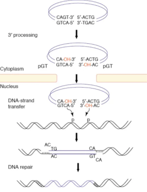
The integration reaction biochemically occurs in two steps (Figure 5) following reverse transcription and binding of IN to the viral DNA ends in the context of the PIC (Vandegraaff and Engelman, 2007). Assembly of the PIC onto the 5’-GCAGT-3’ sequence of the viral DNA ends forms a complex competent for integration, although, in vitro, only IN and DNA are required. The first step, named 3’-end processing, is an endonucleolytic cleavage, which results in the removal of two nucleotides from the 3’-ends of each viral DNA strand immediately 3’ from the conserved CA dinucleotide (5’-GCAGT-3’); a 5’-GT-3’ dinucleotide is generally released, resulting in recessed 3’-OH ends at each terminus of the viral DNA. The phosphodiester bond in the viral DNA strand is hydrolyzed by water as the nucleophile in the presence of Mg2+ (the presumably physiological metal) or Mn2+ (Esposito and Craigie, 1998). Following nuclear translocation of the PIC, IN catalyzes the second transesterification reaction, known under the name of strand transfer, where the recessed 3’-OH ends of the viral DNA act as nucleophiles to attack the DNA phosphodiester backbone of a host chromosome. HIV-1 IN positions the two viral ends to attack both strands of the host acceptor DNA, with a five base pair (bp) gap on each strand. The result is a staggered insertion that is subsequently sealed by host DNA repair enzymes. The HIV-1 provirus is flanked by a 5 bp direct repeat and terminates with the dinucleotides 5′-TG and CA-3′. The direct repeat results from the duplication of the cellular target sequence.
In addition to the linear, double-stranded viral cDNA that serves as the substrate for integration, two types of extrachromosomal viral DNA molecules are observed in the nucleus of HIV-1 acutely infected cells. These are circular DNA forms: 1-LTR circles, generated through homologous DNA recombination, and 2-LTR circles, produced by the nonhomologous end-joining pathway (Brown et al., 1989; Bukrinsky et al., 1993a). Although 1-LTR and 2-LTR circles are not substrates for
integration, they provide useful markers for nuclear import of the viral DNA, since their formation requires the enzymatic machinery present in the nucleus.
Our understanding of the chemistry of the retroviral integration reaction, as well as the screening of HIV-1 IN inhibitors, have been greatly assisted by the development of in vitro integration assays, where purified recombinant IN carries out 3’-end processing and strand transfer reactions in the presence of short synthetic oligonucleotides that mimic the viral DNA ends and a divalent metal ion (Mg2+ or Mn2+) (Craigie et al., 1990; Debyser et al., 2001; Katz et al., 1990; Marchand et al., 2001).
Figure 5. Outline of the integration reaction. (Van Maele et al., 2006).
Integration site selection by HIV-1
To replicate, a retrovirus must integrate a DNA copy of its genomic RNA into a chromosome of the host cell. The selection of cellular integration acceptor sites is crucial for both the retrovirus and the host (Bushman, 2001; Coffin et al., 1997). For the virus, selection of favorable integration target sites is required for efficient viral gene expression (Bisgrove et al., 2005; Jordan et al., 2003; Lewinski et al., 2005). For the host, integration can cause adverse events such as activation of
proto-oncogenes or inactivation of required cellular genes. Indeed, insertional activation of oncogenes has been observed in a human gene therapy trial, in which integration of a therapeutic retroviral vector near the LMO2 proto-oncogene contributed to malignant transformation in several patients (Hacein-Bey-Abina et al., 2003a; Hacein-Bey-Abina et al., 2003b). These adverse events have focused intense interest on the mechanisms mediating retroviral integration site selection. The cellular DNA sequences hosting integration events show modest similarity to one another, indicating that retroviral DNA integration is not tightly sequence-specific (Berry et al., 2006; Carteau et al., 1998; Holman and Coffin, 2005; Stevens and Griffith, 1996; Wu et al., 2005).
Studies using in vitro integration assays allowed the identification of factors influencing integration site selection in simplified models. DNA binding proteins bound to the target DNA block integration by obstructing the access of PICs to the target DNA (Bushman, 1994; Pryciak and Varmus, 1992). In contrast, DNA bending proteins, such as nucleosomes, actually promote integration (Pruss et al., 1994a; Pruss et al., 1994b; Pryciak et al., 1992; Pryciak and Varmus, 1992). On the nucleosome, the positions of maximal DNA distortion are particularly favored for integration (Pruss et al., 1994a; Pruss et al., 1994b), probably because IN distorts its DNA substrates during the reaction cycle (Bushman and Craigie, 1992; Katz et al., 2001; Katz et al., 1998; Scottoline et al., 1997). Consistent with this notion, prior distortion of the integration target DNA promotes the integration reaction. Thus, wrapping of DNA around nucleosomes alone does not inhibit integration (Bushman and Craigie, 1992; Scottoline et al., 1997). However, it should be noted that the nucleosomal templates used in the above-mentioned experiments were all presumed to be in the 10 nm “beads on a string” conformation. The consequences on integration deriving from the incorporation of such structures into the 30 nm chromatin fibers, or the still higher-order structures that comprise the chromosomes, are unknown.
Several groups have investigated HIV-1 integration targeting in vivo by sequencing the junctions between viral and human DNA and analysing their positions in the human genome. Following large-scale sequence analysis, the position of integration sites in the human genome was compared with that of other annotated
features. In the first such study, the distribution of HIV-1 integration sites in the chromosomes of a human lymphoid cell line, SupT1, was investigated (Schroder et al., 2002). This study revealed that genes are favoured targets for HIV-1 integration, and subsequent experiments performed in other cell types reached the same conclusion (Mitchell et al., 2004; Wu et al., 2003). Further studies investigated whether any preferences exist in the location of HIV-1 integration sites along transcription units (TUs) (Mitchell et al., 2004; Schroder et al., 2002; Wu et al., 2003), but no biases have been found. Evidently, the positive influence of TUs on HIV-1 integration extends across their entire length. Gene-rich regions in the human genome are depleted in certain classes of repeat DNA sequences (such as human endogenous retroviruses, HERVs, and long interspersed nuclear elements, LINEs) and enriched in others (such as Alu elements). Therefore, a bias in the rates of integration in these different classes of DNA repeats was expected. Indeed, HIV-1 integration appears to be disfavoured in HERV elements, consistently with favoured HIV-1 integration in genes (Schroder et al., 2002). In addition, HIV-1 integration is strongly disfavoured in alphoid repeats (which, in humans, are composed of α-satellite DNA). This indicates that centromeric heterochromatin, the location of most of the 〈-satellite-containing DNA, is an unfavourable target for HIV-1 integration (Carteau et al., 1998; Schroder et al., 2002). Since centromeric heterochromatin is known to be tightly wrapped by specific DNA-binding proteins, it appears that the packing of DNA into centromeric heterochromatin renders it less accessible, consequently disfavouring integration. Transcriptional profiling analysis has been carried out in some of the cell types studied as integration targets, allowing the influence of transcriptional activity on integration site selection to be assessed. Some of these transcriptional profiling studies were carried out on retrovirus-infected cells (Lewinski et al., 2005; Mitchell et al., 2004; Schroder et al., 2002), so that the results reflected the influence of infection on cellular gene activity (Corbeil et al., 2001; Mitchell et al., 2003; Schroder et al., 2002; van 't Wout et al., 2003). Analysis of the microarray data revealed that the median expression level of genes hosting HIV-1 integration events is consistently higher than that of all the genes assayed on the microarray. Transcriptional profiling studies have also been performed for integration of a
HIV-based vector in SupT1 cells, demonstrating that genes activated upon infection are favoured integration targets (Schroder et al., 2002).
Since gene-rich regions in the human genome correlate with high densities of CpG islands, an analysis of HIV-1 integration frequency near CpG islands was performed. CpG islands are chromosomal regions particularly enriched in the rare CpG dinucleotide, and usually correspond to gene-regulatory elements that contain clustered binding sites for transcription factors. The regions surrounding CpG islands appear to be disfavoured for integration by HIV-1 (Mitchell et al., 2004). Therefore, in the case of HIV-1, gene-dense regions seem to contain interleaved clusters of active genes favouring integration and unfavourable regions that include CpG islands. The mechanism by which CpG islands obstruct HIV-1 integration is unclear: there might be specific proteins bound at these sites that block integration, CpG islands might be located in a nuclear compartment resulting unfavourable for integration, or some other as-yet-unknown mechanism might be responsible. What mechanisms direct retroviral integration target-site selection in the human genome? It has been proposed that retroviral integration is favoured in decondensed chromatin, which would be more accessible to the integration apparatus (Panet and Cedar, 1977). This notion is supported by genome-wide studies, as integration in TUs is favoured in all the data sets for retroviruses and other integrating elements (Lewinski et al., 2005; Mitchell et al., 2004; Nakai et al., 2003; Schroder et al., 2002; Yant et al., 2005). By contrast, integration in highly condensed centromeric heterochromatin is disfavoured (Carteau et al., 1998; Schroder et al., 2002). However, since the integration target preferences of HIV-1, MLV and ASV are so different, it seems unlikely that the accessibility of DNA is the only mechanism determining target-site selection.
Studies on the integration site selection by Ty retrotransponsons in yeast (Boeke and Devine, 1998; Bushman, 2003; Sandmeyer, 2003; Zhu et al., 2003), showing that Ty integration complexes are tethered to their favoured sites in the host genome by interactions with specific cellular proteins, led to the hypothesis that such a tethering mechanism might also operate for retroviruses (Bushman, 2003). Several studies tested in vitro the feasibility of directing integration into specific DNA sites by the use of fusion proteins consisting of HIV-1 IN and a
sequence-specific DNA-binding protein, such as the phage λ repressor (Bushman, 1994), the
Escherichia coli LexA repressor (Goulaouic and Chow, 1996; Katz et al., 1996), the
murine transcription factor Zif268 (Bushman and Miller, 1997), or the synthetic zinc finger protein E2C (Tan et al., 2004). The resulting chimeras proved able to direct integration by recognizing and binding to their cognate target sites on the DNA, causing integration to be mediated into the immediately adjacent regions. Subsequent work also demonstrated that the IN-E2C fusion protein, packaged in
trans into virions, was capable of directing integration of retroviral DNA into a
predetermined chromosomal region in cultured cells (Tan et al., 2006). All these results provided proof of principle that tethering of retroviral IN proteins at target sites can promote nearby integration. For HIV-1 PICs, any viral or cellular protein present in the complex could theoretically act as binding partner in a tethering interaction. Indeed, a number of studies performed in the past few years led to the notion that the target site preferences of HIV-1 integration are in part due to tethering by the host chromatin-binding protein Ledgf/p75, which interacts with HIV-1 IN (Cherepanov et al., 2003; Emiliani et al., 2005) and mediates its binding to chromatin (Llano et al., 2004b; Maertens et al., 2003). In the absence of Ledgf/p75, HIV-1 integration is severely compromised and preferential integration in TUs is diminished (Ciuffi et al., 2005; Marshall et al., 2007; Shun et al., 2007b). Recently, the tethering model for Ledgf/p75 function was strengthened by the finding that fusion proteins containing the IN-binding domain of Ledgf/p75 fused to alternative chromatin binding domains are able to efficiently retarget integration (Ferris et al., 2010; Gijsbers et al., 2010; Silvers et al., 2010). The role of LEDGF/p75 in HIV-1 integration site selection will be furtherly discussed in the following section.
Notably, the existence of a link between nuclear entry of the HIV-1 PIC and integration targeting was recently hypothesized, based on the finding that the cellular proteins TRN-SR2 (TNPO3, transportin 3) and RanBP2 (Nup358), which are components of the nuclear import machinery and of the nuclear pore, respectively, affect the distribution of HIV-1 integration sites in the host chromosomes by regulating the nuclear import of the PIC (Ocwieja et al., 2011). According to the proposed model, nuclear translocation through the pore would first place the PIC in regions of high gene density, and Ledgf/p75 would then tether
the PIC for integration to provide the final distribution in active TUs (Ocwieja et al., 2011).
HIV-1 integration and the host cell
Although purified recombinant IN is necessary and sufficient to carry out 3’-end processing and strand transfer reactions in vitro, numerous studies have demonstrated that the establishment of the integrated provirus in the infected cell also involves a variety of host proteins that can play a role during the different steps of the integration process, including nuclear import, IN catalysis, integration site selection and DNA gap repair.
Cellular cofactors of HIV-1 integration
INI1Integrase Interactor 1 (INI1) is part of the mammalian SWI/SNF complex, which is implicated in ATP-dependent chromatin remodeling during transcriptional activation (Wang et al., 1996); it was first discovered as a binding partner for HIV-1 IN using the yeast two hybrid system (Kalpana et al., 1994). The interaction between the two proteins is HIV-1 specific (Yung et al., 2004), and INI1 was shown to be incorporated into virions (Yung et al., 2001) and to be a component of the reverse transcription and pre-integration complexes in the early steps of the viral replication cycle (Iordanskiy et al., 2006; Turelli et al., 2001). Furthermore, INI1 was found to stimulate the DNA strand transfer catalytic activity of IN in vitro (Kalpana et al., 1994).
There have been conflicting results regarding the role played by INI1 during HIV-1 infection and its necessity. One study found no effects on viral integration in cells depleted for INI1 (Boese et al., 2004), whereas other reports suggested that INI1 might participate in an anti-viral cellular response by interfering with the early steps of HIV-1 replication (Maroun et al., 2006; Turelli et al., 2001). Contrastingly, another work revealed that INI1 indirectly enhances proviral transcription (Ariumi et al., 2006), thus providing evidence of an infectivity-promoting effect of the protein. An ectopically expressed dominant negative mutant of INI1, termed S6, containing the minimal IN-interaction domain (amino acids 183-294), was found to potently inhibit HIV-1 assembly and particle production (Yung et al., 2001). S6 also proved
to be an efficient protective agent against HIV-1 infection when stably expressed before viral challenge. The inhibitory effects of S6 required the interaction with IN in the context of the Gag-Pol polyprotein (Yung et al., 2001). INI1 was also shown to be essential for the production of infectious HIV-1 virions (Sorin et al., 2006; Yung et al., 2001). In fact, viral particles produced in the absence of INI1 exhibited a drastic reduction in viral DNA synthesis, suggesting that the protein may be critical for the early events in reverse transcription (Sorin et al., 2006).
In conclusion, at present, INI1 appears to play dual roles in HIV-1 replication in that, while INI1 present in the producer cells seems to be required for HIV-1 replication, INI1 present in the target cells may be inhibitory. One possibility is that cellular INI1 may play a different role than the virally incorporated INI1, explaining the observed effects on HIV-1 replication.
BAF, Lap2α and Emerin
Barrier-to-Autointegration Factor (BAF) is a cellular component of HIV-1 PICs, which blocks suicidal autointegration and stimulates intermolecular integration activity in vitro (Chen and Engelman, 1998) (Lin and Engelman, 2003) (Lee and Craigie, 1998). This 89-aminoacid protein is highly conserved among multicellular eukaryotes and interacts with LEM domain proteins (Lin et al., 2000), which are components of the nuclear lamina (Foisner, 2001). It has been suggested that BAF mediates the association of the LEM proteins Lamina-associated polypeptide 2 α (LAP2α) and emerin to HIV-1 PICS, and that these two proteins, in their turn, target the PICs to chromatin, thus promoting integration (Jacque and Stevenson, 2006). Depletion of emerin from human cells impaired infection by HIV-1 expressing wild-type Env or the G glycoprotein from vesicular stomatitis virus (VSV-G), whereas depletion of LAP2α reduced infection by HIV-1 harboring wild-type Env, but not by HIV-1 pseudotyped with VSV-G (Jacque and Stevenson, 2006). Emerin, in particular, appeared to be critical for HIV-1 infection of primary macrophages. However, the requirement of LAP2α and emerin for HIV-1 replication has been questioned. In fact, subsequent reports demonstrated that cells from emerin knockout, LAP2α knockout, or emerin and LAP2α double knockout mice supported the same level of HIV-1 infectivity as cells from wild-type littermate mice, indicating
that these proteins do not function as universally important regulators of the PIC trafficking to chromatin for integration (Shun et al., 2007a) (Mulky et al., 2008). Thus, the roles played by emerin and LAP2α in HIV-1 pre-integration trafficking and integration still await further clarification.
HMGA1
High mobility group chromosomal protein A1 (HMGA1), a non-histone DNA binding protein that can modulate transcriptional regulation and chromatin structure (Farnet and Bushman, 1997), was proposed as a cofactor of HIV-1 integration due to its ability to restore the in vitro integration activity of salt-stripped PICs (Farnet and Bushman, 1997). Mechanistic studies pointed to a possible role of the DNA binding activity of HMGA1 in approximating the two LTR ends of the viral DNA molecule and facilitating IN binding by unwinding the LTR termini (Hindmarsh et al., 1999; Li et al., 2000). However, in coimmunoprecipitation experiments, no interaction between HMGA1 and HIV-1 IN could be detected (Hindmarsh et al., 1999). Consistently, chicken cells lacking HMGA1 are not deficient for retroviral integration, suggesting that HMGA1 is not absolutely required for integration (Beitzel and Bushman, 2003). In contrast, evidence was provided pointing to an important role of HMGA1 during transcription (Henderson et al., 2000; Henderson et al., 2004) and splice site regulation of HIV-1 (Tsuruno et al., 2011).
LEDGF/p75
Lens epithelium-derived growth factor (LEDGF/p75) was first identified as an interaction partner of HIV-1 IN by coimmunoprecipitation experiments in human cells expressing IN from a synthetic gene (Cherepanov et al., 2003). Independent screens exploiting the same method (Turlure et al., 2004) and subsequent use of the yeast two-hybrid assay (Emiliani et al., 2005) confirmed this result.
LEDGF/p75 is a ubiquitously expressed chromatin-associated protein that is thought to normally function in transcriptional regulation, stress response and apoptosis pathways (Ganapathy et al., 2003).
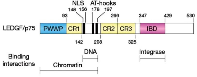
LEDGF/p75 is a multidomain protein (Figure 6), which not only comprises an IN binding domain (IBD) situated C-terminally (Cherepanov et al., 2004; Vanegas et al., 2005), but also several motifs involved in DNA and chromatin association
(Llano et al., 2006b). These include a NLS, two AT-hook elements (so named for their preferential binding to AT-rich DNA), and a PWWP motif that mediates chromatin binding (Llano et al., 2006b; Turlure et al., 2006).
Figure 6. LEDGF/p75 domain organization. (Vandegraaff and Engelman, 2007).
Upon ectopic expression, HIV-1 IN localizes to the nucleus and tightly binds to mitotic chromosomes (Cherepanov et al., 2000; Devroe et al., 2003), properties that can be attributed to its interaction with endogenous LEDGF/p75. In fact, RNAi-mediated knockdown of LEDGF/p75 results in the disruption of IN nuclear localization, redistributing the vast majority of the protein to the cytoplasm (Maertens et al., 2003) (Llano et al., 2004b). Moreover, a mutant IN form, with reduced binding affinity to LEDGF/p75 in vitro, fails to interact with chromosomes (Emiliani et al., 2005). Based on these results, a possible role for LEDGF/p75 in chromosomal tethering of HIV-1 PICs has been proposed.
Initial studies on the effect of LEDGF/p75 depletion on HIV-1 infectivity produced conflicting and inconsistent results (Llano et al., 2004b) (Vandegraaff et al., 2006) (Vandekerckhove et al., 2006). However, more complete knockdowns, which were able to eradicate detectable LEDGF/p75 from chromatin, or the use of mouse embryo fibroblasts (MEFs) derived from LEDGF/p75 knockout mice, revealed a significant loss in HIV-1 infectivity in the absence of this cellular factor (Llano et al., 2006a; Shun et al., 2007b). Consistently, overexpression of LEDGF/p75 IBD, which competes with the endogenous protein for the interaction with IN but is unable to interact with chromatin, imposes a severe defect in HIV-1 infectivity (De Rijck et al., 2006). The defect imposed by both LEDGF/p75 depletion and LEDGF/p75 IBD overexpression specifically occurs at the step of integration (Llano et al., 2006a)
(De Rijck et al., 2006). Taken together, these results highlighted an essential role for the interaction between IN and LEDGF/p75 during the HIV-1 replication cycle, suggesting that this interaction might function by docking the PICs to chromatin. Given its proposed role in the tethering of HIV-1 PICs to chromatin, the hypothesis was made that LEDGF/p75 could potentially function in integration target site selection. Indeed, analysis of the integration site distribution in human cells depleted of LEDGF/p75 by RNAi or in MEFs derived from LEDGF/p75 knockout mice revealed that the residual integration, which can still be found in these cells, is reduced in TUs (Ciuffi et al., 2005; Marshall et al., 2007; Shun et al., 2007b). Furthermore, analysis of the GC content of HIV-1 integration sites in LEDGF/p75-expressing cells indicated that AT-rich regions are favored for integration than GC-rich regions, consistently with the presence of an AT-hook motif in LEDGF/p75. Thus, LEDGF/p75 binding to AT-rich sites appears to direct integration into these sequences. Another approach to investigate the role of LEDGF/p75 in HIV-1 integration site selection examined the frequency of integration events in LEDGF/p75-modulated genes. Since LEDGF/p75 modulates gene expression by binding to DNA, LEDGF/p75-regulated genes should have more bound LEDGF/p75 than randomly selected genes, and thus be preferential integration targets. As expected, LEDGF/p75-modulated genes proved to be favored targets in cells expressing LEDGF/p75, while this preference was abolished in LEDGF/p75 depleted cells (Ciuffi et al., 2005). These data collectively suggested a role for LEDGF/p75 in promoting HIV-1 DNA integration in TUs and AT-rich regions, thus providing the first example of an apparent tethering protein for retroviral integration. Recently, the tethering model for Ledgf/p75 function was strengthened by the finding that expression of fusion proteins containing the IBD of Ledgf/p75 fused to alternative chromatin binding domains in LEDGF/p75-depleted cells redirects viral DNA integration to sites in the genome that reflect the chromatin binding patterns of the chimeric protein (Ferris et al., 2010; Gijsbers et al., 2010; Silvers et al., 2010).
The demonstrated importance of the association between IN and LEDGF/p75 in HIV-1 integration raised the possibility that this interaction could be exploited as an antiviral target. The proof-of-concept of the feasibility of this approach was provided
by the overexpression of the LEDGF/p75 IBD in human cells (De Rijck et al., 2006). In fact, this LEDGF/p75 fragment lacking the chromatin-binding domain proved able to efficiently compete with the endogenous cofactor, thus inhibiting HIV-1 replication and integration to nearly undetectable levels (De Rijck et al., 2006). Moreover, resistance to the LEDGF/p75 IBD fragment arose during virus passaging; this resistance was conferred by mutations in IN mapping to the IN/IBD binding interface. Notably, replication of the IBD-resistant virus was even more sensitive to LEDGF/p75 depletion than was that of the wild-type virus, indicating that the resistant mutant did not replicate in a LEDGF/p75-independent manner (Hombrouck et al., 2007). The development of small molecules that specifically disrupt the interaction between IN and LEDGF/p75 to block HIV-1 replication will be discussed in the following section.
TRN-SR2
The cellular protein transportin-SR2 (TRN-SR2, TNPO3, transportin 3) was recently identified as a cofactor of HIV-1 replication, which is required for nuclear import of the PIC both in cycling cell lines and in macrophages (Brass et al., 2008; Christ et al., 2008; Konig et al., 2008). TRN-SR2 is an importin-β-like karyopherin that plays the role of transporter of serine/arginine-rich splicing factors (SR proteins) into the nucleus (Kataoka et al., 1999). TRN-SR2 was also identified as a binding partner of HIV-1 IN by yeast two-hybrid screenings using IN as a bait, and rebound screenings using a HIV-1 library confirmed that TRN-SR2 interacts solely with IN and none of the others viral proteins (Christ et al., 2008; Rain et al., 2009). It was therefore proposed that TRN-SR2 mediates nuclear import of the HIV-1 PIC through the interaction with IN. However, whereas a direct interaction between HIV-1 IN and TRN-SR2 was confirmed by different studies (Christ et al., 2008; Krishnan et al.; Rain et al., 2009), its role during the PIC nuclear import is currently under debate. Given that MLV replication is independent of TRN-SR2 expression (Christ et al., 2008), the finding that a chimeric HIV/MLV clone carrying MLV CA is insensitive to TRN-SR2 knockdown (Krishnan et al., 2010), together with the identification of a HIV-1 clone bearing the N74D mutation in CA whose replication is not affected by TRN-SR2 depletion (Zhou et al., 2011), led to the hypothesis that the TRN-SR2 dependency of HIV-1 nuclear import is mediated by CA rather than
IN, although no evidence was provided that TRN-SR2 and CA physically interact. A subsequent report provided a potential explanation for the phenotype of the N74D CA mutant HIV-1 clone, showing that the mutant virus was insensitive to TRN-SR2 depletion only when pseudotyped with VSV-G, while the same mutant clone proved to be still dependent on TRN-SR2, although to a somewhat lesser extent, when retaining the HIV-1 envelope (Thys et al., 2011). It was therefore concluded that, while HIV-1 CA appears to modulate the nuclear entry of VSV-G pseudotyped viruses, its role in the nuclear import of viruses which retain their natural envelopes is less clear: CA may exert an indirect effect on the process of nuclear import, probably through its involvement in the steps of uncoating, trafficking and docking of the PICs to the nuclear pore, which all precede the interaction between IN and TRN-SR2 (Thys et al., 2011). However, a recent study aimed at assessing the role of the interaction between IN and TRN-SR2 during HIV-1 infection found that IN mutations disrupting the association with TRN-SR2, while impairing HIV-1 replication, do not significantly affect the amounts of 2-LTR circles formed in the nucleus of the target cells (Cribier et al., 2011). According to the authors, the lack of correlation between a defect in TRN-SR2 binding by IN and the formation of 2-LTR circles in the nucleus strongly supports the notion that the interaction between the two proteins is not directly involved in the nuclear import step of the viral replication cycle (Cribier et al., 2011). This latter study also raises the hypothesis that a direct interaction between IN and TRN-SR2 may take place after the entry of the PIC in the nucleus and proposes a model in which, beside its role in nuclear import, TRN-SR2 may additionally be involved in the nuclear events of HIV-1 replication. Indeed, another report recently proposed a role for TRN-SR2, together with NUP358/RanBP2, in regulating the passage of the PIC through the nuclear pore, so as to place the complex in gene-dense regions of the genome (Ocwieja et al., 2011), a process that may require a direct interaction between IN and TRN-SR2. Therefore, at present, both the mechanism of TRN-SR2-mediated nuclear import of HIV-1 PICs and the role played by the interaction between IN and TRN-SR2 during the viral replication cycle still await further clarification.
p300
was found to interact with HIV-1 IN both in vitro and in vivo, resulting in the acetylation of three lysine residues, located at positions 264, 266 and 273 in the CTD of the viral protein (Cereseto et al., 2005; Topper et al., 2007). Interestingly, a mutant IN carrying arginine substitutions at these residues also proved to be acetylated in vivo, thus suggesting that cellular HATs other than p300 might acetylate IN at different sites. Acetylation by p300 was shown to increase IN binding affinity to DNA and enhance its strand transfer activity in vitro (Cereseto et al., 2005). The role of IN acetylation at lysines 264, 266 and 273 during HIV-1 replication has been debated. One study first reported a severe replication impairment for a HIV-1 clone expressing the triple mutant Flag-tagged IN (Flag-IN K264,266,273R), due to a specific block at the step of integration (Cereseto et al., 2005). However, the results presented in a subsequent report, while confirming the acetylation of IN CTD by p300 in vitro, found no effect on viral replication when arginine substitutions were intoduced at the p300-targeted lysine positions of IN (Topper et al., 2007). The discrepancy was explained by noting that the earlier in
vivo experiments were performed with a HIV-1 clone encoding a Flag-tag epitope
fused downstream and in frame with the C-terminus of IN. Subsequently, in the context of a study aimed at developing HIV-based non-integrating lentiviral vectors (NILVs) as gene therapy tools, the integration efficiency of a GFP-expressing vector harboring mutations at IN lysines acetylated by p300 was determined (Apolonia et al., 2007). The IN K264,266,273R mutant vector exhibited a five-fold lower GFP expression level than the vector encoding wild type IN, which was paralleled by a fourteen-fold lower residual integration rate. The overall importance of IN CTD acetylation by p300 to HIV-1 integration and replication therefore awaits further clarification. For sure, given the observed in vivo acetylation of the IN triple mutant, an interesting issue for the future would be to determine whether additional IN lysines are targets for acetylation by HATs different from p300 and whether IN is subject to other post-translational modifications by as-yet-unidentified cellular cofactors.
Post-translational modifications of IN
modulate its activities.
The intracellular stability of IN is regulated through polyubiquitination and subsequent proteasome-dependent degradation (Devroe et al., 2003; Emiliani et al., 2005; Llano et al., 2004a; Mulder and Muesing, 2000; Tasaki et al., 2005). Notably, the IN cellular binding partners LEDGF/p75 (Llano et al., 2004a), hRad18 (Mulder et al., 2002) and Ku70 (Zheng et al., 2011) have all been reported to protect the viral protein from the host ubiquitin-proteasome system during HIV-1 replication.
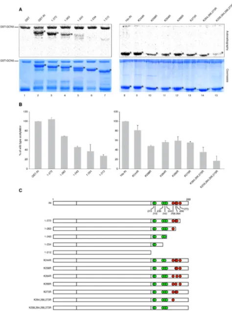
Modulation of the efficiency of integration can be achieved through the reversible acetylation of IN, which has been found to enhance its strand transfer activity in
vitro (Cereseto et al., 2005; Terreni et al.). Consistently, arginine substitutions
introduced at the IN lysines targeted by acetylation have been shown to negatively affect the infectivity of the mutant viruses by specifically reducing their integration level (Cereseto et al., 2005; Terreni et al.).
SUMOylation of IN may also contribute to regulate the efficiency of HIV-1 integration. In fact, it has been recently reported that IN is SUMOylated and the functional relevance of this post-translational modification has been highlighted by the finding that mutant viruses harboring a SUMOylation-defective IN are less infectious than their wild-type counterparts (Zamborlini et al., 2011). However, differently from acetylation, SUMOylation of IN does not seem to affect the catalytic activity of the protein, since SUMOylation-defective IN mutants still retain wild-type levels of enzymatic activity. Therefore, the possibility is currently being explored that SUMOylation may regulate the binding affinity of IN to cofactors required for the efficient completion of the viral replication cycle.
Finally, IN phosphorylation proved to be involved in the regulation of the
permissiveness of resting CD4+ T cells to HIV-1 infection (Manganaro et al., 2010).
In activated T lymphocytes, IN is phosphorylated by c-Jun N-terminal kinase (JNK), and then becomes a substrate for the cellular peptidyl prolyl-isomerase enzyme Pin1, leading to a conformational change in the viral protein (Manganaro et al., 2010). These concerted enzymatic activities lead to increased IN stability and are required for efficient HIV-1 integration and infection (Manganaro et al., 2010). Since JNK is not expressed in resting CD4+ T cells, lack of the above-mentioned
modifications likely contributes to the restriction of HIV-1 infection observed in these lymphocytes.
Restriction of infection at the integration step: KAP1
A recent study extended the family of host factors able to restrict HIV-1 infection through the identification of a cellular protein belonging to the TRIM family, KAP1, which interferes with the integration step of the viral replication cycle (Allouch and Cereseto, 2011). KAP1 was identified in a yeast two-hybrid screening of host proteins that specifically interact with the acetylated form of HIV-1 IN (Allouch and Cereseto, 2011). Indeed, further characterization of the association between the two proteins showed that KAP1 binds with higher affinity to IN upon its acetylation, both in vitro and in cultured cells (Allouch et al., 2011). Investigation on the role of KAP1 during the HIV-1 replication cycle led to the conclusion that KAP1 inhibits viral infectivity by specifically interfering with the integration process, as evidenced by the finding that KAP1 downregulation causes an increase in level of integration, while KAP1 overexpression results in a reduction of provirus formation (Allouch et al., 2011).The antiviral mechanism proposed for KAP1 consists in the recruitment of the histone deacetylase HDAC1 to acetylated IN, thus leading to deacetylation of the viral protein and consequent reduction of the integration efficiency. Consistently, the activity of KAP1 against HIV-1 proved to be fully dependent on deacetylation, as it was found to be ineffective in the absence of HDAC1, even if KAP1 was overexpressed (Allouch et al., 2011). However, since KAP1 is ubiquitously expressed showing no major restriction of HIV-1 infectivity, further studies are required to elucidate the strategy adopted by the virus to counteract this newly discovered host defense mechanism.
IN inhibitors to treat HIV-1 infection
The development of RT and PR inhibitors and the subsequent introduction of combination drug regimens enhancing the overall efficacy and durability of antiretroviral therapy revolutionized the treatment of HIV-1 infection in the mid 1990’s. As the last of the three essential HIV-1 enzymes, IN was considered an equally attractive target for antiretroviral drug development as RT and PR, but it is only a decade later that the first IN inhibitor, raltegravir (RAL, MK-0518) achieved
approval by the US Food and Drug Administration (FDA) (reviewed in (Cahn and Sued, 2007)), while elvitegravir (EVG, GS-9137, JTK303) has reached phase III clinical trials. Both RAL and EVG are derived from the diketo acid (DKA) family of IN inhibitors. DKAs were the first IN inhibitors reported with selectivity for the strand transfer step of the integration reaction (ST-inhibitors), high specificity for IN, and antiviral activity that could be related to IN inhibition (Hazuda et al., 2000). Their selective effect on the strand transfer reaction is a direct result of a mechanism of action in which the inhibitor: 1) only binds to the IN catalytic site in the presence of a viral DNA substrate and not to IN alone (Espeseth et al., 2000) and 2) chelates the two essential magnesium metal ion cofactors from the IN active site, thus rendering the metal-dependent phosphotransferase responsible for strand transfer inactive (Grobler et al., 2002; Marchand et al., 2003; Pais et al., 2002).
Despite its recent success in the clinic, emergence of resistance leading to treatment failure has already been reported for RAL. All the major mutations responsible for decreased susceptibility to RAL appear to localize within the IN active site proximal to the residues involved in coordinating the metal cofactors (Hazuda, 2010), consistent with the mechanism of inhibition based on metal chelation. Distinct subsets of IN mutations have been characterized in the viral sequences of patients who have failed the therapy with RAL. The “primary mutations” conferring high-level resistance to RAL have been identified as Q148R/H/K, N155H, and Y143R/C (Delelis et al., 2009; Kobayashi et al., 2008; Malet et al., 2009; Malet et al., 2008; Marinello et al., 2008; Shimura et al., 2008; Sichtig et al., 2009). Each tends to be associated with “secondary mutations” that appear to restore viral fitness in those primary mutants: for instance, the secondary mutation E92Q invariably clusters with the N155H mutation, while the G140S is mainly found in the Q148H mutants (Cooper et al., 2008; Delelis et al., 2009; Malet et al., 2009). The resistance profile of EVG closely resembles that of RAL, suggesting that EVG is unlikely to overcome resistance to RAL (Marinello et al., 2008). Current research efforts are therefore devoted to the discovery and development of novel compounds effective against viral strains resistant to IN ST-inhibitors. A promising approach that is currently being explored is that of targeting for antiviral therapy the interactions between IN and cellular cofactors, if