RESEARCH NOTE
Evaluation of the effect of a floxed Neo
cassette within the dystroglycan (Dag1) gene
Francesca Sciandra
1*, Bianca Maria Scicchitano
2, Giulia Signorino
3, Maria Giulia Bigotti
4, Barbara Tavazzi
3,
Francesca Lombardi
5, Manuela Bozzi
3, Gigliola Sica
2, Bruno Giardina
3, Sandra Blaess
6and Andrea Brancaccio
1,4*Abstract
Objective: Dystroglycan (DG) is an adhesion complex formed by two subunits, α-DG and β-DG. In skeletal muscle,
DG is part of the dystrophin-glycoprotein complex that is crucial for sarcolemma stability and it is involved in a pleth-ora of muscular dystrophy phenotypes. Due to the important role played by DG in skeletal muscle stability as well as in a wide variety of other tissues including brain and the peripheral nervous system, it is essential to investigate its genetic assembly and transcriptional regulation.
Results: Herein, we analyze the effect of the insertion of a floxed neomycin (Neo) cassette within the 3′ portion of the universally conserved IG1-intron of the DG gene (Dag1). We analyzed the transcription level of Dag1 and the expression of the DG protein in skeletal muscle of targeted mice compared to wild-type and we did not find any alterations that might be attributed to the gene targeting. However, we found an increase of the cross-sectional areas of tibialis anterior that might have some physiological significance that needs to be assessed in the future. Moreover, in targeted mice the skeletal muscle morphology and its regeneration capacity after injury did not show any evident alterations. We confirmed that the targeting of Dag1 with a floxed Neo-cassette did not produce any gross undesired effects.
Keywords: Dystroglycan, Skeletal muscle, Incomplete recombination
© The Author(s) 2017. This article is distributed under the terms of the Creative Commons Attribution 4.0 International License (http://creativecommons.org/licenses/by/4.0/), which permits unrestricted use, distribution, and reproduction in any medium, provided you give appropriate credit to the original author(s) and the source, provide a link to the Creative Commons license, and indicate if changes were made. The Creative Commons Public Domain Dedication waiver (http://creativecommons.org/ publicdomain/zero/1.0/) applies to the data made available in this article, unless otherwise stated.
Introduction
Dystroglycan (DG) is a widely expressed adhesion com-plex encoded by a single gene (Dag1) as a unique precur-sor that undergoes post-translational cleavage to generate two interacting subunits: the extracellular and highly gly-cosylated α-DG and the transmembrane β-DG [1, 2].
DG is crucial for cell membrane stability connect-ing the actin cytoskeleton to extracellular matrix (ECM) proteins [3]. Whenever the DG/ECM axis is perturbed, severe muscular dystrophy phenotypes do arise [4, 5].
Extensive genetic work has been carried out in order to analyze the function of DG in several tissues [6]. It was shown that the knockout of Dag1 in mice is embryonic lethal [7]. This result is in line with what was observed
in some human patients, in which a homozygous loss-of-function mutation in Dag1 results in the absence of DG that leads to early postnatal lethality [8].
Conditional knockouts of Dag1 in several tissues have confirmed the importance of DG for tissue stability. In those studies, no apparent effects of the inserted floxed Neo cassette have been reported [9–11]. Moreover, the knock-in mouse carrying the pathological mutation T190M showed an evident muscular dystrophy pheno-type and possible effects arising from the presence of the exogenous genetic elements might have been covered up [12]. On the other hand, in the knock-in mouse Y890F the floxed-Neo cassette had been already removed in the chimeric mice and no effect arising from the presence of the remaining LoxP element has been reported [13].
Interestingly, the insertion of a floxed Neo-cassette can lead to a significantly reduced expression of the tar-get gene and can also downregulate the transcription of genes located at a distance of several kilobases from it [14–22].
Open Access
*Correspondence: [email protected]; [email protected]
1 Istituto di Chimica del Riconoscimento Molecolare (CNR), c/o Università
Cattolica del Sacro Cuore, Rome, Italy
4 School of Biochemistry, University of Bristol, Bristol BS8 1TD, UK
Considering the lack of experimental data on the effects of the floxed Neo cassette on Dag1 expres-sion, we have decided to assess whether the presence of a floxed Neo cassette or single floxed site in the Dag1 allele might have per se some evident func-tional consequences for Dag1 expression in skeletal muscle.
Main text
Methods
Generation of targeting vector
Genomic DNA from V6.4 murine ES (embryonic stem) cells, (genotype C57BL/6J × 129/SvJae), was used as a template to amplify the right and left hand arms of the DG targeting construct. The 5′ arm of the construct consisted of 3.2 kb of intronic sequences amplified from the Dag1 intron using the following primers: for-ward 5′-GAGTAAGACTTTGTCTATAAAACA-3′ and reverse 5′-AAACCCCAACAACTACGGTCCTCA-3′. The 3′ arm was 4 kb in length, consisted of the exon 3 region, harboring four mutations, flanked by intronic sequences and the 3′-UTR, respectively, was ampli-fied using the following primers: forward 5′-GAGA-AGTGGGATTCATTTAGACAG-3′ and reverse 5′GAATGTAATCTTTAGCTACTGTT-3′. To allow the linearization of the targeting vector that is necessary for the recombination and integration of the construct into the ES cells genome, site-directed mutagenesis was used to mutate a XhoI site in the 5′ arm using the QuikChange site-directed mutagenesis kit (Stratagene®) and the fol-lowing primers: forward 5′-AGTTCCTTCCTGCCTA GAGTGGGTGTTCCCT-3′ and reverse 5′-AGGGAAC ACCCACTCTAGGCAGGAAGGAACT-3′ (mutated nucleotides in bold). Both the right and left hand arms were initially cloned into separate TOPO vectors (Invit-rogen) and then sub-cloned into the final targeting vec-tor, a modified pBluescript KS vector containing a floxed neomycin cassette under the PGK promoter (Fig. 1a). The 5′ and 3′ arms were cloned within a BamHI and HindIII sites respectively. Both enzymes (New England Biolabs® Inc) were incubated at 37 °C for 1.5 h. The Her-pes simplex thymidine kinase (HSVtk) cassette acts as a negative selection marker. The targeting vector was used at the Core Facility for Conditional Mutagenesis, Dibit-San Raffaele Scientific Institute, Milan to electropo-rate and analyze murine ES cells by Southern blot. One homologous recombinant ES clone was microinjected into blastocysts from 129 J mice. The resulting chimeras were then bred with C57BL6 mice to generate germ line-transmitted heterozygous mice (Dag1Neo/+). Dag1Neo/+ mice were backcrossed into the C57BL/6 background for ten generations and then used to create the experimental colonies. Mice from different colonies were crossbred to
generate wild-type (Dag1+/+), heterozygous (Dag1Neo/+) and homozygous mice (Dag1Neo/Neo).
To remove the Neo cassette, homozygotes were cross-bred with male transgenic mice expressing the transgene Cre under the CMV promoter (Jackson Laboratories) to generate Dag1Neo/+ mice. Female F1 animals were bred again with homozygous Dag1Neo/Neo mice to generate Dag1ΔNeo/ΔNeo mice.
Creatine kinase assay
Fresh serum was collected by retro-orbital bleeding. The creatine kinase (CK) level was determined according to the International Federation of Clinical Chemistry with commercial reagents on the Cobas 8000/c701® (Roche Diagnostics).
Histological and immunofluorescence analysis
All the analyses described below were not performed blind. Tibialis anterior (TA) and gastrocnemius (GA) muscles of 5 months-old female mice (n = 4 per geno-type) were embedded in Jung tissue freezing medium and frozen in liquid nitrogen-cooled isopentane. Frozen sections (7 μm) were stained with hematoxylin and eosin (HE) using standard methods [23]. Fiber cross-sectional areas (CSA) of muscle sections were counted manually using ImageJ (NIH, Bethesda, MD) from 6 to 12 ran-domly selected fields per section. At least 150 fibres per mouse from three mice per genotype were counted.
For the immunofluorescences analysis, TA muscle fro-zen sections of 5 months-old female mice were incubated with the following antibodies diluted in PBS containing 1% BSA (Sigma-Aldrich): mouse monoclonal anti-α-DG IIH6 (Millipore, code 05–593) (1:100), rabbit polyclonal anti-β-DG (Abcam, code ab43125) (1:300) and rat mono-clonal anti-laminin-α2 clone 4H8-2 (Sigma-Aldrich, code L0663) (1:100). The primary antibodies were followed by the appropriate secondary goat anti-mouse IgM (for IIH6), goat anti mouse, anti rat or anti rabbit IgG antibod-ies conjugated with Alexa®488 or Alexa®633 fluorophores (1:500) (Invitrogen-Thermo Fisher Scientific). Muscle sec-tions incubated with the only secondary antibody were used as control to validate the primary antibody specific-ity. Sections were viewed with a Leica SP2 microscope and all images were analyzed using ImageJ software (NIH).
Cardiotoxin injury
TA muscles of female 5 months-old mice (four mice per group) were injured along the entire length of the muscle with cardiotoxin (CTX) (LATOXAN SAS, code L8102) injections, 5 μL of 10 μM CTX in saline solution. These conditions ensured muscle damage a uniform manner. The muscles were harvested 10 days after cardiotoxin-induced damage. Frozen sections were processed for
histological analysis. Contralateral intact muscles were used as a control. In our previous study, regeneration was fully active at 7 days after injury [24]. Sections were viewed with a Leica SP2 microscope and all images were analyzed using ImageJ software (NIH).
Western blotting
TA muscle proteins from 5 months-old mice were extracted with PBS 1% Triton-X100 containing pro-tease inhibitors. 20 μg of soluble proteins were analyzed in Western blot as described elsewhere [24] using the following antibodies: anti mouse α-DG IIH6 (Millipore)
(1:1000), anti rabbit β-DG (Abcam) (1:1000) and anti-tubulin HRP-conjugated (Abcam) antibodies.
RT‑PCR
RNA was isolated from frozen muscle using the RNeasy Fibrous Tissue Mini Kit. Reverse Trascriptase PCR reac-tions were carried out using 3 μg of RNA. Quantitative RT-PCR was performed in triplicate using a standard TaqMan® PCR protocol on a StepOne Real time PCR System (Applied Biosystems). Probes were specific for mouse Dag1 and mouse Gadph genes. Data analysis was performed as described elsewhere [25].
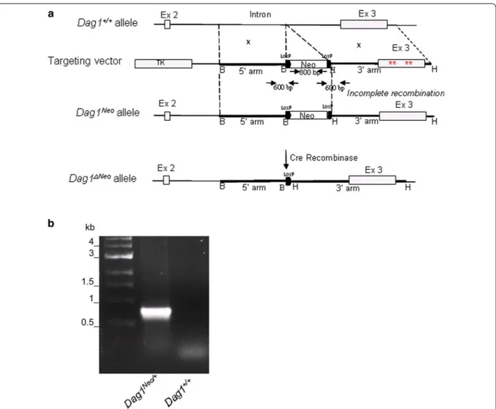
Fig. 1 Organization of the dystroglycan gene and position of the floxed Neo cassette. a Schematic representation of the Dag1+/+ allele, the
targeting vector, the Dag1 allele after incomplete homologous recombination (Dag1Neo) and the neo-deleted allele (Dag1ΔNeo). The 5′ arm of the
construct consisted of a 3.2 kb fragment amplified from the Dag1 intron. The 3′ arm was 4 kb in length and consisted of the exon 3′ region, har-bouring four mutations (red asterisks) hitting the α/β-DG interface, and flanked by intronic sequences and the 3′-UTR, respectively. A XhoI (X) site in the 5′ arm was mutated for cloning purposes. The floxed neomycin cassette (Neo) and the Herpes simplex thymidine kinase (TK) acted as positive and negative selection markers, respectively. Selected restriction enzyme cleavage sites are indicated above the gene (B: BamHI; H: HindIII and X:
XhoI). Primers used for genotyping are also indicated as black arrows. b A representative PCR genotyping analysis of the Dag1Neo/+ mouse. The PCR
Statistical analysis
Data were expressed as mean ± standard error of the mean (SE). Comparisons among groups were performed using an analysis of variance (One-way ANOVA) fol-lowed by a Bonferroni’s post hoc multiple comparison test to determine statistical significance of the differ-ences among the four experimental mice groups; p value of < 0.05 was considered to be statistically significant. Statistical analyses were performed with SPSS software (version 17.0, Chicago, IL, US).
Results and discussion
Insertion of the floxed Neo cassette into Dag1 and removal
by Cre recombinase: the Dag1Neo/Neo and Dag1ΔNeo/ΔNeo mouse
lines
In an attempt to produce a novel DG knock-in mouse line harboring four mutations at the α/β-DG interface [26], an incomplete homologous recombination at the 3′ arm (Fig. 1) prevented the generation of the desired
mutated mouse line. Indeed, we generated and studied a mouse line with a Dag1 allele in which a Neo cassette, flanked by two LoxP sites, was inserted into the second intron (Dag1Neo allele) (Fig. 1).
The Neo cassette was introduced in the second intron (also termed IG1-intron since it interrupts the struc-ture of the IG1 domain within the structurally autono-mous N-terminal region of α-DG [27]). In addition, we have also crossbred our mice with a mouse line that expresses the Cre recombinase under a CMV-promoter and results in Cre-mediated recombination in germ line. This resulted in the Cre-driven excision of the floxed Neo cassette in the targeted Dag1 (Dag1ΔNeo allele, Fig. 1a, b).
Histological and molecular analysis of skeletal muscle
Animals with two wild-type alleles are denoted by “Dag1+/+”, mouse lines in which one allele carries the
floxed Neo cassette are designated “Dag1Neo/+”, whilst their homozygous counterpart is denoted by “Dag1Neo/
Dag1
+/+Dag1
Neo/+Dag1
Neo/NeoDag1
∆Neo/∆Neo_
_
_
_
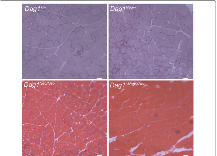
Fig. 2 Histological analysis of skeletal muscle. Hematoxylin and eosin staining of transverse frozen sections of tibialis anterior muscle of 5 months
old Dag1+/+, Dag1Neo/+, Dag1Neo/Neo, and Dag1ΔNeo/ΔNeo female mice (4 mice per group). No dramatic abnormalities were observed in the skeletal
__
__
__
__
__
__
__
__
__
__
β-DG Laminin Merge__
__
Dag1
+/+Dag1
+/+Dag1
+/+Dag1
Neo/+Dag1
Neo/+Dag1
Neo/+Dag1
Neo/NeoDag1
Neo/NeoDag1
Neo/NeoDag1
∆Neo/∆NeoDag1
∆Neo/∆NeoDag1
∆Neo/∆Neoa
b
Fig. 3 Immunofluorescence staining of skeletal muscle tissue sections. Immunolabeling of a α-DG and b β-DG and laminin-α2 of 5 months old
Neo”. Finally, “Dag1ΔNeo/ΔNeo” indicates the mouse line obtained upon the excision of the Neo cassette by Cre recombinase.
At 5 months of age, body weights of Dag1Neo/+ (male 31.5 g ± 2.3; female 25 g ± 1.7), Dag1Neo/Neo (male 31.7 g ± 2.6; female 24.5 g ± 1.5) and Dag1ΔNeo/ΔNeo (male 31.8 g ± 2.2; female 26 g ± 1.1) mice were not significantly different from wild-type controls (male 31.8 g ± 2.5; female 25 g ± 1.5).
Histology of tibialis anterior (TA) and gastrocnemius (GA) muscles of 5 months-old female mice, derived from crosses between heterozygotes (intercross) belonging to different colonies, examined by HE staining did not show any gross morphological sign indicating skeletal muscle alterations in Dag1Neo/+, Dag1Neo/Neo, and Dag1ΔNeo/ΔNeo mice compared to Dag1+/+ controls (Fig. 2). In all the Dag1 targeted mice, the levels of serum CK, a marker for skeletal muscle damage, did not significantly differ from those of wild-type animals (female: Dag1+/+ 366
U/L ± 0.8; Dag1Neo/+ 370 U/L ± 0.8; Dag1Neo/Neo 355 U/L ± 0.7; Dag1ΔNeo/ΔNeo 360 U/L ± 0.85). CK was meas-ured in 3 and 5-months old mice. Immunofluorescence analysis showed the expected localization of the two DG subunits as well as of the DG major skeletal muscle binding partner laminin to the cell membrane and cell surface, respectively (Fig. 3). Western blot analysis con-firmed that the DG precursor is correctly cleaved into its two subunits and that α-DG is highly glycosylated in all
the lines (Fig. 4a). The levels of α- and β-DG were similar compared to wild-type mice (data not shown).
Next, we examined the muscle fiber cross-sectional areas (CSA) by sampling a set of muscle fiber size in TA (Fig. 5a) and GA (Fig. 5b) muscle within the four experi-mental groups (four mice per group). Overall, we found a significant difference of the medians of fiber CSAs between the groups (lines) of mice (p < 0.001) whereas in GA we did not find any difference (p = 0.998). In particu-lar, in TA muscle the CSA in mouse line Dag1ΔNeo/ΔNeo (mean 1826 μm2 ± 37) was significantly increased when
compared with Dag1+/+ mice (mean 1609 μm2 ± 36)
(p < 0.001). At present, we cannot rule out that the observed variability in TA might have significant physi-ological repercussions. Further investigations will be required to better clarify this point.
α-DG Tubulin β-DG Dag1 +/+ Dag1 Neo/+ Dag1 Neo/Neo Dag1 ∆Neo/ ∆Neo
Fig. 4 Western blot analysis. Representative immunoblotting of total
protein lysates from Dag1+/+, Dag1Neo/+, Dag1Neo/Neo, and Dag1ΔNeo/ ΔNeo skeletal muscle. Upper panel; α-DG probed with IIH6 antibody
specific for the α-DG carbohydrates epitopes; middle panel: tubulin used as loading control; lower panel: β-DG probed with the anti 43-DAG antibody showing the correct processing of the DG precursor
2000 1500 1000 * -500 0 0 -1000 -1500 -500 -2000
-Dag1Neo/Neo Dag1∆Neo/∆Neo
Dag1+/+ Dag1Neo/+
a b Fiber size µm 2 (CSA) Fiber size µ m 2 (CSA)
Dag1Neo/Neo Dag1∆Neo/∆Neo
Dag1+/+ Dag1Neo/+
Fig. 5 Analysis of skeletal muscle fiber cross-sectional areas (CSA). a Tibialis anterior (TA) muscle and b gastrocnemius (GA) muscle of
Dag1+/+, Dag1Neo/+, Dag1Neo/Neo, and Dag1ΔNeo/ΔNeo 5-months old
female mice (4 mice per group). *Indicates significant difference (p < 0.001). Error bars represent SEM
The increase of TA muscle fibers was not mirrored by an increase either in DG protein levels or in Dag1 tran-scription (see below) (Figs. 4, 6). The Dag1 transcription levels were measured by Real Time PCR experiments that monitor the changes in mRNA levels of Dag1Neo/Neo, and Dag1ΔNeo/ΔNeo mice relative to Dag1+/+ littermates and no
significant change was detected (p = 0.707) (Fig. 6).
Cardiotoxin‑induced skeletal muscle regeneration
Dystroglycan is essential for postnatal muscle growth, stability and regeneration [9, 28]. To further analyze the possible presence of skeletal muscle defects of the regenerative process, we analyzed the TA muscles of Dag1Neo/+, Dag1Neo/Neo, and Dag1ΔNeo/ΔNeo 5-months old female mice compared to Dag1+/+ mice, 10 days
after CTX-induced damage. HE staining revealed that, although CTX injection caused muscle necrosis with the accumulation of mononucleated infiltrating cells, the regeneration response occurred in all the analysed mus-cles, as demonstrated by the amount of central nucleated fibers present (p = 0.559) (Fig. 7).
Limitations
The targeting of Dag1 with a floxed Neo cassette did not produce any strong undesired skeletal muscle phe-notype. The increase of CSA that we observed in TA of Dag1ΔNeo/ΔNeo mice might be due to some degree of vari-ability between the analyzed mice, but must be further
analyzed. Dag1 second intron is large (10 Kb) and this justifies the absence of relevant effects on Dag1 mRNA upon the introduction of the floxed Neo-cassette [29].
Authors’ contributions
Conceived and designed the experiments: FS, AB. Performed the experi-ments: FS, BMS, GS, MGB, BT, MB. Analysed the data: FS, BMS, FL GS, BG, SB, AB. Wrote the paper: FS, AB. All authors reviewed and contributed to the various draft versions of the manuscript. All authors read and approved the final manuscript. Fold Change -0 -0.7 --0.6 --0.5 --0.4 --0.3 --0.2 --0.1
-Dag1Neo/+ Dag1Neo/Neo Dag1∆Neo/∆Neo
Fig. 6 Quantitative analysis of Dag1 expression in skeletal muscle
of 5 months old Dag1+/+, Dag1Neo/+, Dag1Neo/Neo, and Dag1ΔNeo/ΔNeo
mice. Total mRNA was purified from tibialis anterior skeletal muscle of 5 months old mice and retro-transcribed to cDNA. Real-Time PCR reactions were performed using probes for the Dag1 and Gadph genes. Results are expressed as fold change of Dag1 expression in
Dag1Neo/+ (n = 4), Dag1Neo/Neo (n = 4), and Dag1ΔNeo/ΔNeo (n = 4)
lines relative to the Dag+/+ line (n = 4). No significant difference was
noted between any of the genotypes (p = 0.707). Error bars represent SEM
Centrally nucleated fibers
a
Dag1Neo/Neo Dag1∆Neo/∆Neo
Dag1+/+ Dag1Neo/+
b
Dag1+/+ D ag1+/+
Dag1Neo/+
Dag1Neo/Neo
Dag1∆Neo/∆Neo Dag1∆Neo/∆Neo
_ _ _ _ _ _ _ _ D ag1Neo/Neo Control Injured D ag1Neo/+
Fig. 7 Assessment of skeletal muscle regeneration in Dag1+/+,
Dag1Neo/+, Dag1Neo/Neo, and Dag1ΔNeo/ΔNeo mice. Skeletal muscle
regeneration process after CTX injection was not affected in
Dag-1Neo/+, Dag1Neo/Neo, and Dag1ΔNeo/ΔNeo 5 months old mice compared to Dag1+/+ mice. a Hematoxylin and eosin staining of transverse frozen
sections of tibialis anterior muscle. b Quantification of the number of centrally nucleated fibers in mice 10 days after the injection with CTX. Centrally nucleated fibers were counted from five randomly selected fields per section to evaluate fibre regeneration (four mice per group). No significant difference was found among the four experimental groups (p = 0.559). Error bars represent SEM
Author details
1 Istituto di Chimica del Riconoscimento Molecolare (CNR), c/o Università
Cattolica del Sacro Cuore, Rome, Italy. 2 Istituto di Istologia, Università Cattolica
del Sacro Cuore, Rome, Italy. 3 Istituto di Biochimica e Biochimica Clinica,
Università Cattolica del Sacro Cuore, Rome, Italy. 4 School of Biochemistry,
University of Bristol, Bristol BS8 1TD, UK. 5 Istituto di Clinica delle Malattie
Infet-tive, Università Cattolica del Sacro Cuore, Rome, Italy. 6 Neurodevelopmental
Genetics, Institute of Reconstructive Neurobiology, Life and Brain Center, University of Bonn, Bonn, Germany.
Acknowledgements
We gratefully acknowledge Massimo Aquilina and Fabrizio Fiore from the animal facility of the Catholic University of Rome for their skillful assistance, and Alexandra Joyner, Memorial Sloan Kettering Cancer Center New York, for the PGK-Neo plasmid.
Competing interests
The authors declare that they have no competing interests.
Availability of data and materials
The datasets used and analyzed during the current study are available from the corresponding author on reasonable request.
Consent for publication
Not applicable.
Ethics approval and consent to participate
All animal procedures were approved by the Ethics Committee of the Animal Facility of the Università Cattolica del Sacro Cuore (UCSC, Roma, Prot. Sf 11100/13 and Prot. CE 20353/14 06_01) and by the Italian Ministry of Health, Direzione Generale della Sanità Animale e dei Farmaci Veterinari (196/2015-PR).
Funding
This work was supported by a Wellcome Trust Career Re-entry fellowship to MGB and by an AFM post-doctoral fellowship to FS. This work was in part sup-ported by a PRIN 20109MXHMR_001 Grant to BG.
Publisher’s Note
Springer Nature remains neutral with regard to jurisdictional claims in pub-lished maps and institutional affiliations.
Received: 18 April 2017 Accepted: 10 November 2017
References
1. Bozzi M, Morlacchi S, Bigotti MG, Sciandra F, Brancaccio A. Functional diversity of dystroglycan. Matrix Biol. 2009;28:179–87.
2. Ibraghimov-Beskrovnaya O, Ervasti JM, Leveille CJ, Slaughter CA, Sernett SW, Campbell KP. Primary structure of dystrophin-associated glycoproteins linking dystrophin to the extracellular matrix. Nature. 1992;35:696–702.
3. Moore CJ, Winder SJ. The inside and out of dystroglycan post-transla-tional modification. Neuromuscul Disord. 2012;22:959–65.
4. Endo T. Glycobiology of α-dystroglycan and muscular dystrophy. J Bio-chem. 2015;157:1–12.
5. Zhang QZ. Dystroglycan induced muscular dystrophies—a review. Eur Rev Med Pharmacol Sci. 2016;20:3683–7.
6. Sciandra F, Bigotti MG, Giardina B, Bozzi M, Brancaccio A. Genetic engi-neering of dystroglycan in animal models of muscular dystrophy. Biomed Res Int. 2015;2015:635792.
7. Williamson RA, Henry MD, Daniels KJ, Hrstka RF, Lee JC, Sunada Y, et al. Dystroglycan is essential for early embryonic development: disruption of Reichert’s membrane in Dag1-null mice. Hum Mol Genet. 1997;6:831–41. 8. Riemersma M, Mandel H, van Beusekom E, Gazzoli I, Roscioli T, Eran A,
et al. Absence of α- and β-dystroglycan is associated with Walker-War-burg syndrome. Neurology. 2015;84:2177–82.
9. Cohn RD, Henry MD, Michele DE, Barresi R, Saito F, Moore SA, et al. Disruption of DAG1 in differentiated skeletal muscle reveals a role for dystroglycan in muscle regeneration. Cell. 2002;110:639–48.
10. Moore SA, Saito F, Chen J, Michele DE, Henry MD, Messing A, et al. Dele-tion of brain dystroglycan recapitulates aspects of congenital muscular dystrophy. Nature. 2002;418:422–5.
11. Saito F, Moore SA, Barresi R, Henry MD, Messing A, Ross-Barta SE, et al. Unique role of dystroglycan in peripheral nerve myelination, nodal struc-ture, and sodium channel stabilization. Neuron. 2003;38:747–58. 12. Hara Y, Balci-Hayta B, Yoshida-Moriguchi T, Kanagawa M, Beltrán-Valero
de Bernabé D, Gündeşli H, et al. A dystroglycan mutation associated with limb-girdle muscular dystrophy. N Engl J Med. 2011;364:939–46. 13. Miller G, Moore CJ, Terry R, La Riviere T, Mitchell A, Piggott R, et al.
Preventing phosphorylation of dystroglycan ameliorates the dystrophic phenotype in mdx mouse. Hum Mol Genet. 2012;21:4508–20. 14. Carmeliet P, Ferreira V, Breier G, Pollefeyt S, Kieckens L, Gertsenstein M,
et al. Abnormal blood vessel development and lethality in embryos lack-ing a slack-ingle VEGF allele. Nature. 1996;380:435–9.
15. Meyers EN, Lewandoski M, Martin GR. An Fgf8 mutant allelic series generated by Cre- and Flp-mediated recombination. Nat Genet. 1998;18:136–41.
16. Nagy A, Moens C, Ivanyi E, Pawling J, Gertsenstein M, Hadjantonakis AK, et al. Dissecting the role of N-myc in development using a single target-ing vector to generate a series of alleles. Curr Biol. 1998;8:661–4. 17. Shannon MB, Patton BL, Harvey SJ, Miner JH. A hypomorphic mutation in
the mouse laminin alpha5 gene causes polycystic kidney disease. J Am Soc Nephrol. 2006;17:1913–22.
18. Ackroyd MR, Skordis L, Kaluarachchi M, Godwin J, Prior S, Fidanboylu M, et al. Reduced expression of fukutin related protein in mice results in a model for fukutin related protein associated muscular dystrophies. Brain. 2009;132:439–51.
19. Rijli FM, Dolle P, Fraulob V, LeMeur M, Chambon P. Insertion of a targeting construct in a Hoxd-10 allele can influence the control of Hoxd-9 expres-sion. Dev Dyn. 1994;201:366–77.
20. Fiering S, Epner E, Robinson K, Zhuang Y, Telling A, Hu M, et al. Targeted deletion of 5′HS2 of the murine β-globin LCR reveals that it is not essential for proper regulation of the β-globin locus. Genes Dev. 1995;9:2203–13.
21. Pham CT, MacIvor DM, Hug BA, Heusel JW, Ley TJ. Long-range disruption of gene expression by a selectable marker cassette. Proc Natl Acad Sci USA. 1996;93:13090–5.
22. Meier ID, Bernreuther C, Tilling T, Neidhardt J, Wong YW, Schulze C. Short DNA sequences inserted for gene targeting can accidentally interfere with off-target gene expression. FASEB J. 2010;24:1714–24.
23. Fischer AH, Jacobson KA, Rose J, Zeller R. Hematoxylin and eosin staining of tissue and cell sections. Cold Spring Harbor Protoc. 2008;3(5). https:// doi.org/10.1101/pdb.prot4986.
24. Toschi A, Severi A, Coletti D, Musarò A, Molinaro M, et al. Skeletal muscle regeneration in mice is stimulated by local overexpression of V1a-vaso-pressin receptor. Mol Endocrinol. 2011;25:1661–73.
25. Schmittgen TD, Livak KJ. Analyzing real-time PCR data by the compara-tive CT method. Nat Protoc. 2008;6:1101–8.
26. Sciandra F, Bozzi M, Morlacchi S, Galtieri A, Giardina B, Brancaccio A. Mutagenesis at the α-β interface impairs the cleavage of the dystrogly-can precursor. FEBS J. 2009;276:4933–45.
27. Brancaccio A, Schulthess T, Gesemann M, Engel J. The N-terminal region of α-dystroglycan is an autonomous globular domain. Eur J Biochem. 1997;246:166–72.
28. Rader EP, Turk R, Willer T, Beltrán D, Inamori KI, Peterson TA, et al. Role of dystroglycan in limiting contraction-induced injury to the sarcom-eric cytoskeleton of mature skeletal muscle. Proc Natl Acad Sci USA. 2016;113:10992–7.