Case 9868
Leiomyomatosis Peritonealis Disseminata: a case report
Manenti G, Antonicoli M, Funel V, Bindi A, Simonetti GAbdominal Imaging Section: 2012, Feb. 17 Published: 32 year(s), female Patient:
Authors' Institution
Department of Diagnostic and Molecular Imaging, Interventional Radiology and Radiation Therapy, Fondazione Policlinico Tor Vergata,
Viale Oxford 81, 00133, Rome, Italy
Corresponding Author: Guglielmo Manenti, [email protected]
Clinical History
A 32-year-old woman was referred to our Institution with a persistent abdominal distension lasting for two weeks.
On the admission she showed a normal serum haemoglobin level and thrombocytopenia. She had a history of abdominal discomfort and uncommon pelvic pains.
These symptoms had been diagnosed and treated as a gastro-intestinal infection.
Imaging Findings
Digital radiography revealed a reduced expansion of the right costo-phrenic angle.
The ultrasound examination showed the presence of three heterogeneous masses characterised by hypervascularity and anechoic areas, determining dislocation of the bowel loops.
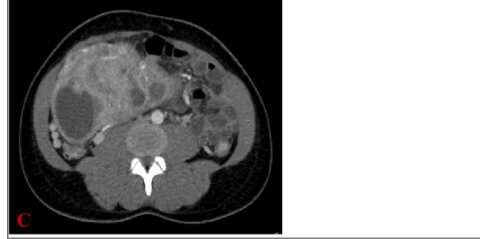
Further Imaging investigations, such as CT, confirmed the presence of many other solid masses in the peritoneal cavity, with heterogeneous attenuation and a c.e. similar to uterine leiomyomas. The tumour in the retro-umbilical region measured 70 mm, the one in the sub-hepatic region reached an extension of 170 x 110 x 180 mm.
On the MR examination these tumours showed low signal intensities on T1 weighted (T1w) images, heterogeneous signals, including numerous hyperintense foci, on T2 weighted (T2w) images, depending on various degrees of oedema and cystic degeneration, and intense and diffuse enhancement after the administration of a Gadolinium contrast material.
Biopsy was performed and the histological diagnosis was smooth muscle tissue without necrosis.
Discussion
Leiomyomatosis Peritonealis Disseminata (LPD) was described first by Wilson and Peale in 1952; Taubert identified LPD as a pathological entity in 1965 and only about 100 cases were described in the English literature since then [1]. It affects mainly women in reproductive age, even if few cases affecting males have been described. Recently a familial case of LPD has been reported [2]. LPD resemble typical uterine leiomyomas at both gross and microscopic level and represent a rare benign proliferative process, characterised by multiple smooth muscle nodules developing through the peritoneum probably from Mueller's epithelium, which is distributed throughout the
subperitoneal mesenchyme. The main hypothesis is that the smooth muscle nodules are derived from metaplasia of submesothelial cells.
A hormonal influence has been hypothesised (contraceptives, hormone replacement therapy, tamoxifen) [3]. Pregnancy has been reported as an independent factor. Many patients have a history of previous fibroid excision, with possible intra-abdominal deposition during surgery and a parasitic mechanism involved. Nevertheless the exact pathophysiology of LPD remains uncertain.
Most cases are asymptomatic and discovered incidentally during surgery. Symptoms include abdominal and pelvic pain, abdominal distension, urinary frequency and less commonly palpable mass, gastrointestinal bleeding and peritonitis.
Ultrasound and CT imaging features are useful to identify and stage the disease. The typical signal intensity patterns on MR images can be used to confirm the diagnosis: the masses of LPD are isointense relative to muscle on T1w sequences, have low signal intensity on T2w sequences and enhance heterogeneously following intravenous administration of Gadolinium-based contrast media. Like uterine fibroid tumors, large masses can show hemorrhagic, necrotic, myxoid and cystic degeneration.
Biopsy is requested and the histopathologic examination shows oxyphilic spindle-shaped cells without signs of malignancy, namely cytological atypia, mitotic features, coagulative type necrosis or invasiveness. Tumour cells are positive for vimentin, desmin and -SMA (Smooth muscle actin), and negative for keratin, S-100, CD34, C-kit.
Once the diagnosis is histologically confirmed, withdrawal of hormonal influence is often sufficient; alternatively patients can undergo surgical excision and/or therapy with Gonadotropin releasing hormone (GnRH) agonists.
Recurrence is possible and each recurrence is more likely to produce sarcomatous degeneration, which is uncommon (10%) [4]. In most reports of malignant LPD, no history of oestrogenous exposure or relationship with uterine leiomyomas was found, suggesting a different pathogenesis with a higher risk of malignant transformation. In such cases, surgical treatment is mandatory; adjuvant chemotherapy, bilateral salpingo-oophorectomy and hormonal therapy can be chosen [5].
The final diagnosis was Leiomyomatosis Peritonealis Disseminata.
Differential Diagnosis List
Primary peritoneal serous carcinoma, Peritoneal carcinomatosis, Malignant mesothelioma
Figures
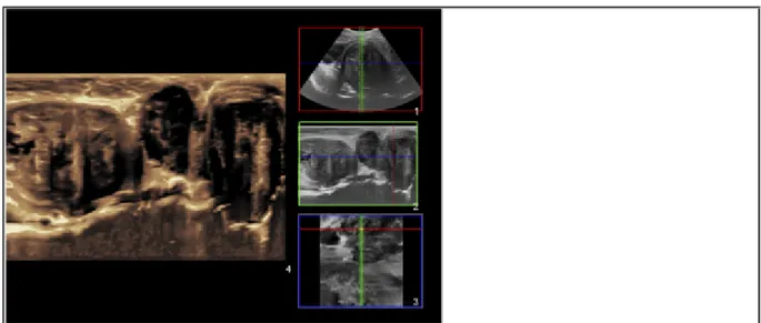
Figure 1 Ultrasonography
Panoramic imaging of the three main lesions showing the spatial relationship of nodules in
the midsagittal plane.
© Manenti G, Department of Radiology, Tor Vergata University, Rome, Italy
Area of Interest: Abdominal wall;
Imaging Technique: Ultrasound;
Procedure: Imaging sequences;
Special Focus: Neoplasia;
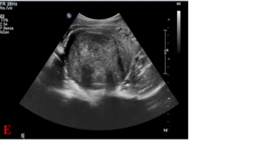
Transverse sonogram of the biggest nodule (13.6x9.7 cm) in the subhepatic region.
© Manenti G, Department of Radiology, Tor Vergata University, Rome, Italy
Area of Interest: Abdominal wall;
Imaging Technique: Ultrasound;
Procedure: Imaging sequences;
Special Focus: Neoplasia;
Grey-scale transverse sonogram revealing a heterogeneous structure and anechoic areas.
© Manenti G, Department of Radiology, Tor Vergata University, Rome, Italy
Area of Interest: Abdominal wall;
Imaging Technique: Ultrasound;
Procedure: Imaging sequences;
Special Focus: Neoplasia;
Power-Doppler sonogram of the same lesion showing high vascular supply.
© Manenti G, Department of Radiology, Tor Vergata University, Rome, Italy
Area of Interest: Abdominal wall;
Imaging Technique: Ultrasound-Power Doppler;
Procedure: Imaging sequences;
Another mass arising from the uterus with the same echo-pattern.
© Manenti G, Department of Radiology, Tor Vergata University, Rome, Italy
Area of Interest: Abdominal wall;
Imaging Technique: Ultrasound;
Procedure: Imaging sequences;
Special Focus: Neoplasia;
The elasticity image of one of the lesions shows a low tissue stiffness, typical of soft tissues.
© Manenti G, Department of Radiology, Tor Vergata University, Rome, Italy
Area of Interest: Abdominal wall;
Imaging Technique: Elastography;
Procedure: Imaging sequences;
Special Focus: Neoplasia;
MPR reconstruction in the sagittal plane showing the continuity of the peritoneal lesions
with the uterine ones.
© Manenti G, Department of Radiology, Tor Vergata University, Rome, Italy
Area of Interest: Abdominal wall;
Imaging Technique: MR;
Procedure: Diagnostic procedure;
Special Focus: Neoplasia;
Here the lesion in the subhepatic region shows a heterogeneous attenuation and contrast
enhancement with dislocation of the bowel loops.
© Manenti G, Department of Radiology, Tor Vergata University, Rome, Italy
Area of Interest: Abdominal wall;
Imaging Technique: CT;
(cfr 1c-d) The lesion shows an extremely heterogeneous pattern with fluid collections.
© Manenti G, Department of Radiology, Tor Vergata University, Rome, Italy
Area of Interest: Abdominal wall;
Imaging Technique: CT;
Procedure: Imaging sequences;
Special Focus: Neoplasia;
A lesion in the uterus mimicking a leiomyomatous uterus.
© Manenti G, Department of Radiology, Tor Vergata University, Rome, Italy
Area of Interest: Abdominal wall;
Imaging Technique: CT;
Procedure: Imaging sequences;
Special Focus: Neoplasia;
(cfr 2b) T2-weighted sequence showing heterogeneous hypointensity of the lesion and
hyperintense fluid areas, representing cystic degeneration.
© Manenti G, Department of Radiology, Tor Vergata University, Rome, Italy
Area of Interest: Abdominal wall;
Imaging Technique: MR;
Procedure: Diagnostic procedure;
Special Focus: Neoplasia;
(cfr 2b) Fast GE "THRIVE" sequence in the portal-venous phase showing heterogeneous c.e.
of the lesion with dislocation of the bowel loops.
© Manenti G, Department of Radiology, Tor Vergata University, Rome, Italy
Area of Interest: Management;
Imaging Technique: MR;
Procedure: Imaging sequences;
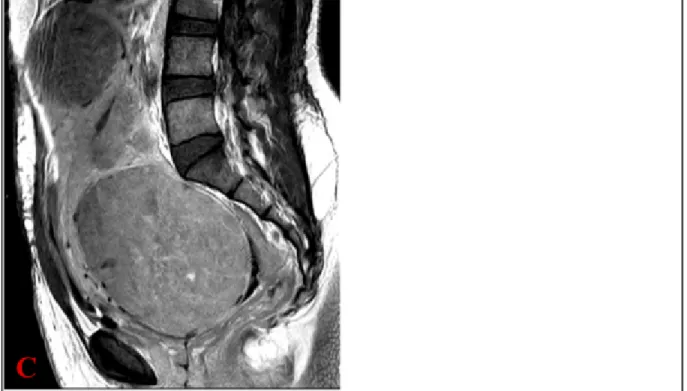
T1-weighted sequence after Gadolinium injection in the sagittal plane showing the lesion
within the uterus and other lesions in the peritoneal cavity
© Manenti G, Department of Radiology, Tor Vergata University, Rome, Italy
Area of Interest: Management;
Imaging Technique: MR;
Procedure: Diagnostic procedure;
Special Focus: Neoplasia;
(cfr 2d) T1-weighted sequence after Gadolinium injection showing the lesion in the uterus.
© Manenti G, Department of Radiology, Tor Vergata University, Rome, Italy
Area of Interest: Abdominal wall;
Imaging Technique: MR;
Procedure: Imaging sequences;
Special Focus: Neoplasia;
Venous phase CT showing the lesion occupying most of the abdomen, with dislocation of
the bowel loops.
© Manenti G, Department of Radiology, Tor Vergata University, Rome, Italy
Area of Interest: Abdominal wall;
Imaging Technique: CT;
Procedure: Imaging sequences;
Special Focus: Neoplasia;
Coronal T2-weighted MR image demonstrating well defined multiple hypointense nodules
with a heterogeneous pattern and fluid collections. No signs of infiltration are seen.
Imaging Technique: MR;
Procedure: Imaging sequences;
Special Focus: Neoplasia;
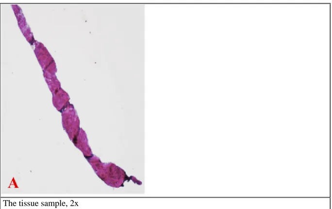
Figure 5 Histological examination, haematoxilin and eosin
The tissue sample, 2x
© Department of Anatomical Pathology, Tor Vergata University, Rome, Italy
Area of Interest: Abdominal wall;
Imaging Technique: Experimental;
Procedure: Biopsy;
Special Focus: Neoplasia;
Microscopic appearance showing interdigitating fascicles of smooth muscle cells, 10x
© Department of Anatomical Pathology, Tor Vergata University, Rome, Italy
Area of Interest: Abdominal wall;
Imaging Technique: Experimental;
Procedure: Biopsy;
No cytological atypia and few mitoses are seen, 20x
© Department of Anatomical Pathology, Tor Vergata University, Rome, Italy
Area of Interest: Abdominal wall;
Imaging Technique: Experimental;
Procedure: Normal variants;
Special Focus: Neoplasia;
MeSH
[C04.557.450.590.450.465]
Leiomyomatosis
The state of having multiple leiomyomas throughout the body. (Stedman, 25th ed) [C04.588.033.513]
Peritoneal Neoplasms
Tumors or cancer of the PERITONEUM.
References
[1] Fasih N, Prasad Shanbhogue AK (2008) Leiomyomas beyond the uterus: unusual locations, rare manifestations Radiographics 28:1931-48
[2] Halama N, Grauling-Halama SA et al (2005) Familial clustering of Leiomyomatosis peritonealis disseminate: an unknown genetic syndrome? BMJ Gastroenterol 5:33
[3] Tanaka YO, Tsunoda H et al (2009) MR and CT findings of leyomiomatosis peritonealis
disseminate with emphasis on assisted reproductive technology as a risk factor Br J Radiol 82:e44-7 [4] Bekkers RL, Willemsen WN et al (1999) Lwiomyomatosis peritonealis disseminate: does malignant transformation occur? A literature review Gynecol Oncol 75:158-63
[5] Levy AD, Arnàlz J et al (2008) From the archives of the AFIP: primary peritoneal tumors: imaging features with pathologic correlation Radiographics 28:583-607
29:347-73
Citation
Manenti G, Antonicoli M, Funel V, Bindi A, Simonetti G (2012, Feb. 17)
Leiomyomatosis Peritonealis Disseminata: a case report {Online}