UNIVERSITÀ DEGLI STUDI DI CATANIA
International PhD
in
“TRANSLATIONAL BIOMEDICINE”
XXVII CYCLE
DEVELOPMENT OF EFFECTIVE LUNG CANCER THERAPIES BASED
ON LUNG CANCER STEM CELLS TARGETING
Dr.ssa Valentina Salvati
Coordinator of PhD Tutor
Prof. Daniele Condorelli Prof.ssa Vincenza Barresi
Co-Tutor
2 INDEX Abstract... 4 1 Introduction ... 6 1.1 Lung Cancer ... 6
1.2 Molecular alterations in lung cancer ... 7
1.2.1 KRAS ... 8 1.2.2 ALK ... 9 1.2.3 TP53 ... 10 1.2.4 BRAF ... 10 1.2.5 PI3K/AKT/mTOR ... 11 1.2.6 PTEN ... 12 1.2.7 MET ... 12 1.2.8 HER2 ... 13 1.2.9 EGFR ... 13
1.3 Cancer stem cells (CSCs) ... 19
1.4 Human Lung Cancer stem cells ... 21
2 Aims of the thesis ... 23
3 Material and Methods ... 24
3.1 Isolation and culture of lung cancer spheres ... 24
3.2 Cell culture ... 24
3.3 Western Blot ... 25
3.4 PCR and sequencing methods for genomic DNA ... 25
3.5 Chemotherapy resistance studies ... 26
3
3.7 Cell Cycle Analysis by Propidium Iodide (PI) Staining ... 26
3.8 Immunohistochemistry ... 27
3.9 Fluorescence in situ hybridization ... 27
3.10 Generation of subcutaneous lung cancer xenograft into NSG mice ... 27
3.11 In vivo treatment of Lung CSC-generated Xenografts ... 28
3.12 Reverse Phase Protein Microarrays Technique ... 28
3.13 Clinical-pathological characteristics of patients ... 29
4 Results ... 30
4.1 Generation and validation of lung cancer stem cells (CSCs) ... 30
4.2 Molecular characterization of lung CSCs ... 31
4.3 Responsiveness to chemotherapy compared to Erlotinib and EGFR pathway activation in lung CSCs ... 33
4.4 Effects of Erlotinib treatment in lung CSCs ... 37
4.5 Antitumor activity of Erlotinib in lung CSCs-derived xenografts ... 39
4.6 Antitumor activity of Erlotinib compared to chemotherapy in lung CSCs- derived xenografts ... 41
4.7 Analysis of EGFR phosphorylation in advanced NSCLC patients ... 43
4.8 Expression of CSCs markers to improve the identification of Erlotinib sensitive and resistant lung CSCs ... 46
5Discussion and conclusion ... 48
4
ABSTRACT
Non-small cell lung cancer (NSCLC) accounts for approximately 80% of lung cancer subtypes and it is the most common and the most deadly cancer worldwide.
The conventional treatment for advanced NSCLC has consisted of chemotherapy but the impact of traditional chemotherapy on progression-free survival (PFS) and overall survival (OS) remains small.
The improved understanding of the molecular pathways involved in tumorigenesis and the increased ability to detect specific genetic alterations as targets, have led to significant advances in the development of newer and more directed therapies.
For instance, the epidermal growth factor receptor (EGFR) is frequently over-expressed in NSCLC and is taken as a promising target for NSCLC treatment.
Several studies have indicated that the presence of EGFR mutation is a robust predictor of response to EGFR inhibitors (EGFRi). Although first-generation EGFR inhibitors show encouraging clinical responses in lung tumors, almost all patients develop resistance to these inhibitors over time.
Two general mechanisms of acquired resistance to EGFR tyrosine kinase inhibitors (TKIs) treatment in EGFR mutant NSCLC patients have been described: secondary alterations in EGFR that prevent inhibition of EGFR by EGFR TKIs (drug resistant EGFR mutation), and additional genetic alterations that can co-occur with an EGFR activating mutation in EGFR mutant NSCLC cells.
However, tumor resistance to treatments could be also dependent on the presence of chemoresistant cancer stem cells (CSCs), a subpopulation of cells capable of self-renewing and undergoing asymmetric division, thereby giving rise to differentiated progeny that comprises the bulk of the tumor. Thus, exploring CSCs in lung cancer may help define responses to conventional and innovative therapies, and contribute to the introduction of relevant improvements in clinical oncology, toward the direction of more effective and less destructive personalized therapies.
Therefore, the EGFR targeted therapy in NSCLC continues to evolve since the discovery of EGFR driver mutations and utilization of EGFR-specific TKIs. However, about 10-20% of advanced NSCLC patients with wild-type EGFR also benefit from EGFRi treatment, suggesting that other determinants of outcome, besides EGFR mutation, might exist.
5
Thus, this project has been focused on the analysis of EGFR signaling and on the study of the sensitivity to EGFR inhibitors of lung CSCs and of lung CSCs derived xenografts, with the aim of identifying possible predictive bio-markers of response to EGFRi, in EGFR mutation-negative cells. This study led to the identification of phosphorylated EGFR at tyrosine 1068 residue but not pEGFR-tyr1173, as predictive biomarker of response to Erlotinib in lung CSCs in vitro and in lung CSCs-derived xenografts. Furthermore, also commercial lung cancer cell lines sensitive to Erlotinib expressed pEGFR-tyr1068 despite EGFR mutations, thus the expression of pEGFR-tyr1068 in lung CSCs was associated to positive Erlotinib response.
Assessing EGFR phosphorylation status by immunohistochemistry (IHC) in EGFR mutated and EGFR-wt NSCLC patient tumors, it was shown that anti-ptyr1068 positivity correlated with EGFR mutation while anti-ptyr1173 did not. Based on these data, it has been possible to hypothesize that the level of pEGFR-tyr1068 detected in patient tumors, would identify both mutation positive and mutation negative tumors with activated receptor and could be correlated with positive response to Erlotinib treatment in NSCLC patients.
These studies might have relevant therapeutic implications for the treatment of lung cancers and could add another option to cure NSCLC patients.
6 1 INTRODUCTION
1.1 Lung cancer
Lung cancer is one of the most common human cancers and the leading cause of cancer death worldwide; the survival rate remains stable at 15% (1).
Lung cancer is a heterogeneous disease clinically, hystologically, biologically, and molecularly (2). It is defined as a malignancy arising from the cells of the respiratory epithelium and is divided into two broad categories, Small cell lung cancers (SCLC) and Non-Small cell lung cancers (NSCLC), identified based on histological, clinical and immunohistochemical characteristics.
SCLCs are a highly malignant tumor type derived from cells exhibiting neuroendocrine characteristic and account for approximately 15% of lung cancer cases (3,4).
The remaining 85% of cases are NSCLCs, which are further divided into three major subtypes: adenocarcinoma (ADC) constituting 40-70% of cases, squamous cell carcinoma (SCC) with 10-15% and large cell carcinoma (LCC) accounting for 5% of NSCLCs.
In general, ADCs arise in more distal airways, whereas SCCs arise in more proximal airways, and the letters are more strongly associated with smoking and chronic inflammation than ADCs (5,6). ADCs often have glandular histology and express biomarkers that are consistent with an origin in the distal lung, by contrast, SCCs are characterized by squamous differentiation, which is more reminiscent of the pseudostratified columnar epithelium that lines the trachea and upper airways (5-7).
The prognosis for lung cancer patients is poor, in part due to late diagnosis (4).
Whilst SCLC can be highly responsive to chemotherapy, offering the possibility of long-term survival when combined with radiotherapy, NSCLC remains considerably more refractory to treatment (8). Although approximately 20% of NSCLC are operable at presentation, recurrence rates remain high at 30–50%.
Locally advanced NSCLC can be treated with radical chemo-radiotherapy with intent to cure; however, overall 5-year survival rates remain low at around 7–20%. For most cases of advanced NSCLC, systemic chemotherapy response rates are below 20–50%, and the 5-year mortality is in the order of 95%.
7
The low response rate of lung cancer patients may be a consequence of an homogeneity of therapeutical approaches (including platinum-based doublet chemotherapy and radiation therapy alone or in combination), that doesn't take into account for lung cancer biological heterogeneity. The strategy for a more successful lung cancer treatment should be the identification of clinical biological properties of each lung cancer subtype and possibly, within each form of lung cancer, to aim at personalized therapy for each patient-specific molecular pattern.
1.2 Molecular alterations in lung cancer
Despite advances in cytotoxic chemotherapy and combined treatment schedules in the clinical settings, lung cancer prognosis remains inadequate.
Major advances in the understanding of the molecular pathogenesis of lung cancer have led to new strategies for early detection, diagnosis, staging and therapy that hold promise for improving lung cancer outcomes.
Tobacco smoke is certainly a predominant etiologic risk factor for lung cancer. Carcinogens present in tobacco smoke or their intermediate metabolites can bind covalently to DNA at certain specific sites, forming bulky adducts and leading to gene mutations (9). However, a fraction of lung cancers arise in patients who have never smoked, suggesting the importance of host genetic/epigenetic susceptibility in the occurrence and development of lung cancer. In fact, lung cancers develop through a multistep process involving microenvironment and development of multiple genetic and epigenetic alterations, particularly activation of growth promoting pathways and inhibition of tumor suppressor pathways.
A variety of genetic abnormalities are encountered in lung cancer, with different incidences in distinct histological subtypes of the disease (4).
Understanding of the molecular biology of lung cancer has been revolutionized by next-generation sequencing technologies that provide a comprehensive means of identifying somatic alterations in entire cancer and have led to significant advances in the development of newer and more directed therapies.
For instance, several molecular alterations have been defined as “driver mutations” in NSCLC as mutations in Kirsten-rous avian sarcoma (KRAS), mutations in TP53 gene, a chromosome 2p inversion producing an EML4-ALK fusion gene (echinoderm microtubule-associated protein-like 4
8
fused with the anaplastic lymphoma kinase) and mutations in epidermal growth factor receptor gene (EGFR) (10). Other key signaling pathways such as RAS/RAF/MEK, PI3K/AKT/mTOR, PTEN, mesenchymal-epithelial transition (MET) and human epidermal growth factor receptor 2 (HER2) have also been identified as novel targets for NSCLC treatment (11,12).
In Table 1 are indicated the frequencies of the most important genetic alterations reported for the different forms of lung cancer and the therapeutic agents.
Table 1
1.2.1 KRAS
KRAS is part of the RAS family of proto-oncogenes (KRAS, NRAS and HRAS occurring in humans) and encodes a G-protein with a critical role in controlling signal transduction pathways which regulate cell proliferation, differentiation and survival (13).
Ras proteins are guanosine diphosphate (GDP) bound and inactive in normal quiescent cells.
There is a switch to the activated guanosine triphosphate (GTP) bound form following activation of upstream growth factor receptors. The activated Ras-GTP subsequently binds and activates a number of downstream pathways including mitogen-activated protein kinase (MAPK),
GENE FREQUENCY ACCORDING TO NSCLC HISTOLOGIC SUBTYPE
ADC SCC
THERAPEUTIC AGENT
KRAS 25-40% mutation Rare non canonical mutation, 7% amplification
None
EGFR 10-40% mutation Rare Erlotinib,Gefitinib,
Afatinib
ALK 4% translocation 0% Crizotinib
BRAF 2% mutation 0% Vemurafenib
MET 5% amplification 10% amplification Crizotinib,
Cabozantinib
HER2 2% mutation <1% mutation Trastuzumab,
Afatinib, Dacomitinib
PI3KCA <2% mutation 10% mutation Rapamycin and
rapalogs
PTEN 2% inactivation 10% inactivation Vandetanib
TP53 50% mutation 20% mutation Advexin a p53
9
RAS/RAF/MEK/MAPK pathway and the PI3-K [PI3K/AKT/mammalian target of rapamycin (mTOR)] pathways.
KRAS plays a critical role in downstream signal transduction induced by a variety of growth factor receptors including EGFR and constitutive activation of the protein circumvents the need for growth factor mediated signaling.
Activating mutations alter the GTPase activity of the protein hindering inactivation of the active RAS-GTP to GDP leading to increased signaling through multiple downstream growth promoting pathways (14).
Activating mutations in the KRAS oncogene are the commonest oncogenic alteration in lung ADC occurring in about 25-40% of cases, while HRAS and NRAS mutations are very rare (15).
In keeping with the role of KRAS alterations as driver mutations, they do not occur in association with EGFR mutations (16-19). Differences in the prevalence of KRAS mutations in lung ADC most likely relate to different patient populations as KRAS mutations are more common in Western populations compared to Asian populations (19,20) and are more frequent in males and smokers (17,19,21). ADC in never smokers have been reported to harbour KRAS mutations in between 0-15% of cases (22,23). In addition, KRAS mutations are very rare or absent in SCCs and SCLC (15,24).
The high frequency of KRAS mutations in lung cancer makes it an ideal therapeutic target but unfortunately clinical trials of targeted agents have generally been disappointing.
1.2.2 ALK
The anaplastic lymphoma kinase (ALK) is a receptor tyrosine kinase that is aberrant in a variety of malignancies. Multiple different ALK rearrangements have been identified in approximately 3-7% of unselected NSCLC (25) although some studies have found a slightly lower prevalence (26).
The majority of these ALK fusion variants result most commonly in fusions of portions of the
echinoderm microtubule-associated protein-like 4 (EML4) gene with chromosome 2p the ALK gene. At least nine different EML4-ALK fusion variants have been identified in NSCLC (27-29). The fusion proteins created by these rearranged genes have functions of both anaplastic lymphoma kinase and the partner protein. The presence of the partner protein allows phosphorylation of anaplastic lymphoma kinase without dimerization.
10
The oncogenic EML4-ALK fusion protein has a constitutively activated kinase and is linked to cell proliferation and inhibition of apoptosis mediated through the RAS/RAF/MAPK, PI3K/AKT and JAK3-STAT3 signaling pathways (30).
ALK rearrangements are more commonly found in ADC from younger patients who are never smokers or light smokers (26,27).
ALK rearrangements are usually mutually exclusive with EGFR and KRAS mutations (16,26). The United States Food and Drug Administration approved the ALK Break Apart fluorescence in
situ hybridization (FISH) Probe Kit (Abbott Molecular, Des Plaines, IL, USA) as a companion
diagnostic for targeted therapy with Crizotinib in lung cancers (31).
1.2.3 TP53
TP53 located on chromosome 17p13 encodes a nuclear phosphoprotein of 53 kDa that identifies and binds to regions of damaged DNA (32) and acts as a transcription factor controlling the expression of a multitude of different genes. Damaged DNA or carcinogenic stress induces TP53 leading to cell cycle arrest by inducing expression of cyclin dependent kinase inhibitors to enable DNA repair or apoptosis.
Inactivating mutations in TP53 (mostly missense mutations within the DNA-binding domain) have been reported in 80-100% of SCLCs (32) and about 65% of NSCLCs (33).
The mutational spectrum of different types of TP53 mutations differs between smokers and non-smokers with smoking related cancers having a significantly higher frequency of G to T transversions compared to G to C transversions (thought to be induced by polycyclin aromatic hydrocarbons in tobacco smoke) and G to A transitions at CpG dinucleotides more commonly seen in never smokers. Genetic alterations of TP53 have also been associated with treatment resistance (32) and TP53 gene mutations can occur in association with EGFR and KRAS mutations (20). Numerous studies have led to the development of pharmacological methods of reactivating p53, such as by gene replacement therapy.
1.2.4 BRAF
BRAF encodes a serine/threonine protein kinase that is the downstream effector protein of KRAS and activates the mitogen-activated protein kinase (MAPK) signal transduction pathway involved in
11
the regulation of cell proliferation and survival. While activating BRAF mutations are common in melanoma (34), they occur in only about 2-3% of NSCLC (21,34,35).
The mutations in NSCLC differ from those in melanoma and colorectal carcinoma with a lower proportion of V600E mutations that affect the kinase domain of the protein. In lung ADC, V600E mutations in exon 15 account for up to about 50% of BRAF mutations followed by G469A in exon 11 and D594G in exon 15 (32,35). Some of the BRAF mutations in NSCLC occur in the kinase domain (such as V600E, D594G and L596R) while others occur in the G-loop of the activation domain of the gene (such as G465V and G468A) (34). As BRAF and KRAS genes are part of the signaling pathway mediated by EGFR, it is not surprising that mutations in these genes are almost always mutually exclusive, in keeping with a common downstream pathway to transformation. Non-V600E BRAF mutations have been associated with current or former smokers while V600E mutations appear to be more common in female never smokers (35). Although uncommon, BRAF mutations represent an important therapeutic target due to the availability of targeted therapies already in clinical use for melanoma, for instance have been reported BRAF-mutated NSCLC cases to respond to Vermurafinib, however there are only limited data about the clinical response to this approach in NSCLC (36).
1.2.5 PI3K/AKT/mTOR
The PI3K/AKT/mTOR pathway is involved in regulation of cell proliferation, survival, differentiation, adhesion and motility (37,38).
The pathway is activated through activation of a variety of membrane tyrosine kinase receptors including EGFR, HER2, insulin-like growth factor receptor (IGFR), vascular endothelial growth factor receptor (VEGFR) and platelet derived growth factor receptor (PDGFR) (39,40).
The PI3K/AKT/mTOR pathway is frequently deregulated in many tumors including 50-70% of NSCLC (39).
Pathway activation in lung carcinogenesis occurs through a variety of mechanisms including activating mutations in EGFR, KRAS, PI3K or AKT (39) as well as PIK3CA amplification, or loss of negative regulation by the tumor suppressor gene PTEN.
The PI3K protein family (phosphatidylinositol 3-kinases) are intracellular lipid kinases and the main catalytic subunit, the p110alpha isoform, is encoded by the PIK3CA gene (39,40).
12
Activating mutations and amplification of PIK3CA cause constitutive ligand-independent pathway activation (41). PIK3CA mutations mostly involve the catalytic domain and have been identified in approximately 1-3% of NSCLCs (41) and are more common in SCCs than ADCs.
Unlike most oncogenic driver mutations, PIK3CA mutations may occur in association with EGFR or KRAS mutations (41) suggesting they may not represent true driver mutations.
PIK3CA may also be amplified in NSCLC, especially in SCCs (41,42) and increased copy number of PIK3CA has been reported in ~5% of SCLCs (41).
Several inhibitors of the PI3K pathway are undergoing evaluation in preclinical and clinical studies. These include pan and selective inhibitors of PI3K, AKT, Rapamycin and rapalogs for mTOR inhibition, dual mTORC1-mTORC2 inhibitors and dual PI3K-mTOR inhibitors (39).
1.2.6 PTEN
PTEN encodes a lipid and protein phosphatase on chromosome 10 that plays a significant role in cell cycle progression, apoptosis, growth, proliferation and migration via negative control of the PI3K/AKT pathway.
PTEN mutations and loss of PTEN protein expression are relatively common in squamous cell carcinoma. Mutations of PTEN occur only rarely in about 5% of NSCLC (43) being more common in SCC than ADC (10.2% vs. 1.7%) and associated with a history of smoking. By contrast, reduced protein expression has been reported in about 75% of NSCLC (44).
The TKI Vandetanib has shown efficacy against EGFR mutation-positive lung cancer cell lines with loss of PTEN (45).
1.2.7 MET
MET is a proto-oncogene located on chromosome 7q21−q31 that encodes a transmembrane tyrosine kinase receptor for its ligand hepatocyte growth factor (HGF) (46).
Mutations in MET are rare, but a high MET gene copy number has been detected in 1−11% of NSCLC cases. High gene copy numbers are more common in squamous cell carcinoma than adenocarcinoma and are often associated with high MET protein expression and poor prognosis (46). MET amplification that drives and maintains the phosphatidylinositol 3-kinases (PI3K)/AKT
13
pathway, bypassing EGFR blockade by tyrosine kinase inhibitors (TKIs), has emerged as one of the critical events for secondary resistance to EGFR-TKIs in patients with EGFR-mutated NSCLC (47). A number of therapeutic agents targeting the MET/HGF pathway are in clinical development, including Cabozantinib, Crizotinib and Rilotumumab (antagonistc antibodies against HGF)(48).
1.2.8 HER2
The human epidermal growth factor receptor 2 (HER2/ERBB2) gene encodes a membrane bound receptor tyrosine kinase that is a member of the ERBB family of receptors, along with EGFR. Unlike other ERBB receptors, it does not bind ligand directly but can form heterodimers with other ligand-bound members of the receptor family (49). Activation leads to signaling through a variety of signal transduction pathways including PI3K, MAPK and JAK/STAT pathways (50).
Activation of HER2 occurs in a small proportion of lung cancers with overexpression in approximately 20% of cases, gene amplification in 2% (51) and activating mutations in 1.6-4% of NSCLC (52,53). Activating mutations of HER2 are exon 20 in frame insertions of 3 to 12 base pairs in length (52).
Alterations of HER2 occur mostly in ADC (52,53) and mutations occur in tumors that are wild-type for EGFR and KRAS (52) and in some studies, are associated with female gender, Asian ethnicity and non-smoking status (52,53), similar to the clinical profile of EGFR mutant tumors.
HER2-mutated adenocarcinoma cases have been reported to respond to Trastuzumab and Afatinib (54).
1.2.9 EGFR
The epidermal growth factor receptor 1 (EGFR/ERBB1) gene encodes a transmembrane tyrosine kinase with an extracellular ligand-binding domain and an intracellular component including a tyrosine kinase domain (55). Binding of the ligand epidermal growth factor leads to receptor homo or heterodimerization with other members of the EGFR family and activation of the tyrosine kinase domain (56,57).
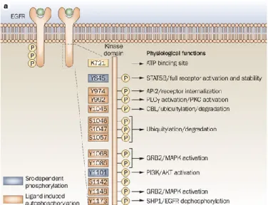
EGFR can be phosphorylated at tyrosine 1068 and 1173 that are major sites, whereas Tyr 992, 1148 and 1086 are minor phosphorylation sites. Distinct downstream signalling cascades are initiated by EGFR depending on its phosphorylation patterns: 1068 can bind Grb2 or Gaba1 and subsequently
14
activate their downstream signalling pathways; phosphorylation at Tyr 1173 leads to interaction with Shc and phospholipase Cγ (PLCγ), which are involved in activation of MAPK signalling pathway (58,59)(Fig. 1). Numerous studies suggest that there may be apparent differences between EGFR phosphorylation pattern and function of different tyrosine phosphorylation sites.
Thus, EGFR is involved in regulation of numerous oncogenic functions such as cell proliferation, survival, differentiation, invasion and metastasis (57) and signal transduction stimulated by EGFR occurs through the PI3K/AKT/mTOR, RAS/RAF//MAPK and JAK/STAT signaling pathways (56).
Figure 1: Ligand binding to EGFR causes receptor homodimerization or heterodimerization, which leads to transphosphorylation of the cytoplasmic tail tyrosine residues. Tyrosine phosphorylation in the C-terminus includes Y974, Y992, Y1045, Y1068, Y1086, Y1148 and Y1173 (shown in orange), or SFKs can phosphorylate Y845 and Y1101 (shown in purple). These phosphorylated tyrosines serve as docking sites for various proteins which contain Src Homology domains (SH2) and phosphotyrosine binding domains (PB). These events lead to the activation of several signaling cascades most notably the MAPK, PI3K/AKT, STAT and PLCγ pathways which ultimately result in proliferative signals to the cell nucleus. From: (59)
15
Activating mutations in EGFR lead to constitutive tyrosine kinase activation; other mechanisms of increased EGFR signaling include increased protein expression or increased gene copy number (60,61).
Improper activation of EGFR has been implicated in the pathogenesis and progression of many malignancies including NSCLC, as well as in the poor prognosis of patients.
In malignant cells, including NSCLC cells, the activity of the receptor may become dysregulated and no longer under the control of inherent inhibitory mechanisms. Spontaneous EGFR mutations often are oncogenic; that is, they activate the EGFR signaling pathway in the absence of ligand and promote cell proliferation, survival and anti-apoptotic signals.
These types of mutations can be found in 10 to 20% of patients with NSCLC at an advanced stage and in more than 50% of adenocarcinomas and tumors from East Asians, never smokers and young women (62).
The described mutations generally interest the exons of the EGFR gene between the 19th and the 21th, which correspond to the portion of the receptor with kinase activity, nearby the binding site of ATP (that is fundamental to the activation of the receptor by autophosphorylation). As a result, the receptor is blocked into a state of constitutive activation, thus signaling to the cell to proliferate and to resist apoptosis.
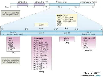
The two most frequent genetic mutations responsible for this anomaly in cell cycle are the substitution of arginine for leucine at codon 858 (L858R) at exon 21, and in-frame deletions at exon 19 (del E746_A750 is the most common). L858R accounts for 45-50% of mutations while deletions at exon 19 account for another 45-50%. Other rarer mutations (5%) associated with EGFR constitutive activation are insertions in exon 20 and substitutions at the glycine residue at codon 719 in exon 18 (as the G719S mutation) (63) (Fig. 2).
Furthermore, in about 30-75% of samples from NSCLCs, an EGFR over-expression due to epigenetic causes (transcriptional hyper-activation), gene amplification or oncogenic viruses can be detected.
16
Figure 2: A cartoon representation of epidermal growth factor receptor (EGFR) showing the distribution of
exons in the extracellular domain (EGF binding), transmembrane domain (TM) and intracellular domain (comprising the tyrosine kinase and autophosphorylation regions). The cysteine-rich regions in the extracellular domain (EGF binding; purple shaded region) and the tyrosine kinase region in the intracellular domain (cyan shaded region) are also represented. Exons 18–21 in the tyrosine kinase region where the relevant mutations are located are expanded (represented by the cyan bar), and a detailed list of EGFR mutations in these exons that are associated with sensitivity (magenta boxes) or resistance (yellow boxes) to EGFRi is shown. From: (63)
EGFR expression seen in advanced lung cancers made this an attractive target for molecular intervention in this disease (64).
Thus, EGFR has emerged as a critical tumorigenic factor in the development and progression of NSCLC and recently, a significant advancement is made in the treatment of NSCLC following the observation that somatic mutations in the kinase domain of EGFR strongly correlated with sensitivity to EGFR tyrosine kinase inhibitors (TKIs) (65,66). Exquisite sensitivity and marked tumor response have been shown with EGFR TKIs such as Erlotinib (OSI-774, Tarceva) and Gefitinib (ZD1839, Iressa) in EGFR mutant tumors, an example of oncogene addiction in lung cancer where tumors initiated through EGFR mutation-activation of EGFR signaling rely on continued EGFR signaling for survival (65-67).
17
There are two major approaches for inhibiting EGFR signaling: (A) prevent ligand binding to the extracellular domain with a monoclonal antibody and (B) inhibit the intracellular tyrosine kinase activity with a small molecule TKI.
Monoclonal antibodies, such as Cetuximab and Panitumumab, are either chimeric mouse-human or fully humanized antibodies targeting the EGFR extracellular domain, leading to blockade of ligand activated signal transduction and receptor dimerization. Binding of the antibody initiates EGFR internalization and degradation, which leads to signal termination.
The treatment has shown consistent improvement of clinical outcome when added to chemotherapy. However, this class of treatment only inhibits ligand-dependent activation of EGFR and not autophosphorylation of the tyrosine kinase domain via constitutive activation. These mutations may still activate the downstream pathways, and upregulate cell cycle progression, cell growth, and angiogenesis.
TKIs are synthetic small molecules that block the magnesium-ATP-binding pocket of the intracellular tyrosine kinase domain.
Gefitinib and Erlotinib are approved by the US Food and Drug Administration for use in advanced cases of NSCLC refractory to conventional chemotherapeutic drugs (68,69).
A series of studies have indicated presence of EGFR mutation is a robust predictor of increased sensitivity to tyrosine kinase inhibitors (TKIs) and is associated with longer progression-free survival (PFS) with TKIs (70-73).
Thus, first-line treatment with EGFR-TKIs in EGFR mutation-positive NSCLC patients is associated with better outcomes during initial therapy and with less toxicity than that observed with standard platinum-based doublet chemotherapy.
Subsequent clinical trials have been developed using EGFR mutation as pre-requisite for patient selection and inclusion criteria. However, excited about the impressive impact of EGFR mutation, patients with EGFR wild-type tumors have been deemed to be not suitable for TKIs therapy, in fact clinical practice guidelines exclude EGFR wild-type patients from TKI therapy as a matter of fact (74-76).
EGFR wild-type patients constitute 80-90% of all NSCLC and 60-85% of adenocarcinoma; these patients are deprived of these treatment options leaving them to the classic chemotherapy, which is more toxic and has limited benefits. Interestingly, a fraction of advanced NSCLC patients with wild-type EGFR also benefit from EGFR-TKIs (77).
18
Wu et al. (78) retrospectively summarized the effectiveness of Erlotinib in patients with EGFR wild-type NSCLC and described the response rate (RR) of 13.9%. In addition, Schneider et al. (79) also analyzed the patients from German Center in TRUST study and reported a 3% response to Erlotinib in EGFR wild-type cases. However, the value of Erlotinib in second-line and third-line treatment of patients with wild-type or unknown EGFR mutation status is still controversial.
Some studies have suggested that significant portion of the Erlotinib response in NSCLC patients without detectable EGFR mutations is related to the limitations of detection methods (80); but also with higher sensitivity detection methods, some patients who are EGFR wild-type respond to Erlotinib treatment thus, it is possible that there are determinants, other than EGFR mutations, of Erlotinib efficacy that have yet to be investigated.
Potential biomarkers include EGFR amplification and/or presence of EGFR ligands.
In fact, emerging data indicate that the EGFR allele amplification also may play an important role; high copies of EGFR (amplification or high polysomy) have been detected in approximately 30% of NSCLC patients using fluorescence in situ hybridization (FISH), and it is usually associated with poor clinical prognosis (81,82). Patients who have increased EGFR copy number show significant survival benefit from EGFR-TKIs treatment (81,82).
Despite this, treatment option in wild-type EGFR is first-line therapy with platinum doublets. Moreover, the data from the TAILOR phase III study, demonstrate that Docetaxel is more effective than Erlotinib in EGFR wild-type, in fact, patients treated with Docetaxel had a significantly higher response rate and progression-free survival than those assigned to receive Erlotinib (83).
Indeed, this study doesn’t necessary suggest that chemotherapy remains the only option for EGFR wild-type patients, but that further molecular subtyping of wild-type EGFR tumors might reliably identify patients with wild-type EGFR who could benefit from TKIs therapy.
Although first-generation EGFR inhibitors show encouraging clinical responses in lung tumors, almost all patients develop resistance to these inhibitors over time.
Two general mechanisms of acquired resistance to EGFR TKI treatment in EGFR mutant NSCLC patients have been described to date: secondary alterations in EGFR that prevent inhibition of EGFR by an EGFR TKI (drug resistant EGFR mutation), and additional genetic alternations that can co-occur with an EGFR activating mutation in EGFR mutant NSCLC cells.
The characterization of the biological basis of EGFR TKI resistance is leading to development of new therapeutic strategies to optimize responses to EGFR inhibition in EGFR mutant lung cancer
19
patients by delaying or preventing the emergence of dominant drug resistant subclones that exist or are induced in an EGFR mutant lung cancer (e.g. BIBW2992, PF00299804)(84,85).
Moreover, recent reports reveal a critical role of tumor heterogeneity in the development of drug resistance. It is now believed that cancer is a process of clonal evolution and that every single tumor is a complex hierarchy of tumor subclones resulting from distinctive microenvironmental adaptation. These subclones can survive through the therapeutic intervention and dominate regrowth of the tumor.
Thus, tumor resistance to treatments could be also dependent on the presence of chemoresistant cancer stem cells, a subpopulation of cells capable of self-renewing and undergoing asymmetric division, thereby giving rise to differentiated progeny that comprises the bulk of the tumor (86).
1.3 Cancer stem cells (CSCs)
It has long been known that many types of tumors are composed of a heterogeneous population of cells that differ in morphology, proliferative capability and tumorigenic potential (87).
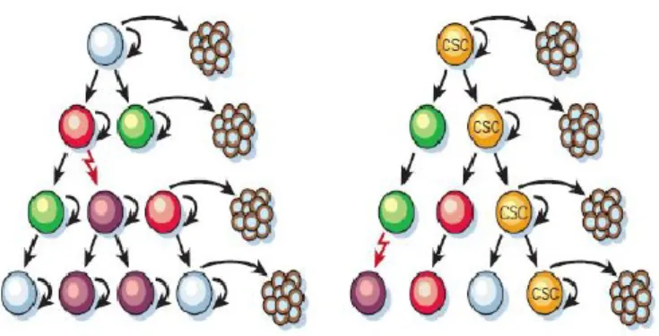
According to traditional models of carcinogenesis, this heterogeneity was explained by stochastic genetic events that, under the influence of the microenvironment, would generate a series of events of clonal selection (88,89). The Cancer Stem Cells (also termed tumor-initiating cells) hypothesis, formulated in recent years, asserts that individual tumors are organized into a hierarchy composed of subsets of tumorigenic stem cells and their non-tumorigenic progeny cells (86,87,88,90)(Fig. 3). Heterogeneous subclonal lineages in solid tumor are branched from distinctive CSCs and are dynamically maintained by these regenerating cells.
Similarly to non-transformed stem cells, CSCs have not only the ability to self-renew but also to undergo asymmetric divisions and low rate of replication, expression of genes related to stem cell, resistance to chemotherapeutic drugs and radiation.
20
Figure 3: Two general models of heterogeneity in solid cancer cells. a, Cancer cells of many different
phenotypes have the potential to proliferate extensively, but any one cell would have a low probability of exhibiting this potential in an assay of clonogenicity or tumorigenicity. b, Most cancer cells have only limited proliferative potential, but a subset of cancer cells consistently proliferate extensively in clonogenic assays and can form new tumors on transplantation.
From: (86)
The first direct experimental evidence supporting the idea of the hierarchical model of CSC-driven tumorigenesis came from acute myeloid leukemia (AML) (91).
These works demonstrated that AML-initiating cells have an immature phenotype (CD34+; CD38-)
similar to normal hematopoietic stem cells (92).
In addition the self-renewal capacity of these cells, a key hallmark of CSCs, was proven by the fact that leukemia could be propagated when implanted into a secondary recipient mouse (90).
Despite technical limitations and some controversies, currently, a growing number of reports support the existence of cancer stem cells in solid tumors (93), including lung cancer.
The capacity of CSC to undergo self-renewal and differentiation has been demonstrated experimentally in various assays.
Purified cancer cells isolated from freshly dissected tissues are able to grow as spheres in non-adherent cultures, form colonies more efficiently than their differentiated progeny and can be continually re-passaged at clonal density.
21
However, the current gold standard assay for CSC tumourigenic potential is their ability to grow in
vivo as serially transplantable tumors in immunodeficient hosts.
This strategy has successfully identified CSCs in several tumor types, such as brain, breast, hematological malignancies as well as in lung cancer.
1.4 Human Lung Cancer stem cells
The existence of CSCs in lung cancer is supported by the fact that only a small population of tumor cells (1.5%) from adenocarcinoma samples possess clonal forming and tumorigenic ability (94). Moreover, indirect evidence suggests the possible presence of cancer stem cells in pulmonary tumors. Stem-like cells have been identified in mouse lung, such as a cell population able to drive the malignant transformation in experimentally-induced neoplasia (95).
Normal lung tissue is composed by a variety of cell types, such as basal mucous secretory cells of the trachea and bronchi, Clara cells of bronchioles, type 1 and type 2 pneumocytes of alveoli. These mature cells derive from the differentiation of lineage-restricted lung progenitor cells, which in turn originate from undifferentiated multipotent lung stem cells (96).
Multipotent, long-lived cells have been identified throughout the airways and give rise to both transiently amplifying and terminally differentiated daughter cells.
Like stem cells of other tissues, lung stem cells are responsible for local tissue maintenance, injury and repair (97).
Some reports have described lung stem cells as cells expressing antigens typical of undifferentiated cells, such as CD34 and BCRP1(98,99). However, only recently, following the growing enthusiasm for the model of “cancer stem cells” in solid tumors and new insights in the biology of normal and cancer stem cells in the mouse airway, the interest of the research is starting to raise for investigation on stem cells in human lung cancer.
Although there are controversies regarding lung cancer CSC markers, CD133, CD44, aldehyde
dehydrogenase isoform 1 (ALDH1), octamer-binding transcription factor 4 (Oct4) and Nanog have
been associated with NSCLC. Importantly, the first isolation and expansion of lung CSC from primary patient tumors was reported by De Maria and Eramo’s research group (100).
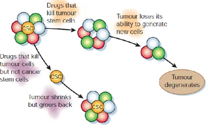
Significant studies have highlighted a key role of CSCs in development, maintenance, metastasis, chemoresistance and relapse of solid tumors, indicating these undifferentiated transformed stem cells as primary targets for more effective anti-cancer therapies (91,101) (Fig. 4).
22
Thus, exploring CSCs in lung cancer may help define responses to conventional and innovative therapies and contribute to the introduction of relevant improvements in clinical oncology, toward the direction of more effective and less destructive personalized therapies. Hence, the development of innovative therapeutic tools targeting lung CSCs may provide promising challenges for increasing lung cancer patient survival.
Figure 4: Conventional therapies may shrink tumours by killing mainly cells with limited proliferative
potential. If the putative cancer stem cells are less sensitive to these therapies, then they will remain viable after therapy and re-establish the tumor. By contrast, if therapies can be targeted against cancer stem cells, then they might more effectively kill the cancer stem cells, rendering the tumors unable to maintain themselves or grow. Thus, even if cancer stem cell-directed therapies do not shrink tumors initially, they may eventually lead to cures.
23 2 AIMS OF THE THESIS
The aim of this study has been the development of innovative therapeutic tools for killing lung Cancer Stem Cells (CSCs) by inhibiting altered signaling pathways.
The lung CSCs previously isolated (100) and new ones that were expanded from patient tumors that routinely are obtained from clinical centers, have been used for these studies.
Although conventional anti-cancer treatments might eradicate most malignant cells in a tumor, they may be ineffective against chemoresistant CSCs, thus this cellular subpopulation should represent the primary target for innovative and most effective experimental therapeutic strategies.
Recent perspectives in cancer therapy consider altered protein signaling networks as the main signature underlying neoplastic transformation. In addition, they emphasize the importance of studying signaling pathways active in tumor cells to develop effective antitumor drugs.
The epidermal growth factor receptor (EGFR) is frequently over-expressed in NSCLC and is taken as a promising target for NSCLC treatment.
Several studies have indicated that the presence of EGFR mutation is a robust predictor of response to EGFR inhibitors (EGFRi). However, about 10-20% of advanced NSCLC patients with wild-type EGFR also benefit from EGFRi treatment, suggesting that other determinants of outcome, besides EGFR mutation, might exist. Thus, this work has been focused on the analysis of EGFR signaling and on the study of the sensitivity of lung CSCs to EGFR inhibitors, with the aim of identifying possible predictive bio-markers of response to EGFRi, in EGFR mutation-negative cells.
These studies might be of crucial importance in developing innovative diagnostic and therapeutic tools for the treatment of lung cancers.
24 3 MATERIALS AND METHODS
3.1 Isolation and culture of lung cancer spheres
Tumor samples were obtained in accordance with consent procedures approved by international Review Board of Department of Laboratory Medicine and Pathology, Sant’Andrea Hospital (University La Sapienza, Rome). Surgical specimens were washed several times in DMEM-F12 medium supplemented with high doses of penicillin/streptomycin and amphotericin B (100U/ml, Gibco,BRL, Rockville,MD) to prevent contamination. Tissue dissociation was carried out by mechanical mince with scissors and tweezers coupled to enzymatic digestion with collagenase II (1.5 mg/ml) (Gibco-Invitrogen, Carlsbad,CA) and DNaseI (20 μg/ml) (Roche,Basilea,SW), for 2 h in a thermostatic shaker at 37°C. When the cell suspension obtained contained high amounts of red blood cells, hypotonic cell lysis with ammonium chloride was performed. Recovered cells were cultured in serum-free medium containing 100 μg/ml insulin, 100 μg/ml apo-transferrin, 10 μg/ml putrescine, 0.03 μM sodium selenite, 2 μM progesterone, 0.6% glucose, 5mM hepes, 0.1% sodium bicarbonate, 0.4% BSA, glutamine and antibiotics, dissolved in DMEM-F12 medium (Gibco-Invitrogen, Carlsbad, CA) and supplemented with 20 μg/ml EGF and 10 μg/ml bFGF.
Flasks non-treated for tissue culture were used to reduce cell adherence and support growth as undifferentiated tumor spheres. The medium was replaced or supplemented with fresh growth factors twice a week until cells started to growth forming floating aggregates. Cultures were expanded by mechanical dissociation of spheres, followed by re-plating of both single cells and residual small aggregates in complete fresh medium.
3.2 Cell culture
The EGFR mutant NSCLC cell lines H1975 (L858R/T790M), H1650 (del ex19) and the EGFR wild type cell lines A549, H460, Calu-3, Calu-1, H1299, H292, were purchased from ATCC and maintained in DMEM or RPMI 1640 medium supplemented with 10% FBS, penicillin and streptomycin.
25
3.3 Western Blot
Proteins were resolved on 4–12% polyacrylamide gels (Invitrogen) and transferred to nitrocellulose membranes. Rabbit polyclonal anti-Phospho-Akt (Ser473), -Phospho-EGFR (Tyr 1068), -Phospho- EGFR (Tyr 1173),-Phospho-Stat3 (Ser727), EGFR and pro-caspase 3 were purchased from Cell Signaling. Mouse monoclonal anti-PTEN was purchased from BD, anti-Phospho-ERK (clone E-4), rabbit polyclonal anti-Bcl-2, anti-BAX (N20), caspase-3 (E-8), ERK, AKT, STAT3 were purchased from Santa Cruz. Mouse monoclonal -Tubulin and α-Actin were purchased from Sigma-Aldrich (St. Louis, Mo, USA).
3.4 PCR and sequencing methods for genomic DNA
DNA or cDNA of all lung cancer stem cell clones were extracted with Pure Link Genomic DNA purification kit purchase from Invitrogen (Gibco-Invitrogen, Carlsbad, CA) or RNeasy mini kit (Qiagen, Hilden, Germany) and reverse transcribed into cDNA by using SuperScript II RT with oligo(dT) as primers (Invitrogen, Grand Island, NY). PCR was performed with the following primers ampliying specific gene region known to be mostly mutated in lung cancer:
KRAS: forward CGTCTGCAGTCAACTGGAAT, reverse CCCTGACATACTCCCAAGGA. These primers amplify a cDNA region encompassing the sites of KRAS mutations (codon 12 and 13 of the exon 1, codon 61 of exon 2).
EGFR: forward TGAGGATCTTGAAGGAAACT, reverse ATGGTATTCTTTCTCTTCCGC. These primers amplify a gene region including exon 18 through 21.
P53: exon 5 forward GCTGCCGTGTTCCAGTTGC, exon 5 reverse CAGCCTGTCGTCTCTCCA; exon 7 forward TGCCACAGGTCTCCCCAAGG, exon 7 reverse GTGTGCAGGGTGGCAAGTG; exon 8 forward CCTTACTGCCTCTTGCTTCT, exon 8 reverse ATAACTGCACCCTTGGTCTC These primers amplify gene regions encompassing codon 157 ,158,175 and 179 of exon 5, codon 248 and 249 of exon 7, codon 273 of exon 8.
EML4-ALK fusion: Fusion-genome-S(5’-CCACACCTGGGAAAGGACCTAAAG-3’) Fusion genome-AS (5’-AGCTTGCTCAGCTTGTACTCAGGG-3’).
PTEN: exon 1-exon 9 HER2: exon 23-exon 24
26
After PCR, the amplification products were run in a 2% agarose gel to verify the purity and then 10g were sent to MWG (Ebersberg,Germany) for sequencing with the same forward and reverse primers used for PCR amplification.
3.5 Chemotherapy resistance studies
Three thousand cells obtained from dissociation of lung cancer spheres were plated in triplicate in 96-well flat-bottom plates.
Chemotherapeutic agents were added at the following final concentrations: Gemcitabine 50 μM, Paclitaxel 30ng/ml, Cisplatin 1 μg/ml, Etoposide 5 μg/ml, Pemetrexed 100 μg/ml. Cell viability was evaluated after 48 h of treatment by Cell Titer Glo luminescent assay (Promega, Madison, WI, USA) based on the ATP quantity evaluation.
3.6 Inhibitor activity studies
The compounds with tyrosine kinase inhibition activity (Biomol International, USA) were tested for the ability to kill lung cancer stem cells. Three thousand cells obtained from lung cancer spheres dissociation were plated in triplicate in 96-well flat-bottom plates. Erlotinib (Tarceva, OSI Pharmaceuticals, Genentech) and Gefitinib (Iressa, AstraZeneca) were added at the final concentration of 10 μM; Cetuximab (Erbitux, Imclone/Merck/Bristol-Myers Squibb) and Afatinib (Giotrif, Boehringer-Ingelheim) were added at the final concentration of 100μg/ml and 1 μM, respectively. After 48, 72, 96 hours we evaluated the cytotoxic effect with cell viability assay (Cell Titer Glo, Promega).
3.7 Cell Cycle Analysis by Propidium Iodide (PI) Staining
Cells (1x10⁵) were washed with PBS and re-suspended in 0.1% sodium citrate, pH 7.4/0.1% Triton
X100, containing 50g/ml propidium iodide and 200 g/ml RnaseA. After 2 hrs of incubation at 4°C, samples were analyzed with FACSCAN BD (Franklin Lake, NJ, USA).
27
3.8 Immunohistochemistry
Formalin-fixed paraffin-embedded 3 micron tissue sections were deparaffinized in xylene and rehydrated in a graded series of alcohol. Immunohistochemical stainings were performed using avidin-biotin-peroxidase complex (UltraTek HRP, Scy Tek Laboratories).
Slides were counterstained with hematoxylin and permanently mounted.
3.9 Fluorescence in situ hybridization
FISH was performed using the LSI EGFR (SpectrumOrange™), a locus-specific probe for the EGFR human gene locus (7q12) and the chromosome enumeration probe (CEP 7, SpectrumGreen™) for alpha-satellite DNA located at the centromere (7q11.1-q11.1) (Vysis, Inc., Downers Grove, IL). The assay was carried out according to the manufacturer's instructions. Shortly after deparaffinization, the FFPE specimens were incubated in the pre-treatment solution (82°C, 30 minutes) and then digested with protease (37°C, 15 minutes). After washing, the slides were counterstained with 4',6-diamidino-2 phenylindole (DAPI) and analyzed using a fluorescent microscope. An average of 30 nuclei was counted for each case. The EGFR gene copy number, chromosome 7 copy number, and the average EGFR gene to chromosome 7 signal ratio were reported as FISH genetic variables.
3.10 Generation of subcutaneous lung cancer xenograft into NSG mice
For mice xenografts, cells were mechanically dissociated to obtain single-cell suspensions, diluited in growth factor-containing medium mixed 1:1 with matrigel (BD Pharmigen, San Jose, Ca) and subcutaneously injected in NOD SCID or NSG mice. When tumor diameters reached at least 5 mm in size, mice were killed and tumor tissue was collected, fixed in buffered formalin and subsequently analyzed by immunohistochemistry. Hematoxylin and eosin staining followed by immunohistochemical analysis were performed to analyze tumor histology and to compare mouse with patient tumors.
28
3.11 In vivo treatment of Lung CSC-generated Xenografts
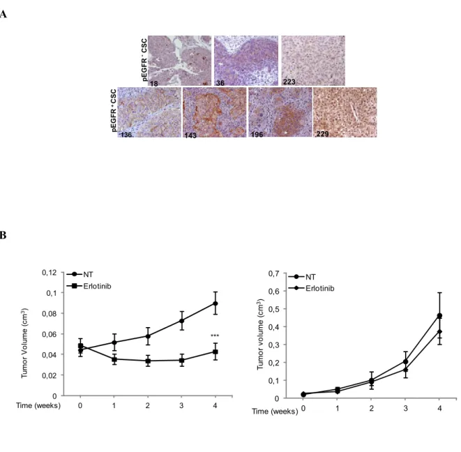
Cell suspensions were mixed 1:1 with growth factor reduced Matrigel (BD) and injected subcutaneously in the flanks of four week-old female NSG mice (Charles River). Two in vivo experiments were performed. In the first in vivo experiment, when tumors reached a mean of 0.5 cm diameter, mice were assigned into 2 treatment groups: a) control (vehicles only); b) Erlotinib (100 mg/kg/5 days on and 2 days of/gavage). In the second in vivo experiment, mice were assigned into 4 treatment groups: a) control (vehicles only); b) Erlotinib; chemotherapeutic agent combinations: c) Cisplatin (3 mg/kg /biweekly/I.P.) + Pemetrexed (200 mg/kg /biweekly/I.P); d) Cisplatin (3 mg/kg /biweekly/I.P.) + Gemcitabine (60 mg/kg /biweekly/I.P). Tumors were measured once a week for the 4 weeks using a caliper, and mice were monitored for signs of drug-induced toxicity and weighed. Of note, chemotherapy treatment determined systemic toxicity particularly severe when Gemcitabine was used. At the end of treatments tumors were collected, dissociated or fixed in formalin and embedded in paraffin for IHC. In each group two mice were left alive in order to check tumor growth at the end of treatment. The dose of Erlotinib was determined on the basis of the plasma concentration peak in vivo.
3.12 Reverse Phase Protein Microarrays Technique
The RPPMA (reverse phase microarrays) technique performs quantitative analysis of specific cellular proteins that can exist in a phosphorilated or non-phosphorilated form. Such a type of protein microarrays could be used to investigate cell cultures, serum samples or biological fluids. For RPPA analysis samples were lysed using T-PER (Tissue Protein Extraction Reagent, Thermo Scientific, Waltham, MA, USA) and diluted up to 0.5 mg/mL with Novex Tris-Glycine SDS Sample Buffer 2X (Invitrogen Corporation, Carlsbad, CA, USA). Subsequently, protein lysates were spotted in a two-fold 5 point dilution curve onto nitrocellulose-coated microscope slides via Aushon Arrayer 2470 (Billerica, MA, USA). Then each slide underwent incubation with a single validated primary antibody using DAKO Autostainer Plus (DAKO Corporation, Glostrup, Denmark). Signal amplification was performed by using DAKO CSA kit (Catalyzed Signal Amplification, DAKO) and diamminobenzidine was used for colorimetric detection of the primary antibody signal. Total protein quantification was performed using Sypro Ruby Protein Staining solution (Invitrogen) and total protein slides were scanned using a Vidar Revolution 4200
29
microarray scanner (Vidar Systems Corporation, Herndon, VA, USA). Antibody slides were scanned using a flatbed scanner and raw images were loaded into MicroVigene software (VigeneTech Inc., Boston, MA, USA) for secondary antibody subtraction and normalization to total protein.
3.13 Clinical-pathological characteristics of patients
The retrospective study included 55 patients from Regina Elena National Cancer Institute; 32
30 4 RESULTS
4.1 Generation and validation of lung cancer stem cells (CSCs)
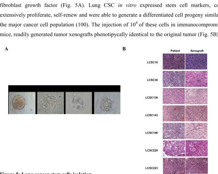
De Maria and Eramo’s research group has been the first to report the identification of lung cancer stem cells (100) through optimizing a reproducible and efficient method that allows to isolate and expand CSCs from lung cancer surgical specimens. This research group isolated and characterized lung cancer undifferentiated cells from the major subtypes of malignant lung tumors, including adenocarcinoma (AC), Squamous Cell Carcinoma (SCC), Large Cell Neuroendocrine Carcinoma (LCNEC) and Small Cell Lung Carcinomas (SCLC). All lung CSCs isolated were able to grow indefinitely as tumor spheres in serum-free medium containing epidermal growth factor and basic fibroblast growth factor (Fig. 5A). Lung CSC in vitro expressed stem cell markers, could extensively proliferate, self-renew and were able to generate a differentiated cell progeny similar to
the major cancer cell population (100). The injection of 104 of these cells in immunocompromised
mice, readily generated tumor xenografts phenotipycally identical to the original tumor (Fig. 5B).
Figure 5: Lung cancer stem cells isolation
(A) Phase contrast photographs of lung cancer spheres obtained from the indicated tumor subtypes. (B) Hematoxylin-eosin (H&E) performed on tumor specimens derived from parental tumor (Patient) or
from tumors generated by subcutaneous injection of lung cancer spheres in NSG mice (Xenograft).
A B LCSC18 LCSC36 LCSC136 LCSC143 LCSC196 LCSC229 LCSC223 Patient Xenograft 20X
31
4.2Molecular characterization of lung CSCs
In the first part of the study, in order to define the molecular background of lung CSCs, these cells were investigated for the presence of the most common alterations associated with NSCLC by DNA sequence analysis.
NSCLC is associated with EGFR over-expression in up to 80% of the patients and mutation of the EGFR proto-oncogene is found in 10-20% of lung carcinomas, therefore, the mutational status of EGFR and others lung cancer associated genes as KRAS, EML4-ALK fusion gene, PTEN, PI3K and HER2 was determined. Moreover, the EGFR and HER2 copy number was determined by FISH analysis.
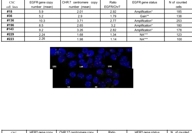
All lung CSC lines were wild type for the region encompassing the most common and clinically relevant EGFR gene mutations (ex 18-,ex 21) and mutation in different sites were excluded by sequencing the whole gene (Fig. 6A).
KRAS G12C mutation was detected in two lung-CSC lines and the EML4-ALK fusion gene was never detected as well as mutations in PTEN or PI3K; HER2 was not mutated in the clinically relevant region (exon 20, ins774) (Fig. 6A).
The evaluation by FISH analysis of the EGFR genomic status in lung CSCs (Fig. 6B), showed increased EGFR gene copy number or low amplification in all lung CSCs analyzed, often associated with increased polysomy at different degrees. HER2 was weakly or not amplified in lung-CSC lines (Fig. 6C).
A
Tumor type KRAS EGFR EML4 -ALK fusion PTEN PI3K HER2 LCC -NE (pT2pN2pMx -IIIA) Mut Ex4 (A146X) wt no wt
SCC (pT2pN2pMX(IIIA)/G2) wt wt no wt SCC (pT3pN0pMx- IIB- G3) wt wt no wt SCC (pT2 N0-IB) mut G12C ( ggt >tgt ) wt no wt AC (pT2pN2pMx-IIIA-G2) wt wt no wt AC (pT4pN1 IIIA-G3) mut G12C (ggt>tgt) wt no wt AC (pT2a pN0 M1 G3) wt wt no wt wt wt wt wt wt wt wt wt wt wt wt wt wt wt #18 #36 #136 #196 #143 #229 #223
32
Figure 6: Genetic pattern of lung cancer associated genes in lung CSCs
(A) Mutational status of lung cancer therapeutic targets in lung-CSCs. (B) Descriptive table (up) and
representative images (down) of EGFR FISH status in lung cancer stem cells (EGFR -orange signal- and centromere 7 -green signal-). (C) HER2 FISH analysis of lung CSCs. Amplification: Ratio > di 2;
** Increase gene copy number and chr 7 polysomy; *** No amplification , no polysomy. CSC
cell lines
EGFR gene copy number (mean)
CHR 7 centromere copy number (mean)
Ratio EGFR/Chr7
EGFR gene status N of counted cells 5.9 2.01 2.92 Amplification* 185 5.2 2.9 1.79 Gain** 138 10.3 3.71 2.77 Amplification* 253 8.5 2.65 3.2 Amplification* 180 9.2 3.26 2.82 Amplification* 178 2.24 1.68 1.34 NA*** 123 18 2.26 1.98 1.14 NA*** 100 #18 #36 #136 #196 #143 #229 #223 B A C CSC cell lines
HER2 gene copy number
CHR 17 centromere copy number
Ratio HER2/Chr17
HER2 gene status N of counted cells 3 2 1.5 NA*** 100 4 2 >2 Amplified 120 >10 2 >2 Amplified 115 3 2 1.5 NA*** 80 2 2 1 NA*** 100 3 2 1.5 NA*** 60 2 3 0.6 NA*** 100 #143 #18 #36 #136 #196 #143 #229 #223
33
4.3 Responsiveness to chemotherapy compared to Erlotinib and EGFR pathway activation in lung CSCs
It was evaluated the response of lung-CSCs to Erlotinib treatment and, in view of a hypothetical therapeutic assignment of a subgroup of EGFR wild type patients to Erlotinib instead of chemotherapy, it has been thought to be relevant to compare the cytotoxic effect determined by Erlotinib on lung-CSCs with that induced by chemotherapeutic agents used as first line therapy for adenocarcinomas (Cisplatin, Gemcitabine, Pemetrexed), squamous cell carcinomas (Cisplatin, Gemcitabine, Docetaxel) or large cell neuroendocrine carcinoma (Cisplatin, Etoposide, Gemcitabine, Docetaxel) as single agents or in the clinically used combinations, mostly platinum doublets. Some lung CSCs were highly resistant to either Erlotinib or chemotherapy, while others displayed a marked Erlotinib sensitivity, even at higher activity than to Cisplatin doublets (Fig. 7A). These results were highly encouraging, particularly for the squamous cell carcinoma (SCC) type of lung cancer, as this subgroup of cancer patients does not have valid alternative options to chemotherapy, currently.
NSCLC patients with sensitizing mutations in EGFR experience improved progression-free survival and response rates with first-line Erlotinib therapy relative to traditional platinum-based chemotherapy. Instead, the patients with EGFR-mutation negative tumors gain greater benefit from platinum-based chemotherapy.
However, at least 10% of patients with wild-type EGFR are responsive to Erlotinib, suggesting that other determinants of outcome might exist besides EGFR mutation.
In order to find molecular predictors of Erlotinib sensitivity, the EGFR pathway activation and the response to Erlotinib treatment (after 48-72h) of lung CSCs were analyzed and it was found that the lines highly sensitive to Erlotinib displayed phosphorylation of EGFR at tyr1068 (LCSC136, LCSC143, LCSC196, LCSC229) despite lack of EGFR mutation, while in the Erlotinib resistant lung-CSCs (LCSC18, LCSC36, LCSC223) EGFR phosphorylation at tyr1068 was not detected (Fig. 7B-D). The results of immunoblot showed also activation of the EGFR downstream pathway (Fig. 7D).
The EGFR can be phosphorylated at tyrosine 1068, 1173 that are major sites, whereas tyr 992, 1148 and 1086 are minor phosphorylation sites. Distinct downstream signalling cascades are initiated by EGFR depending on its phosphorylation, thus it was evaluated also the phosphorylation of EGFR at
34
tyr1173 in all lung CSCs (EGFR WT) and it was found that, differently from pEGFR-tyr1068, pEGFR-tyr1173 was not associated with lung CSCs response to Erlotinib treatment (Fig. 7D). Thus, these results suggested that pEGFR-tyr1068 may be a predictive biomarker to Erlotinib response in lung CSCs with wild type EGFR.
New generation irreversible EGFR inhibitors have shown encouraging antitumor activity in lung cancers, thus, the sensitivity of lung CSCs to others EGFR-TKIs as Gefitinib (Iressa), the monoclonal antibody Cetuximab and the irreversible EGFR inhibitor Afatinib, has been evaluated. A similar correlation was observed among EGFR phosphorylation and cellular response with Gefitinib and Afatinib, while Cetuximab did not display cytotoxic or cytostatic activity against any lung-CSCs (Fig. 7C). Thus, p-EGFR levels correlated with lung-CSCs sensitivity to both reversible and irreversible EGFR inhibitors confirming that EGFR phosphorylation could represent a predictive biomarker for EGFR-TKIs response in lung-CSCs with wild type EGFR.
LCSC36 LCSC136 LCSC196 ** ** ** * ** C on tro l C is G em D o c tx C is + G em C is + D o c tx E rl o ti n ib C on tro l C is G em D o c tx C is + G em C is + D o c tx E rl o ti n ib C on tro l C is G em D o c tx C is + G em C is + D o c tx E rl o ti n ib 0 20 40 60 80 100 120 140 C e ll V ia b ili ty (% vs c on tr o l) * * LCSC18 C on tro l C is E to C is+ Et o G em G em + D o c tx D o ct x E rl o ti n ib ** ** ** LCSC223 C is C on tro l G em D o ct x P em C is + G em C is + D o ct x C is + P em tr x E rl o ti n ib ** ** LCSC229 C is C on tro l G em D o ct x P em C is + G em C is + D o ct x C is + P em tr x E rl o ti n ib * 0 20 40 60 80 100 120 140 0 20 40 60 80 100 120 140 **** ** LCSC143 C is C on tro l G em D o ct x P em C is + G em C is + D o ct x C is + P em tr x E rl o ti n ib A B A 0 20 40 60 80 100 120 140 control 48h 72h 0 20 40 60 80 100 120 140 C e ll vi ab ili ty (% v s c o n trol ) ** *** ** ** ** *** *** * 18 36 136 196 143 229 223
35
control Iressa Erlotinib Cetuximab Afatinib
** *** ** * ** *** * ** C e ll v ia b ili ty (% v s c o n tr o l) 20 40 60 80 100 120 140 * * ** *** 18 36 136 196 143 229 223 0
Figure 7: Comparison of chemotherapy and Erlotinib antitumor activity against lung CSCs
(A) Cell viability of control or treated lung CSCs. Erlotinib (10µM), Cisplatin (1µg/ml), Pemetrexed
(100µg/ml), Gemcitabine (50µM), Docetaxel (0.5µ/ml,) Etoposide (5µ/ml) were added to partially dissociated spheres and cell viability evaluated at the indicated time points by cell viability assay (Cell Titer Glo, Promega). (B) Cell viability of control or Erlotinib-treated lung cancer spheres. (C) Cell viability of control or indicated EGFR inhibitors. (D) Immunoblot for the indicated phosphoproteins (left) or proteins
(right) of the EGFR pathway in lung CSCs or control (H1975) cells. C D H1975 LC SC 18 LC SC 22 9 LC SC 14 3 LC SC 19 6 LC SC 13 6 LC SC 36 LC SC 22 3 pEGFR Tyr 1068 pEGFR Tyr 1173 pAkt pErk pStat3 PTEN -Actin EGFR Akt Erk Stat3 -Actin H1975 LC SC 18 LC SC 22 9 LC SC 14 3 LC SC 19 6 LC SC 13 6 LC SC 36 LC SC 22 3
36
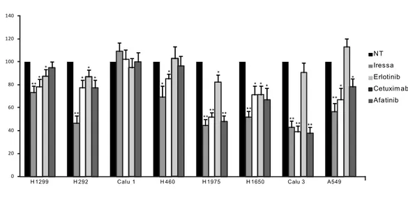
In order to increase reliability of the results obtained, the effects of Erlotinib (Fig. 8B) and others EGFR-inhibitors (Fig. 8D), were analyzed on several commercial lung cancer cell lines with different genetic background (Fig. 8A) and it was observed that the cells that responded to Erlotinib expressed tyr1068 regardless the presence of EGFR sensitizing mutations, while pEGFR-tyr1173 did not correlate with Erlotinib sensitivity (Fig. 8B-C). In fact, the lines with EGFR mutations (H1975, H1650) responded to Erlotinib treatment, as expected, and expressed tyr1068, but also the EGFR wild type line (Calu3) was sensitive to Erlotinib and expressed pEGFR-tyr1068; on the contrary, p-EGFR-tyr1173 was not always expressed in EGFR mutated or wild type sensitive cells.
Thus, the expression of pEGFR-tyr1068 in lung cancer cells was associated with EGFR mutation/sensitivity as well as with sensitivity in wild type receptor context, while p-EGFR-tyr1173 was not correlated with sensitivity.
The data suggested that only pEGFR-tyr1068 may be a predictive biomarker to Erlotinib response in lung cells with wild type EGFR or with EGFR mutation.
A
B C
Cell Line Cell ID EGFR mutation Ras mutation PTEN Her-2 amplification Her-2 mutation PI3KC p53
Calu3 NSCLC (ADC) WT WT WT amplification WT WT mutant A549 NSCLC (ADC) WT G12S (KRAS) WT balanced low polysomy WT mutant WT H1975 NSCLC (ADC) L858R,T790M WT WT 0 WT mutant mutant H1650 NSCLC (BAC) deletion ex 19 WT deletion 0 WT mutant mutant H460 NSCLC (La) WT Q61H (KRAS) WT balanced low polysomy WT mutant WT H292 NSCLC (mucoepidermoid) WT WT WT 0 WT WT WT
H1299 NSCLC WT Q61K (NRAS) WT 0 WT WT WT
Calu1 NSCLC (mucoepidermoid) WT G12C (KRAS) WT 0 WT WT WT
0 20 40 60 80 100 120
H1299 H292 Calu 1 H460 H1975 H1650 Calu3 A549 NT Erlotinib ** ** * * * * * 129H 9 C a lu 3 H 1 6 5 0 H 1 9 7 5 H 4 6 0 C a lu 1 H 2 9 2 A 5 4 9 EGFR p-EGFR Tyr 1068 p-EGFR Tyr 1173 -actin
37
Figure 8: Erlotinib and commercial lung cancer cell lines
(A) Genetic background of commercial lung cancer cell lines. (B) Cell viability of control or
Erlotinib-treated commercial lung cancer cell lines (Erlotinib 10µM). (C) Immunoblot analysis for EGFR and phospho-EGFR sites of commercial lung cancer cell lines. (D) Cell viability of control or indicated EGFR inhibitors evaluated following 72 hours of drug exposure by cell viability assay (Cell Titer Glo, Promega).
4.4 Effects of Erlotinib treatment in lung CSCs
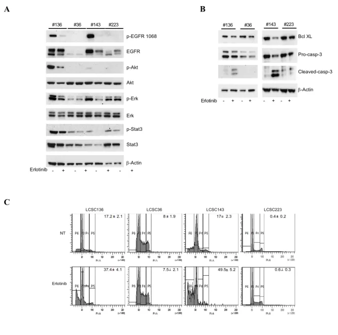
EGFR mutation-negative lung CSCs with activated receptor, were efficiently killed by Erlotinib in
vitro and Erlotinib cytotoxicity occurred mainly through decrease of p-EGFR, its downstream
pathway (p-STAT3, p-AKT, p-ERK) and decrease of the anti-apoptotic protein Bcl-XL (Fig. 9A-B). Besides downregulation of anti-apoptotic Bcl-XL (Fig. 9B), Erlotinib treatment induced cleavage of caspase 3 as demonstrated by the decreased levels of pro-caspase 3 and increased levels of cleaved caspase 3 observed by immunoblot (Fig. 9B).
Moreover, the presence of DNA fragmentated-subG1 apoptotic cells was evaluated by propidium iodide staining and flow cytometry analysis. Erlotinib treatment determined an increase of the cells with a sub-diploid DNA content (representing the apoptotic cells), specifically in lung CSCs with EGFR activation (Fig. 9C).
D ** 0 20 40 60 80 100 120 140
H 1299 H 292 Cal u 1 H 460 H 1975 H 1650 Calu 3 A549 ** * * ** * * * * * * ** * * * ** ** ** ** * ** ** *
control Iressa Erlotinib Cetuximab Afatinib
** 0 20 40 60 80 100 120 140
H 1299 H 292 Cal u 1 H 460 H 1975 H 1650 Calu 3 A549 ** * * ** * * * * * * ** * * * ** ** ** ** * ** ** *
control Iressa Erlotinib Cetuximab Afatinib
** 0 20 40 60 80 100 120 140
H 1299 H 292 Calu 1 H 460 H 1975 H 1650 Calu 3 A549 NT Iressa Erlotinib Cetuxim ab Afatinib *** * ** * * * * * * ** * * * ** ** ** ** * **** * ** 0 20 40 60 80 100 120 140
H 1299 H 292 Cal u 1 H 460 H 1975 H 1650 Calu 3 A549
** * * ** * * * * * * ** * * * ** ** ** ** * ** ** *
