UNIVERSITÀ DEGLI STUDI DI MESSINA
Dipartimento di Scienze Chimiche, Biologiche, Farmaceutiche e Ambientali
DOTTORATO DI RICERCA IN SCIENZE CHIMICHE
XXXII CICLO
Thermodynamic investigation on As(III) and
As(V) in aqueous solution and extraction
from natural water samples by Polymer
Inclusion Membranes (PIMs)
Donatella Chillé
Supervisor
Prof. Claudia Foti
Coordinator
Prof. Paola Dugo
This thesis was finantial supported by FSE (Fondo Sociale Europeo) regional
fund.
Project: Dottorati FSE XXXII Ciclo Unime
CIP 2014.IT.05.SFOP.014/3/10.5/9.2.02/0006
“Tenta l’incompiuto, lo straordinario
Vivi in eccesso, cominciando adesso
Goditi il trionfo, crea il tuo miracolo
Cerca il vero amore dietro ad ogni
ostacolo!”
Acknowledgements
Three years could appear like an infinitely long period of time but now that I am going to write my doctoral thesis, I realize that they have passed really quickly. During this period, I had the pleasure to meet so many people who have enriched me from a professional but mainly human point and to them I address my thanks. However, there are people who deserve to be mentioned as they were pillars for me during this experience. In particular, my supervisor Prof. Claudia Foti, a very important person who, every day, guided my research activity always providing me with valuable advices. Also every single member of the analytical research group and especially Anna, Rosalia and Antonio with whome, having to share the office, I also shared “joys and sorrows”. Thank you very much for EVERYTHING.
I spent part of the second year of my PhD at the University of Girona (Spain), guest of the Prof. Clàudia Fontàs. A kind person, always available both at work and out, who allowed me to know, appreciate and even "export" a research line very far from mine. Receiving compliments, especially in the context of an international congress, was a great satisfaction for me! Many thanks also to Laura, Gemma and Ruben. You were really important figures for me because you made me feel like home from the first day I have been there, alleviating the lack of all the affections I had left in my beloved Sicily (or Chichilia), where you will always be welcome. Thanks also to Soetkin, Ibrahim, Marta and Mayra, who accompanied my days in the laboratory between a PIM and a laugh! Of course, I cannot fail to mention Enriqueta, always friendly and present for any suggestion, and Eva who patiently explained to me the principles of EDXRF, helping to carry out part of my research work. All of you will always have a special place in my heart!
Among the special thanks, I cannot fail to include Fausta, my so-called left arm (since she is left-handed). We shared high school, graduation years and part of the PhD period, thus we can say that we have been supporting each other for more than half of our lives! Thanks for all the tips, relative to work and life, and to always being there even when we have been located in different countries.
A deserved thanks goes to my FAMILY. My parents and my sister know that I am not a person of many words, but on this occasion I want to say that a good part of my success is due to them, to their advices, to their continuous encouraging me to overcome the limits that I have often imposed myself unjustifiably.
Especially, Prof. Anna Napoli and Dr. Donatella Aiello for the mass spectrometry measurements; Prof. Carmelo Sgarlata and Prof. Ottavia Giuffré for the calorimetric titrations; Dr. Franz Sajia, Dr. Sebastiano Trusso, Dr. Rosina Ponterio, Dr. Viviana Mollica Nardo and Dr. Fausta Giacobello for the computationl and Raman studies, as well as Dr. Giuseppe Cassone for the molecular dynamics simulations.
Index
Aim of the work ... 1
Introduction ... 5
Section I: Experimental Section ... 13
1 Chemicals, analytical techniques, mathematical and computational approaches used for thermodynamic studies ... 14
1.1 Chemicals ... 14
1.2 Potentiometry ... 16
1.2.1 General aspects ... 16
1.2.2 Potentiometric equipment and procedure ... 17
1.3 UV-Vis spectrophotometry ... 20
1.3.1 General aspects ... 20
1.3.2 UV-Vis equipment and procedure ... 21
1.4 Calorimetry ... 23
1.4.1 General aspects ... 23
1.4.2 Instrumental equipment and procedure ... 23
1.5 Raman Spectroscopy ... 27 1.5.1 General aspects ... 27 1.5.2 Instrumental equipment ... 27 1.6 Mass Spectrometry ... 28 1.6.1 General aspects ... 28 1.6.2 Instrumental equipment ... 29 1.7 Computational methods ... 30
1.7.1 Ab initio molecular dynamics ... 30
1.7.2 Ab initio molecular dynamics simulations of acid-base properties of As(III) and As(V) 30 1.7.3 Ab initio molecular dynamics simulation of As(III)- tla, tma and dmsa systems ... 31
1.8 Calculations ... 32
1.8.1 Computer programs ... 32
1.8.2 Equilibrium constants ... 34
1.8.3 Dependence of the stability constants on the ionic strength ... 35
1.8.4 Dependence of the stability constants on the Temperature ... 37
2 Analytical techniques for arsenic determination in water samples ... 39
2.1 Chemicals ... 39
2.2 Inductive Coupled Plasma - Optical Emission Spectroscopy (ICP-OES) ... 40
2.2.1 General aspects ... 40
2.2.2 Instrumental equipment and procedure ... 41
2.3 Energy Dispersive X-Ray Fluorescence (EDXRF) Spectrometry ... 42
2.3.1 General aspects ... 42
2.3.2 Instrumental equipment and procedure ... 43
Section II: Arsenic(III): acid-base properties and interaction with different classes of ligands in aqueous solution ... 44
1 As(III) acid-base properties: results and discussion ... 45
1.1 Computational results ... 46
1.2 Experimental results ... 47
1.2.1 Protonation constants determination ... 47
1.2.2 Dependence on ionic strength ... 48
1.2.3 Dependence on temperature ... 48
1.3 Final remarks ... 51
2 As(III)-thiols interactions: results and discussion ... 52
2.1 Ligand protonation constants ... 55
2.2 As(III)-tla, -tma and -dmsa complexes ... 57
2.2.1 Thermodynamic results... 57
2.2.2 Dependence on temperature ... 62
2.2.3 Computational results... 64
2.2.4 Raman spectroscopy ... 67
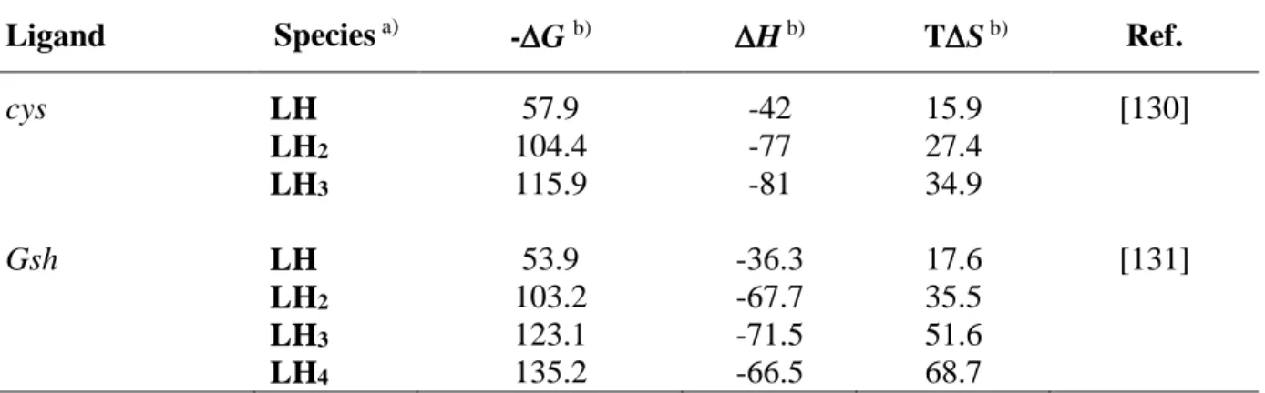
2.3 As(III)-cys and -gsh complexes ... 69
2.3.1 Thermodynamic results... 69
2.3.2 Dependence on ionic strength ... 73
2.3.3 Dependence on temperature ... 73
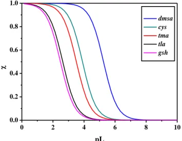
2.4 Sequestering ability ... 75
2.5 Literature comparison ... 76
2.6 Final remarks ... 77
3 As(III)-carboxylic acids, -amino acids and -nucleotides interactions: results and discussion ... 78
3.1.1 General aspects ... 79
3.1.2 Ligand protonation constants ... 81
3.1.3 Thermodynamic results... 81
3.2 As(III)-amino acids complexes ... 84
3.2.1 General aspects ... 84
3.2.2 Ligand protonation constants ... 86
3.2.3 Thermodynamic results... 86
3.3 As(III)-nucleotides complexes ... 89
3.3.1 General aspects ... 89
3.3.2 Ligand protonation constants ... 91
3.3.3 Thermodynamic results... 91
3.4 Sequestering ability ... 94
3.4.1 As(III)-carboxylate complexes ... 94
3.4.2 As(III)-amino acid complexes ... 95
3.4.3 As(III)-nucleotide complexes ... 96
3.5 Final remarks ... 98
4 As(III)-phosphonic acids interactions: results and discussion ... 99
4.1 Ligand protonation constants ... 101
4.2 As(III)-NTAP, -NTA2P and -NTA3P complexes ... 102
4.2.1 Thermodynamic results... 102
4.2.2 Dependence on ionic Strength ... 105
4.2.3 Dependence on temperature ... 107
4.2.4 Mass spectrometry results ... 112
4.3 Sequestering ability ... 117
4.4 Final remarks ... 118
5 Empirical relationships ... 119
Section III: Arsenic(V): acid-base properties and interaction with alkaline-earth and trivalent metal cations in aqueous solution ... 121
1 As(V) acid-base properties: results and discussion ... 122
1.1 Computational results ... 123
1.2 Experimental results ... 124
1.2.1 Protonation constants determination ... 124
1.2.3 Dependence on temperature ... 127
1.3 Final remarks ... 128
2 As(V) interaction with alkaline-earth metals: results and discussion ... 129
2.1 Ligand protonation and metal hydrolytic constants ... 131
2.2 Ca2+, Ma2+ and Sr2+ -arsenate complexes ... 131
2.2.1 Thermodynamic results... 131
2.2.2 Dependence on ionic strength ... 133
2.2.3 Dependence on temperature ... 134
2.2.4 Speciation in natural waters ... 136
2.3 Final remarks ... 137
3 As(V) interaction with Fe3+ and Al3+: results and discussion ... 138
3.1 Ligand protonation and hydrolysis constants ... 140
3.2 Fe3+ and Al3+-arsenate complexes ... 140
3.2.1 Thermodynamic results... 140
3.2.2 Dependence on ionic strength ... 144
3.2.3 Dependence on temperature ... 145
3.3 Sequestering ability ... 147
3.4 Final remarks ... 148
Section IV: Arsenic speciation in natural waters by polymer inclusion membrane preconcentration and X-ray fluorescence detection ... 150
1 Polymer Inclusion Membranes (PIMs)... 151
1.1 General aspects ... 151
1.2 PIMs composition and preparation ... 153
2 Arsenic extraction by PIMs and detection by EDXRF ... 155
2.1 Preliminary PIM extraction experiments... 156
2.2 As(III) preconcentration in PIMs ... 157
2.3 PIMs preconcentration combined with EDXRF for arsenic detection ... 158
2.3.1 Testing PIM abilities to perform As determination by EDXRF ... 158
2.3.2 Analytical figures of merit of the developed method ... 160
2.3.3 Effect of water matrix on As extraction ... 161
3.1 Reduction of As(V) to As(III) and determination with the developed methodology 164
3.2 Speciation studies ... 165
3.3 Application to natural waters containing arsenic ... 166
3.4 Final remarks ... 168
Section V: Conclusions ... 169
1
Aim of the work
Arsenic is considered a powerfull poison, used from ancient times for committing suicides and murders. Its toxicity is well known and it represents an important hazard for human health. It must be remembered that the toxicity, as well as the mobility and bioavailability of an element are related to the chemical form in which the element itself is present and for this reason it is important to distinguish among the different forms of arsenic in order to have awareness of the risk connected to its exposure. Despite this, very few studies are reported in literature about the thermodynamic behaviour of arsenic, especially when it combines with molecules of biological importance. Accordingly, this research work stems from the desire to enrich the literature database regarding the arsenic speciation in aqueous solution. The speciation studies play an important role in the present-day analytical research, especially in the environmental field where the increase of pollution due, for example, to heavy metals reflects in a world problem for human health and ecosystem. The total determination of an element is not sufficient to explain the possible produced effect and thus the identification of the individual chemical and physical forms, called speciation analysis, is needed. This kind of study also reveals very useful in medical field when chelating therapy is necessary for removing metals from human body. In this case, the knowledge of the exact chemical form of the metal, for example the oxidation state, is crucial since the chelating agent could act differently depending on it.
In the present thesis, arsenic was treated considering two different oxidation states, in particular +5 and +3, using arsenate and arsenite respectively as starting molecules. The introduction describes the history of arsenic, its chemical and physical properties as well as the distribution in the environment (air, soil, water), the effects on human health and the application fields.
After a first section in which all the chemicals, analytical techniques, mathematical and computational approaches are described, the second one (Section II) is focused on the metallic behavior of arsenic in the trivalent oxidation state. The first investigation, regarding the acid-base properties of arsenite, was performed in aqueous solution but also by exploiting ab initio molecular dynamic simulations. Its interaction with different types of ligands was analysed at T = 298.15 K and different ionic strength values, by employing NaCl as background salt. In this section, a wide space was reserved for thiol ligands, given the high affinity of As(III),
2 often cited in literature [1-3], for the -SH groups of proteins. In addition to the classic thermodynamic approach, conducted through different analytical techniques including potentiometry, spectrophotometry and calorimetry, which allowed to determine the best speciation models together with the thermodynamic parameters of the obtained species, also
ab initio molecular dynamic studies were performed in order to better understand the
coordination mode of the metalloid with the functional groups present in the molecules. In order to complete the study relative to the interaction of As(III) with ligands of biological interest, also carboxylic acids, as well as amino acids and nucleotides were taken into account, performing an investigation less extensive, since the formation of weakly complexes was registered. In order to evaluate the possible use of phosphonic ligands for removing As(III) from environmental matrices, the binding ability of phosphonic acids derived from nitrilotriacetic acid by replacing the carboxyl groups with phosphonic ones were investigated in a wide range of ionic strength (0.1 < I/mol L-1 <1) by means of potentiometry and calorimetry and the sequestering ability was examined in conditions simulating sea water (I = 0.7 mol L-1 e pH = 8.1) and a fresh water (I = 0.001 mol L-1 e pH = 7). For these systems, also mass spectrometry experiments (LD MS/MS) were carried out in order to gain information about the structures of the complexes.
For the metal-ligand systems considered more interesting from the thermodynamic point of view, also the dependence of the stability constants on the ionic strength was taken into consideration by using the Debye-Hückel type equation. It is a mathematical model that allows to determine the stability constants at ionic strength values different from the experimental ones, by knowing the infinite dilution constant (log Tβ) and C, an empirical parameter linked to the charges involved in the formation reaction. This approach is particularly important for predictive purposes in real matrices which are characterized by very different ionic strength conditions. For all the systems, the sequestering ability of the ligands towards the metalloid was evaluated by using an empirical parameter known as pL0.5 that will
be well discussed later.
Section III is dedicated to the study of arsenate (As(V)) as ligand molecule, by analyzing its non-metallic behavior with respect to bi- and trivalent cations. Specifically, the acid-base properties were determined in a wide range of ionic strength and T = 298.15 K and, also in this case, the study was completed with the application of molecular dynamic simulations. Successively, considering that in sea water arsenic is mostly present as As(V), speciation studies were performed in presence of the most representative alkaline-earth metals of this fluid, i.e. Ca2+, Mg2+ and Sr2+. The speciation of arsenate with alkaline-earth metals was
3 evaluated by means of potentiometry, in order to define the speciation models, and through calorimetry for establishing the thermodynamic parameters for the dependence on temperature. Trivalent cations, specifically Fe3+ and Al3+, were chosen as a consequence of the pronounced affinity of arsenate towards these metals, widely discussed in terms of sorption processes onto mineral surfaces, such as metal oxides of Al, Fe and Mn, but poorly investigated as metal-ligand complexes in aqueous solution. Also for this system, the dependence of the stability constants on ionic strength and temperature was dealt with and, to complete the study, the sequestering capability of arsenate towards Fe3+ and Al3+ was investigated.
Section IV is focused on the research work performed during my PhD abroad at the University of Girona (Spain), under the supervision of Prof. Clàudia Fontàs of the Department of Chemistry. With respect to the previous sections, this one is more applicative and based on the extraction of arsenic, in particular As(III), from natural waters by means of Polymer Inclusion Membranes (PIMs), incorporating the extractant Bis (2,4,4-trimethylpentyl) dithiophosphinic acid, commercially known as Cyanex 301. After preliminary solvent extraction experiments (using a 0.1 mol L-1 Cyanex 301 solution in toluene), performed in 0.1 mol L-1 HCl solutions containing 10 mg L-1 of As(III) or As(V), that demonstrated the
complete extraction of As(III) and As(V) after 30 minutes and 24 hours respectively, the extraction was carried out by using Polymer Inclusion Membranes (PIMs). This type of membranes, based on the incorporation of an extractant within a polymeric matrix possess a series of chemical-physical characteristics that will be discussed in the section, are easy to prepare and need a small amount of solvent for the preparation, in contrast with the previous solvent extraction experiments. Membranes with different compositions, in terms of polymer and extractant amount present in the membrane were tested and, finally, the best performance in terms of As(III) extraction was obtained with a PIM containing 50% CTA (Cellulose triacetate) as the polymer and 50% Cyanex 301 (% in mass). It was found that this PIM was not able to extract As(V), and, therefore, can be useful for speciation studies. In order to apply the extraction system to natural waters, some extraction experiments were performed using mineral water, sea water, tap water and spring water samples, all spiked with known concentrations of As(III). Then, the extraction efficiency was evaluated by analyzing the initial and final solutions by emission spectroscopy (ICP-OES). Based on the good efficiency of the PIM as a sorbent to collect As(III), a new analytical methodology was developed by combining the extraction by PIMs followed by detection by Energy Dispersive X-Ray Fluorescence (EDXRF) spectrometry, that allows the direct determination of the arsenic
4 extracted in the membrane, without any elution step or any other pre-treatment. Also, the detection of As(V) was achieved by applying a previous reducing treatment with sodium thiosulfate and potassium iodide prior its extraction by PIMs and detection by EDXRF. Finally, the developed methodology was successfully used to measure As species in naturally occurring arsenic water samples.
5
Introduction
Arsenic is a substance well known since ancient time for its poison properties. The name derives from the Persian zarnikh, meaning “yellow orpiment”. Then it was incorporated into ancient Greek as arsenikon, which also means “masculine” or “potent”. The term was translated as arsenicum in latin and arsenic in old French from which the current English word derived [4]. Throughout history, arsenic, thanks to its characteristics of odourless and tasteless, was used to commit several barbaric murders, especially in royal families in order to facilitate rich inheritance. Nero, for example, murdered his stepbrother Britannicus in order to become Emperor of Rome. Because of these events and its potency, arsenic was indicated as “Poison of Kings and King of Poisons”. Among the prominent families engaged in poisoning, the Borgias were the most notorious. In Italy, a lady which stood out for her poisoning skills was Giulia Toffana. She made “Aqua Toffana” and sold it both as a cosmetic and a devotionary object in vials, basically to women trapped in difficult marriages who wanted to get rid of their husbands. The sale always was accompanied by appropriate instructions in order to avoid accidental poisonings. Giulia and her daughter Girolama were executed in Campo dei Fiori in Rome for the death of hundreds of men.
Moreover, in the 19th century arsenic powder was used by women in order to whiten their faces, their hair and scalp to destroy vermin [5, 6]. The fame of the toxic properties of arsenic was also described in many literary and theatrical works. For example, it was employed by Madame Bovary for her suicide in the novel of the French writer Gustave Flaubert, published in 1856, but also in “Arsenic and Old Lace”, a play by American playwright Joseph Kesselring, written in 1939. It became best known through the subsequent film adaptation starring by Cary Grant and directed by Frank Capra. In the comedy, the aunts of the leading actor, by renting rooms to lonely old bachelors, put an end to their suffering by serving them a cup of elderberry wine spiked with arsenic, strychnine and "just a pinch of cyanide" while they getting acquainted. Unfortunately, arsenic compounds, such as Lewisite and Adamsite, were also used in the World Wars as chemical weapons. The first one has an odour of geraniums and is a potent respiratory and eye irritant as well as extremely poisonous if absorbed. Adamsite is a vomiting agent. Its effects on health are less dangerous than that produced by Lewisite since they become significant just when the exposure verifies in enclosed space or under adverse weather conditions. In these cases, it may result in more
6 severe adverse health effects as well as death [7, 8]. Despite its toxicity, arsenic was widely employed in medical fields from ancient times. Hippocrates recommended an arsenic paste for treating ulcers as well as Paracelsus [8, 9]. The use of arsenic as a drug therapy reached the glory in the 18th century when Thomas Fowler, a British physician, prepared the “Fowler solution” made by arsenic trioxide in potassium bicarbonate (1% w/v). This remedy was used empirically for the treatment of several diseases such us chorea, asthma, eczema and psoriasis. The discovery of a decrease in white blood cells counts in subject affected by leukemia when treated with Fowler solution led to the use of this mixture for the treatment of leukemia until the 20th century when it was replaced by radiation therapy [9].
The arsenic decline in medicinal field could be attributed to its toxicity and carcinogenicity, especially skin cancer. The International Agency for Research on Cancer included arsenic and certain arsenic compounds in group 1, namely agents that are carcinogenic to humans [10, 11].
Chemical properties and redox behavior. Arsenic is the 20th most abundant element in the earth’s, 14th in the sea and 12th in the human body. It belongs to Group 15 of
the Periodic Table with atomic number 33 and atomic mass equal to 74.921. Its more stable isotope is the 75As. Arsenic is part of the metalloids and, for this reason, it exhibits both metallic and non-metallic properties. Elementary arsenic exists in three solid modifications:
Yellow arsenic is unstable since it is obtained by sudden cooling of arsenic vapours to below 0°C; it is converted in the more stable grey form thanks to heat, light or catalysts such as iodine and bromine.
Black arsenic is more stable than yellow form but less than Grey and its individuality is not certain.
Grey arsenic is metallic and represents the ordinary stable form.
Arsenic shows various valences (-3; 0; +3; +5) and it can be present as organic and inorganic forms. It displays -3 oxidation state, for example, in the arsenic trihydride (arsine, AsH3)
compound. It is a colorless gas with a high poisonous power and a disagreeable garlic odor. The +3 oxidation state is shown in arsenic trioxide (As2O3), a tasteless and odourless
compound used in the past as a lethal poison. Also arsenous acid and arsenites are trivalent compounds. Examples of compounds in which arsenic presents +5 oxidation state are arsenic pentaoxide (As2O5), arsenic pentasulfide (As2S5) as well as arsenates. Organic compounds
7 occur as a result of biological activity and some of them, such as 2-chlorovinyldichlorarsine (Lewisite), were used as chemical weapons [12-14].
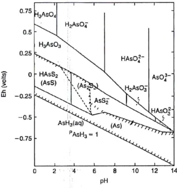
Depending on the redox potential and pH, arsenic in water mainly exists in trivalent or pentavalent form, since the -3 and 0 valence states rarely occur in nature. Figure i shows the Eh-pH diagram for inorganic species of arsenic in natural environment. Under oxidizing
conditions (high Eh values) the As(V) form predominates and the distribution of the
oxoanions is related to the pH. In particular, at pHs lower than 2 the principal species is the H3AsO4 one; from pH = 7 to pH = 11 the H2AsO4- and HAsO42- species prevail and they are
progressively substituted by the AsO43- for higher pH values. Under reducing conditions (low
Eh values) arsenic mainly exists as As(III). Also in this case, the oxoanions distribution is
associated to the pH. For pH < 9, the prevalent species is the undissociated one H3AsO3 which
becomes H2AsO3- once crossed this value. At Eh values below -250 mV, if in presence of
sulphur or hydrogen sulphide, arsenic compounds such as As2S3 can be found. Arsine and
elemental arsenic could be formed but in very strong reducing conditions [2, 15].
Figure i. Eh-pH diagram for arsenic at T = 298.15 K. Conditions: CAs = 0.00001 mol L-1 and
CS = 0.001 mol L-1 [15].
The redox behavior of arsenic in acidic medium (pH = 1) was well analysed by means of cyclic voltammetry by Yang et al. The results showed that the over-potential of As(III) oxidation to As(V) is very high (~ 0.9 V) in the experimental conditions under investigation. This aspect could indicate that on-site peroxidation in acidic wastewater is very difficult [16].
8
Arsenic in the environment. Arsenic is an ubiquitous element easily found in the environment both for natural and anthropogenic reasons. As mentioned above, it is a component of the earth’s crust but it also occurs in minerals and soils and thus, it can reach water and air through wind-blown dust and runoff. Arsenic is naturally present in over 200 different mineral forms but only a few are encountered in relevant amounts, such as realgar (AsS), orpiment (As2S3) and arsenopyrite (FeAsS) which represents the most abundant As
mineral [17]. The rock erosion together with weathering and volcanic eruptions are events that could contribute to the release of arsenic. In soil, arsenic can be found in three main forms: As(III), As(V) and organic arsenic. The As content is affected, basically, by climate, organic and inorganic components of the soils but also by the redox potential. [18, 19]. As an example, during the wet season, the soil conditions become reducing with the consequence of arsenic release from iron hydroxide sites. In the dry season, it remains fixed in the soil texture and therefore the arsenic amount results to be higher [20]. It should be noticed that in oxidizing conditions arsenate is the most stable form and it is easily adsorbed into clay, iron and manganese oxides/hydroxides, avoiding its mobility into the soil [21, 22]. In particular, on amorphous iron hydroxide, As(V) absorption seems to be independent of pH at low As concentrations but it increases when pH decreases (4 < pH < 9) at high As concentrations. Instead, the best adsorption of As(III) verifies at around pH = 7 [23]. In reducing conditions the most mobile arsenite prevails; accordingly, its immobilization could be favored by oxidation. The arsenic mobility is also influenced by the presence of ions defined as competitors with respect to arsenic. Inorganic and organic anions, such as (PO43-), silicate
(SiO22-), sulphate (SO42-), nitrate (NO3-), bicarbonate (HCO3-) and natural organic matter
(NOM) can compete with arsenic for adsorption sites [18, 24-26]. Among these ones, the effect due to the presence of the phosphate is the most pronounced, since the arsenate structure is very similar to the phosphate molecule. Moreover, phosphorus is present in considerable amount in the soil, probably because of its use as component in fertilizers in agricultural field. Hence, PO43- could be able to substitute the adsorbed AsO43- favoring the
mobility or, otherwise, prevent its adsorption. Clays and inorganic arsenic compounds can be methylated by microorganisms with the aim to produce, under oxidizing conditions, monomethylarsonic acid (MMA), dymethilarsinic acid (DMA) and trimethylarsine oxide (TMAO). Generally, arsenic levels in untreated and uncontaminated soils are between 1-40 mg kg-1, while larger concentrations are observed in alluvial and organic soils [17]. The recommended value by the European Community is at most 20 μg g-1 [27].
9 In natural waters arsenic should be present in low concentrations; the World Health Organization fixed a regulatory value equal to 10 μg L-1 in drinking waters. Normally, the
highest concentration is observed in ground waters as a consequence of a strong water-rock interaction and favorable physical and geochemical conditions for the dissolution and mobilization of arsenic [27]. The main forms that can be found in natural waters are the same previously mentioned as regards the soil, namely arsenate and arsenite. Also organic species, such as dimethylarsinate (DMA) and monomethylarsonate (MMA), have been identified but in very low concentrations and, especially, in surface water as a result of biological activity. [27, 28]. Biomethylation is made by various species of micro-and higher organisms and may affect the mobility of arsenic species. In anoxic conditions, in fact, it causes the reduction of As(V) to soluble As(III) species and mobilizes As from the aquifers into groundwater [20, 29]. The seawater, normally, contains 0.001-0.008 mg L-1 of arsenic. The thermodynamically most stable form is arsenate and its pentavalent oxyanions (H2AsO4-, HAsO42- and AsO43-). At
lower redox potential and reducing conditions, arsenic in the trivalent oxidation state (H3AsO3) prevails [17]. In unpolluted fresh and ground waters, the amount of arsenic ranges
from 1-10 μg L-1, reaching values of 5000 μg L-1 in areas of sulfide mineralization and mining [17, 27, 30].
In the atmosphere, arsenic is mainly present as a mixture of As(V) and As(III) absorbed on particulate matters. Both natural phenomena (volcanic eruptions, weathering) and anthropogenic activities (pesticides, smelting, fossil fuel combustion, mining activity) promote the arsenic release in the air [27]. Normally, the human exposure to arsenic through air is negligible. The estimated levels in Europe range from 0.2 – 1.5 ng m-3 in rural areas, 0.5 – 3 ng m-3 in urban areas and 50 ng m-3 in industrial areas [14]. The daily respiratory intake of
arsenic is approximately 120 ng m-3 of which just 30 ng m-3 are absorbed [17].
Arsenic toxicology and health effect.
Arsenic may reach human organism through
many pathways, as explained in the previous paragraphs. However, it seems that the preferential way is represented by the direct consumption of As contaminated drinking water [31]. Groundwater is the most important source of drinking water and it is often employed for the irrigation of vegetables and fruits. Thus, the consumption of agricultural products cultivated by using arsenic contaminated groundwater represents another source of arsenic exposure [32]. Elevated concentrations of arsenic are registered primarily for natural reasons, such as rock erosion, but the use in industrial processes, mining activities, pesticides and fertilizers contributes to the global contamination, basically as a consequence of leaching10 processes. In 1963, the World Health Organization fixed a recommended value for arsenic in drinking water equal to 50 μg L-1, but successively it was further reduced to 10 μg L-1 in 1992,
following the suspicion of carcinogenicity [33]. The FAO/WHO provisional tolerable weekly intake (PTWI) of 15 μg kg-1 body weight for a 60-kg man is 2.1 μg kg-1. However, many countries of the world, such as Bangladesh [34, 35], India [35], Argentina [36], Canada [37] and Vietnam [38], present high levels of arsenic in drinking waters; thus, the daily arsenic intake, often, exceed the PTWI [14]. The arsenic poisoning can be differentiated in acute and chronic. The acute toxicity is often related to the accidental ingestion of insecticides or pesticides and rarely as a consequence of a suicide attempt. The prevalent features are nausea, vomiting, diarrhea and excessive salivation. The haematological picture changes and renal and respiratory failure occurs as well as pulmonary edema. Urinary arsenic concentration is the best indicator to detect a recent poisoning.
Chronic toxicity is the most common form and it occurs in people daily exposed to the contaminant for several reasons. Absorbed arsenic is accumulated in liver, heart, kidney and lungs and deposited in the ketarin-rich tissues. The normal amount of arsenic in hair is around 0.08-0.25 μg g-1 until 1.0 μg g-1 in case of poisoning. In nails the normal concentration is 0.34 μg g-1, while in urine it can vary from 5 to 40 μg per day. A concentration of 100 μg per day
is registered in case of acute and subacute poisoning [17]. The effects of the chronic intake of arsenic on human organism are various, ranging from hyperpigmentation, hyperkeratosis, increase of risk of cardiovascular and respiratory disease as well as diabetes mellitus, until malignant change in almost all the organs of the body [39]. A ten-year study, conducted by Choudhury et al., was recently published. In the investigation, a sample of 960 patients coming from different arsenic contaminated districts of Bangladesh and showing cutaneous malignancy (single lesion), was tested from January 2004 to December 2015. The results of the study are very alarming, if we think that only a small portion of the nation was examined. In fact, the 58.65% of the patients (N = 563) was affected by Basal Cell Carcinoma (BCC), Squamous Cell Carcinoma (SCC) was found in 40% (N = 384) of the study population, 1.15% (N =11) resulted to be the melanoma patients and 0.21% (N = 02) of the people showed Merkel Cell Carcinoma (MCC) [40].
In relation to the chemical form in which the element occurs, the exhibited toxicity may be different. As(III) and As(V), in fact, act differently in human organism. For the first one, a high affinity towards sulfhydryl groups of the proteins is documented as well as the resulting toxicity due to the inhibition of important biochemical events [1]. Arsenate presents a very
11 similar structure to phosphate molecule and this similarity could involve an incorporation of arsenate in some metabolic reactions in which phosphate is normally required [3].
When ingested, inorganic arsenic is readily absorbed in the gastrointestinal tract. It seems that it is badly absorbed through intact human skin but can bind externally to skin and hair. Absorbed arsenic is transported through the blood to the other organs of the body, bound to SH groups of proteins and low-molecular-weight compounds such as GSH or cysteine. The major part of arsenic in blood is cleared with a half-time of about 1 hour, while the whole-body half-time of ingested arsenite is about 4 days and the urine is the prevalent excretory pathway [41]. In blood, in fact, the concentration is elevated only for short time after absorption. However, in case of chronic exposure, such as contaminated drinking water intake, arsenic levels can reach a constant state which reflects the degree of exposure [14]. In human organism, alternative steps of reduction of the pentavalent arsenic compounds come in succession with oxidative methylation processes which allow to the formation of differently methylated arsenicals, in particular monomethyl and dymethyl arsenic compounds, generically indicated as MAs and DMAs when the oxidations state cannot be determined [42, 43]. The Figure ii, taken from Ref. [44], well shows the metabolic conversions of arsenic species. The predominant metabolite of inorganic arsenic, rapidly excreted by most mammals, is represented by the dimethylarsinic acid (DMAsV) [3].
12
Applications. Probably, the most pronounced use of arsenical compounds is to be attributed to the formulations of pesticides, fungicides and herbicides. Paris Green, a copper acetoarsenite compound, appeared around 1860 and was employed as standard insecticide for about 30 years. Later, it was replaced by lead arsenate, considered less toxic for plants, and it was widely used for many years as pesticide for apple and cherry orchards [8, 45]. Calcium arsenate was used for dusting cotton in order to control boll weevil and the cotton leaf worm [45]. The use of these compounds was limited with the introduction of DDT, dichlorodiphenyltrichloroethane, in 1947, although, ultimately it became infamous for its environmental impacts [2]. Inorganic arsenic compounds, in particular sodium arsenite, were employed since 1890 as weed killers [17]. The application of chromated copper arsenate (CCA) in combination with ammonical copper arsenate (ACA) as wood preservatives dates back to 1940. Nowadays, its employment is approved for nonresidential constructions, such as utility poles or marine facilities [8, 46]. Arsenic is also employed as decolorizing agent in the glass industry, as additive in the production of alloys in order to improve hardness and heat resistance, in textile and tanning industries, in manufacture of fireworks and pigments. Lately, gallium arsenide and indium arsenide play an important role in the fabrication of semiconductors, solar cells, integrated circuits and much more [14, 30].
The importance of arsenic in medical field should be remembered since it was used for more than 2400 years for a several diseases including ulcers, the plague, and malaria [47]. Known medicinal preparations, containing arsenic, include Fowler’s solution (potassium arsenite) [48], Donovan’s solution (arsenic and mercuric iodides), arsphenamine (Salvarsan), supplanted by neoarsphenamine (Neosalvarsan), easier to prepare than the last one [49] [17]. In the modern era, researchers evidenced that As(III) produces anticancer effects in leukemic [50] and human glioblastoma cells [51] and induces autophagy and apoptosis in human fibrosarcoma [52] and breast cancer cells [53]. Arsenic-containing compounds substituted with organic groups such as modified phenylarsine oxides were examined in order to test their citotoxic abilities on human leukemic cells and breast cancer cells in culture. Among them, p-GSAO (para 4-[N-(S-glutathionylacetyl)amino]phenylarsenoxide seems to produce a series of effects that lead to apoptosis of angiogenic endothelial cells and inhibition of tumor growth in mice with no apparent toxicity. Arsenic trioxide was approved by FDA in 2000 as chemotherapeutic agent for the treatment of APL (Acute Promyelocytic Leukemia). Employed as single agent, it allowed the 86% complete remission in APL patients showing- minimal toxicity equal to any other drug or combined treatment exploited for the same purpose [54].
13
Section I
Experimental Section
This section is divided in two brief chapters. The first one is dedicated to the description of the chemicals, analytical techniques, computational and mathematical approaches used to perform speciation studies in aqueous solution. The second one deals with the chemicals and analytical techniques employed for arsenic determination in water samples after its extraction by means of PIMs.
Section I
Chapter 1
14
1
Chemicals, analytical techniques,
mathematical and computational
approaches used for
thermodynamic studies
1.1 Chemicals
Sodium (meta)arsenite was prepared by weighing the corresponding salt NaAsO2 purchased
by Sigma-Aldrich with a purity of 90% and used as it is as well as sodium arsenate dibasic heptahydrate, Na2HAsO4•7H2O,(Sigma Aldrich, ≥ 98%). All the compounds employed for the
investigation are summarized in Table 1.1. They were used without any purification and their purity grade was checked by alkalimetric titrations by means of a potentiometer. The employed alkaline-earth metal cations were magnesium chloride hexahydrate, MgCl2·6H2O,
(Fluka ≥ 99%), calcium choride dehydrate, CaCl2·2H2O, (Fluka ≥ 99%) and strontium
chloride hexahydrate, SrCl2·6H2O, (Merck ≥ 99%). They were prepared by weighing the
corresponding solids and their solutions titrated by a standard EDTA (ethylendiaminotetraacetic acid) solution. As regards trivalent metal cations, iron chloride hexahydrate, FeCl3·6H2O, (Sigma Aldrich ≥ 99%) and aluminium chloride hexahydrate,
AlCl3·6H2O, (Sigma Aldrich ≥ 99%) were used. Both were prepared by weighing the
respective salts and titrated by a standard EDTA solution for determining their concentrations. In particular, the second one was standardized by a back-titration with EDTA, using a standard CuSO4 solution as titrant. Sodium hydroxide and hydrochloric acid solutions,
prepared from concentrated Fluka ampoules, were titrated with potassium hydrogen phthalate and sodium carbonate respectively, previously dried in an oven at 383.15 K for at least one
15 hour. Moreover, NaOH was stored in dark bottles equipped with soda lime traps, in order to preserve it from CO2. Also the sodium chloride solution, employed as ionic medium, was
prepared by weighing the pure salt (Fluka, puriss.), pre-dried in an oven at 383.15 K. Ultrapure water (conductivity < 0.1 μS cm-1) and grade A glassware were employed for the
preparation of each solution.
Table 1.1.List of compounds under investigation and related information.
Ligands
Abbreviation or acronym
used
Sellers
Sodium (meta)arsenite - Sigma-Aldrich
Sodium arsenate dibasic heptahydrate ars Sigma-Aldrich
2-mercaptopropanoic acid or thiolactic acid tla Sigma-Aldrich
2-mercaptosuccinic acid or thiomalic acid tma Fluka
meso-2,3-Dimercaptosuccinic acid dmsa Sigma-Aldrich
L-Cysteine cys Fluka
L-Glutathione reduced gsh Sigma-Aldrich
1,3-propanedicarboxylic acid or malonic acid mal Fluka
Propane-1,2,3-tricarboxylic acid or tricarballylic acid tca Fluka
Butane-1,2,3,4-tetracarboxylic acid btc Fluka
Benzenehexacarboxylic acid or mellitic acid mlt Fluka
Glycine gly Sigma-Aldrich
L-Aspartic Acid asp Fluka
L-Lysine lys Fluka
n-(Phosphonomethyl)iminodiacetic acid hydrate NTAP Sigma-Aldrich
n,n-bis(phosphonometyl)glycine NTA2P Sigma-Aldrich
Nitrilotri(methylphosphonic acid) NTA3P Sigma-Aldrich
Adenosine 5′-monophosphate sodium salt AMP Sigma-Aldrich
Adenosine 5′-diphosphate sodium salt ADP Sigma-Aldrich
Section I
Chapter 1
16
1.2 Potentiometry [55]
1.2.1 General aspects
Potentiometric methods are based on the measurement of the potential of electrochemical cells when no current passage occurs. The system is composed by:
A reference electrode, whose potential value is constant over time and independent from the solution composition in which the analyte is dissolved;
A working electrode, also known as indicator electrode, whose response depends on the activity of the ion of interest.
An instrument for measuring potential, such as a potentiometer.
In particular, potentiometric titrations allow to measure the potential change when the titrant is added in the cell and they can be applied to redox, acid-base, precipitation and complexation reactions. For this purpose, a typical glass electrode, normally indicated as ISE-H+, since it is selective towards H+ ions, was used to study proton exchange in acid-base and complexation equilibria. The operating principle is based on the different ion activity inside and outside the membrane. In fact, the indicator electrode, putted inside a glass tube in which can be also present the reference electrode (combined electrode), is immersed in HCl solution with constant activity, namely 0.1 mol L-1, saturated with AgCl. This internal solution is
separated from the solution containing the analyte by the selective membrane. The different H+ activity between the internal and external solutions generates the potential difference that
is only function of the H+ activity of the external solution since the internal H+ concentration
is constant. The dependence of the activity by the potential can be explained by the following Nernst equation: (ext) a (int) a log E E H H 0 + + s -= (1.1)
where E represents the measured potential value, E0 is the formal potential and s is the nernstian slope, up to pH ≈ 11. It corresponds to 2.303RT/nF and at T = 298.15 K it is equal to 59.16 mV and 29.58 mV for monovalent and bivalent ions respectively. The formal potential, in addition to the standard potential, is composed by other contributions, in particular the junction potential and asymmetry potential. The first one is caused by the charge separation due to the diffusion processes of the ions as a consequence of the concentration gradients. The last one is produced by the no perfect homogeneity of internal and external membrane surfaces.
17
1.2.2 Potentiometric equipment and procedure
Potentiometric measurements were carried out by means of different systems composed by a Metrohm model 809 Titrando potentiometer equipped with an Orion-Ross 8102 combined glass electrode and an automatic dispenser Metrohm Dosino 800. The system were interconnected with a PC provided with a software named TIAMO 2.2 able to control several parameters very useful for a correct acquisition of experimental data. In particular:
maximum number of reading cycles, in other words the number of readings of the potential value for each experimental point;
maximum and minimum adding of titrant
time interval between the successive readings of the potential; final value of potential of the titration.
For the potentiometric systems, the estimated error is 0.15mV for e.m.f., while for titrant volume readings is 0.002mL. All the measurements were performed in thermostated glass jacket cells, generally at T = 298.15 and rarely at T = 288.15, T = 310.15 and T = 318.15 ± 0.1K. In the reaction cell was bubbled nitrogen in order to eliminate the interference due to CO2 and O2 and the solution was kept under magnetic stirring, in order to ensure the
homogeneity after each titrant adding. In this thesis, first of all, the acid-base properties of arsenate (As(V)) and arsenite (As(III)) were studied. In particular, 25 mL of solution containing arsenate or arsenite, NaCl as ionic medium and hydrochloric acid (in order to investigate a large pH range) were added in the cell and titrated with NaOH. When the interactions of arsenate with metals or the speciation behaviour of As(III) with ligands were investigated, the potentiometric measurements were carried out as previously described but by adding in cell a specific amount of metals or ligands in order to have a prefixed concentration. Indipendent titrations of HCl with NaOH were carried out in order to establish the formal potential, E0, and pKw values at the same experimental conditions of ionic strength and
Section I
Chapter 1
18 Table 1.2. Experimental conditions for the acid-base properties of arsenate and arsenite in NaCl and T = 298.15 K.
CArsenate a) CArsenite a) CH+ b) I range c) pH range Nd)
1-3 - 5-10 0.1-1 2-11 15 - - - - 1-6 5-10 0.1-1 2-11 12 - - -
a) mmol L-1; b) mmol L-1; c) mol L-1d) number of titrations.
Arsenate was studied only as ligand molecule and its interaction with several metal cations was evaluated a T = 298.15 K, in NaCl as background salt, considering a large range of ionic strength (0.1 < I/mol L-1 < 1) and different metal-ligand ratios. All the experimental conditions are included in the Table 1.3.
Table 1.3. Experimental conditions for the Mn+ -arsenate (ars) systems in NaCl and T =
298.15 K.
System cMa) cLa) CM/CL ratio Ionic Strength rangeb) pH
range n c) Ca2+-ars 1-5 1-10 0.5-2 0.1-1 2-10 12 Mg2+-ars- 1-5 1-10 0.3-2 0.1-1 2-10 12 Sr2+-ars 1-5 1-10 0.5-2 0.1-1 2-10 12 Fe3+-ars 0.3-1 0.5-3 0.1-0.2 0.1-0.7 2-4.5 14 Al3+-ars 0.5-1 1-4 0.125-0.5 0.1-0.7 2-4.5 12
a)mmol L-1; b)mol L-1; c) number of titrations.
As regards As(III), it was investigated from the metallic point of view and for this aim, different classes of ligands were taken into account in order to perform speciation studies. The list of the ligands under investigation is reported in Table 1.4 together with the employed experimental conditions.
19 Table 1.4. Experimental conditions for the As(III) – ligands systems in NaCl and T = 298.15 K.
System cMa) cLa) CM/CL ratio Ionic Strength
rangeb) pH range nb) As(III)-tla 1-10 1-20 0.3-2 0.15 2-11 12 As(III)-tma 1-10 1-15 0.3-2 0.15 2-11 8 As(III)-dmsa 1-2 1-3 0.5-2 0.15 2-11 14 As(III)-mal 1-5 1.5-10 0.2-2 0.15 2-10 8 As(III)-tca 3-8 2-10 0.3-2.5 0.15 2-10 8 As(III)-btc 2-8 2-10 0.3-4 0.15 2-10 6 As(III)-mlt 2-8 2-8 0.2-4 0.15 2-10 6 As(III)-cys 1-3 1-9 0.2-2 0.15-1 2-10 20 As(III)-gsh 1-5 2-9 0.2-1 0.15-1 2-10 20 As(III)-gly 3-6 2-10 0.5-2 0.15 2-10 6 As(III)-asp 3-6 2-10 0.3-2 0.15 2-10 6 As(III)-lys 3-6 2-10 0.3-2 0.15 2-10 6 As(III)-NTAP 1-5 1-8 0.5-2 0.1-1 2-10 14 As(III)-NTA2P 4-5 2-8 0.5-2 0.1-1 2-10 12 As(III)-NTA3P 3-5 2-8 0.5-2 0.1-1 2-10 12 As(III)-AMP 1-3 1-4 0.5-2 0.15 2-10 5 As(III)-ADP 1-2 1-4 0.3-2 0.15 2-10 5 As(III)-ATP 1-5 1-5 0.5-2 0.15 2-10 7
a)mmol L-1; b)mol L-1; c) number of titrations.
As concerning the dimercaptosuccinic acid, measurements of metal-ligand systems were preceded from the study of the acid-base properties at the same ionic strength (I = 0.15 mol L-1) and temperatures (T = 288.15 K, T = 298.15 K, 310.15 K and T = 318.15 K) used for mixed systems. Ligand concentrations ranging from 1 to 3 mmol L-1 and metal concentration between 1 and 2 mmol L-1 were employed and a wide range of pH (2-11) was investigated. For other systems, the determination of the acid-base properties of the ligand was not necessary since they were previously obtained.
Section I
Chapter 1
20
1.3 UV-Vis spectrophotometry [55]
1.3.1 General aspects
Spectroscopy methods are based on the absorption or emission, from an atom or a molecule, of electromagnetic radiation. This one can be viewed both as a particle, called photon, and a wave that propagate through oscillating electric and magnetic fields, perpendicular to each other. To the electromagnetic radiation is associated an energy that follows the Plank’s law:
•
= h
E
(1.2)In particular, in absorption spectroscopy of visible light, the energy of the photon is transferred to the analyte, with the consequent relocation of a valence electrone in a higher-energy, or excited, state. This transition is possible only if the radiation has an energy equal to the difference in energy between the two quantized energy states of the analyte. The possible electronic transitions are several, as can be seen in Table 1.5, but the most employed in chemical analysis are the last two, since they involve functional groups very useful for identifying a molecule and also because the interested wavelengths are easily accessible.
Table 1.5. Permissible Electronic Transitions.
Transition Wavelength Range (nm) Examples
σ σ* < 200 C-C, C-H
n σ* 160-260 H2O, CH3OH, CH3Cl
π π* 200-500 C=C, C=O, C=N, C≡C
n π* 250-600 C=O, C=N, N=N, N=O
The functional groups and the bonds that allow that a molecule absorb ultraviolet and visible radiation are called chromophores. Also transition metal ions can absorb energy and it is due to valence electrons in d-orbitals. Normally, these ones are iso-energetic; however, the interaction with a ligand or solvent perturbs this condition and causes a split in two o more groups with different energy. The absorption, also, can take place as a consequence of a partial electron transfer in an excited state between a donor and an acceptor. In analytical chemistry, the UV-Vis spectroscopy is widely used inasmuch the absorbed radiation allows to obtain qualitative and quantitative information about an analyte. Graphically, the result is a spectrum obtained by representing the intensity of the absorbed radiation as a function of the wavelength. In particular, the qualitative analysis is carried out by comparing the spectra under investigation with literature data, while for the quantitative one is necessary to follow the Lambert-Beer Law that linearly correlates the intensity of the absorbed radiation with the
21 analyte concentration. In particular, when a sample absorbs energy, a decrease in terms of intensity of the radiation is registered. This aspect can be linked with the concentration of the molecules that absorb (higher is the concentration of the particles higher is the attenuation) and with the optic path of the radiation (longer is it, higher is the absorbed light and greater the attenuation phenomenon). This one can be explained by the term Transmittance
0 P P
T = T (1.3)
where P0 is the intensity of the incident radiation and PT is the transmitted radiation intensity
through the solution. An alternative way to express the attenuation of the electromagnetic radiation is the Absorbance, defined as:
T 0 0 T P P log P P log -T log -A= = = (1.4)
As mentioned before, absorbance is directly proportional to the concentration and to the optical path, according to the following relation:
C b
A=• • (1.5)
where:
ε = molar extinction coefficient (L mol-1 cm-1). It depends on the wavelength of the absorbed radiation, on the solvent and chemical species that lead to the absorption;
b = thickness of the cell or optical path of the solution (cm), generally equal to 1 cm; c = concentration of the absorbent species (mol L-1)
However, this is a limiting law because its validity ceases at high concentrations, i.e. for concentrations ≤ 0.01 mol L-1.
1.3.2 UV-Vis equipment and procedure
Spectrophotometric measurements were performed by using a Varian Cary 50 UV-Vis spectrophotometer equipped with an optical fiber probe with a path length equal to 1 cm. The system is interconnected with a PC provided with a software named Varian Cary WinUV, able to register the spectra in a wavelength interval ranging from ultraviolet region to visible one. The titrant was added in the cell through a 665 Metrohm automatic burette and, at the same time, the pH change was monitored by means of a combined glass electrode (Thermo/Orion Ross type 8102) connected to a Metrohm 713 potentiometer. Accordingly, two sets of data is possible to collect: absorbance values vs (nm) and pH vs titrant volume
Section I
Chapter 1
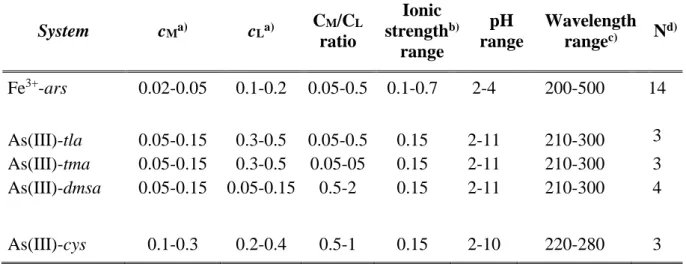
22 (mL). Each experiment was carried out in thermostated glass cell following the same procedure previously described for potentiometric measurements. The metal-ligand systems were investigated on 25 mL of solution containing the appropriate amount of metal, ligand and background salt (for reaching a prefixed ionic strength value) at T = 298.15K. When necessary, independent titrations of metals or ligands solutions were performed in order to calculate the molar absorption coefficients. All the specific experimental conditions are reported in Table 1.6.
Table 1.6. Experimental conditions for the metal-ligands systems investigated by spectrophotometry in NaCl and T = 298.15 K.
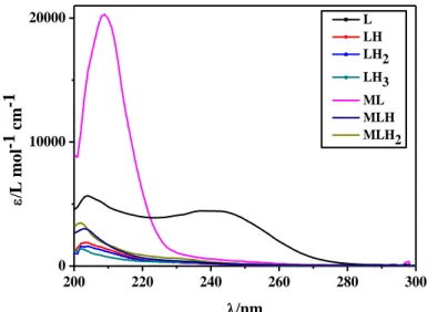
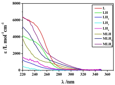
System cMa) cLa) CM/CL ratio Ionic strengthb) range pH range Wavelength rangec) Nd) Fe3+-ars 0.02-0.05 0.1-0.2 0.05-0.5 0.1-0.7 2-4 200-500 14 As(III)-tla 0.05-0.15 0.3-0.5 0.05-0.5 0.15 2-11 210-300 3 As(III)-tma 0.05-0.15 0.3-0.5 0.05-05 0.15 2-11 210-300 3 As(III)-dmsa 0.05-0.15 0.05-0.15 0.5-2 0.15 2-11 210-300 4 As(III)-cys 0.1-0.3 0.2-0.4 0.5-1 0.15 2-10 220-280 3
23
1.4 Calorimetry
[56]1.4.1 General aspects
The titration calorimetry or direct calorimetry is a technique in which the heat, developed or absorbed in a chemical reaction, is measured as a function of the titrant volume added during the measurement. Most of the experiments were performed by an isoperibolic calorimeter constituted by a reaction cell quasi-adiabatic, immersed in a thermostated bath. The temperature change in the reaction cell, proportional to the difference between the temperature of the bath and the cell itself, could be caused by a chemical reaction or a physical interaction between the sample and the titrant which lead to the formation of new species in solution. With this technique the resulting data, expressed as heat produced as a consequence of the added titrant, were analysed in order to calculate thermodynamic parameters such as K, H0 e S0. Also a nano-isothermal calorimeter was employed and it
will be examined in the next subparagraph.
1.4.2 Instrumental equipment and procedure
The instrumental equipments employed in this study were a CSC isoperibolic calorimeter, model 4300, visible in Figure 1.1 and nano-isothermal titration calorimeter Nano-ITC (TA Instruments).
Section I
Chapter 1
24 For the first instrument, titration was performed continuously since the titrant was added with constant rate. The reaction cell, constituted by a glass vessel covered with a silver layer with a capacity of 25 or 50 mL, is immersed in 25mL thermostated bath, thermically isolated and with a temperature stability of 0.0005°C. The titrant adding took place by means of a 2.5 mL Hamilton syringe in glass and Teflon, model 1002TLL. The stirring of the solution in the dewar was guaranteed by a glass stirrer whose rate is easily adjustable, while the temperature was controlled by a thermistor also immersed in the reaction cell and part of the Wheatstone bridge. The thermistor is crossed by a small current. When the temperature in the dewar changes, as a consequence of the titrant adding, the resistence and, consequently, the voltage through the Wheatstone bridge proportionally vary. This voltage is amplified in the electrical circuit and the temperature variations are measured by a multimeter connected with the Wheatstone bridge. A personal computer, managed by a specific software named CALOR8, collected data automatically. This computer program is also able to fix the parameters for the measurement such as:
the rate of buret delivery in mL/min and the delay of the burette in seconds (both measured previously);
the initial volume;
total time and time intervals, expressed in seconds, for the readings of the baselines, the calibrations and the titration;
the values of HTRI and HTRV (the voltage across the standard resistor and the heating resistance calibration, respectively) and the starting potential of the measurement. Daily, before starting experiments, it was necessary to check the voltage across the Wheatstone bridge and set it to zero. Moreover, the determination of the dilution heats for each solution, by titrating pure solvent with the same titrant employed for the subsequent measures, was required in order to subtract this amount of heat to the total determined in the measurements.
Once put the solution in the dewar, this one was allocated in the thermostated bath and the solution was heated up by means of a heater in order to make equal the temperatures of the solution and bath. The good operation of the heating resistance was periodically checked by a chemical calibration which consists in titrating a THAM [tris-(hydroxymethyl)amino-methane] buffer with HCl. The accuracy of the apparatus for the measured heat is Q ± 0.008 J and for the volume v ± 0.001 mL. After inserting all the input data previously described, the measure started. For the acid-base properties of As(III) and As(V), 25 ml of solution
25 containing arsenate or arsenite sodium salts and NaCl as ionic medium were titrated with HCl solution. Details are reported in Table 1.7. The study of As(III)-ligand complexes were performed by adding the ligand under investigation as sodium salt to 25 mL of a solution containing the metal, the ionic medium and sometimes HCl. Details are listed in Table 1.8. Regarding arsenate-metals invetigations, 25 mL of solution featured by the metal cation under study and the necessary amount of ionic medium, in order to obtain a prefixed ionic strength, were titrated by the arsenate salt solution having a pH equal to 11.6. The details of the experimental conditions are shown in Table 1.9.
As regards the second equipment, it was constituted by an active cell volume of 0.988 mL and equipped with a 250 μL injection syringe. The reaction mixture in the sample cell was stirred at 250 rpm during the titrations. The reference cell was always filled with ultrapure water. All solutions were gently degassed under vacuum for about 15 min before each run. Measurements were run in the overfilled mode which does not require any correction for liquid evaporation and for the presence of the vapor phase [57]. The power curve was integrated by NanoAnalyze (TA Instruments) to obtain the gross heat evolved/absorbed in the reaction. The calorimeter was calibrated chemically by a test HCl/TRIS reaction according to the procedure previously described [58]. The instrument was also checked through electrical calibrations.
ITC measurements were carried out by titrating a 12÷15 mmol L-1 aqueous solution of As(III)
(containing HCl) into an aqueous solution of either NTAP (1.8÷2.1 mmol L-1) or NTA2P
(2÷2.5 mmol L-1); both the solutions of the titrant (syringe) and the titrate (cell) were adjusted to I = 0.1 mol L-1 by adding proper amounts of NaCl. Different pH windows, in the range 3.0 ≤ pH ≤ 10.8 were monitored; typically, three titrations were carried out per each pH window. Heats of dilution were determined in separate “blank” experiments by titrating the solution of As(III) (containing suitable amount of HCl and ionic medium) into a solution containing the ionic medium (0.1 mol L-1) only.
The net heats of reaction, obtained by subtracting the heat evolved/absorbed in the blank experiments, were analyzed by HypCal [59]. This software, specifically designed for the determination of equilibrium constants and formation enthalpies of complexes in solution, makes use of a non-linear least-squares minimization of the function:
U = Σ (Qobs. – Qcalc.)2 (1.6)
where Qobs. is the observed heat for a given reaction step, corrected for the dilution (blank)
Section I
Chapter 1
26
Qcalc. = − Σ (δnΔH) (1.7)
where δn is the change in the number of moles of a reaction product and ΔH is the molar formation enthalpy of the reaction product. The summation is carried out over all the reaction steps of the specific chemical system. The squared residuals (Qobs. – Qcalc.)2 are summed over
all the titration points. The parameters were determined by simultaneously analyzing calorimetric data from different titrations carried out at different pH ranges.
Table 1.7. Experimental conditions for the acid-base properties of arsenite and arsenate in NaCl and T = 298.15 K.
System CAs(III)/As(V)a) CHb) Ionic Strength rangeb) pH range
As(III) 20 1 0.1-1 3.2-10.5
As(V) 10 0.5 0.1 and 0.7 2.2-11.3
a)mmol L-1; b)mol L-1.
Table 1.8. Experimental conditions for the As(III)–ligands systems in NaCl and T = 298.15 K.
System cMa) cLa) CHa) Ionic Strength rangeb) pH range
As(III)-tla 4 400 4 0.15 2.4-9.8 As(III)-tma 4 400 4 0.15 2.4-9.8 As(III)-cys 5 500 1 0.15 3-10.3 As(III)-gsh 5 500 5 0.15 2.5-9.5 As(III)-NTAP 12-15 1.8-2.1 0.1 3-10.8 As(III)-NTA2P 12-15 2-2.5 0.1 3-10.8 a)mmol L-1; b)mol L-1.
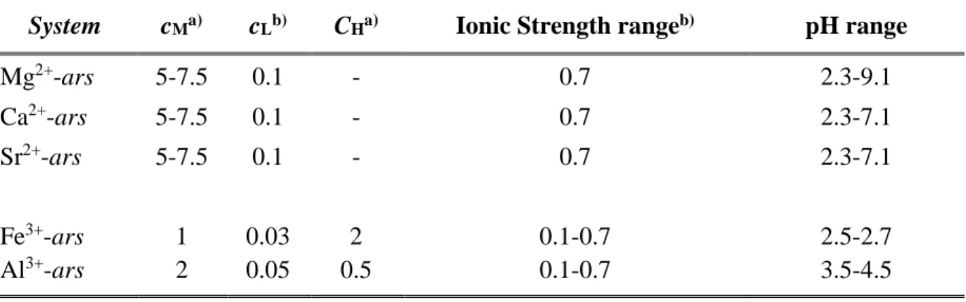
Table 1.9. Experimental conditions for the Mn+ -arsenate systems in NaCl and T = 298.15 K
System cMa) cLb) CHa) Ionic Strength rangeb) pH range
Mg2+-ars 5-7.5 0.1 - 0.7 2.3-9.1 Ca2+-ars 5-7.5 0.1 - 0.7 2.3-7.1 Sr2+-ars 5-7.5 0.1 - 0.7 2.3-7.1 Fe3+-ars 1 0.03 2 0.1-0.7 2.5-2.7 Al3+-ars 2 0.05 0.5 0.1-0.7 3.5-4.5 a)mmol L-1; b)mol L-1.