Relationships of Ligand Binding, Redox Properties, and Protonation
in Coprinus cinereus Peroxidase*
Received for publication, November 26, 2002, and in revised form, February 25, 2003 Published, JBC Papers in Press, March 5, 2003, DOI 10.1074/jbc.M212034200
Chiara Ciaccio‡§, Antonella Rosati§¶, Giampiero De Sanctis储, Federica Sinibaldi‡, Stefano Marini‡, Roberto Santucci‡, Paolo Ascenzi¶, Karen G. Welinder**,
and Massimo Coletta‡ ‡‡
From the ‡Department of Experimental Medicine and Biochemical Sciences, Universita` di Roma Tor Vergata, Via Montpellier 1, I-00133 Roma, Italy, the¶Department of Biology, Universita` Roma Tre, Viale G. Marconi 446, I-00146 Roma, Italy, the储Department of Molecular, Cellular, and Animal Biology, Universita` di Camerino, Via F. Camerini 2, I-62032 Camerino, Italy, and the **Department of Biotechnology, Aalborg University, Sohngaardshol-msvej 49, DK-9000 Aalborg, Denmark
The pH dependence of the redox potentials and kinetics for CO association and dissociation was determined between pH 3.0 and 13.0 at 25 °C for the wild-type Coprinus cinereus fungal peroxidase and for a site-directed mutant in which Asp245, which is H-bonded to N␦ of the imidazole of the proximal His183, was substituted with Asn. The determination of these functional properties allowed this information to be merged in a self-consistent fashion and to formulate for the first time a complete scheme em-ploying the minimum number of groups required to de-scribe the whole proton-linked behavior of both redox and ligand binding properties. The overall pH dependence can be accounted for by four redox- and ligand-linked groups. The proximal H-bond, which is strictly conserved in all peroxidases, will still be present in the site-specific mutant, but will no longer have an ionic character, and this event will bring about an alteration of redox equilibria and CO binding kinetics, envisaging a relevant role played by this H-bond also in modulating redox properties and ligand binding equilibria.
The importance of the protein moiety in controlling the func-tional behavior of the active site in hemoproteins is a wide-spread concept, even though different proteins seem to apply various strategies to accomplish this interrelationship. Thus, in the case of multimeric hemoproteins such as human hemo-globin, the functional control is mostly exerted through a li-gand-linked quaternary conformational transition, which af-fects ligand binding kinetic and thermodynamic parameters in a ligand-dependent fashion (1, 2).
In the case of monomeric hemoproteins, the protein matrix plays important roles through residues that either limit the access of the ligand to the distal portion of the heme pocket (3– 6) or constrain the conformation of the hemoprotein stere-ochemistry in the proximal portion of the pocket (7–9). In the case of myoglobin, the H-bond between Ser92 and N␦ of the
proximal His93appears to be of particular importance (9).
In the case of heme peroxidases, an invariant H-bond be-tween N␦of the proximal imidazole and an aspartate (10 –15) has been proposed to play a central role in the functional modulation of different peroxidases (16 –18). As a matter of fact, we have shown that substitution of the invariant proximal aspartate of manganese peroxidase has a relevant influence on the redox potentials of this enzyme (19), confirming the hypoth-esis previously formulated (17, 18).
Peroxidases are divided into three main structural classes. Class I refers to intracellular peroxidases of prokaryotic origin such as cytochrome c peroxidase. Class II includes secretory fungal peroxidases such as manganese peroxidase, lignin perox-idase, and Coprinus cinereus peroxidase. Class III contains se-cretory plant peroxidases such as horseradish peroxidase (16).
In the last few years, particular attention has been ad-dressed toward fungal peroxidases, also favored by the x-ray resolution of the structural arrangement for several of them and by their potential industrial usefulness. Therefore, a de-tailed description of different functional aspects of this class of peroxidases may be of relevance for better insight into their structure-function relationship. In particular, analysis of the pH dependence of different functional properties provides in-formation on the proton linkage relationships, which are rele-vant for the modulation of functional properties of the protein. Such an approach is very demanding with respect to analysis of individual functional features because it is constrained by the requirement of a general description with the minimum num-ber of parameters. As a matter of fact, it is often difficult to reconcile data concerning spectroscopic, thermodynamic, and kinetic observations, which indeed reflect differing structural and functional aspects of the whole molecule.
In this study, we analyzed some of the functional proton-linked equilibria and kinetics observed in the fungal peroxidase from
C. cinereus (which is indistinguishable from the Arthromyces ramosus peroxidase; therefore, we will refer to CIP1also when
referring to structural information taken from the crystal structure of A. ramosus peroxidase). The crystal structures of both peroxidases have been solved (14, 20), and the protein from C. cinereus has been thoroughly characterized by spectro-scopic techniques (21–23). This study has focused on the role of Asp245H-bonded to the proximal His183in the redox and ligand
binding properties as a function of pH. Analysis of CIP and the site-directed mutant D245N provided insight into the proton-linked equilibria in the oxidized and reduced forms, according to Scheme 1.
* This work was supported by Grant COFIN MM03185591 from the Italian Ministero dell’Universita` e della Ricerca Scientifica e Tecno-logica. The costs of publication of this article were defrayed in part by the payment of page charges. This article must therefore be hereby marked “advertisement” in accordance with 18 U.S.C. Section 1734 solely to indicate this fact.
This paper is dedicated to the memory of Prof. Eraldo Antonini on the occasion of the 20th anniversary of his death.
§ Both authors contributed equally to this work.
‡‡ To whom correspondence should be addressed. Tel.:
39-6-7259-6365; Fax: 39-6-7259-6353; E-mail: [email protected]. 1The abbreviation used is: CIP, C. cinereus peroxidase.
© 2003 by The American Society for Biochemistry and Molecular Biology, Inc. Printed in U.S.A.
This paper is available on line at http://www.jbc.org
MATERIALS AND METHODS
Recombinant wild-type CIP and the D245N mutant were both con-structed and expressed in Aspergillus oryzae (24), and the protein samples were purified and prepared as previously described (21). The reduced Fe(II) form was obtained by addition of a minimum volume of fresh sodium dithionite to a deoxygenated buffered solution.
Cyclic voltammeter measurements were carried out using an AMEL 433 multipolarograph. All redox potentials reported here refer to the normal hydrogen electrode. Direct current cyclic voltammograms were run in different buffers (see below) at a scan rate of 50 mV/s. A pyrolytic graphite electrode (AMEL) was the working electrode; a saturated calomel electrode (⫹244 mV versus the normal hydrogen electrode at 25 °C; AMEL) was the reference electrode; and a platinum ring was the counter electrode. The pyrolytic graphite electrode was modified as follows. A suitable amount of tributylmethylphosphonium chloride polymer bound (polystyrene cross-linked with 1% divinylbenzene) to anion-exchange resin was dissolved in dimethyl sulfoxide to a final concentration of 0.035% (w/v). 8l of this solution were then mixed with 12l of 1.6 mMCIP (or D245N) solution and deposited on the surface of
a pyrolytic graphite electrode (2 mm diameter) previously polished using an alumina (0.3-m particle size)/water slurry, followed by son-ication in deionized water (25). The modified electrode was left to dry overnight under vacuum before measurements.
CO binding kinetics were measured employing a rapid-mixing stopped-flow apparatus (SX.18 MV, Applied Photophysics Co., Salis-bury, UK) with a 1-ms dead time. The protein was kept in one syringe in a deoxygenated solution (N2-flushed with the addition of sodium
dithionite to a final concentration of 1 mg/ml) in very low ionic strength 1.0 mMsodium phosphate buffer (pH 7.0), and it was mixed with a
higher ionic strength buffer (final I⫽ 0.15) at the desired pH value and concentration of CO. Progress curves were recorded at several wave-lengths to follow both the formation of the CO adduct (at 423 nm) and the disappearance of the unliganded form (at 437 nm).
CO dissociation kinetics were followed on a Jasco V-530 spectropho-tometer recording the absorption spectrum in the 380 – 450 nm range. The protein was kept at pH 7.0 in 1.0 mMsodium phosphate buffer in the presence of slightly substoichiometric amounts of CO. The reaction started when the protein solution was mixed in a large excess of oxy-genated reaction mixture at the desired pH (final I⫽ 0.15) because the dissociation of CO was followed by a rapid oxidation of the peroxidase, which was detected by the change of the absorption spectrum.
All experiments were performed at 25 °C in 1.0 mMCaCl2employing
the following buffer systems: 0.15Msodium acetate (pH 4.0 –5.5), 0.15 Msodium phosphate (pH 3.0 – 4.0 and 5.0 –7.5), 0.15MTris-HCl (pH 7.5–9.0), 0.15Msodium borate (pH 8.5–9.5), 0.15Msodium bicarbonate
(pH 9.0 –10.5), and 0.15Msodium carbonate (pH 10.0 –13.0). No buffer-dependent effects were observed at overlapping pH values.
The pH dependence of the redox properties was analyzed according to Equation 1, Eobs⫽ E0䡠
冘
i⫽0 i⫽n写
r⫽0 i 䡠 Kar(ox)䡠 关H⫹兴冘
i⫽0 i⫽n写
r⫽0 i 䡠 Kar(red)䡠 关H⫹兴 (Eq. 1)where Eobsis the observed redox potential at a given pH value; E0is the
redox potential of the form present at very alkaline pH values; Ka(red)and
Ka(ox)are the proton binding constants in the reduced and oxidized forms, respectively (such as Ka ⫽ 10pKa), outlining that, in Equation 1,
Ka0䡠[H⫹]⫽ 1; and n is the total number of protonating redox-linked groups. It must be pointed out that, in Equation 1, we use the formalism Ka to refer to the groups that are sequentially protonated as pH is lowered, as also depicted in Scheme 1; therefore, r follows this sequence, and r⫽ 1 relates to the residue displaying the highest pKavalue. Equa-tion 1 indicates that only residues that change their pKaupon reduction (or oxidation) contribute to the pH dependence of the redox potential.
The pH dependence of the CO binding second-order rate constant was analyzed according to Equation 2,
kobs⫽
冘
i⫽0 i⫽n ki䡠写
r⫽0 i Kar(red)䡠 关H⫹兴冘
i⫽0 i⫽n写
r⫽0 i Kar(red)䡠 关H⫹兴 (Eq. 2)where kobsis the observed CO binding second-order rate constant at a
given pH value, kiis the CO binding second-order rate constant of the
ith-protonated species (i⫽ 0–4), and all other symbols have the same meaning as described for Equation 1.
RESULTS
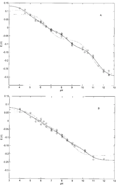
Fig. 1 shows the pH dependence of the redox properties of the wild-type peroxidase from C. cinereus (CIP) (Fig. 1A) and of the site-directed mutant D245N (Fig. 1B), in which Asn was substituted for Asp245, which is H-bonded in CIP to the
proximal His183. Least-squares fitting of experimental data
was carried out according to Equation 1, employing n⫽ 2, 3, or 4. Least-squares fitting of the pH dependence of the redox properties employing n⫽ 2 was clearly inadequate for both CIP and D245N (Fig. 1, A and B, – - – -), whereas n ⫽ 4 appeared to be only slightly better than n⫽ 3. For a phe-nomenological description of the pH dependence of the redox properties of CIP and D245N, n⫽ 3 might be satisfactory, using as few parameters as possible. Data from fitting ac-cording to n⫽ 3 and 4 are reported in Table I both for CIP and the D245N mutant.
FIG. 1. A, pH dependence of the redox potentials for CIP at 25 °C. OO, nonlinear least-squares fitting of the data according to Equation 1 for n⫽ 4 (values reported in Table I); – – –, nonlinear least-squares fitting of the data according to Equation 1 for n⫽ 3; – - – -, nonlinear least-squares fitting of the data according to Equation 1 for n⫽ 2. B, pH dependence of the redox potentials for D245N at 25 °C.OO, nonlinear least-squares fitting of data according to Equation 1 for n⫽ 4 (values reported in Table I); – – –, nonlinear least-squares fitting of the data according to Equation 1 for n⫽ 3; – - – -, nonlinear least-squares fitting of the data according to Equation 1 for n⫽ 2. For further details, see “Results.”
Remarkably, CIP is the first peroxidase reported to display more than two redox-linked protonating groups that change their pKavalues depending on the redox state of the heme iron.
Thus, a similar analysis of horseradish peroxidase (isoenzyme C) and manganese peroxidase showed that two redox-linked residues fully accounted for the pH dependence of the redox properties (19, 25, 26). As in other peroxidases, all redox-linked groups observed in CIP displayed a decrease in pKaupon heme reduction, suggesting that the removal of one electron from the heme iron brings about a situation that renders less easy the protonation of these residues. Such a feature is likely referable to an electrostatic effect because Fe(II) is less positive than Fe(III), even though we cannot rule out redox-linked structural change(s) that induce a pKashift in residues surrounding the
heme pocket.
To extend this analysis, we carried out a similar pH depend-ence investigation of CO binding kinetics, which yielded the information on the right side of Scheme 1. Fig. 2 shows the pH dependence of CO association, and Fig. 3 displays the pH dependence of the CO dissociation kinetics for CIP and the D245N mutant. In both cases, Equation 2 was employed for the analysis, even though it must be stressed that, in the case of the CO association process, the pKavalues refer to the P(II)
populations of Scheme 1, whereas for the CO dissociation proc-ess, pKavalues refer to the PL(II) populations. In this way, the
two types of kinetic information allow a complete description of the proton-linked modulation of the right side of Scheme 1. However, because for the CO association kinetics, the popula-tions coming into play (i.e. P(II)) are in common with those involved in the redox process reported above (i.e. P(II) and P(III)), we also tried to merge these two facts into a self-consistent fitting procedure to obtain from the two sets of data a minimum number of protonating groups accounting for the whole proton-linked equilibria involving the P(III), P(II), and PL(II) species for both CO kinetics and redox processes (Scheme 1).
Fig. 2A shows that an independent fitting of the CO associ-ation kinetic data for CIP could be undertaken using only two protonating residues, whose pKavalues are reported in Table
II. If we compare them with values reported in Table I for the pH dependence of the redox equilibria, we notice that there is a substantial overlap between the pKa2(red)value in Table I (for
n⫽ 3) and the pKa1value in Table II (for n⫽ 2). On the other
hand, the value of pKa2for the kinetic proton linkage appears to be irreconcilable with any of the pKa(red)values reported for
n⫽ 3 in Table I, whereas it is fairly close to the pKa4(red)value
for n⫽ 4. Altogether, these observations seem to indicate that there is a partial overlap between proton-linked residues mod-ulating redox properties and CO association kinetic parame-ters, such that Scheme 1 with four protons (i.e. n⫽ 4) could be required for a complete description of the linkage between protons, redox equilibria, and CO binding behavior.
However, to find out in a more quantitative fashion whether
this is a reasonable assessment, we fitted the pH dependence of the CO association kinetics imposing the three pKa(red)values
reported in Table I for n⫽ 3 and allowing a free flotation of ki values (Equation 2). Fig. 2B (– – –), corresponding to what was obtained by least-squares fitting of data according to this pro-cedure, shows that these values are inadequate to give a sat-isfactory description of the pH dependence of the CO associa-tion kinetics because the transiassocia-tion at lower pH values, corresponding to the value of pKa2 for n ⫽ 2 (Table II), is
missed. On the other hand, imposing values of pKa(red)for n⫽
4 in Table I (for redox potentials) gives a very satisfactory fitting of the pH dependence of the CO association kinetics (Fig. 2B,OO), supporting that four protons indeed are the mini-mum number required for the whole description of proton link-age for redox and kinetic properties, with two values overlap-ping (Table II for n⫽ 4). As a self-consistent test, we also fitted the CO binding data employing Equation 2, leaving a free flotation of both kiand pKa(red)values and obtaining a set of
parameters closely similar (within the uncertainty limits) to those reported in Table II for n⫽ 4.
A similar analysis was carried out for the pH dependence of the CO association kinetics of the D245N mutant; also in this case, two protonating groups were sufficient to give an inde-pendent description of the pH dependence (Fig. 2A and Table II for n⫽ 2). Such pKa values were very close to two of those
obtained by analysis of the pH dependence of the redox prop-erties for n⫽ 4 (viz. pKa2(red)and pKa3(red)) (Table I), but they were irreconcilable with the values obtained for the same anal-ysis for n⫽ 3 (Table I). This appears to be evident also com-paring the fitting of the pH dependence of the CO association kinetics of D245N imposing pKa(red)values from the proton-linked behavior of redox potentials either for n⫽ 3 (Fig. 2C,
SCHEME1. General scheme for the redox-, ligand-, and
proton-linked equilibria investigated in CIP. Horizontal equilibria on the
left side concern the redox processes. Horizontal equilibria on the right side refer to the ligand (L) binding in the reduced forms. Vertical equilibria refer to the protonation processes.
TABLE I
pKavalues for the redox-linked protonating groups in CIP and D245N from the fitting of the pH dependence of the redox potentials
according to Equation 1 employing n⫽ 3, 4
CIP D245N n⫽ 3 n⫽ 4 n⫽ 3 n⫽ 4 pKa1(ox) 11.98⫾ 0.14 12.00⫾ 0.15 10.61⫾ 0.14 10.74⫾ 0.13 pKa2(ox) 8.84⫾ 0.16 8.98⫾ 0.16 8.21⫾ 0.17 8.65⫾ 0.16 pKa3(ox) 6.09⫾ 0.15 4.79⫾ 0.18 5.62⫾ 0.14 4.80⫾ 0.16 pKa4(ox) 6.71⫾ 0.14 6.66⫾ 0.12 pKa1(red) 10.71⫾ 0.14 10.75⫾ 0.16 9.90⫾ 0.13 10.12⫾ 0.12 pKa2(red) 8.02⫾ 0.16 8.24⫾ 0.17 7.38⫾ 0.17 7.97⫾ 0.17 pKa3(red) 5.22⫾ 0.14 6.10⫾ 0.18 4.91⫾ 0.15 6.12⫾ 0.13 pKa4(red) 4.21⫾ 0.15 4.31⫾ 0.11 E0(V) ⫺0.294 ⫾ 0.024 ⫺0.295 ⫾ 0.033 ⫺0.235 ⫾ 0.021 ⫺0.237 ⫾ 0.020
- - - - -) and for n⫽ 4 (Fig. 2C, OO). Therefore, as observed for CIP, the global description of the proton linkage to the redox properties and CO association kinetics of D245N requires four protonating groups, two of which are overlapping.
For the pH dependence of the CO dissociation kinetics (re-ported in Fig. 3) that lead to the determination of pKL(red)
(Scheme 1), we followed a conceptually similar approach, even though it must be stressed that, in this case, our constraint was different from that used for the CO association kinetics because we have no independent determination of pKL(red)values. The
constraint is instead related to the fact that the values of the association and dissociation kinetic constants at each protona-tion stage i and i⫺ 1 (and thus the corresponding equilibrium constants) must be consistent with the values of pKa1(red)and
pKL1(red) so as to obey mass conservation law within each thermodynamic square (Scheme 1), as for the following rela-tionship (Equation 3).
共ki⫺1共off兲/ki(off)兲 䡠 10pKL1⫽ 共ki⫺1共on兲/ki共on兲兲 䡠 10pKa1 (Eq. 3)
FIG. 2. A, pH dependence of the CO association rate constants for CIP (E) and D245N (⫻) at 25 °C. OO, nonlinear least-squares fitting of the data according to Equation 2 employing the values reported in Table II for n⫽ 2. B, pH dependence of the CO association rate constant for CIP at 25 °C. – – –, nonlinear least-squares fitting of the data according to Equation 2 imposing values of pKa(red)for n⫽ 3 (Table I); OO, nonlin-ear least-squares fitting of the data according to Equation 2 imposing values of pKa(red) for n⫽ 4 (Table I). C, pH dependence of the CO association rate constant for D245N at 25 °C. - - - - -, nonlinear least-squares fitting of the data according to Equation 2 imposing values of pKa(red)for n⫽ 3 (Table I); OO, nonlinear least-squares fitting of the data according to Equation 2 imposing values of pKa(red)for n⫽ 4 (Table I). For further details, see “Results.”
FIG. 3. pH dependence of the CO dissociation rate constants
for CIP (E) and D245N (ⴛ) at 25 °C. OO, nonlinear least-squares fitting of the data according to Equation 2. Values of kiand pKLiare
reported in Table III. The outcome of the fitting was constrained by mass law relationships with values of kiand pKa1(red)reported in Table II, such as for each step i (Schemes 1–3), Equation 3 has been used to apply this constraint. For further details, see “Results.”
TABLE II
Values of kiand pKafor the pH dependence of the CO association
kinetics for CIP and D245N from the independent least-squares fitting of the data according to Equation 2 employing n⫽ 2 and/or imposing
values of pKa(red)from the proton-linked redox behavior
reported in Table I (n⫽ 4). CIP D245N n⫽ 2 k1(M⫺1s⫺1) (1.82⫾ 0.21) ⫻ 10 5 (3.25⫾ 0.42) ⫻ 105 k2(M⫺1s⫺1) (2.64⫾ 0.28) ⫻ 10 4 (1.82⫾ 0.24) ⫻ 105 k3(M⫺1s⫺1) (4.30⫾ 0.51) ⫻ 10 4 (3.69⫾ 0.43) ⫻ 104 pKa1 8.05⫾ 0.17 8.08⫾ 0.18 pKa2 4.29⫾ 0.18 6.41⫾ 0.17 n⫽ 4 k1(M⫺1s⫺1) (1.83⫾ 0.20) ⫻ 10 5 (3.24⫾ 0.41) ⫻ 105 k2(M⫺1s⫺1) (1.94⫾ 0.22) ⫻ 10 5 (3.30⫾ 0.42) ⫻ 105 k3(M⫺1s⫺1) (3.16⫾ 0.33) ⫻ 10 4 (1.53⫾ 0.23) ⫻ 105 k4(M⫺1s⫺1) (2.58⫾ 0.27) ⫻ 10 4 (3.55⫾ 0.45) ⫻ 104 k5(M⫺1s⫺1) (4.53⫾ 0.54) ⫻ 10 4 (3.74⫾ 0.47) ⫻ 104 pKa1(red) a 10.75 10.12 pKa2(red) a 8.24 7.97 pKa3(red) a 6.10 6.12 pKa4(red) a 4.21 4.31 aFixed parameters.
In this way, the pH dependence of the redox equilibria and the CO association and dissociation kinetic rate constants all be-come functionally interrelated through a set of proton linkage relationships, giving rise to a complete description of the mod-ulation of the whole system. Therefore, to be consistent with what was already determined for the proton linkage of the redox and CO association kinetic behavior, analysis of the pH dependence of the CO dissociation kinetics was limited to the search for a set of five dissociation rate constants (correspond-ing to the 0 – 4 protonated species) and four pKL(red)values that altogether obey the mass law constraints described above. De-spite the large number of parameters involved, the limitations imposed turned out to be fairly restrictive, as essentially only one solution was found for CIP and D245N that satisfactorily described the pH-dependent behavior of their CO dissociation kinetics (Fig. 3 and Table III).
DISCUSSION
The overall description of the pH dependence of the redox properties and CO association and dissociation kinetics of CIP can be fully accounted for by the redox- and ligand-linked protonation of four groups, and this behavior is depicted in Schemes 2 and 3 for CIP and D245N, respectively. An impor-tant observation is that, unlike for other peroxidases investi-gated up to now, the observed pH dependence cannot be ac-counted for by only two redox-linked groups, suggesting that CIP shows a different environment of the heme pocket com-pared with other peroxidases, bringing about a variation of the protonation properties of residues that affect the redox proper-ties, with the involvement of additional redox-linked group(s). However, it must be stressed that it is the first time (at least to our knowledge) that an attempt has been undertaken to merge in a systematic way the redox, ligand binding, and proton linkage information on a peroxidase. Therefore, we cannot rule out the possibility that such an investigation on other peroxi-dases will give a similar outcome.
In particular, a comparative study has been carried out be-tween wild-type cytochrome c peroxidase and the side-directed mutant D235N for CO association and dissociation kinetics (27, 28), but it was extended over a much more limited pH range. In any case, the behavior observed was qualitatively similar to what was observed in CIP and D245N because the CO associ-ation rate constant was⬃5-fold faster in the case of the D235N mutant with respect to wild-type cytochrome c peroxidase (27), whereas a closely similar CO dissociation rate constant was observed, which, in both proteins, displayed a decrease as the pH was lowered (28).
An additional step to understand from the structural stand-point how redox properties and ligand binding features are modulated in CIP and D245N is to try to identify the four protonating groups or at least to give some hint as to which might be the most plausible candidates to play different proton-linked roles. Although this is not easy, some insight can be obtained by the investigation of the redox properties in the
site-directed mutant D245N because the substitution of Asp245
with Asn weakens the H-bond between the carboxylate of Asp245and N␦of the proximal His183, likely affecting
proton-linked equilibria. We first noticed that one of these residues,
viz. pKa1, was greatly affected by the mutation, which also
sig-nificantly influenced pKa2, whereas pKa3remained unchanged, and pKa4was only slightly altered by the mutation (Table I). In
particular, it is very informative that the substitution of Asp245
with Asn brought about a lower value for pKa1 both in the
oxidized (pKa1(ox)⫽ 10.74 ⫾ 0.13 in D245N and 12.00 ⫾ 0.14 in CIP) and in the reduced (pKa1(red)⫽ 10.12 ⫾ 0.12 in D245N and
10.75⫾ 0.13 in CIP) forms, indicating that the much weaker H-bond and the consequent structural change(s) induce a rele-vant decrease in the protonation features of this residue such that it protonates less easily (Table I). Similar considerations apply to the effect of the substitution on pKa2, even though in this
case the shifts in pKavalues were smaller than for the group
characterized by pKa1(Table I). Thus, upon heme reduction, the pKa2values shifted from 8.98⫾ 0.15 to 8.24 ⫾ 0.17 in CIP and
from 8.65⫾ 0.16 to 7.97 ⫾ 0.17 in D245N. On the other hand, the residue responsible for pKa1showed almost no effect on the CO
association kinetics for both CIP and D245N (Schemes 2 and 3 and Table II), and the same is true for pKL1on the CO
dissocia-tion kinetics (Schemes 2 and 3 and Table III). As a whole, such features might suggest that the residue responsible for pKa1is a
very basic residue. It may be interesting to recall that, over the same pH range, a spectroscopic transition has been reported to occur in the ferric resting state of CIP and D245N, which has been assigned to the binding of the hydroxyl ion to Fe(III) (21, 22). This occurrence appears to be associated with the weakening of the proximal Fe–His183 bond, and it displays a higher pK
a
value in CIP than in D245N (as also observed in our case for pKa1(ox)) (Table I)), a difference that must be associated with a
different interaction mode of OH⫺ with residues of the distal portion of the heme pocket (21, 22). The observed values of pKa
TABLE III
Kinetic rate constants and protonation parameters for the CO dissociation from CIP and D245N
CIP D245N k1(s⫺1) (7.73⫾ 0.63) ⫻ 10⫺3 (6.73⫾ 0.57) ⫻ 10⫺3 k2(s⫺1) (7.73⫾ 0.36) ⫻ 10⫺3 (6.73⫾ 0.31) ⫻ 10⫺3 k3(s⫺1) (1.18⫾ 0.27) ⫻ 10⫺4 (2.73⫾ 0.54) ⫻ 10⫺4 k4(s⫺1) (3.29⫾ 0.09) ⫻ 10⫺5 (1.38⫾ 0.18) ⫻ 10⫺5 k5(s⫺1) (1.14⫾ 0.07) ⫻ 10⫺4 (3.41⫾ 1.03) ⫻ 10⫺5 pKL1(red) 10.78⫾ 0.17 10.13⫾ 0.17 pKL2(red) 9.27⫾ 0.14 9.02⫾ 0.19 pKL3(red) 6.64⫾ 0.15 6.84⫾ 0.17 pKL4(red) 4.06⫾ 0.15 3.94⫾ 0.16
SCHEME2. Overall redox-, ligand-, and proton-linked equilibria of CIP.
for the formation of the Fe(III)–OH⫺adduct in CIP and D245N (21, 22) are compatible with the values of pKa1(ox)reported in
Table I, suggesting that they likely refer to the same phenome-non. In addition, also the Fe(II) form of both proteins displayed a spectroscopic transition over the same pH range (Fig. 4A), sug-gesting a conformational change from a pentacoordinated species to a hexacoordinated one, with pKavalues significantly different between CIP (pKa⫽ 11.70 ⫾ 0.12) and D245N (pKa⫽ 10.68 ⫾
0.13) (Fig. 4B). It must be remarked that the pKavalues for the
spectroscopic transition of the unliganded Fe(II) forms are differ-ent from the pKa1(red) values reported in Table I for the two proteins, clearly indicating that they do not refer to the same phenomenon, even though it appears evident that they seem related because they have a closely similar trend. Altogether, these observations may suggest that the group responsible for the most alkaline transition, corresponding to pKa1, is N␦of the
proximal His183. Thus, the occurrence of the H-bond of this group
with Asp245in CIP is likely to bring about a very high pK
a1value,
which is significantly decreased upon the drastic weakening of the H-bond in the D245N mutant. This assignment is also con-sistent with the observation that the deprotonation of this resi-due induces the formation of a hexacoordinated species. As a matter of fact, the H-bond holds the proximal imidazole, which is thus constrained in a well defined conformation; the deprotona-tion of N␦is then very likely associated with an increase in the degrees of freedom for the proximal imidazole, decreasing the energy barrier for its movement toward the heme plane and thus facilitating the hexacoordination. Therefore, the severe weaken-ing of this H-bond in D245N likely decreases the pKavalue for
the protonation of N␦ of His183, thus facilitating also the
hexacoordination.
Lowering the pH from 10 to 6 brought about in both CIP and D245N an almost 10-fold reduction in the observed second-order rate constant for CO binding (Fig. 2), which was accom-panied by a 100-fold decrease in the CO dissociation rate con-stant (Fig. 3), resulting in a pH-dependent increase in CO
binding equilibrium affinity (which is much more marked than in the case of cytochrome c peroxidase (27, 28)). For the CO association process, such a transition appears to be modulated mostly by pKa2(red)in CIP and by pKa3(red)in D245N (Schemes
2 and 3 and Table II). On the other hand, the CO dissociation rate constant was modulated by both pKL2(red)and pKL3(red)in
both CIP–CO and D245N–CO (Table III). The assignment of the residue responsible for pKa2, which plays a relevant
proton-linked modulatory role in the redox properties as well as in CO association with CIP (but not with D245N) and in CO dissoci-ation from both CIP and D245N (Schemes 2 and 3), is, however, not obvious. The values of both pKa2(red) and pKL2(red) were
affected to a similar extent by the weakening of the H-bond, showing a ⬇0.25-pH unit shift between CIP and D245N (Schemes 2 and 3). As a matter of fact, there are several groups in close proximity that might show a value of pKawithin the
pKa2range; the most likely one might be one of the two water molecules bridging one of the propionates with Arg51(Fig. 5)
SCHEME3. Overall redox-, ligand-, and proton-linked equilibria
of D245N.
FIG. 4. A, absorption spectra of the unliganded Fe(II) form of CIP at
pH 8.0 (spectrum a) and 13.0 (spectrum b). Absorption spectra in the visible region were multiplied by 5. Similar spectral changes were observed for the unliganded Fe(II) form of D245N. B, pH dependence of the absorption at ⫽ 423 nm for the unliganded Fe(II) forms of CIP (E) and D245N (⫻) at 25 °C. OO, least-squares fitting of the experimental data according to the following equation, Aobs⫽ A0⫹ ⌬A䡠(Ka䡠[H]⫹/(1⫹
Ka䡠[H]⫹)), where Aobsis the observed absorbance at ⫽ 423 nm at a
given pH, A0is the absorbance at the lowest limit of the pH values,⌬A
is the total absorbance change corresponding to the pH-dependent transition, Kais the proton binding constant (10pKa), and [H⫹] is the proton concentration (10⫺pH). For further details, see “Discussion.”
(14, 20). Thus, the close proximity with the negative charge of the propionate could give this H2O molecule(s) a strong ionic
character with a very high pKavalue. Furthermore, its location
renders it very likely that the oxidation state of the heme iron has a relevant effect on the pKa value of such an H-bond
network, and the same can be true for the CO-bound form. This being true, it may be very likely that the alteration of the heme geometry, induced by the severe weakening of the proximal H-bond in D245N, brings about a variation of the interaction between the propionate and Arg51(mediated by this H
2O
mol-ecule) and thus a decrease in the pKavalue and/or a
displace-ment of Arg51such that it plays a less important role in the
modulation of CO association in favor of the residue responsi-ble for pKa3(see above).
On the other hand, the identification of the residue respon-sible for pKa3appears to be less problematic because its proton-linked redox behavior leads to its identification as the distal His55. Thus, the value of pK
a3(ox) is very low, as has been
indicated by several observations to be the case for the distal His in peroxidases (29), displaying a remarkable pKaincrease upon reduction of the heme iron. This behavior might suggest that the environment of the distal His55changes significantly
when Fe(III) is reduced to Fe(II), possibly because of a struc-tural movement of Arg51, which has been thought to be mainly
responsible (with its positive charge) for the very low pKavalue
displayed by His55in the Fe(III) species (29). Conversely, the
mutation that occurs on the proximal side upon the substitu-tion of Asp245with Asn does not seem to affect at all the pK
a3(ox)
and pKa3(red)values, indicating that the environment of His55is
not perturbed by this mutation. Unfortunately, site-directed mutants of His55are not available for CIP to prove that this
assignment is true, but under this very reasonable assumption, it appears that His55indeed is an important modulator of CO
binding to D245N, turning out to be much less crucial in the case of CIP. This clearly indicates that the mutation that oc-curs on the proximal side, which brings about the severe weak-ening of the H-bond, induces a conformational change that is
transmitted also to the distal side of the heme pocket, render-ing much more important the functional role of His55in CO
binding (or else reducing the role played by some other deter-minant, such as that characterized by pKa2(red)). In this respect,
it can be emphasized that the faster CO binding rate constant of D245N with respect to CIP can be fully attributed to a decrease in the energy barrier for ligand access to the heme, an observation that indeed confirms that the mutation that occurs in D245N alters the shield between the bulk solvent and the heme. However, this structural change does not appear to affect the ionic properties of His55, which maintains unchanged
pKavalues (Schemes 2 and 3 and Table I), but instead of some
other residue(s), such as the one(s) characterized by pKa2,
whose role in CO binding is drastically reduced in D245N, this being also associated with a variation of pKa values in the
mutant (Schemes 2 and 3 and Tables I and II). On the other hand, this assignment being true, His55seems to play a main
role in the control of the CO dissociation from both CIP–CO and D245N–CO, suggesting that the conformational change mainly affects the unliganded Fe(II) form.
In the case of the residue responsible for pKa4(red), it must be noted that its protonation appears to have an accelerating effect on the CO association with CIP, whereas in D245N, although showing a closely similar value (Schemes 2 and 3), it has almost no effect on the analogous process. On the other hand, the protonation of this residue, being characterized by pKL4(with values lower than the corresponding pKa4(red) val-ues) in the CO-bound form, has a similar effect on the CO dissociation from both CIP and D245N. This behavior and the pKa4(red)and pKL4values suggest that this residue is likely N⑀ of the imidazole of the proximal His183, which is involved in the
proximal axial bond. This being correct, the raising of the observed CO dissociation rate constant as pH is decreased below 6 (for CIP) and 5 (for D245N) (Fig. 3) is directly related to the appearance of the pentacoordinated CO complex. This assignment, which implies that the protonation event is asso-ciated with the weakening of the proximal bond, appears to be consistent with spectroscopic observations by electronic ab-sorption, resonance Raman spectroscopy, and NMR (21, 22). Furthermore, the pKa4(ox) value, measured from the pH
de-pendence of the redox properties (Fig. 1 and Table I), is closely similar to the value determined independently for the ferric form of CIP (30).
As a whole, this analysis allows us to envisage a general mechanism for the linkage among protons, ligand binding, and redox equilibria where four protons are required for an overall description of both CIP and D245N, with different effects that also point out the role of the H-bond between Asp245and N␦of
the proximal His183. First of all, the severe weakening of the
proximal H-bond in D245N brings about an alteration of pKa
for two of the four redox-linked protonating residues (likely being N␦of His183and an H
2O-bound network of charges in the
distal side of the heme pocket) (Schemes 2 and 3). Furthermore, although three of the four redox-linked protonating groups are also ligand-linked for both CIP and D245N (Schemes 2 and 3), the kinetic effects are different in the two molecules, reflecting the alteration that occurs on the distal side of the heme pocket as a consequence of the weakening of the proximal H-bond in D245N. Thus, the role played by the H2O-bound network of charges, which is predominant in CIP, is overwhelmed in D245N by the role of His55, and this is likely referable to a
structural change that occurs in the distal side upon the mu-tation in the proximal portion of the heme pocket (and possibly involving Arg51and the H
2O-mediated bridge with one of the
heme propionates). In addition, the drastic weakening of the proximal H-bond seems to alter the kinetic control of CO
bind-FIG. 5. Structural diagram of the heme pocket of CIP
accord-ing to the crystal structure of Ref. 20. Key residues of heme
sur-roundings are indicated. Seven distal water molecules in the heme-linked H-bond network are also shown (black spheres). The image was generated using the Swiss-PdbViewer Version 3.7 visualization pro-gram. For further details, see “Discussion.”
ing by the proximal axial bond of the heme, corresponding to the protonation of N⑀of His183(see pK
a4(red)in Schemes 2 and
3), because, unlike for CIP, no effect is observed on CO binding to D245N upon protonation of the residue characterized by pKa4(red)(Scheme 3). However, it must be pointed out that, in
peroxidases, the role of this protonation in the modulation of CO binding is significantly reduced with respect to myoglobins and hemoglobins, where the protonation of N⑀of the proximal histidine is associated with a large increase in the CO binding rate constant (7, 31). This behavior might be correlated, at least in CIP and D245N, with the fact that the stereochemical ar-rangement of the heme is held in a more fixed position, which renders the intrinsic reactivity with CO more favorable and less influenced by the protonation of the proximal imidazole. In conclusion, CIP appears to display a much tighter connection between the proximal and distal sides of the heme pocket with respect to other peroxidases such as horseradish peroxidase (isoenzyme C) and manganese peroxidase (as already outlined by spectroscopic observations (21 and 22)), and this appears to be particularly evident in that the weakening of the proximal H-bond brings about a conformational transition on the distal side of the heme pocket that renders faster access of the ligand to the heme pocket.
Acknowledgments—We thank Prof. G. Smulevich and Dr. A. Feis for several stimulating discussions and Novozymes Co. for the recombinant wild-type and mutant proteins.
REFERENCES
1. Unzai, S., Eich, R., Shibayama, N., Olson, J. S., and Morimoto, H. (1998)
J. Biol. Chem. 273, 23150 –23159
2. Gibson, Q. H. (1999) Biochemistry 38, 5191–5199 3. Perutz, M. F. (1989) Q. Rev. Biophys. 22, 139 –237
4. Lambright, D. G., Balasubramanian, S., Decatur, S. M., and Boxer, S. G. (1994) Biochemistry 33, 5518 –5525
5. Brunori, M. (2000) Biophys. Chem. 86, 221–230
6. Scott, E. E., Gibson, Q. H., and Olson, J. S. (2001) J. Biol. Chem. 276, 5177–5188
7. Coletta, M., Ascenzi, P., Traylor, T. G., and Brunori, M. (1985) J. Biol. Chem. 260, 4151– 4155
8. Murray, L. P., Hofrichter, J. H., Henry, E. R., Ikeda-Saito, M., Kitagishi, K., Yonetani, T., and Eaton, W. A. (1988) Proc. Natl. Acad. Sci. U. S. A. 85, 2151–2155
9. Decatur, S. M., Belcher, K. L., Rickert, P. K., Franzen, S., and Boxer, S. G. (1999) Biochemistry 38, 11086 –11092
10. Finzel, B. C., Poulos, T. L., and Kraut, J. (1984) J. Biol. Chem. 259, 13027–13036
11. Poulos, T. L., Edwards, S. L., Wariishi, H., and Gold, M. H. (1993) J. Biol.
Chem. 268, 4429 – 4440
12. Sundaramoorthy, M., Kishi, K., Gold, M. H., and Poulos, T. L. (1994) J. Biol.
Chem. 269, 32759 –32767
13. Kunishima, N., Fukuyama, K., Matsubara, H., Hatanaka, H., Shibano, Y., and Amachi, T. (1994) J. Mol. Biol. 235, 331–344
14. Petersen, J. F. W., Kadziola, A., and Larsen, S. (1994) FEBS Lett. 339, 291–296
15. Gajhede, M., Schuller, D. J., Henriksen, A., Smith, A. T., and Poulos, T. L. (1997) Nat. Struct. Biol. 4, 1032–1038
16. Welinder, K. G. (1992) Curr. Opin. Struct. Biol. 2, 383–393
17. Welinder, K. G., Bjørnholm, B., and Dunford, H. B. (1995) Biochem. Soc.
Trans. 23, 257–262
18. Mukai, M., Mills, C. E., Poole, R. K., and Yeh, S.-R. (2001) J. Biol. Chem. 276, 7272–7277
19. Santucci, R., Bongiovanni, C., Marini, S., Del Conte, R., Tien, M., Banci, L., and Coletta, M. (2000) Biochem. J. 349, 85–90
20. Fukuyama, K., Itakura, H., Sato, K., Takahashi, S., and Hosoya, T. (1997)
J. Biol. Chem. 272, 5752–5756
21. Smulevich, G., Neri, F., Marzocchi, M. P., and Welinder, K. G. (1996)
Biochem-istry 35, 10576 –10585
22. Veitch, N. C., Gao, Y., and Welinder, K. G. (1996) Biochemistry 35, 14370 –14380
23. Neri, F., Indiani, C., Welinder, K. G., and Smulevich, G. (1998) Eur. J.
Bio-chem. 251, 830 – 838
24. Welinder, K. G., and Andersen, M. B. (December 9, 1993) U. S. Patent WO93/24618
25. Ferri, T., Poscia, A., and Santucci, R. (1998) Bioelectrochem. Bioenerg. 44, 177–181
26. Harbury, H. A. (1957) J. Biol. Chem. 225, 1009 –1024
27. Miller, M. A., Coletta, M., Mauro, J. M., Putnam, L. D., Farnum, M. F., Kraut, J., and Traylor, T. G. (1990) Biochemistry 29, 1777–1791
28. Miller, M. A., Mauro, J. M., Smulevich, G., Coletta, M., Kraut, J., and Traylor, T. G. (1990) Biochemistry 29, 9978 –9988
29. Dunford, H. B. (2001) J. Biol. Inorg. Chem. 6, 819 – 822
30. Abelskov, A. K., Smith, A. T., Rasmussen, C. B., Dunford, H. B., and Welinder, K. G. (1997) Biochemistry 36, 9453–9463
31. Coletta, M., Ascenzi, P., and Brunori, M. (1988) J. Biol. Chem. 263, 18286 –18289
![[3] F. Carpi and D. De Rossi: Electroactive Polymer-Based Devices for e-Textiles in Bio- medicine. IEEE Transaction on Information technology in Biomedicine, Sept. 2005, Vol. 9, ISSN 1089-7771.](data:image/gif;base64,R0lGODlhAQABAIAAAP///wAAACH5BAEAAAAALAAAAAABAAEAAAICRAEAOw==)