UNIVERSITÀ DEGLI STUDI DELLA TUSCIA DI VITERBO
DIPARTIMENTO DI SCIENZE E TECNOLOGIE PER L'AGRICOLTURA, LE FORESTE, LA NATURA E L'ENERGIA
Corso di Dottorato di Ricerca in
SCIENZE DELLE PRODUZIONI VEGETALI E ANIMALI - XXIX Ciclo
Investigations on the transmission of the bacterium Xylella fastidiosa by insect vectors (Settore Scientifico-disciplinare AGR/12)
Tesi di dottorato di: Dott. ISSAM EDDINE BEN MOUSSA
Coordinatore del corso Tutore
Prof. STEFANIA MASCI Prof. LEONARDO VARVARO
Prof. ANNA MARIA D'ONGHIA
Co-tutore
Prof. Caroline Roper Dott. Franco Valentini Prof. Stefano Speranza
ii
Dedicace
To my beloved parents, my darling sister
and to Safsoufa
iii
AKNOWLEDGEMENT
It is my pleasure to express my heartfelt thanks to all the people who helped me to
accomplish my PhD thesis and those who had great contribution in my life.
First of all, I would like to thank the coordinator of the PhD program, Pr. Stefania
Masci and the coordinator of IAMB's IPM department, Dr. Anna Maria
D‟ONGHIA for giving me the opportunity to work on this interesting subject, their
direction and for their guidance.
Furthermore, I would like to gratefully and sincerely thank my supervisor Pr.
Leonardo Varvaro and my advisors Dr. Valerio Mazzoni, Dr. Franco Valentini and
Dr. Stefano Speranza for their assistance, patience and their critical comments in
reviewing my paper.
Special thanks to my co-supervisors during my training at the university of
California Riverside, Pr. Caroline Roper, Pr. Elaine Backus and Dr. Rodrigo
krugner for their collaboration, for giving me valuable remarks and most
importantly for their friendship.
My gratitude for all my friends who helped me to achieve this end and supported
me during critical moments.
My deepest thanks to my beloved parents who raise me up, educated me and still
continue to take care of me.
iv
Table of contents
Introduction ... 1
Chapter I: Literature review ... 3
I.1. Background on Xylella fastidiosa ... 3
I.1.1. History ... 3
I.1.2. Biology ... 4
I.1.3. Taxonomy and classification ... 6
I.1.4. Geographical distribution ... 7
I.1.5. Symptoms ... 10
I.1.6. Vectors ... 12
I.2. The Olive Quick decline syndrome ... 15
I.2.1. Current situation in Apulia ... 15
I.2.2. Host plants ... 16
I.2.3. Vectors ... 17
Chapter II: Identification of potential insect vectors of Xylella fastidiosa in Apulia region and study of their seasonal dynamics in relation with disease diffusion. ... 18
II.1. Background ... 18
II.2. Material and Methods ... 18
II.2.1. Capture and Identification of Insects ... 18
II.2.2. Extraction and amplification of X. fastidiosa DNA ... 20
II.2.3. Cloning and sequence analysis ... 20
II.2.4. Data Analysis ... 22
II.3. Results ... 23
II.3.1. Identification and abundance of insects ... 23
II.3.2. Detection and seasonal fluctuations of X. fastidiosa infection in captured insects ... 25
II.3.3. Sequences analysis ... 30
II.4. Discussion ... 32
Chapter III: Evaluation of the possibility to utilize potential vectors as indicators of the presence of the bacterium in apparently uncontaminated areas so called "Spy Insect approach" ... 35
III.1. Background ... 35
III.2. Material and methods ... 35
III.2.1. Insects capture and identification ... 35
v
III.3. Results ... 37
III.3.1. Insects capture and identification ... 37
III.3.2. PCR assays ... 38
III.4. Discussion ... 41
Chapter IV: Effect of X. fastidiosa endoglucanases on the acquisition, retention of the bacteria by an efficient insect vector ... 42
IV.1. Background ... 42
IV.2. Material and methods ... 42
IV.2.1. Bacterial strains and growth conditions ... 42
IV.2.2. Insect maintenance ... 43
IV.2.3. Insect acquisition experiments ... 43
IV.2.4. Quantification of X. fastidiosa ... 44
IV.2.5. Scanning electron microscopy ... 45
IV.2.6.Data analysis ... 45
IV.3. Results ... 45
IV.3.1. Quantitative comparison (qPCR) ... 45
IV.3.2. Qualitative comparison (SEM) ... 46
IV.4. Discussion ... 49
Conclusions ... 52
vi
List of figures
Figure 1: Scanning electron micrographs of tangential sections through grapevine and associated xylem vessels. (A) Water-conducting xylem vessels of a grapevine cross section serve as the habitat for X.
fastidiosa cells. (B) View into a sectioned xylem vessel walls (Meng et al., 2005). ... 5
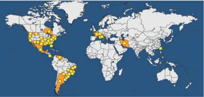
Figure 2: Cells of X. fastidiosa in biofilms on cuticle of insect foregut (Source: Almeida) ... 5 Figure 3: Geographical distribution of Xylella fastidiosa (EPPO/PQR 2016). ... 9 Figure 4: A. Typical leaf scorch symptoms, caused by Xylella fastidiosa subsp. fastidiosa on grapevine leaves; B. Typical symptoms on leaves (variegated spots) and fruits (dwarf growth) of Citrus caused by
Xylella fastidiosa subsp. pauca; C. Severe symptoms of leaf scorch on a Platanus occidentalis (sycamore)
caused by X. fastidiosa subsp. fastidiosa; D. Reduced growth of peach (on the left) affected by phony peach disease (Janse and Obradovic, 2010). ... 12 Figure 5: Phylogenetic tree of the Apulian isolate of X. fastidiosa derived from multilocus sequence typing (MLST) based on the concatenated sequences of seven genes. The Italian CoDiRO strain is
indicated by the green circle (olive) (Maria Saponari, CNR, Bari, Italy) ... 16 Figure 6: Distribution of olive quick decline in Apulia region (situation in October 2013). ... 19 Figure 7: LS Mean of total number of individuals captured for each species during two years of study. Letters above each bar indicate significant differences using one-way ANOVA with Tukey's HSD test (F= 8.05, dF= 14, P < 0.001). Vertical bars denote 0.95 confidence intervals. ... 25 Figure 8: Seasonal population dynamics of Xylella fastidiosa-infected and non-infected adults of
Philaenus spumarius (A), Neophilaenus campestris (B), and Euscelis lineolatus (C) in olive orchards. .. 28
Figure 9: Relationship between seasonal abundance (Mc) of Xylella fastidiosa-infected insects (overall abundance) and the relative incidence of infected specimens (Mi) along a single season. (A) Philaenus
spumarius. (B) Neophilaenus campestris. (C) Euscelis lineolatus. ... 30
Figure 10: Position of the three X. fastidiosa strains from 3 insect species (P. spumarius, N. campestris and E. lineolatus) in the phylogenetic tree. ... 31 Figure 11: Design of the area of study in Trepuzzi. ... 36
vii
Figure 12: Mean of X. fastidiosa cells number detected per 1μL of DNA extracted from BGSS head along 3 retention time intervals after acquisition from artificial diets containing suspensions of wild type bacteria (Temecula 1) compared to those fed on suspensions of ∆engXCA2 mutant. ... 46 Figure 13: Scanning electron micrograph illustrating X. fastidiosa biofilms within the precibarial trough of BGSS fed on artificial diet containing bacterial suspensions. Bar = 5 μm. ... 47 Figure 14: Scanning electron micrographs of the epipharyngeal surface of the foregut of BGSS fed on artificial diets containing ∆engXCA2 mutant (A), wild type X. fastidiosa (B) and artificial diet only (C) for 6 hours. Images are obtained after 7 days of multiplication period and are representative of 20 total replicates per treatment (10 replicates per time interval, i.e. 4 and 7 days of multiplication period). Diet-fed insects represent negative controls. Micrographs illustrate bacterial colonization along the insect's cibarium and precibarium, Bar = 50 μm (left) and details of bacterial biofilms in the precibarium channel and cibarium's ventral surface, Bar = 20 μm (right). ... 48 Figure 15: Scanning electron micrographs of the hypopharyngeal surface of the cibarium of BGSS fed on artificial diets containing ∆engXCA2 mutant (A), wild type X. fastidiosa (B) and artificial diet only (C) for 6 hours. Images are obtained after 7 days of multiplication period on basil and are representative of 20 total replicates per treatment (10 replicates per time interval, i.e. 4 and 7 days of multiplication period). Diet-fed insects represent negative controls. Bar = 50 μm. ... 49
viii
List of tables
Table 1. Host plants of different X. fastidiosa subspecies ... 7 Table 2. Classification and total number of captured species during the two-year survey. ... 24 Table 3. Average monthly abundance of Auchenorrhyncha species captured during a two-years survey. 26 Table 4. Average monthly incidence of Xylella fastidiosa-infected Auchenorrhyncha specimens collected during a two-year survey. ... 27 Table 5. Incidence of Xf-positive Hemiptera insects captured from the buffer zone in May and June 2014. ... 38 Table 6. Incidence of infected specimens of the three Xf-positive species during the two months of survey throughout the area of study. ... 40
1
Introduction
Olive (Olea europaea L.) is a typical Mediterranean crop. The olive was one of the first species to be cultivated, and even during the Bronze Age, it represented economic wealth for many Mediterranean societies. The olive has contributed to the development of all the cultures in the Mediterranean Basin. Man has learnt to utilize every part of the plant: the foliage for animal feed, the fruit for table oil and lighting and the wood for fuel.
Among cultivated species, olive is the sixth most important oil crop in the world, due to the beneficial nutritional properties of its oil and high economic value. The Mediterranean basin is the traditional area of cultivation and has 95% of the olive production of the world. Average olive oil production in the EU in recent years has been 2.2 million tonnes, representing around 73% of world production. Spain, Italy and Greece account for about 97% of EU olive oil production. Italian olive cultivation covers approximately 1 700 000 ha, 80% of which located in southern Italy, where Apulia represents the most important region, with about 370,000 ha.
Xylella fastidiosa is an economically important pathogen of several commercial crops, olive
included, on which it induces leaf scorch symptoms and dieback. After the first report of this bacterium on olive in California, quick decline of aged olive trees has also taken place for the first time in the Mediterranean basin, over a large area of Apulia (southern Italy). The outbreak has caused some panic and concern among olive growers because the disease was spreading very rapidly, posing an economic and environmental threat to an entire region where olive production is the staple agricultural activity and centuries-old olive trees form an inestimably valuable part of the landscape and natural heritage. Several investigations to ascertain the causes of this severe disease were performed and still are underway. As first results, symptomatic trees were found to be infected by several fungal species belonging to the genus Phaeoacremonium and
2
Phaemoniella. In addition, PCR assays on diseased trees gave positive reactions to X. fastidiosa.
Consequently, the disease was called "Olive Quick decline Syndrome" (OQDS) since many pathogens were associated with it.
Even though the exact etiological role of X. fastidiosa in the “rapid decline” disease in Italy is still not fully demonstrated, the quick dissemination of the bacterium in Apulia region prompted an investigation to prevent its spread. Xylella fastidiosa is a vector-borne pathogen transmitted by several species of Auchenorrhyncha, belonging mainly to Cicadellidae (sharpshooters) and Aphrophoridae (spittlebugs). Therefore, considering that containment of X. fastidiosa spread is mainly based on vector control, the overall goal of this thesis was to study the transmission of the bacterium in the region and determine effective control measures.
To this aim, the main objectives of this thesis were: (i) to identify potential insect vector(s) of
X. fastidiosa in Apulia region and to study their seasonal dynamics in relation with disease
diffusion; (ii) to evaluate the possibility to utilize these insects as indicators of the presence of the bacterium in apparently uncontaminated areas so called " Spy Insect approach"; (iii) to explore the effect of X. fastidiosa endoglucanases on the acquisition, retention and transmission of the bacteria by an efficient insect vector, as a biological control measure.
3
Chapter I: Literature review
I.1. Background on Xylella fastidiosa
I.1.1. History
In the 1880‟s a „mysterious‟ vine disease destroyed ca. 14,000 ha of grapes (Vitis spp.) and ca. 50 wineries had to close down in the Los Angeles area in California. This disease was described in detail in 1887 by N.B. Pierce (1856-1916) and was therefore named “Pierce‟s disease” (PD) of grapevine. A similar disease was recorded on peach (Prunus persica) in 1890 in Georgia (USA), with outbreaks in 1929, 1951 and 1976 and was named “phony peach disease” (PPD). The causal agent was described variously as a virus (Hutchins et al, 1953), a rickettsia-like bacterium (Goheen et al, 1973), until the organism was shown to be a thin, rod-shaped, Gram-negative bacterium and Koch‟s postulates were satisfied (Davis et al., 1978). In 1987, Wells et al. proposed to include the agent of these diseases in a new genus, Xylella, and named the organism
X. fastidiosa based on its fastidious growth. Already in the 1940‟s sharpshooter leafhoppers and
spittlebugs were identified as vectors of PD and PPD (Severin, 1949). Once the bacterium was described and more easily cultured, X. fastidiosa was found, with or without symptoms, in a large number of other hosts, among which the most important are Prunus spp. (almond leaf scorch, ALS in Prunus amygdalus and plum leaf scald, PLS in P. domestica), Acer spp., Carya
illinoinensis (pecan), Coffea arabica (coffee leaf scorch, CLS, also pathogenic to Citrus), Hedera helix, Morus rubra, Nerium oleander (oleander leaf scorch, OLS), Platanus occidentalis, Quercus spp., Ulmus americana. Furthermore the bacterium was found in Medicago sativa
4
Many wild plants were found to carry the pathogen (often latently), such as grasses, sedges and trees (Freitag, 1951; Raju et al., 1982).
In 1987 in Brazil, a rapidly spreading disorder, called citrus variegated chlorosis (CVC) or X-disease of Citrus, was observed and X. fastidiosa was isolated from X-diseased trees. Characterization of X. fastidiosa culminated in whole genome sequencing, and the citrus strain of
X. fastidiosa actually was the first plant pathogenic bacterium for which the whole genome was
sequenced (Chang et al., 1993).
I.1.2. Biology
X. fastidiosa (Wells et al., 1987) is a gram negative fastidious xylem limited bacterium with
single (occasionally filamentous), non-motile, aflagellate straight rods (0.25 to 0.35 by 0.9 to 3.5 pm) cells. Colonies are gram negative bacteria, catalase positive, and oxidase negative, utilize hippurate, and produce gelatinase and often beta-lactamase but not beta-galactosidase, coagulase, lipase, amylase, phosphatase, indole, or H2S. The bacterium is strictly aerobes with optimum
growth at 26-28°C and pH 6.5-6.9 (Wells et al., 1987). It colonizes the water-conducting xylem vessels of plants (Figure 1) and develops biofilms that contribute to the blockage of sap flow, resulting in plant stress and disease. It moves downstream, but also is able to migrate upstream, via twitching motility, using type IV pili (Meng et al., 2005). Twitching motility is a mean of flagellar-independent bacterial movement over moist surfaces. Biofilm formation and cell-cell aggregation of X. fastidiosa requires the presence of type I pili while type IV pili are essential for motility (Li et al., 2007).
5
Figure 1: Scanning electron micrographs of tangential sections through grapevine and associated xylem vessels. (A) Water-conducting xylem vessels of a grapevine cross section serve as the habitat for X.
fastidiosa cells. (B) View into a sectioned xylem vessel walls (Meng et al., 2005).
In advanced stages of infection, sap blocking biofilms are formed both in the host plant and in the foregut of the vectors (Figure 2). X. fastidiosa proliferates only in xylem vessels, in roots, stems and leaves. The vessels are ultimately blocked by bacterial aggregates and by tyloses and gums formed by the plant. In the vector biofilm the bacterial cells are pearly attached (Newman et al., 2004).
6
The bacterium is efficiently acquired by insect vectors, with no latent period, and persists in infective adults indefinitely (Severin, 1949). Bacterial colonization of plants is based on xylem vessels and movement through the xylem network via pit membranes. The bacterium can enter neighboring vessels through pits, after degradation of the pit membranes, which is apparently also triggered by a diffusible signal from the bacterium (Newman et al., 2004). Several bacterial components; such as type I pili, hemagglutinin, adhesins and finbriae; are implicated in X.
fastidiosa biofilm formation (Chatterjee et al., 2008). Various vascular pathogens, such as Xanthomonas and Clavibacter spp. produce polysaccharides while colonizing plants.
Interestingly, X. fastidiosa secretes exopolysaccharides (EPS) in both, plant vessels and insect vector foregut, which allow the formation of biofilms confirming that they are essential for plant virulence and vector transmission of X. fastidiosa (Killiny et al., 2013). EPS of X. fastidiosa contain a back-bone of β-1,4-glycosidic linkages which can be cleaved by endoglucanases (Roper
et al., 2016).
I.1.3. Taxonomy and classification
Based on 16S ribosomal ribonucleic acid, Wells et al. (1987) found that different strains of the bacterium were related to xanthomonads. These bacteria form a distinct group, for which the name Xylella fastidiosa was proposed and a new genus was established with one species in the gamma subgroup of the eubacteria. The taxonomic relationship among 26 strains of X. fastidiosa from 10 hosts, conducted on sequenced 16S–23S intergenic spacer (ITS) region, showed a DNA relatedness ≥70% (Schaad et al., 2004). However, at high stringency (Tm –8 °C), three distinct genotypes (A, B, and C) were revealed. Taxon A included strains from cultivated grape, alfalfa, almond (two), and maple, interrelated by 85% (mean); taxon B included strains from peach, elm, plum, pigeon grape, sycamore, and almond (one), interrelated by 84%; and taxon C included only strains from citrus, interrelated by 87%. The mean reciprocal relatedness between taxons A and
7
B, A and C, and B and C, were 58, 41, and 45%, respectively. ITS results also confirmed the same grouping: taxons A and B, A and C, and B and C had identities of 98.7, 97.9, and 99.2%, respectively. Previous and present phenotypic data support the molecular data. Taxon A strains grow faster on Pierce‟s disease agar medium whereas B and C strains grow more slowly. Taxon B and C strains are susceptible to penicillin and resistant to carbenicillin whereas A strains are opposite. Each taxon can be differentiated serologically as well as by structural proteins and the names X. fastidiosa subsp. piercei, subsp. multiplex and subsp. pauca were respectively proposed for taxons A, B, and C. So far, 5 subspecies of X. fastidiosa are registered (Table 1).
Table 1. Host plants of different X. fastidiosa subspecies
Subspecies Host species Family References
multiplex
Prunus dulcis (almond), P. armeniaca (apricot), P. persica (peach)
Rosaceae
Nunney et al., 2013
Olea europaea (Olive) Oleaceae
fastidiosa Vitis vinifera Vitaceae
Janse and Obradovic, 2010
sandyi Nerium oleander Apocynaceae
tashke Chitalpa tashkentensis Bignoniaceae
pauca Citrus spp. Rutaceae Schaad et al., 2004
8
The distribution below is given for all host plants (Figure 3). For many years,X. fastidiosaremained confined to the Americas. In 1994, it was first noticed in Asia, in Taiwan causing leaf scorch on Asian pear (Pyrus pyrifolia). In the 2000s, it was also reported causing Pierce's disease in Taiwanese vineyards. In the EPPO region, the finding in Apulia (Southern Italy) represented the first confirmed detection in Europe. The introduction pathways of X. fastidiosainto Asia or Europe are unknown. However, it can be noted that EPPO member countries have intercepted several timesX. fastidiosa on imported coffee plants from south America.
Asia: Iran, Taiwan (introduced, first found in Asian pears and then in grapevine).
North America: Canada (Ontario), Mexico, USA (Alabama, Arizona, Arkansas, California, Delaware, District of Columbia, Florida, Georgia, Indiana, Kentucky, Louisiana, Maryland, Mississippi, Missouri, Montana, Nebraska, New Jersey, New Mexico, New York, North Carolina, Oklahoma, Oregon, Pennsylvania, South Carolina, Tennessee, Texas, Virginia, Washington).
South America: Argentina, Brazil (Bahia, Espirito Santo, Goias, Minas Gerais, Parana, Rio de Janeiro, Rio Grande do Sul, Santa Catarina, Sao Paulo, Sergipe), Costa Rica, Paraguay, Venezuela.
Europe: X. fastidiosa is an EPPO A1 quarantine pathogen. The occurrence of Pierce‟s disease of
grapevine was reported in Kosovo, although this report remained dubious because of the lack of further recoveries (EPPO RS N°98/157, 1998). In France, X. fastidiosa was detected in a single blemished apricot plant by serological assays based on immunofluorescence. All further serological and molecular tests failed to detect X. fastidiosa (Anses, 2012). In 2012, X. fastidiosa was isolated from coffee plants (Coffea arabica and C. canephora) growing in a confined glasshouse near Tours, France.
9
In October 2013, the occurrence of X. fastidiosa was reported in Southern Italy (Apulia region), associated to quick decline symptoms on olive trees (Olea europea) and leaf scorch on oleander and almond (Saponari et al., 2013).
In October 2015, the bacterium (X. fastidiosa subsp. multiplex) was discovered in France on the island of Corsica on Polygala myrtifolia plants (ornamentals). It was then found on the mainland, first in the municipalities of Nice and Mandelieu-la-Napoule (Alpes-Maritimes), and then other foci were found in Alpes-Maritimes and Var departments.
In April 2016, The Institute for National and International Plant Health of the Julius Kühn-Institut (JKI) has notified the first identification of an oleander sample found positive to X. fastidiosa in Saxony (Germany).
Finally, in November 2016, the Government of the Balearic Island (GOIB) has officially announced the first detection of the quarantine pathogen Xylella fastidiosa in Spain, in the town of Manacor on the island of Mallorca. The MLST analysis identified the three isolates as belonging to the X. fastidiosa subsp. fastidiosa ST-1 strain.
10
I.1.5. Symptoms
Most disease symptoms are associated with bacterial blockage of xylem fluid transport through the plant (water and nutrients). In susceptible host plants symptoms consist of marginal leaf scorching, wilting of foliage and withering of branches, dieback and stunting with eventual plant death.
Early symptoms appear as slight chlorosis or bronzing along the leaf margin or tip that intensify and that may become water-soaked before browning and drying.
These symptoms are first found on a few branches, later on almost all foliage. The affected area is delineated by a narrow chlorotic band that becomes clear in autumn. A premature defoliation takes place with new malformed leaves formed. Abnormally shaped fruit may also be formed and stems may show internal and external discoloration, dieback and abnormal growth, leading to eventual death of the host.
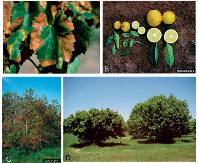
Grapevines. The most characteristic symptom of primary infection is leaf scorch. An early sign
is sudden drying of part of a green leaf, which then turns brown while adjacent tissues turn yellow or red (Figure 4, A). The desiccation spreads and the whole leaf may shrivel and drop, leaving only the petiole attached. Diseased stems often mature irregularly, with patches of brown and green tissue. In later years, infected plants develop late and produce stunted chlorotic shoots. They rarely survive more than one or two years, despite any signs of recovery.
Peaches. Young shoots are stunted and bear greener and denser foliage than healthy trees. Lateral
branches grow horizontally or droop, so that the tree seems uniform, compact and rounded (Figure 4, D). Leaves and flowers appear early, and leaves remain on the tree longer than on
11
healthy trees. Affected trees yield increasingly fewer and smaller fruits until, after 3-5 years, they become economically worthless.
Citrus. Trees can start showing the symptoms of variegated chlorosis from nursery size up to
7-10 years of age. These younger trees become systemically affected by X. fastidiosa. Usually, trees more than 15 years old are not totally affected, but rather have one or two major scaffold branches showing symptoms. Affected trees show foliar chlorosis resembling zinc deficiency with interveinal chlorosis. The chlorosis appears on young leaves as they mature and may also occur on older leaves. Newly affected trees show sectoring of symptoms, whereas trees which have been affected for a period of time show the variegated chlorosis throughout the canopy. As the leaves mature, small, light-brown, slightly raised gummy lesions (becoming dark-brown or even necrotic) appear on the underside, corresponding to the yellow chlorotic areas on the upper side (Figure 4, B). Fruit size is greatly reduced. The sugar content of affected fruit is higher than in non-affected fruit, and the fruit has a hard rind, causing damage to juicing machines. Blossom and fruit set occur at the same time on healthy and affected trees, but normal fruit thinning does not occur on affected trees and the fruits remain small but open earlier. Since more fruits remain, total production is not greatly reduced. On affected trees of cv. Pera and other orange cultivars, fruits often occur in clusters of 4-10, resembling grape clusters. Affected trees show stunting and slow growth rate. Twigs and branches die back and the canopy thins, but affected trees do not die.
Olive. The outbreak in Italy is characterized by extensive leaf scorch and dieback of olive trees
(Olea europaea), some of which are over 100 years old, over a large area estimated in ca. 8 000 hectares. A disease with similar symptoms was reported on olive trees from South California in 2010 and X. fastidiosa was associated with it. Later on, it was found that X. fastidiosa did not cause olive leaf scorch/branch dieback, but olive may serve as an alternative, albeit suboptimal,
12
host that may contribute to the epidemiology of X. fastidiosa-elicited diseases in California as a source of inoculum (Krugner et al., 2014).
Figure 4: A. Typical leaf scorch symptoms, caused by Xylella fastidiosa subsp. fastidiosa on grapevine leaves; B. Typical symptoms on leaves (variegated spots) and fruits (dwarf growth) of Citrus caused by
Xylella fastidiosa subsp. pauca; C. Severe symptoms of leaf scorch on a Platanus occidentalis (sycamore)
caused by X. fastidiosa subsp. fastidiosa; D. Reduced growth of peach (on the left) affected by phony peach disease (Janse and Obradovic, 2010).
I.1.6. Vectors
X. fastidiosa is a xylem-limited bacterium that in nature is exclusively transmitted by xylem-fluid
feeding insects. These insects belong to the order Hemiptera, sub-order Cicadomorpha (de Jong, 2013). They have sucking mouthparts (mandibular and maxillary stylets) that allow them to reach
13
the xylem of plants, from which they ingest sap. Due to the very poor nutritional value of xylem fluid, xylem-fluid feeders ingest large amounts of crude sap and produce big amounts of liquid excretions. They are generally not direct pests unless present at very high population levels. Within Cicadomorpha, the three superfamilies, Cercopoidea, Cicadoidea and Membracoidea include xylem-fluid feeding groups, but, while all Cercopoidea (known as spittlebugs or froghoppers) and Cicadoidea (cicadas) are regarded as xylem-fluid feeders, only the subfamily
Cicadellinae (known as sharpshooters) within the family Cicadellidae includes actually
xylem-fluid feeders. Even though other sap-sucking insects also feed on xylem, only these three groups of „specialists‟ in xylem-fluid feeding have been shown to be vectors of X. fastidiosa. Spittlebugs, cicadas and sharpshooters are heterometabolous insects that develop through egg, five nymphal and one adult (winged) stages. Nymphs of cicadas and of spittlebugs of the family Cercopidae are subterranean root feeders, while nymphs of spittlebugs of the family Aphrophoridae and of sharpshooters develop on the parts of host plants above the ground. All adults feed and live on the aerial parts of host plants. The transmission of X. fastidiosa by insects is peculiar in that it does not require a latent period and is of persistent type (Almeida et al., 2005). Bacteria are restricted to the alimentary canal and do not infect systemically the insect body. They adhere to and multiply in the cibarium and precibarium (part of the foregut). This implies that vectors lose the infectivity with moulting, as the foregut is of ectodermal origin and is renewed with it. Therefore, newly emerged adults must feed on an infected plant to become infectious. Once infected, adult vectors can transmit during their whole lifetime, because the bacterium multiplies and persists in the vector foregut (Almeida et al., 2005). The bacterium is not transovarially transmitted to the progeny of the vector (Freitag, 1951). For all these reasons and also because of their high mobility, winged adults are mostly responsible of X. fastidiosa spread. It is important to remember that, since the bacterium is restricted to the foregut (Purcell and Finlay, 1979), the number of bacterial cells per insect is low and therefore a very sensitive diagnostic tool, like
14
PCR, is needed to detect the presence of X. fastidiosa in the vectors. Although restricted to xylem-fluid feeding insects, transmission of X. fastidiosa is known to be poorly specific and therefore all xylem-fluid feeding insects are considered potential vectors, until proven otherwise (Frazier, 1944; Purcell, 1989; Almeida et al., 2005). However, transmission efficiency varies substantially depending on insect species, host plant, and X. fastidiosa genotype (Redak et al. 2004).
Non-European vectors
Since X. fastidiosa has been found and studied primarily in the Americas and causes disease in different crops in the Nearctic and Neotropics regions, its vectors have been identified and studied in these biogeographical areas only. Known vectors of X. fastidiosa are listed by Redak et
al. (2004).
Potential European vectors
With the exception of Philaenus spumarius (Aphrophoridae), an old world species introduced in North America, all the known American vectors are absent in Europe (de Jong, 2013). According to Janse and Obradovic (2010) only two species, Cicadella viridis and Philaenus spumarius, are possible vectors for Europe. Other xylem-fluid feeders, should indeed be considered as potential vectors according to Frazier (1944) and Purcell (1989).
A list of potential vectors of X. fastidiosa in Europe was drawn from the Fauna Europaea database (de Jong, 2013) using the following criteria: all the xylem-fluid feeding insects were included, provided that their presence was certain. As stated before, cicadas are xylem-fluid feeders and are thus expected to be potential vectors, although their role in X. fastidiosa transmission is poorly understood. In Italy, about 16 species of cicadas of families Cicadidae and
Tibicinidae are known, out of 60 species reported in Europe, most having a very restricted area of
15
most important vectors of X. fastidiosa in the Americas and only few species are present in Europe. C. viridis is widespread in Europe but only in humid areas. On the contrary, a relatively high number of spittlebug species, which are less important as vectors in America, occur in Europe and some, like P. spumarius, are very common. It has to be noted that, while the sharpshooters in America overwinter as adult and can maintain X. fastidiosa during winter, the European sharpshooters and most of the European spittlebugs (Aphrophoridae, but not Cercopidae) overwinter as egg (Nickel and Remane, 2002) and therefore, if infected, cannot sustain overwintering of X. fastidiosa, since transovarial transmission does not occur (Freitag, 1951).
I.2. The Olive Quick decline syndrome
I.2.1. Current situation in Apulia
In 2013, X. fastidiosa was reported in southern Italy associated with quick decline symptoms on olive trees, oleander and almond (Saponari et al., 2013). X. fastidiosa was found initially in the area of Gallipoli (around 8 000 ha of olive trees, with a significant part severely affected) and it was subsequently found in many other sites, first to the north and later also to the east of the initially reported outbreak areas. X. fastidiosa has been identified from olive plants based on PCR detection, ELISA, indirect immunofluorescence, electron microscopy and immunogold labelling, as well as by laboratory culture. The genotype of the strain of X. fastidiosa present in Italy is considered to be a new genetic variant within the subspecies pauca (Cariddi et al., 2014). It has been shown that the strain present in Italy is very homogeneous, and identical to a variant infecting oleander in Costa Rica. This also represents the first report of subspecies pauca in Costa Rica (Nunney et al., 2014). It was assigned a new sequence type (ST) profile, ST 53, and named CoDiRO for “Complesso del Disseccamento Rapido dell‟ Olivo”. Concatenated sequences of the
16
seven MLST genes (Figure 5) showed that the CoDiRO strain is a “divergent” variant within the subspecies pauca.
Figure 5: Phylogenetic tree of the Apulian isolate of X. fastidiosa derived from multilocus sequence typing (MLST) based on the concatenated sequences of seven genes. The Italian CoDiRO strain is indicated by
the green circle (olive) (Maria Saponari, CNR, Bari, Italy)
I.2.2. Host plants
The bacterium was isolated on periwinkle wilt gelrite and buffered cysteine–yeast extract media, from symptomatic natural infected oleander and periwinkle infected by X. fastidiosa-positive spittlebugs. Later on, it was isolated from Olea europea, Olea oleaster, Prunus dulcis, Prunus
avium, Polygala myrtifolia, Westringia fruticosa (Maria Saponari and Donato Boscia, CNR,
Institute for Sustainable Plant Protection, personal communication, October 2014). The current list ofX. fastidiosahost plant species consists of 359 plant species (including hybrids) from 204 genera and 75 different botanical families. Compared to the previous database, 44 new species
17
and 2 new hybrids, 15 new genera and 5 new families were found. The majority of the additional species (70%) were reported in Apulia, Corsica and southern France (EFSA, 2016).
In olive trees, symptoms are found on all known varieties. Old varieties, such as Ogliarola Salentina, Cellina di Nardò and the common varieties Frantoio and Coratina, appear quite susceptible while the variety Leccino seems less susceptible, although there is much uncertainty about such indications because such records are based on field observations and still have to be fully demonstrated. Although the disease was more frequently found in old trees, presumably because of the severity of symptoms, it has also been observed on young plants (Cariddi et al., 2014). First leaf scorch or, more often, desiccation symptoms generally appear on one or two branches, and then appear randomly on the rest of the canopy. It is thought that the dieback symptoms take several years to extend to the whole plant.
I.2.3. Vectors
Since the discovery of the X. fastidiosa -associated epidemics in olive groves in 2013, field surveys and transmission experiments have been carried out throughout the year, mainly by sweep nets, in the infested areas, both on olive trees and on grasses. Collected insects were further identified and tested in the laboratory for the presence of X. fastidiosa by PCR. These investigations were part of the work performed in this thesis. Thereby, further information on vector situation in Apulia are reported in the results of objectives II and III of this dissertation.
18
Chapter II:
Identification of potential insect vectors of Xylella fastidiosa in Apulia region and study of their seasonal dynamics in relation with disease diffusion.II.1. Background
The widespread of the bacterium in Apulia region prompted an investigation for the identification of the local insects, mainly leafhoppers (Cicadellidae) and spittlebugs (Aphrophoridae), which could be responsible of its transmission in Apulia. No species of sharpshooters, which are by far the most important vectors in the Americas, was previously reported in Europe. Putative vectors for Europe are considered Cicadella viridis L. (Cicadellidae) and Philaenus spumarius L. (Aphrophoridae) (Janse and Obradovic, 2010), even though the list of potential vectors should be extended to all the xylem-fluid feeders according to Purcell (1989) but also phloem feeders since they can feed occasionally on xylem (Pompon et al., 2011). To this aim, all Auchenorrhyncha species present in the contaminated area were considered. Captured Auchenorrhyncha individuals were identified and quantified in order to study their seasonal dynamics then tested for the presence of X. fastidiosa.
II.2. Material and Methods
II.2.1. Capture and Identification of Insects
From October 2013 to September 2015, Auchenorrhyncha insects were collected monthly from four olive groves located in the Lecce province of Apulia and known to have high incidence of X.
fastidiosa (Figure 6). As negative controls for the bacteria detection assays described below,
additional insects were captured from an olive grove located in Bari province, where X. fastidiosa was known to be absent based on preliminary surveys. A circular sweep net (38-cm diameter) was used to collect insects from 10 randomly selected locations per grove. In each location,
19
samples were collected from an olive tree and ground vegetation under the tree canopy. Trees were sampled by shaking four branches, from four different sides of the tree in the sweep net, whereas samples from the ground vegetation were collected by 10 sweeps. Captured insects were collected on site using an aspirator, placed in vials containing 70% ethanol and transported to the laboratory for identification. All adults of Auchenorrhyncha species collected were identified. The taxonomic classification of captured insects was based on Ribaut (1952), Della Giustina (1989) and Holzingher et al. (2003). For species identification, male genitalia was dissected and kept in KOH (10%) for 24 hrs; then mounted on glass slide in Faure‟s liquid and observed under stereoscopic microscope. Due to the multiplication and accumulation of X. fastidiosa cells in the foregut (Janse and Obradovic 2010), only the head of each adult specimen was detached from the body, as described by Bextine et al. (2004) and used for the DNA extraction method described below.
High incidence Low to medium incidence
Gallipoli
Trepuzzi
Lecce
20
II.2.2. Extraction and amplification of X. fastidiosa DNA
Insect heads were macerated individually using a sterile pestle in a tube containing 200 μl of extraction buffer (Triton X 1%, Tris-HCl 20 mM, EDTA 2 mM). After the homogenization, 180 μl of suspension were transferred to a new tube and left to incubate at 60° C for 30 min to allow complete lysis of cells. Two hundred microliters of chloroform-isoamyl alcohol (24:1) were added to the suspension and centrifuged at 12,000 g for 10 min. DNA was precipitated by adding 1 vol of cold isopropanol and incubated overnight at -20° C. After a new centrifugation at 20,000 g for 20 min, the pellet was washed with 70% ethanol, dried and finally resuspended in 20 μl of sterile water. DNA extracts from insect samples were tested by PCR with RST31 (GCGTTAATTTTCGAAGTGATTCGATTGC) and RST33 (CACCATTCGTATCCCGGTG) specific primers targeting the RNA polymerase sigma-70 factor genomic area and generating an amplicon of 733 bp (Minsavage et al. 1994). DNA extracts from midveins of X. fastidiosa-infected olive leaves were used as positive controls. Each PCR mixture contained template DNA at 10 ng/μl, 1.25 U of Go Taq polymerase (Promega, Madison, WI, USA), 1× Go Taq Flexi DNA Buffer, 0.2mM dNTPs, 0.3 μM forward and reverse primers, all in a final reaction volume of 25 μl. Samples were put in the thermocycler (IQ5 Thermocycler, BioRad, USA) with the following program: initial denaturation at 94° C for 4 min, 35 cycles at 94° C for 30 s, 55° C for 30 s and 72° C for 40 s, and a final extension at 72° C for 7 min. Products were visualized by ethidium bromide staining after electrophoresis in 1.2 % agarose gel.
II.2.3. Cloning and sequence analysis
PCR amplicons were transformed in StrataCloneTM PCR Cloning vector pSC-A (Stratagene, CA, USA), subcloned into Escherichia coli DH5α or SoloPACK cells and custom sequenced (Primm, Milan, Italy). Four copies of each DNA clone were sequenced bi-directionally to eliminate any
21
sequence ambiguity. The sequences obtained were cleaned from vector with the assistance of the DNA Strider 1.1 program (Marck, 1988). Sequence similarities were analyzed with BLASTN at the National Center for Biotechnology Information website (Altschul et al., 1990). Multiple sequence alignments were generated by using the default options of CLUSTALX 1.8 (Thompson
et al., 1997). Sequences obtained from this study were deposited in GenBank. II.2.3.1. Ligation
PCR amplicons corresponding to different insect species were considered. Two μl of each PCR product were mixed with 3 μl of cloning buffer and 1 μl of vectors and the mix was incubated at room temperature for 15 min.
II.2.3.2. Transformation of competent cells
Fifty microliters of bacterial suspension were added to the mix and immerged in ice for 20 min. After a heat shock at 42°C for 45 sec, the mix was replaced in ice. Then the samples were added to 800 μl of LB without ampicilline and shaked at 37°C for 90 min. Samples were centrifuged at 5 000 rpm for 3 min, and the pellet (100 μl) was plated using sterile glass rod onto solid LB plate previously prepared adding 35 μl of Xβ galactosidose. Plates were incubated overnight at 37°C.
II.2.3.3. Extraction of DNA plasmid
From each plate, single white colonies, likely containing the recombinant plasmids, were collected and diluted in bacterial tubes containing 3 ml of LB with ampicilline and incubated overnight at 37°C with agitation. An aliquot (1.5 ml) from each tube was transferred to another eppendorf tube and centrifuged at 13 000 rpm for 30 sec. The supernatant was discarded and the pellet resuspended in the remaining 1.5 ml in the bacterial tube. After a new centrifugation at 13 000 rpm for 30 sec, the supernatant was discarded.
22
The isolation of the plasmid DNA was done using Quick lyse Qiagen Mini prep kit. Thereby, 400 μl of lysis buffer (QLL buffer) and the mix were homogenized on vortex. This mixture was transferred onto collection tubes with filters and centrifuged at 13 000 rpm for 1 min; then 400 μl of washing buffer (QLW buffer) and the mix were added and again centrifuged at 13 000 rpm for 1 min. The liquid was discarded keeping the covering filters, and again centrifuged to eliminate the remaining ethanol. Then, the covering filters containing the DNA were transferred on new eppendorf tubes where 50 μl of elution buffer (QLE buffer) were added and centrifuged at 13 000 rpm for 1 min. Covering filters were then discarded and the DNA kept into the eppendorf tubes.
II.2.3.4. Enzymatic digestion
Three microliters of DNA were mixed with 1 μl of restriction enzyme buffer H (10pb), 0.2 μl of EcoRI and 5.8 μl of sterile water. The mix was incubated at 37°C for 1h until used for electrophoretic analyses.
II.2.4. Data Analysis
Olive orchards were selected based on three characteristics: short distance from each other, same olive tree variety, and absence of insecticide applications. Data on insect abundance and prevalence of X. fastidiosa-infected insects from each orchard were combined for analysis. Species dominance was characterized according to the categories described by Tischler (1949): eudominant, more than 10% of the total number captured; dominant, between 5 and 10%; subdominant, between 2 and 5%; rare, between 1 and 2%; sub-rare, less than 1 %. Mean separation for species abundance comparison was made with one-way analysis of variance (ANOVA) followed by Tukey's honestly significant difference (HSD) test.
23
The mean of each monthly collection (Mc) and X. fastidiosa-infection (Mi) were calculated to
compare abundance of captured species and correlation with the rate of infection along a single vegetative season (12 months), applying the following formulas:
where N1 and X1 were, respectively, the number of individuals captured and the number of
individuals infected per species per month in the first year, while N2 and X2 were the number of
individuals captured and the number of individuals infected of the same species and same month in the second year of study. In addition, the total monthly collection of insects (McT), which was
the sum of Mc per each species, was calculated. The correlation between seasonal abundance
(Mc) and incidence of infected specimens (Mi) for species found to harbour the bacterium was
evaluated on the basis of Pearson's (multiple) correlation coefficient (r). Data analysis described above and graphs were treated using the statistical software STATISTICA (version 7.0.61.0).
II.3. Results
II.3.1. Identification and abundance of insects
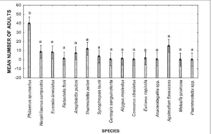
Overall, 2398 adults of Auchenorrhyncha were captured. Fifteen species were identified, belonging to two infraorders (Cicadomorpha and Fulgoromorpha) and five different families (i.e., Aphrophoridae (two species), Cercopidae (one species), Cicadellidae (10 species), Issidae (one species), and Flatidae (one species)) (Table 2). The Tischler‟s abundance classification in classes of abundance revealed three species as eudominant, i.e. P. spumarius (39.8%), Agalmatium
flavescens (Olivier) (Hemiptera: Issidae) (15.1%) and Thamnotettix zelleri (Kirschbaum)
(Hemiptera: Cicadellidae) (11.9%); three as dominant, i.e. N. campestris (8.6 %), E. lineolatus Mc= (N1+N2)/2
24
(8.1%) and Anoplotettix putoni Ribaut (Hemiptera: Cicadellidae) (7.2%); one as subdominant, i.e.
Synophropsis lauri (Horvart) (Hemiptera: Cicadellidae) (3.6%); three as rare, i.e. Exitianus capicola (Stäl) (Hemiptera: Cicadellidae) (1.6%), Allygus modestus Scott (Hemiptera:
Cicadellidae) (1.2%) and Fieberiella florii (Stäl) (Hemiptera: Cicadellidae) (1.1%), while the remainder of the captured species were classified as sub-rare with less than 1% abundance (Table 2). Philaenus spumarius was clearly the most captured species in the olive groves (Figure 7).
Table 2. Classification and total number of captured species during the two-year survey.
Infra order Superfamily Family Species Na P
(%)b Cicadomorpha Cercopoidea Aphrophoridae Philaenus spumarius (L.) 955 39.8
Neophilaemus campestris
(Fallèn)
207 8.6 Cercopidae Cercopis sanguinolenta
(Scopoli)
12 0.5 Cicadoidea Cicadellidae Allygus modestus Scott 28 1.2
Anoplotettix putoni Ribaut 172 7.2
Fieberiella florii (Stäl) 26 1.1
Synophropsis lauri (Horvart) 86 3.6
Euscelis lineolatus Brullé 195 8.1
Conosanus obsoletus (Kirschbaum) 13 0.5 Exitianus capicola (Stäl) 39 1.6 Thamnotettix zelleri (Kirschbaum) 286 11.9 Psammnotettix spp. 2 0.1 Anaceratagallia spp. 13 0.5 Fulgomorpha Fulgoroidea Issidae Agalmatium flavescens (Olivier) 363 15.1 Flatidae Metcalfa pruinosa Say 1 0.04
Total 2398
a
N: number of individuals captured, b P (%): Percentage of individuals of each species among the total number of insects collected.
25
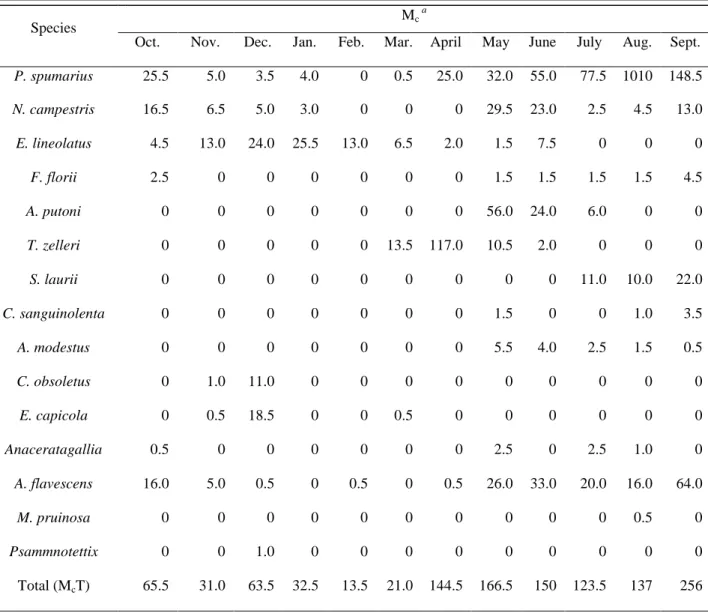
Occurrence of Auchenorrhyncha was mainly concentrated in the period from March to October with the only exception being E. lineolatus, which was prevalent from November to February. During two years of the study, the highest number of Auchenorrhyncha adults captured was in September (McT= 256), whereas May was the month with the highest species richness (10 out of
15 species collected) (Table 3).
Figure 7: LS Mean of total number of individuals captured for each species during two years of study. Letters above each bar indicate significant differences using one-way ANOVA with Tukey's HSD test (F=
8.05, dF= 14, P < 0.001). Vertical bars denote 0.95 confidence intervals.
II.3.2. Detection and seasonal fluctuations of X. fastidiosa infection in captured insects
Only three out of 15 species collected were found positive for presence of X. fastidiosa by PCR:
26
out of 2398 adults (9.3%) of these three species were positive for presence of X. fastidiosa. In contrast, none of 460 adults of nine species, including P. spumarius, N. campestris and E.
lineolatus, captured from the X. fastidiosa-free area in Bari were positive by PCR.
Table 3. Average monthly abundance of Auchenorrhyncha species captured during a two-years survey.
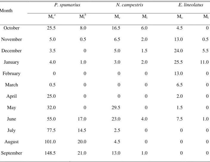
In the X. fastidiosa-infected areas, PCR assays showed that X. fastidiosa was detected in adults of
P. spumarius, N. campestris and E. lineolatus, starting from June until January, whereas all insect
samples collected from February to May were negative for presence of X. fastidiosa (Table 4).
Species Mc
a
Oct. Nov. Dec. Jan. Feb. Mar. April May June July Aug. Sept.
P. spumarius 25.5 5.0 3.5 4.0 0 0.5 25.0 32.0 55.0 77.5 1010 148.5 N. campestris 16.5 6.5 5.0 3.0 0 0 0 29.5 23.0 2.5 4.5 13.0 E. lineolatus 4.5 13.0 24.0 25.5 13.0 6.5 2.0 1.5 7.5 0 0 0 F. florii 2.5 0 0 0 0 0 0 1.5 1.5 1.5 1.5 4.5 A. putoni 0 0 0 0 0 0 0 56.0 24.0 6.0 0 0 T. zelleri 0 0 0 0 0 13.5 117.0 10.5 2.0 0 0 0 S. laurii 0 0 0 0 0 0 0 0 0 11.0 10.0 22.0 C. sanguinolenta 0 0 0 0 0 0 0 1.5 0 0 1.0 3.5 A. modestus 0 0 0 0 0 0 0 5.5 4.0 2.5 1.5 0.5 C. obsoletus 0 1.0 11.0 0 0 0 0 0 0 0 0 0 E. capicola 0 0.5 18.5 0 0 0.5 0 0 0 0 0 0 Anaceratagallia spp. 0.5 0 0 0 0 0 0 2.5 0 2.5 1.0 0 A. flavescens 16.0 5.0 0.5 0 0.5 0 0.5 26.0 33.0 20.0 16.0 64.0 M. pruinosa 0 0 0 0 0 0 0 0 0 0 0.5 0 Psammnotettix spp. 0 0 1.0 0 0 0 0 0 0 0 0 0 Total (McT) 65.5 31.0 63.5 32.5 13.5 21.0 144.5 166.5 150 123.5 137 256 a
27
Results of PCR assays are reported in Table 4, sub-divided into three periods per year based on presence or absence of infected individuals: Period 1. October to January, Period 2. February to May, and Period 3. June to September. The first period was characterized by increasing occurrence of X. fastidiosa-infected E. lineolatus adults, while presence of X. fastidiosa-infected
P. spumarius and N. campestris decreased steadily, being almost negligible in winter. In the first
year, 50 out of 154 adults (32.5%) of P. spumarius, N. campestris and E. lineolatus collected during Period 1 tested positive for X. fastidiosa. Incidence of infection for E. lineolatus was 23 of 76 (30.2%), for N. campestris 16 of 37 (43.2%), and for P. spumarius 11 of 41 (26.8%).
Table 4. Average monthly incidence of Xylella fastidiosa-infected Auchenorrhyncha specimens collected during a two-year survey.
Month
P. spumarius N. campestris E. lineolatus
Mc a Mi b Mc Mi Mc Mi October 25.5 8.0 16.5 6.0 4.5 0 November 5.0 0.5 6.5 2.0 13.0 0.5 December 3.5 0 5.0 1.5 24.0 5.5 January 4.0 1.0 3.0 2.0 25.5 11.0 February 0 0 0 0 13.0 0 March 0.5 0 0 0 6.5 0 April 25.0 0 0 0 2.0 0 May 32.0 0 29.5 0 1.5 0 June 55.0 17.0 23.0 4.0 7.5 1.0 July 77.5 14.5 2.5 0 0 0 August 101.0 20.0 4.5 0 0 0 September 148.5 21.0 13.0 1.0 0 0 a
MC: Mean of captured individuals,
b
28
Similarly, during the same time period in the second year , 26 of 118 (22%) adults of the same three species tested positive for X. fastidiosa with 11 (42.3%) being E. lineolatus that was the most abundant (49.1% of the total adults) species. Incidence of X. fastidiosa infection in N.
campestris and P. spumarius was, respectively, 28.0% (7 of 25) and 22.8% (8 of 35) (Figure 8 A,
B).
Figure 8: Seasonal population dynamics of Xylella fastidiosa-infected and non-infected adults of
Philaenus spumarius (A), Neophilaenus campestris (B), and Euscelis lineolatus (C) in olive orchards.
The second period (February to May) was characterized by absence of adult individuals until April, when there was a rapid increase in the number of newly emerged adults. None of the
29
insects collected during this period (P. spumarius, n = 115, N. campestris, n = 59 and E.
lineolatus, n = 46) were found positive for X. fastidiosa by PCR (Figure 8). Finally, the third
period (June to September) was characterized by a dominant presence of P. spumarius, with 445 of 507 (87.7%) and 319 of 358 (89.1%) individuals captured in the first and second year, respectively, with about 20% of these insects infected with X. fastidiosa (Figure 8, A).
A positive correlation between seasonal abundance of insects and relative incidence of X.
fastidiosa was noticed for P. spumarius and E. lineolatus, for which the number of infected adults
increased as abundance increased. Specifically, in January the number of E. lineolatus captured and incidence of infected insects were at maximum with Mc = 25.5 and Mi = 11 (Table 4), which
was supported by the high coefficient of correlation (r = 0.90) (Figure 9, C). Similarly, the highest number of infected P. spumarius individuals coincided with greatest abundance in September with Mc = 148.5 and Mi = 21 (Table 4), resulting in a high coefficient of correlation (r
= 0.92) (Figure 9, A).
Unlike P. spumarius, the peak of N. campestris population was in May with Mc = 29.5, when
incidence of infected adults was null, while highest incidence of infected adults occurred in October (Mi = 6). Moreover, the number of infected adults of N. campestris was clearly
independent from abundance of this species, as supported by the low coefficient of correlation (r= 0.42) (Figure 9, B). Population density was high in May (Mc = 29.5) and June (Mc = 23),
decreased steadily from July to August and then increased again in September (Mc = 13) and
30
Figure 9: Relationship between seasonal abundance (Mc) of Xylella fastidiosa-infected insects (overall abundance) and the relative incidence of infected specimens (Mi) along a single season. (A) Philaenus
spumarius. (B) Neophilaenus campestris. (C) Euscelis lineolatus.
31
Six PCR amplicons, two from each one of the 3 Xf-positive insect species (P. spumarius, N.
campestris and E. lineolatus), were chosen for cloning and sequencing. The 6 sequences obtained
were 99.3-100% identical among them at nucleotide level, and 3 of them (one sequence from each insect) were deposited in GenBank (accession no. HG939491, HG939505 and HG939506). BlastN analysis showed that all these sequences had the highest nucleotide identity (99.6-99.7%) with X. fastidiosa DNA fragments OL-X4-5 (accession no. HG532021) and OL-G (accession no. HG532022) from Apulian olive trees. However, they shared 96-98% identity with homologous fragments of isolates from other countries, and in particular 98% with the strains of subspecies fastidiosa GB514 (CP002165 from Texas-USA) and M23 (CP001011 from California-USA), and 97% with the strain of subspecies pauca 9a5c (AE003849).
Figure 10: Position of the three X. fastidiosa strains from 3 insect species (P. spumarius, N. campestris and E. lineolatus) and 3 strains obtained from olive plant tissue (Xf. subsp. olive) in the phylogenetic tree.
32
Based on this sequence analysis, a phylogenetic tree (Figure 10) was deduced, which confirmed that the 3 sequences from insects were in the same clade with the strains of X. fastidiosa from Apulian olive trees, thus supporting that a single X. fastidiosa strain is present in that area. Whether the insects acquired the bacterium directly from olive plants or by alternative hosts, still remains to be established.
II.4. Discussion
Results of the present study provided new insights to X. fastidiosa epidemiology in the Apulia region of Italy. This study describes seasonal abundance of Auchenorrhyncha species and temporal dynamics of X. fastidiosa infection of insects in olive groves. The surveys determined that among 15 species of Auchenorrhyncha captured during the two years of study, P. spumarius was the most abundant species in olive groves in Lecce province. Adult P. spumarius began to molt to the adult stage between April and May and were particularly abundant in summer. In contrast, E. lineolatus was the most abundant species during winter (November to February) with very few adults captured later in the season (i.e., June), which suggests that individuals of this species overwinter as adults in olive groves.
Over the two-year study, X. fastidiosa was detected by PCR in P. spumarius, N. campestris and E. lineolatus. Detection of X. fastidiosa coincided with presence of adult P. spumarius, N.
campestris and E. lineolatus, with incidence of X. fastidiosa-infected specimens highly correlated
with seasonal abundance, except for N. campestris. One hypothesis to explain the lack of correlation between N. campestris abundance and peak in X. fastidiosa incidence in insects is that adult N. campestris prefers grasses as a feeding host plant, which were mostly absent in the studied environment throughout the summer. Consequently, host plants used for feeding also could have influenced correlation between the infection incidence and population size since
33
adults of N. campestris may not frequently come in contact with infected plants during the summer.
Detection of infected specimens of P. spumarius from June to January was not surprising. Adults likely move from herbaceous plants and shrubs in late spring to olive trees in summer, from which acquisition of X. fastidiosa may occur and persist until insect death (Hill and Purcell 1995). Therefore, incidence of X. fastidiosa-positive insects was directly influenced by the life cycle of each species. None of the few adult P. spumarius and N. campestris captured in April and May were found infected. Likely, these individuals belonged to a new (first) adult generation because no adults of these species were captured in the previous period. In contrast, E. lineolatus captured in October and November were not infected by X. fastidiosa, which suggest that: (1) adults were collected prior to coming in contact with X. fastidiosa-infected olive trees and/or (2) acquisition efficiency from olive trees during winter months is low for this species.
Philaenus species, including P. spumarius, are vectors of X. fastidiosa in California (Severin
1950), but little is known about its role in Pierce‟s disease epidemiology. In southern Italy, P.
spumarius has been considered the most important vector of X. fastidiosa in olive groves.
However, given similar trophic characteristics of Philaenus sp. and Neophilaenus sp., further studies are needed to evaluate transmission efficiency of X. fastidiosa to olive by these species. While N. campestris must be considered a candidate vector species, Euscelis spp. are phloem-fluid feeding insects unlikely to transmit a xylem-limited bacterium. For instance, E. maculipenis DeLong and Davidson failed to transmit X. fastidiosa to grapevines in California (Severin 1949). However, considering the high numbers of X. fastidiosa-infected adults, it is clear that individuals of E. lineolatus acquire bacteria from infected host plants and retains X. fastidiosa with unexpectedly high efficacy for a phloem-fluid feeding insect. Although transmission of X.
fastidiosa by phloem-fluid feeding insects has not been demonstrated, our study clearly confirms
34
2011). One possible explanation of why these insects cannot successfully inoculate the bacterium into xylem vessels may be related to specific feeding behaviors (e.g., ingestion, egestion, salivation) that are performed in phloem but not in xylem tissue (Backus et al. 2012).
Our findings suggest that preventive control measures against juvenile stages of the univoltine vector, P. spumarius, may reduce spread of X. fastidiosa by reducing the number of adult insects. Since nymphs are stationary and primarily use ground vegetation as feeding hosts, managing alternative host plants in olive groves by mechanical and/or chemical practices and/or use of insecticides may help suppress P. spumarius populations. If adopted, treatments should target nymphal populations present between March and mid-April, or when signs of nymph development are visible (foamy substances present on the herbaceous plants). However, P.
spumarius is a polyphagous insect that feeds and reproduces on plants in many habitats, including
cultivated and non-cultivated hosts near olive groves. Little is known about mobility of adult P.
spumarius and potential to colonize olive groves. Therefore, additional research is needed to
identify host plants of P. spumarius to guide landscape management strategies targeting key reproductive and feeding hosts. In addition, it is important to know which environmental conditions influence movement of vectors from other plants (in particular from herbaceous plants) to olive trees. Considering the complex olive-X. fastidiosa-insect pathosystem, a study on efficacy of different control approaches is warranted, taking particular consideration of the role of natural predators in regulating vector population density.
35
Chapter III:
Evaluation of the possibility to utilize potential vectors as indicators of the presence of the bacterium in apparently uncontaminated areas so called "Spy Insect approach"III.1. Background
The quick dissemination of X. fastidiosa prompted an effective strategy for quickly monitoring this bacterium in a certain area in order to limit its spread. Since X. fastidiosa is a vector-borne bacterium, a survey on potential insect vectors present in the areas surrounding the infection sites (buffer zone) was carried out in order to evaluate the possibility to detect Xf-positive insects (so called “spy insects”), which can be used as indicators of the bacterium presence in apparently uncontaminated areas.
III.2. Material and methods
III.2.1. Insects capture and identification
From May to June 2014, adult insects were collected from 9 olive orchards from Trepuzzi area, where an isolated Xf-outbreak was present (Figures 6). All around the severely infected olive orchard (C), eight other orchards, apparently Xf-free, were identified symmetrically distributed from 4 sides [North (N1, N2), South (S1, S2), East (E1, E2) and West (W1 and W2)], at a distance of 500mt and 1000mt, respectively (Figure 11). A sweeping net was used to manually trap the insects from both olive canopy and ground vegetation. The insects captured were carefully collected by aspiration directly in loco, put in small tubes containing 70% ethanol and brought to the laboratory for identification. The classification and nomenclature of captured leafhoppers and froghoppers were based on Ribaut (1952), Della Giustina (1989) and Holzingher
36
KOH (10%) for 24 hrs. Then, they were mounted on glass slide in Berlese‟s liquid and observed under stereoscopic microscope.
C
N
S
E
W
E1 S1 W1 N1 E2 N2 W2 S2Figure 11: Design of the area of study in Trepuzzi.
III.2.2. Extraction of X. fastidiosa DNA from insects and PCR assays
Only adults were considered and the total of captured specimens were tested. Due to the multiplication and accumulation of bacteria particles in the foregut (Janse and Obradovic, 2010), the heads of single adult specimens were detached from the body as described by Bextine et al. (2004) and used for DNA extraction as follows. Each head was grinded using sterile pestles in a tube containing 200 μl of extraction buffer (Triton-X 1%, Tris-HCl 20 mM, EDTA 2 mM). After homogenization, 180 μl of suspension were transferred to a new tube and left to incubate at 60° for 30 min to allow full cell lysing to take place. Two hundred microliters of chloroform-isoamyl alcohol (24:1) were added to the suspension and centrifuged at 10,000 rpm for 10 min. DNA was