UNIVERSITY OF CATANIA
INTERNATIONAL DOCTORAL PROGRAM IN
ONCOLOGICAL SCIENCES
- CYCLE XXVIII –
Saverio Candido
Neutrophil gelatinase-associated lipocalin (NGAL) and
matrix metalloproteinases (MMPs) as biomarkers of
bladder cancer development and progression
DOCTORAL THESIS
Tutor: Prof. Massimo Libra
Coordinator: Prof. Ferdinando Nicoletti
CONTENTS
ABSTRACT ... 1
1. INTRODUCTION ... 2
1.1 NGAL ... 2 1.2 MMP-9 ... 62. RESULT ... 8
2.1 COMPUTATIONAL ANALYSES OF NGAL IN DIFFERENT TUMOR TYPES ... 8
2.1.1 mRNA expression ... 8
2.1.2 Protein expression ... 12
2.1.3 Clinical impact ... 14
2.2 VALIDATION OF COMPUTATIONAL RESULTS IN BLADDER CANCER ... 26
3. DISCUSSION ... 31
3.4 FUTURE CHALLENGES ... 34
4. MATERIAL AND METHODS ... 36
4.1 COMPUTATIONAL ANALYSES ... 36
4.2 PATIENTS RECRUITMENT AND BLOOD SAMPLE COLLECTION ... 37
4.3 ELISA ASSAY ... 37
4.4 STATISTIC ANALYSIS ... 38
1
ABSTRACT
Neutrophil gelatinase-associated lipocalin (NGAL), also called lipocalin-2, is a secreted protein belonging to the lipocalin family proteins and actively participates into the proliferation, differentiation, and development of human tissues, including tumours. It positively modulates the activity of the matrix metalloproteinases-9 (MMP-9) that are involved in the enzymatic remodelling of the extracellular matrix. MMP-9 regulates the degradation of extracellular matrix in processes such as angiogenesis, tumour growth, and metastasis. By forming the NGAL/MMP-9 complex, NGAL protects MMP-9 from proteolytic degradation, a fundamental mechanism in controlling the activity of the proteins, and enhances its enzymatic activities.
As a secreted protein, NGAL is detectable in many biologic fluids, including urine, where several neoplastic cells and other tumor microenvironmental factors can be directly released from the cancer. Our in silico analysis suggested an active role of NGAL in tumour development of several cancer types. Validation of these findings is here described in bladder cancer as a good tumor model in which investigate the role of this protein because urine is in direct contact with the primary tumor.
On these bases, the release of NGAL in both urine and serum samples from 89 bladder cancer patients was measured. Further investigations, aimed to emphasize the role of NGAL in cancer, were performed by analysing MMP-9 and NGAL/MMP-9 complex levels in the same subset of bladder cancer patients. Control experiments were performed in 119 cancer-free controls, previously enrolled in a case-control study. Urinary concentrations were standardized on creatinine level. The performance of these proteins as cancer biomarkers was evaluated through the receiver operating characteristic (ROC) analysis.
In conclusion, the present study deepens the knowledge of the molecular mechanisms sustaining NGAL expression in tumor cells and its effects on cancer metastatic behaviour. Furthermore, NGAL/MMP-9 pathway is associated with an aggressive phenotype of transitional cell carcinoma of the bladder (TCC). The elevated negative predictive value of MMP-9 and NGAL/MMP-9 complex make them candidate markers of exclusion test for TCC. These findings suggest that these proteins may be integrated in the surveillance of bladder cancer, thus improving patients complaints and diminishing their discomfort.
2
1. INTRODUCTION
Metastatic spreading is the major cause of death in cancer patients. Several molecules have been shown to contribute to tumor invasion and spreading. Recently, the role of neutrophil gelatinase-associated lipocalin (NGAL) has been associated with cancer development and progression [See Candido S et al, 2015 for a review]. However, its specific role for each tumor type was lacking and then better clarified in the present study.
NGAL is a secreted protein detectable in many biologic fluids, including urine, where several neoplastic cells and other tumor microenvironmental factors can be directly released into the urine. Therefore, bladder cancer may represent a good tumor model in which investigate the role of this candidate marker as urine is in direct contact with the primary tumor. Among tumor microenvironmental factors, matrix metalloproteinase-9 plays an important role, especially in tumor growth and spreading. Notably, NGAL's ability to combine in a dimeric complex with MMP-9, results in a protective action of MMP-9 from its auto-degradation and consequently results in a higher gelatinolytic action of MMP-9 on extracellular matrix [Yan L et al, 2001]. By this function, it has been shown that NGAL may promote cancer development in a variety of different cancer types [Fernández CA et al, 2005; Kubben FJ et al, 2007; Zhang H et al, 2007; Smith ER et al, 2008].
The analysis of NGAL transcript levels and its potential clinical implications in different cancer types, explored by bioinformatic approaches, represents the first part of the study included in this Ph.D. Program and it was already published in Oncotarget [See Candido S et al, 2014]. In detail, NGAL transcript levels were explored in different cancer types by analysing public available microarray datasets. NGAL protein expression were performed by analyzing the Human Protein Atlas. These computational data are thoroughly discussed below.
On these bases, validation of our computational data, representing the second part of the study included in this Ph.D. Program, was performed in a consecutive series of both blood and urine samples from bladder cancer patients and from age and sex-matched normal samples.
1.1 NGAL
Members of the lipocalin protein family are characterized by their ability to bind small hydrophobic molecules (such as prostaglandins, retinoids, arachidonic acid, hormones and fatty acids). They often bind to specific cell-surface receptors and form macromolecular
3 complexes. Highly conserved lipocalin crystal structures consist of a single eight-stranded continuously hydrogen-bonded antiparallel β-barrel delineating a calyx shape, which represents the internal ligand-binding site (Figure 1). Members of the lipocalin family, in the past classified exclusively as transport proteins, have now been described to carry out a variety of different functions. Some of these functions include: retinol transport, cryptic coloration, olfaction, pheromone transport, and the enzymatic synthesis of prostaglandins, moreover the lipocalins are also involved in the regulation of the immunoresponse and the mediation of cell homoeostasis [Flower DR, 1996].
Figure 1. Highly conserved lipocalin crystal structures consist of a single eight-stranded continuously hydrogen-bonded antiparallel β-barrel (A) delineating a calyx shape, which represents the internal ligand-binding site (B). Hydrophobicity surface (C). Images were created from the RCSB PDB database
(http://www.rcsb.org) (ID: 1NGL) using the UCSF Chimera package UFCS Chimera package that is developed by the Resource for Biocomputing, Visualization, and Informatics at the University of California, San Francisco (supported by NIGMS P41-GM103311). Ref: The solution structure and dynamics of human neutrophil
gelatinase-associated lipocalin by Coles M, et al. J Mol Biol. 1999; 289: 139-57.
NGAL, also called lipocalin 2, siderocalin and 24p3, was identified in several forms: a monomer (25-kDa), a disulfide-linked homodimer (46-kDa), and a disulfide-linked heterodimer with human neutrophil gelatinase B (135-kDa) [Kjeldsen L et al, 1993]. NGAL has several functions. In early studies NGAL was described as a factor of innate immune system. NGAL is released by neutrophils at sites of infection and inflammation to sequester bacterial ferric siderophores, participating in the antibacterial iron-depletion strategy of innate immune system [Goetz DH et al, 2002]. Subsequently, it was shown that NGAL is responsible for iron delivery to the cytoplasm where it is accumulated and activates or represses iron-responsive genes. Iron unloading depends on the cycling of NGAL through acidic endosomes [Yang J et al, 2002]. In contrast, Devireddy LR et al have shown that NGAL is also involved in apoptosis-dependent deprivation of trophic factors. Apo-NGAL, after binding to its putative receptor, 24p3R, is internalized and associates with an intracellular siderophore, transferring chelated iron to the extracellular medium, thereby
4 reducing intracellular iron concentration which leads to the expression of the pro-apoptotic protein Bim, leading to the induction of apoptosis [Devireddy LR et al, 2005] (Figure 2).
Figure 2. Effects of iron concentration on NGAL activity. NGAL in the presence of siderophores and ferrous
iron (Fe++) is involved in iron uptake which is important in controlling the iron-dependent growth pathways, drug resistance and inhibiting the expression of 1alpha. In contrast, under conditions of iron depletion, HIF-1alpha expression is stimulated, iron-dependent growth pathways are inhibited and apoptosis is induced. Abbreviations: Neutrophil gelatinase-associated lipocalin (NGAL), NGAL receptor (NGAL-R), hypoxia-inducible factor-1alpha (HIF-1-alpha), Bcl-2-like protein 11 (Bim).
NGAL was originally identified as a protein covalently associated with 92-kDa gelatinase/MMP9 from human activated neutrophils [Kjeldsen L et al, 1993]. NGAL is expressed in many other types of cells in response to various injuries, especially in kidney diseases. Serum NGAL levels correlate clearly with the severity of renal injury, reflecting the degree of tissue damage. For this reason, NGAL may become one of the most promising next-generation biomarkers in clinical nephrology and as well as other diseases and pathological states [Bolignano D et al, 2010].
NGAL is up-regulated by IL-1 beta, but not by TNF-alpha, in type II pneumocyte-derived cell line through the induction of the NF-kB pathway [Cowland JB et al, 2003]. IL-1 beta selectivity in inducing NGAL is due to the synthesis of IkB-zeta, a NF-kB-binding cofactor, elicited specifically by IL-1beta stimulation which is required for transcriptional activation of NGAL [Cowland JB et al, 2006]. Stimulation with TNF-alpha in the presence of IL-17, which
5 stabilizes the IkB-zeta transcript, is able to induce NGAL expression by IkB-zeta protein binding to NF-kB on the NGAL promoter [Karlsen JR et al, 2010]. It has been also demonstrated that activation of the NF-kB pathway is associated with up-regulation of NGAL-ErbB2-mediated signaling [Li SH et al, 2009] (Figure 3).
Figure 3. Effects of cytokines and growth factor receptors on NGAL gene expression.
Abbreviations: Matrix metalloproteinase 9 (MMP-9), v-Erb-B2 Avian Erythroblastic Leukemia Viral Oncogene Homolog (Erb-B2 = HER2), tumor necrosis factor-alpha (TNF-α), interleukin-17 17), interleukin-1 beta (IL-1β), focal adhesion kinase (FAK), nuclear factor kappa B (NF-κB) inhibitor of NF-κB zeta subunit (IκBζ), ferrous iron (Fe++).
Its complex with MMP-9 results in a higher gelatinolytic activity of MMP-9 on extracellular matrix [Yan L et al, 2001]. By this function, it was suggested that NGAL induce cancer development in several cancer types [Fernández CA et al, 2005; Kubben FJ et al, 2007; Zhang H et al, 2007; Smith ER et al, 2008]. Conversely, anticancer activities of NGAL have been demonstrated by its ability to inhibit the pro-neoplastic factor 1a, the synthesis of HIF-1a-dependent VEGF [Venkatesha S et al,2006; Tong Z et al, 2008], and phosphorylation of FAK kinase [Tong Z et al, 2008], as shown in colon [Lee HJ et al, 2006], ovarian [Lim R et
6 al, 2007] and pancreatic [Tong Z et al, 2008] cancers (Figure 3). Further evidences indicate that NGAL plays key roles in the inflammation and in the regulation of cell growth and adhesion in both normal and tumor tissues [Friedl A et al, 1999; Yang J et al, 2009; Bolignano D et al, 2010; Chakraborty S et al, 2012;].
1.2 MMP-9
Matrix metallopeptidase 9 (MMP-9) is a proteolytic enzyme belonging to the zinc-metalloproteinases family involved in the degradation of the extracellular matrix. MMP-9 was extensively studied in several cancer types, due to their dominant role in both tumor invasion and metastatic process [Shay G et al, 2015].
MMP-9 is involved in the degradation of extracellular matrix in normal physiological processes such as embryonic development, reproduction, angiogenesis, bone development, wound healing and cell migration [Vandooren J et al, 2013]. In these processes, the expression of MMP-9 is strictly regulated at transcriptional and post-transcriptional levels. Several factors such as cytokines, extracellular matrix proteins, cell/cell interactions and cell/matrix interactions are involved in MMP-9 transcriptional regulation, while at post-transductional level, the cleavage of MMP-9 proenzyme form (proMMP-9) is required to the full proteolytic activity [Sternlicht MD et al, 2001; Chen J et al, 2015] (Figure 4).
7 In the presence of pathologic conditions, the expression of MMP-9 is up regulated by several inflammatory cytokines and growth factors that trigger the mitogen-activated protein kinases (MAPK) pathways leading to the trans-activation of the factors AP-1/PEA3 (Activator Protein-1/A Polyoma Enhancer Binding Protein-3), required for transcription of MMP-9 [Chen J et al, 2015]. In addition, MMP-9 transcription is regulated by the transcription factor NF-kB that is able to bind the MMP-9 promoter in response to inflammatory stimuli sustained by several factors, such as TNF-α [Shin SY et al, 2010].
Previous studies tried to determine which MMP-9 genetic modifications might be responsible of its upregulation. Among these, both polymorphisms and methylations of MMP-9 promoter have been considered [Yan C et al, 2007; Kulis M et al, 2013].
8
2. RESULT
2.1 COMPUTATIONAL ANALYSES OF NGAL IN DIFFERENT TUMOR TYPES 2.1.1 mRNA expression
Gene expression patterns of NGAL mRNAs present in different tumor types were obtained from several datasets (Table 1). Significant differences between tumor tissues and relative normal counterparts for each cancer type are reported in Table 1.
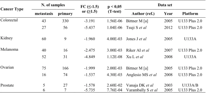
Significant differences between tumor tissues and relative normal counterparts for each cancer type are reported in Table 1. This analysis showed that NGAL transcript levels were significantly higher in the majority of solid tumors compared to the relative normal tissues for every dataset analyzed. While, lower levels of NGAL were observed in each dataset of cervical cancer, esophageal cancer, head and neck cancer and in haematological malignancies. In Table 2 are presented the significant differences of NGAL transcript levels observed in metastatic tissues compared to those of the relative primary tumor. The results showed that in all dataset analyzed the levels were significantly lower in the metastatic tissues than in primary tumors from 5 different tumor types, including colorectal, kidney, melanoma, ovarian and prostate (Table 2).
In Figure 5 the distribution of NGAL transcript levels among cancer cases and normal samples is shown. The percentage of tumor cases showing NGAL transcript levels below the 25th percentile and above the 75th percentile of the “normal” samples is also reported.
9
Table 1. Gene expression patterns of NGAL in different cancer types from 29 datasets
Cancer Type Cancer of samples Normal Fold Change (≤-2) or (≥2) p < 0.01 (T-test) Author (ref.) Dataset Year Platform Solid tumor
Bladder 109 48 4.13 2.75E-05 Sanchez-Carbayo M et al 2006 U133A
Cervix 32a 24 -3.21 4.06E-04 Scotto L et al 2008 U133A
Colon 95 5 2.62 1.23E-02 Kaiser S et al 2007 U133 Plus 2.0
81 24 4.15 7.04E-08 Skrzypczak M et al 2010 U133 Plus 2.0
70 12 4.78 7.06E-06 Hong Y et al 2010 U133 Plus 2.0
Esophagus 17b 17 -5.41 9.27E-05 Hu N et al 2010 U133A
53b 53 -2.92 1.05E-06 Su H et al 2011 U133A/B
Head and
Neck 6
d 4 -18.58 7.33E-03 Schlingemann J et al 2005 U133A
31c 10 -12.44 3.60E-09 Sengupta S et al 2006 U133 Plus 2.0
kidney 51e 5 4.15 4.98E-05 Yusenko MV et al 2009 U133 Plus 2.0
Liver 35 10 8.66 2.32E-05 Wurmbach E et al 2007 U133 Plus 2.0 22 21 3.48 5.14E-04 Roessler S et al (1) 2010 U133 Plus 2.0 225 220 2.94 6.86E-22 Roessler S et al (2) 2010 HT U133A
Lung 30f 30 2.79 1.72E-03 Su LJ et al 2007 U133A
58f 49 2.28 2.64E-06 Landi MT et al 2008 U133A
226f 20 3.707 1.53E-7 Okayama H et al 2012 U133 Plus 2.0
Ovary 185g 10 5.84 1.54E-06 Bonome T et al 2008 U133A
99g 4 3.03 6.98E-04 Hendrix ND et al 2006 U133A
Pancreas 36h 16 14.05 5.15E-06 Pei H et al 2009 U133 Plus 2.0
11h 6 10.00 9.03E-05 Segara D et al 2005 U133A
39i 39 7.70 1.64E-10 Badea L et al 2008 U133 Plus 2.0
Thyroid 9j 9 3.77 9.76E-4 He H et al 2005 U133 Plus 2.0
14j 4 2.33 0.001 Vasko V et al 2007 U133 Plus 2.0
26j 4 2.05 9.34E-7 Giordano TJ et al 2006 U133A
Hematologic tumor
ALL 750 74 -11.77 7.57E-152 Haferlach T et al 2010 U133 Plus 2.0 AML 542 74 -16.94 2.32E-165 Haferlach T et al 2010 U133 Plus 2.0
285 8 -4.91 5.00E-03 Valk PJ et al 2004 U133A
CLL 448 74 -42.98 7.73E-194 Haferlach T et al 2010 U133 Plus 2.0
Myeloma 9k 5 -3.90 0.002 Agnelli L et al 2009 U133A
Legend: Solid tumor: Cervix: aCervical Squamous Cell Carcinoma. Esophagus: bEsophageal Scquamous Cell
Carcinoma; Head and Neck: cNasopharyngeal Carcinoma; dSquamous Cell Carcinoma. Kidney: eRenal
Carcinoma; Liver: (1), dataset 1; (2) dataset 2; Lung: fLung adenocarcinoma; Ovary: gOvarian Carcinoma;
Pancreas: hPancreatic Carcinoma; iPancreatic Ductal Adenocarcinoma; Thyroid: jThyroid Gland Papillary
Carcinoma.
Hematologic tumor: ALL, Acute Lymphoblastic Leukemia; AML, Acute Myeloid Leukemia; CLL, Chronic
10
Table 2. NGAL transcripts in metastatic tissues compared to the relative primary tumor
Cancer Type N. of samples FC (≤-1.5) or (≥1.5) p < 0,05 (T-test) Data set
metastasis primary Author (ref.) Year Platform
Colorectal 43 330 -3.191 1.56E-06 Bittner M [a] 2005 U133 Plus 2.0 27 56 -5.437 1.04E-06 Tsuji S et al 2012 U133 Plus 2.0
Kidney 60 9 -1.960 4.00E-03 Jones J et al 2005 U133A
Melanoma 40 16 -2.475 3.00E-03 Riker AI et al 2007 U133 Plus 2.0
52 31 -4.849 1.12E-08 Xu L et al 2008 U133A
Ovarian 75 166 -1.999 2.00E-03 Bittner M [a] 2005 U133 Plus 2.0 16 74 -1.537 4.30E-03 Anglesio MS et al 2008 U133 Plus 2.0 Prostate 5 27 -1.578 2.60E-02 Vanaja DK et al 2003 U133A/B
6 7 -5.735 7.76E-04 Varambally S et al 2005 U133 Plus 2.0 [a] GEO Series GSE2109; FC, Fold change.
11 Figure 5. Distribution of NGAL transcript levels among cancer cases and normal samples. The percentage
of tumor cases, indicated for each tumor setting, shows NGAL transcript levels below the 25th percentile (Cyan
12
2.1.2 Protein expression
To understand if there was an association between mRNA and protein NGAL expression, an immunohistochemistry evaluation was performed by analysis of Human Protein Atlas web site. In Figure 6 immunohistochemistry analysis of NGAL in 15 solid cancer types are shown. Cancer types with negative immunostaining are not shown. The data demonstrated that concordance of NGAL at both mRNA and protein levels was obtained for the following cancer types: bladder, colorectal, liver, lung, ovarian, and pancreatic (Table1 and Figure 6 Panels: A, E, I, J, H and L). Cervical, esophageal/stomach and head and neck cancers showed a moderate positive immunostaining (Figure 6, panel D, F and G), while mRNA expression levels were lower in cancer tissue compared to the normal counterpart (Table 1).
Conversely, weak immunostaining (<10% of cases) or negative protein expression levels were observed in brain, breast, melanoma and prostate cancers. Accordingly, no significant differences in NGAL mRNA expression were observed between cancer and relative normal counterpart in all datasets analysed for each of these 4 cancer types (brain, breast, melanoma and prostate) (Figure 6, Panel B, C, K and M).
Although, higher mRNA levels were detected in renal and thyroid cancers than in relative normal counterparts (Table 1), a negative immunostaining was observed (Figure 6, Panel H and N). Finally, concordant negative mRNA and protein levels of NGAL were observed in the majority of haematological malignancies (data not shown).
13 Figure 6. Immunohistochemistry analysis of NGAL expression in human cancer. The data were obtained
from the Human Protein Atlas. A single representative case for each cancer type (total 15) is shown along with its normal counterpart. Expression of NGAL in cancer sample was evaluated as strong, moderate, weak and negative immunostaining. The percentage is referred to the total cancer samples analyzed for each tumor type.
14
2.1.3 Clinical impact
The role of NGAL for each tumor type is described below and summarized in Table 3.
SOLID TUMORS Bladder
The current study reveals that both mRNA transcript and protein levels were higher in bladder cancer tissues than in the normal counterparts. Accordingly, 50.5% of cases displayed NGAL transcripts above the 75th percentile of the “normal” values suggesting its role as a diagnostic marker (Figure 5A). These data are in agreement with previous investigations in which NGAL along with MMP-9 were overexpressed in urothelial bladder carcinomas suggesting their role as early diagnostic markers for this tumor type [Roy R et al, 2008; Monier F et al, 2000]. Reduced protein levels of both NGAL and MMP-9 have been detected in urine samples from bladder cancer patients with clinical relapse suggesting that reduced levels of these proteins may be used as indicators of tumor progression [Monier F, et al 2002].
Brain
A significant association of MMPs and NGAL expression was detected in urine from brain cancer patients and in tumor specimens. Additionally, surgical resection of the tumor resulted in a reduction of both MMPs and NGAL in urine samples [Smith ER et al, 2008]. The identification of new potential molecular targets in this cancer type may be very helpful to discover new therapeutic strategies. Immunohistochemistry analysis reveals that NGAL expression is frequently up-regulated in gliomas and is associated with poor clinical outcome [Liu MF et al, 2011]. Barresi et al show that NGAL was overexpressed in primary high grade brain tumors and not in the metastatic cases [Barresi V et al, 2010 (4)]. According to the exclusion criteria designed for the purpose of our present study, no data on brain cancer were generated by computational analysis. Accordingly, NGAL protein expression was not detected through the Human Protein Atlas evaluation.
Breast
Similar observations acquired for brain tumors were obtained for breast cancer as no data were generated by our computational analysis. The expression pattern of NGAL has been evaluated by several authors and generated conflicting results on the clinical significance of this protein in breast cancer.
15 A heterogeneous pattern of expression at the mRNA and protein levels was observed in breast cancer patients in a study conducted by Stoesz SP et al [Stoesz SP et al 1998]. The authors also described a significant correlation between NGAL expression and other markers of poor prognosis, including estrogen and progesterone receptor-negative status and high proliferation (S-phase fraction) [Stoesz SP et al 1998]. These studies were also confirmed by Bauer M et al [Bauer M et al, 2008]. Similarly, Shen ZZ et al showed that MMP-9 and MMP-9/NGAL complex expression were higher in breast cancer than in benign breast and/or normal tissues [Shen ZZ et al, 2003]. While, in the study conducted by Provatopoulou X et al. in a large series of breast cancer patients, MMP-9 and NGAL were overexpressed in cancer and this overexpression was associated with the severity of disease but no significant correlation was found for the complex formation [Provatopoulou X et al 2009].
In a transgenic mouse model of breast cancer, Berger et al. demonstrated that lack of NGAL in mice leads to a reduction of tumor growth. This reduction was attributed to an NGAL-dependent decrease of MMP-9 activity and to a lack of high molecular weight MMP activity [Berger T et al, 2010]. Accordingly, Li et al have shown that NGAL expression is associated with increased metastasis and poor prognosis in breast cancer patients [Li SH et al 2009]. The results obtained by Leng et al in an animal model of breast cancer suggest that the suppression of NGAL function, by an inhibitory monoclonal antibody, has a great potential for breast cancer therapy, particularly by interfering with metastasis in aggressive types of breast cancer [Leng X et al, 2009]. Conversely, in a previous study it was shown that NGAL overexpression promotes in vivo the development of lung metastasis [Shi H et al, 2008]. Our recent studies indicate that increased NGAL expression did not alter the sensitivity of the MCF-7 breast cancer cell line to the chemotherapeutic drug doxorubicin [Chappell WH et al, 2012 (1)]. However, ectopic NGAL expression did alter the sensitivity of breast cancer cells to targeted therapy [Chappell WH et al, 2012 (2)]. Furthermore, NGAL was found to be a predictive marker for complete response after neo-adjuvant chemotherapy in low-risk subgroups of breast cancer patients and may be considered as an independent prognostic factor for decreased disease free survival in primary human breast cancer [Wenners AS et al, 2012].
16
Cervical cancer
A recent analysis conducted by immunohistochemistry on a set of cervical biopsy specimens from 225 women showed a close relationship between NGAL expression levels, HPV lesion grade and detection of high risk HPV types. Up-regulation of NGAL in higher grade lesions is likely to from the suppression of wild-type p53 by the HPV E6 oncoprotein. Suppression of p53 results in elimination of p53 block of NGAL transcription [Syrjänen S et al, 2010]. These data were in agreement with our IHC evaluation (Figure 6D) but not in line with our computational analysis as only 6.3% of cervical cases displayed NGAL transcripts above the 75th percentile of “normal” values, while 59.4% below the 25th percentile (Figure 5B).
Colorectal cancer
In 1996 Nielsen et al analyzed the role of NGAL by both immunohistochemistry and mRNA by in situ hybridization in colon cancer and in inflammatory colorectal diseases. Increased expression of NGAL was detected both in non-malignant epithelium such as diverticulitis, inflammatory bowel disease and in malignant colonic lesions. In adenocarcinomas, NGAL overexpression was observed in the transitional mucosa and in the superficial ulcerated area. On the other hand, NGAL expression was not detected in lymph node metastases from this adenocarcinoma [Nielsen BS et al, 1996]. The authors speculate that NGAL is predominantly involved in inflammatory reaction and in tumor transformation, while it does not appear to play a prominent role in metastatic process.
However, Lee et al have shown that NGAL may function as a metastasis suppressor in colon cancer cells. In their studies, they genetically manipulated highly metastatic human colon cancer cell lines, which normally express low NGAL protein levels, to overexpress NGAL. Ectopic expression of NGAL suppressed, in vivo, the liver metastasis of metastatic human colon cancer cell lines in experimentally-driven metastasis assays [Lee HJ el al, 2006]. Hu et al examined the potential molecular mechanism of NGAL involvement in colorectal cancer. They demonstrated that NGAL overexpression altered the subcellular localization of E-cadherin and catenins, decreased E-E-cadherin-mediated cell to cell adhesion, enhanced cell– matrix attachment, and increased cell motility and in vitro invasion. They proposed that NGAL exerted these effects through the alteration of the subcellular localization of Rac1, one of Rho small GTPases, in an extracellular matrix-dependent manner, but not by MMP-9 [Hu L et al, 2009]. Recently, Bousserouel S et al have shown, in a preclinical model of colon
17 carcinogenesis, that NGAL is significantly upregulated only in advanced stages of tumor progression [Bousserouel S et al, 2010].
Real time PCR and zymographic analysis on visceral adipose tissue (VAT) biopsies from 11 colon cancer patients revealed increased levels of NGAL and other inflammation associated factors like osteopontin, tumor necrosis factor-α (TNF-α), and chitinase-3 like-1 compared to control subjects, suggesting their involvement in cancer development and progression [Catalán V et al, 2011].
Recently, Martí J et al showed the prognostic utility of NGAL mainly in metastatic CRC [Martí J et al, 2010; Martí J et al 2013]. Higher levels in colon cancer cases then controls were observed by Fung KY et al but the authors concluded that it was not a promising biomarker for the diagnosis of CRC as the sensitivity of NGAL was found to be 24% at 90% specificity [Fung KY et al, 2013]. Accordingly, although NGAL is still expressed by the majority of human neoplastic colorectal lesions, the author it is not a useful biomarker for determining disease progression from adenomatous to malignant colorectal neoplasia [McLean MH et al, 2013]. Our analysis, in line with the majority of previous studies, shows the upregulation of NGAL in adenocarcinoma tumor samples and reduced expression in metastatic samples.
Esophageal cancer
Esophageal cancer is the eighth most common incident cancer in the world and ranks sixth among all cancers in mortality. Esophageal cancers are classified into two histological types; esophageal squamous cell carcinoma (ESCC), and adenocarcinoma, and the incidences of these types show a striking variety of geographic distribution, possibly reflecting differences in exposure to specific environmental factors. Both alcohol consumption and cigarette smoking are major risk factors for the development of ESCC [Koshy M et al, 2004].
Zhang et al performed immunohistochemistry, western blot and gelatin zymography on 81 paraffin sections including ESCC, normal mucosa, simple hyperplasia and dysplasia, and on 73 fresh specimens of ESCC to evaluate the role of NGAL in ESCC. Immunohistochemical studies revealed that ESCC have a diverse and obvious whole-cytoplasmic staining pattern for NGAL, while normal esophageal epithelium presented a weak positive signal within a restricted cytoplasmic area. On western blot analysis, NGAL expression level was found to be significantly higher in ESCC than in normal mucosa, and positively correlated with cancer cell differentiation. No significant association was observed between NGAL expression and cell proliferation. Finally, the authors showed higher enzymatic activity of MMP-9/NGAL
18 complex in ESCC than in normal mucosa. These findings suggest that NGAL is involved in differentiation pathways and invasive progression of ESCC [Zhang H et al, 2007]. Similar data were reached by Du et al, [Du ZP et al, 2011]. Accordingly our IHC evaluations show that NGAL is overexpressed in tumor and not in normal tissue. However, different trend is observed by analyzing the transcript levels as 71.4% of cases displayed NGAL transcripts below the 25th percentile of “normal” values, while 8.6% above the 75th percentile (Figure 5D).
Fang et al. identified a new NGALR isoform designated as NGALR-3, that results from alternative splicing. Interestingly, it was shown that the NGALR-3 isoform was overexpressed in 70% of esophageal carcinoma cases in comparison with those of normal adjacent epithelium. These findings suggest that the new NGALR-3 variant could play a more important role in esophageal carcinoma. The authors proposed that the novel NGALR-3 isoform could mediate a unilateral intracellular iron-delivery pathway which increased intracellular iron levels. This could be involved in the tumor growth of esophageal carcinomas [Fang WK et al, 2007].
Li EM et al studied the role of NGAL in invasion, division and proliferation of an esophageal carcinoma cell line. Their results demonstrated that the antisense blocking of NGAL transcription not only decreased effectively the activity of MMP-9 and MMP-2 secreted by SHEEC cells, but also suppressed significantly the invasion of these cells in nude mice. However, it was shown that NGAL was not apparently related with division and proliferation of SHEEC tumor cells [Li EM et al, 2003 (1)]. Furthermore, the same authors, in attempt to demonstrate the regulation mechanism of NGAL overexpression in SHEEC, analyzed the structural characters of 5'-untranslated region (5'-UTR) and 3'untranslated region (3'-UTR) of NGAL. Upon cloning and DNA sequencing of 69 bp 5'-UTR and 147 bp 3'-UTR of NGAL gene they did not observe any base pair mutations [Li EM et al, 2003 (2)].
Gastric cancer
Gastric cancer (GC) is the final result of a multistep process initiated by environmental factors, including diet and Helicobacter pylori infection [Stemmermann GN et al, 2002]. H. pylori infection is one of the most important risk factors for this malignancy [Uemura N et al, 2001; Huang JQ, 2003].
During infection, H. pylori synthesize siderophores, which chelate Fe3+ with high affinity and facilitate its transport into the pathogen [Neilands JB, 1995; Braun V et al, 2002;
19 Krewulak KD et al, 2008]. The host cells respond to infection by increasing the secretion of NGAL that binds the bacterial siderophores and prevents their uptake into bacteria. The iron depletion results in inhibitingH. pylori growth [Holmes MA et al, 2005].
NGAL and MMP9/NGAL complex were shown to be upregulated in GC tissue (mainly in neutrophils and epithelial cells) compared to adjacent normal gastric mucosa, confirming the hypothesis that the association of NGAL with MMP9 could prevent extracellular autodegradation of the proteinase. Enhanced levels of the MMP9/NGAL complex, but not of MMP-9 and NGAL, have been related to worse clinical outcome in cancer patients and significantly associated with the classifications of Lauren and WHO, suggesting that MMP9/NGAL complex could be considered as a novel prognostic factor for gastric cancer [Kubben FJ et sal, 2007].
Wang HJ et al [Wang HJ et al, 2010] proposed NGAL as a potential biomarker for prognosis and an ancillary diagnostic test of gastric cancer. In this study, they showed high levels of NGAL expression in 333 GC patients by immunohistochemistry. NGAL was correlated with size of tumor, Lauren's classification, lymph node metastasis, vascular invasion, distant metastasis and TNM stage. The multivariate analysis indicated that NGAL can be used as an independent prognostic factor. Serum NGAL levels were determined in blood samples from 63 healthy donors and 60 GC patients and analyzed according to TNM. NGAL blood levels were higher than those of CA19-9 in TNM I patients, and higher than those of CEA and CA19-9 in TNM II. Therefore, serum NGAL has a great potential as a tumor marker for GC and could be associated with a poor prognosis [Wang HJ et al, 2010].
According to the exclusion criteria designed for the purpose of our present study, no data on GC were generated by our analyses.
Head and neck
Our “in silico” analysis showed a significant down-regulation of NGAL in head and neck cancer compared to the normal counterpart. In fact, 94.6% of head and neck cancer displayed NGAL transcripts below the 25th percentile of “normal” values, while no samples showed mRNA levels of NGAL above the 75th percentile (Figure 5E). However, our IHC evaluation is in line with previous data [Lin CW et al, 2012] as expression of NGAL appeared to be moderate in 50% of cases and absent in normal tissue.
20
Hepatocellular Carcinoma (HCC)
Zhang Y et al demonstrated the up-regulation of NGAL expression in HCC was significantly correlated with unfavorable clinic-pathologic features and independent poor prognostic factor for overall survival in patients [Zhang Y et al, 2012]. In agreement, our analysis showed a significant up-regulation of NGAL mRNA levels in HCC from 2.3- to 8.6-fold changes in comparison to normal hepatic tissues in the three datasets analyzed (Table 1). Similar data were obtained by IHC considerations.
Lung cancer
At the present, few studies have been performed to evaluate the role of NGAL in lung cancer. However, Friedl A et al found high NGAL levels in lung adenocarcinoma [Friedl A et al, 1999]. Accordingly, in the present study we detected a constant NGAL upregulation both at mRNA and protein levels for adenocarcinoma histotype. In contrast, NGAL was not overexpressed in carcinoid lung cancer.
Melanoma
In melanoma a substantial down-regulation of NGAL was observed only in metastatic disease versus primary tumor while no statistic differences were observed in NGAL mRNA levels between primary tumor and normal tissue. Protein expression of NGAL was not detected by IHC (Human Protein Atlas web site) (Figure 6K). To our knowledge no previous data were generated on NGAL in melanoma tissue samples.
Ovarian cancer
Epithelial ovarian cancer is one of the most aggressive cancers diagnosed in women with high mortality rate. This cancer is usually asymptomatic and often diagnosed late in the disease process [Paley PJ, 2001]. Unfortunately, there are no specific markers for early diagnosis. Lim R et al analyzed NGAL by IHC in a total of 59 ovarian tissues including normal, benign, borderline and malignant (grades 1, 2 and 3). NGAL expression was weak or moderate in benign tissues. Both borderline and grade 1 tumors displayed the highest amount of NGAL expression with moderate to strong staining, whereas in grade 2 and 3 tumors, the extent of staining was significantly less (p < 0.01) and staining intensity was weak to moderate. Additionally, the authors analyzed, by ELISA, NGAL levels in 62 serum samples from normal individuals and ovarian cancer patients (grade 1). The NGAL concentration was 2 and
21 2.6-fold higher in patients with benign tumors and cancer patients (grade 1) [Lim R et al, 2007]. In line with results provided by Lim et al, all datasets here examined showed an upregulation of NGAL in ovarian cancer samples, with a range from 3 to 6 fold. Furthermore, the analysis of both Bittner and Anglesio datasets (Ref. in Table 2) revealed a significant reduction of NGAL mRNA levels in metastatic disease (see Table 2). In the same study, Lim and collaborators analyzed NGAL in ovarian cancer cell lines treated with epidermal growth factor (EGF) indicating that NGAL expression was downregulated in ovarian cancer cell lines undergoing the epithelial to mesenchymal transition (EMT) induced by EGF. Downregulation of NGAL expression correlated with the upregulation of vimentin, enhanced cell dispersion and downregulation of E-cadherin expression, some of the hallmarks of EMT. These data indicate that NGAL may be a good marker to monitor transformation of benign lesions to premalignant and malignant ovarian tumors and that the molecule may be involved in the progression of epithelial ovarian malignancies [Lim R et al, 2007].
NGAL, as well as other proteins, was shown to be regulated in ovarian cancer cell lines by 17 beta-estradiolestrogen [Walker G et al, 2007]. More recently Cho et al. described the upregulation of NGAL in a panel of 54 ovarian cancers, 15 borderline and 53 benign ovarian tumors, and 90 healthy controls by real time PCR and immunohystochemical analysis. NGAL levels were significantly higher in ovarian tissues and particularly in well-differentiated tumors. Similar results were obtained by analyzing NGAL serum levels from ovarian cancer patients showing highest levels in differentiated cancer [Cho H et al, 2009]. Notably, according to previous data, the results of the present study show that 91,2% of ovarian cases displayed NGAL transcripts above the 75th percentile of the “normal” values, while 0.4% below the 25th percentile. Similar trend was obtained for the IHC analyses. While transcript levels of NGAL were significantly lower in metastatic setting compared to primary tumor.
Pancreatic cancer
Pancreatic cancer (PaCa) is the fifth leading cause of cancer death in both men and women [Ahmedin J et al, 2002]. Early detection of this disease is not possible in spite of significant diagnostic tools. The effective therapy, surgery, is limited to about 25% of the cases and often is unable to prevent cancer recurrence in these patients [Cooperman AM, 2001]. Much remains to be understood about the natural course and biology of this disease.
Using microarray analysis, many laboratories have reported the differential expression of several novel genes, including that of NGAL, associated with the progression of pancreatic
22 cancer [Argani P et al, 2001; Han H et al, 2002; Terris B et al, 2002]. Moniaux et al detected NGAL levels by immunohistochemistry on tissue samples from normal patients, pancreatitis, and pancreatic adenocarcinoma patients. Their results revealed higher levels in pancreatic adenocarcinoma than in normal and pancreatitis samples [Moniaux N et al, 2008]. These findings are in agreement with the results of the present study in which we observe a constant upregulation of NGAL at both mRNA and protein expression levels in pancreatic adenocarcinoma samples vs normal tissues. Moniaux et al also evaluated NGAL levels in pancreatic cancer cell lines with varying grades of differentiation documenting a positivity for NGAL expression in both well and moderately differentiated cells. In contrast, NGAL expression was uniformly negative in poorly differentiated adenocarcinoma. Further, they examined NGAL levels in serum samples. They used ELISA to detect NGAL. The authors concluded that NGAL was fairly accurate in distinguishing between pancreatic cancers and non-cancer cases. In conclusion, NGAL is highly expressed in early pancreatic dysplastic lesions, suggesting a possible role as an early diagnostic marker for pancreatic cancer [Moniaux N et al, 2008].
NGAL is also detected in bile and may be useful as a novel biomarker to distinguish benign from malignant biliary obstruction [Zabron AA et al, 2011].
The biological roles of NGAL in pancreatic adenocarcinoma have been studied both in vitro and in vivo by Tong et al [Tong Z et al, 2008]. The authors transfected PaCa cells with NGAL and demonstrated no effects on cell viability or sensitivity to chemotherapy, but decreases in cell adhesion, invasion and angiogenesis was observed both in vitro and in vivo. The negative effects on tumor progression and metastasis were due to alteration of FAK phosphorylation and decrease of VEGF production [Tong Z et al, 2008]. We can gather from this study that modulation of NGAL activity could control PaCa angiogenesis and metastasis.
Prostate cancer
According to the exclusion criteria designed for the purpose of our present study, no differences were identified between prostate cancer tissue and the normal counterparts. Similarly, IHC evaluation did not display any differences between normal and tumor. While, significant differences were observed in metastatic disease of prostate cancer when compared with primary tumors as NGAL transcripts were lower in metastatic setting compared to primary tumor. However, in vitro studies showed that NGAL plays a significant role in the progression of prostate cancer by regulating MMP2 and MMP-9 [Tung MC et al, 2013].
23 Therefore further studies are needed to better clarify the role of NGAL in prostate cancer, as until now conflicting results have been generated between “in silico” and “in vitro” data.
Renal tumor
The incidence of renal cell carcinoma (RCC) is growing [Rathmell WK et al, 2010]. Most of the patients initially diagnosed with localized disease are cured by surgery, but over 30% of them die from relapse. The molecular basis of a great diversity in clinical behavior of RCC is still unclear and makes it a target to investigate the nature of these heterogeneities [Banks RE et al, 2006].
A recent immunohistochemical study on a set of 30 surgically-resected renal tumors revealed that NGAL is expressed in several histotypes of renal tumors especially in papillary and chromphobe histotypes. NGAL expression is highest in the higher histological grade of papillary and clear cell RCC and in its peritoneal metastasis [Barresi V et al, 2010 (3)].The authors suggested that the upregualtion of NGAL in the above-mentioned tumor histotypes could be related to an increased requirement of iron uptake and could justify the use of iron chelators for renal cancer therapies. In agreement, our analysis revealed an upregulation of NGAL in RCC. Conversely, IHC evaluation did not show any immunostaining for NGAL. It was further shown that NGAL, as detected by ELISA, had predict value for progression free survival in RCC patients treated with sunitinib malate [Porta C et al, 2010]. While, most recent data conducted by Di Carlo does not reach any conclusive results on the usefulness of NGAL as a diagnostic marker [Di Carlo A, 2013].
Thyroid cancer
Our data show that transcript levels of NGAL are higher in tumor thyroid cancer (papillary carcinoma) when compared with normal counterpart. In fact, 83.7% of case displayed NGAL transcripts above the 75th percentile of “normal” values, while 6.1% of cases showed mRNA levels of NGAL below the 25th percentile (Figure 5K). In contrast, no protein expression was detect by IHC (Human Protein Atlas web site). According to mRNA expression data, previous studies showed that NGAL is overexpressed in thyroid cancer [Barresi V et al, 2012 (1,2); Iannetti A et al, 2008].
24
HEMATOLOGICAL MALIGNANCIES
Different then for solid tumor, NGAL expression in hematological malignancies displays a very homogeneous behavior: in fact, all the datasets that reach the levels of significance p<0.01 exhibit an uniform downregulation of NGAL compared to controls. Datasets analyzed included myeloma and leukemias (ALL, acute lymphoblastic leukemia; AML, acute myeloid leukemia, CLL; chronic lymphocytic leukemia). Accordingly, recent data show that highest mRNA NGAL levels were associated with better prognosis in AML [Yang WC et al, 2013]. According to the exclusion criteria designed for the purpose of the present study, no data on chronic myeloid leukemia (CML) were generated by computational analysis. However, NGAL was identified in an expression profiling study on CML cells. In this work, the authors analyzed gene expression profiles of cancer cells from 27 patients using a cDNA microarray. Among the 150 genes up-regulated, they observed an increase of NGAL mRNA levels in CML patients [Kaneta Y et al, 2003]. Subsequently, Villalva C et al have performed RT-PCR for NGAL expression in a large cohort of CML patients. Their results indicate that NGAL is expressed in parallel with the BCR-ABL oncoprotein at the early stage of leukemia process and it is secreted at high levels in these patients. The authors have excluded the possibility that the large increase of NGAL expression in CML patients at diagnosis resulted from the presence of circulating myelocytes in blood, which constitutively secrete NGAL protein during maturation [Villalva C et al, 2008]. Leng X et al have proposed two activities associated with NGAL in a mouse model of CML tumorigenesis. On the one hand, NGAL induces apoptosis of normal hematopoietic cells resulting in the replacement of these with leukemic cells and on the other NGAL facilitates tissue invasion through stabilization of MMP-9 activity [Leng X et al, 2008].
25
Table 3. Clinical impact of NGAL expression pattern in different cancer type according to previous studies
TUMOR TYPE METHODS CLINICAL IMPACT OF NGAL EXPRESSION AUTHOR [REF.] YEAR
SOLID TUMORS
BLADDER CM, GZ,
MS Diagnostic marker Roy R et al Monier F et al (1) 2008 2000 GZ, IHC,
ELISA Early marker of tumor progression Monier F et al (2) 2002
BRAIN IHC, GZ,
ELISA Diagnostic marker Smith ER et al 2008
IHC Positive correlation with high proliferation index in
primary tumor Barresi V et al (4) 2010
IHC Associated with poor outcome Liu MF et al 2011
BREAST IHC Associated with poor outcome Stoesz SP et al 1998
ELISA Positive correlation with lymphatic node metastasis Shen ZZ et al 2003 ELISA Positive correlation with breast cancer aggressiveness Provatopoulou X et al 2009 IHC Positive correlation with poor prognosis in primary
human breast cancer Bauer M et al 2008
IHC Positive correlation with poor outcome Li SH et al 2009 IHC Positive correlation with poor outcome Wenners AS et al 2010 CERVICAL IHC Positive correlation with HPV type Syrjänen S et al 2012 COLORECTAL IHC, FISH Positive correlation with tumor trasformation Nielsen BS et al 1996
ELISA Prognostic utility in metastatic patients Martì J et al (1,2) 2010,2013
PCR, GZ Diagnostic marker Catalán V et al 2011
ELISA Not suitable as a diagnostic marker Fung KY et al 2013 ELISA Not useful marker of progression McLean MH et al 2013 ESOPHAGEAL GZ, IHC,
WB
Positive correlation to cancer differentiation Zhang H et al 2007 IHC Positive correlation with progression Du ZP et al 2011 GASTRIC IHC,
ELISA, WB Positive correlation with poor outcome Kubben FJ et al 2007 IHC, ELISA Diagnostic marker and positive correlation with poor
outcome Wang HJ et al 2010
HEAD & NECK ELISA Positive correlation with poor outcome Lin CW et al 2012 HCC IHC Positive correlation with poor outcome Zhang Y et al 2012 LUNG IHC Positive correlation with poor outcome Friedl A et al 1999
PANCREATIC ELISA Diagnostic marker Moniaux N et al 2008
OVARIAN IHC, ELISA, WB
Promotion of epithelial to mesenchymal transition Lim R et al 2007 PCR, IHC,
ELISA
Positive correlation to cancer differentiation Cho H et al 2009 RENAL IHC Positive correlation with malignant phenotype Barresi V et al (3) 2010 ELISA Positive correlation with poor outcome Porta C et al 2010 THYROID IHC, Real
time
Positive correlation with malignant phenotype Iannetti A et al 2008
IHC Diagnostic marker Barresi V et al (1,2) 2012
HEMATOLOGICAL MALIGNANCIES
AML RT-PCR Positive correlation with better prognosis Yang WC et al 2013 CML RT-PCR Positive correlation with early stage of disease and
BCR-ABL positivity Villalva C et al 2008
Legend: AML, Acute Myeloid Leukemia, CM, Chromatography; CML, Chronic Myelogenous Leukemia; GZ,
26
2.2 VALIDATION OF COMPUTATIONAL RESULTS IN BLADDER CANCER
The majority of cases with TCC were by far men and aged ≥65 years (Table 4). Ever smoking was reported by 85.4% of cases and 66.4% of controls, whereas no difference was observed for education and drinking habit. Non-muscle invasive tumors (i.e. Ta/is-T1) represented 78.4% of cases, whereas papillary features was reported in 79.8% of TCCs.
Table 4. Distribution of 89 cases of transitional cell carcinoma of the bladder (TCC) and 119
hospital controls according to socio-demographic characteristics, tobacco smoking, alcohol drinking, and clinical pathological factors
Variables TCCs (n=89) Controls (n=119) χ2 test n (%) n (%) Sex Men 75 (84.3) 99 (83.2) Women 14 (15.7) 20 (16.8) 0.04; p=0.84 Age (years) < 65 34 (38.2) 47 (39.5) 65-74 39 (43.8) 49 (41.2) ≥ 75 16 (18.0) 23 (19.3) 0.15; p=0.93 Education < 7 42 (47.2) 53 (44.5) 7-11 31 (34.8) 43 (36.1) ≥ 12 16 (18.0) 23 (19.3) 0.15; p=0.93 Smoking status Never 13 (14.6) 40 (33.6) Former 37 (41.6) 64 (53.8) Current 39 (43.8) 15 (12.6) 27.76; p<0.01
Alcohol drinking status
Never/Former 7 (7.9) 17 (14.3) Current 82 (92.1) 102 (85.7) 2.05; p=0.15 Invasivenessa Ta/is 50 (56.8) T1 19 (21.6) T2-T4 19 (21.6) Gradinga Well/moderately differentiated 40 (45.5) Poorly diff./undifferentiated 48 (54.6) Histological subtype Papillary 71 (79.8) Non-papillary 18 (20.2)
aThe sum does not add up to the total because of missing values.
Urinary NGAL concentrations were significantly higher in cases than in controls (median: 18.35 vs. 7.75 ng/mg Cr; p<0.01); likewise, higher urinary levels of MMP-9 (median: 6.54 vs 1.18 ng/mg Cr; p<0.01) and MMP-9/NGAL complex (median: 0.00 vs 1.11 ng/mg Cr;
27 p<0.01) were observed in cases compared to controls (Table 5). Figure 7A shows increasing urinary concentrations of NGAL, MMP-9, and MMP-9/NGAL according to invasiveness, with markers concentration significantly higher in T2-T4 cases (p<0.01). Interestingly, MMP-9/NGAL complex was undetectable in 53.8% of controls, but only in 30.3% of all TCCs (5.3% of T2-T4 cases). In the univariate analysis, the expression of the three molecules was higher in poorly differentiated/undifferentiated than in well/moderately differentiated TCCs and in non-papillary than in papillary subtype (Table 5).
Table 5. Median urinary and serum NGAL, MMP-9, and MMP-9/NGAL complex
concentrationsa in 119 hospital controls and in 89 cases of transitional cell carcinoma of the bladder (TCC) according to clinical pathological features
n Median urinary concentrations (ng/mg Cr) Median serum concentrations (ng/mL)
NGAL MMP-9 MMP-9/NGAL complex NGAL MMP-9 MMP-9/NGAL complex
Controls 119 7.75 1.18 0.00 90.49 717.94 51.86 All TCCs 89 18.35 6.54 1.11 83.70 738.51 53.44 KW test p<0.01 p<0.01 p<0.01 p=0.50 p=0.32 p=0.58 Invasivenessb Ta/is 50 16.18 3.30 0.55 78.78 610.01 38.27 T1 19 15.24 5.98 0.29 84.09 822.82 57.24 T2-T4 19 68.55 24.29 6.64 139.32 943.55 113.85 KW test p<0.01 p<0.01 p<0.01 p<0.01 p=0.01 p<0.01 Adjusted KW test p=0.56 p=0.02 p=0.08 p=0.02 p=0.21 p=0.03 Gradingb Well/moderately differentiated 40 13.49 4.33 0.51 78.78 615.97 38.48 Poorly diff./undifferentiated 48 20.25 10.71 1.30 100.26 825.47 68.49 KW test p<0.01 p<0.01 p<0.01 p=0.04 p=0.06 p=0.03 Adjusted KW test p=0.47 p=0.04 p=0.31 p=0.76 p=0.50 p=0.71 Histological subtype Papillary 71 14.84 3.97 0.52 78.91 611.63 43.90 Non-papillary 18 49.88 44.03 10.67 113.66 937.75 75.36 KW test p<0.01 p<0.01 p<0.01 p<0.01 p<0.01 p=0.04 Adjusted KW test p=0.01 p=0.02 p<0.01 p=0.04 p<0.01 p=0.14
aUrinary concentrations were standardized on creatinine level (Cr) and expressed in ng/mg Cr; bThe sum does
not add up to the total because of missing values; cMutually adjusted for tumour invasiveness, grading and
histological subtype plus age and smoking habits. KW= Kruskal-Wallis test
However, after mutual adjustment for tumour characteristics, no association persisted with grading, whereas only MMP-9 and MMP-9/NGAL complex were still associated to tumour invasiveness.
28 No significant differences between TCC cases and controls emerged in serum (Table 5). Nonetheless, higher levels of NGAL and MMP-9/NGAL complex were observed in patients with muscle-invasive tumors (Figure 7B). These differences were statistically different after taking into account the other tumor characteristics (Table 5).
Stronger correlations were observed between urinary and serum levels of NGAL and MMP-9 in TCC patients. These correlations were higher in patients with muscle invasive TCCs (r=0.72 and r=0.54, respectively) than in those with non-muscle invasive TCCs (r=0.22 and r=0.27, respectively). The correlation was less marked for MMP-9/NGAL complex. Among controls, urinary concentrations of NGAL, MMP-9, MMP-9/NGAL complex did not correlate with those in serum (Supplementary Figure 1).
Table 6. Sensitivity (Se), specificity (Sp), positive predictive value (PPV), and negative
predictive value (NPV) for urinary NGAL, MMP-9, and MMP-9/NGAL complex as biomarkers of transitional cell carcinoma of the bladder (TCC)
Optimal cut-off (ng/mg Cr) Se Sp PPV NPV All TCCs NGAL 11.59 61% 61% 53% 67% MMP-9 2.20 65% 66% 59% 72% MMP-9/NGAL complex 0.265 65% 67% 60% 72% Non muscle-invasive TCCs NGAL 11.59 57% 61% 45% 71% MMP-9 1.66 62% 58% 46% 73% MMP-9/NGAL complex 0.19 61% 61% 48% 73% Muscle-invasive TCCs NGAL 17.1 74% 76% 33% 95% MMP-9 6.5 84% 82% 43% 97% MMP-9/NGAL complex 0.89 84% 83% 44% 97%
ROC curves were used to determine the optimal cut-off for the three molecules (Table 6). Low sensitivity was reported for all the three urinary markers in all TCCs, which were able to correctly classify approximately 65% of cases. For MMP-9 and MMP-9/NGAL complex, the sensitivities and the negative predictive values greatly increased among muscle-invasive cancers (Se=84% and NPV=97%). According to ROC analysis (Figure 8), 9 and MMP-9/NGAL complex were the best markers among all TCCs (AUC=0.68). For T2-T4 TCCs, MMP-9 and MMP-9/NGAL complex were still the best markers showing similar diagnostic performances (AUC=0.90 and 0.88, respectively – Table 6). Diagnostic performances in serum were generally lower than in urine (data not shown).
29 Figure 7. Distribution of NGAL, MMP-9, and MMP-9/NGAL complex concentrations in urine (A) and serum
(B) in hospital controls and in cases of transitional cell carcinoma of the bladder (TCC) according to tumour invasiveness. Median values are represented by horizontal lines. P-values computed by non-parametric Kruskal-Wallis and Mann-Whitney U tests. * indicates a p-value < 0.05; ** indicates a p-value < 0.01.
NGAL Controls Ta/is T1 T2-T4 Urin ary NG A L c o n ce n tra tio n (n g /m g Cr) 0 0.1 1 10 100 1000 10000 NGAL Controls Ta/is T1 T2-T4 S eru m NG A L c o n ce n tra tio n (n g /m L ) 0 25 100 250 1000 MMP-9 Controls Ta/is T1 T2-T4 Urin ary M M P -9 c o n ce n tra tio n (n g /m g Cr) 0 0.1 1 10 100 1000 10000 MMP-9 Controls Ta/is T1 T2-T4 S eru m M M P -9 c o n ce n tra tio n (n g /m L ) 0 25 100 250 1000 2500 10000 NGAL/MMP-9 complex Controls Ta/is T1 T2-T4 Urin ary NG A L /M M P -9 c o m p le x c o n ce n tra tio n (n g /m g Cr) 0 0.1 1 10 100 1000 10000 NGAL/MMP-9 complex Controls Ta/is T1 T2-T4 S eru m NG A L /M M P -9 c o m p le x c o n ce n tra tio n (n g /m L ) 0 10 25 100 250 1000 A B I n v a s i v e n e s s ** * ** ** * * ** * ** I n v a s i v e n e s s I n v a s i v e n e s s I n v a s i v e n e s s I n v a s i v e n e s s I n v a s i v e n e s s * ** ** * *
30 Figure 8. Receiver operating characteristic (ROC) curves for urinary MMP-9 and MMP-9/NGAL complex
31
3. DISCUSSION
In the present study, computational and IHC evaluations were performed to understand whether differences in NGAL transcript or protein levels occur in different cancer types when compared with the relative normal tissues or the metastatic counterparts.
Our computational analysis suggest an active role of NGAL in early stages of tumor development, reason that many authors proposed NGAL as a diagnostic and prognostic marker. While a decrease of NGAL expression in metastatic samples was detected when compared to matched primary tumors. This observation suggests a common inactivation pathway of NGAL gene during distant tumor dissemination and leads us to venture the hypothesis that NGAL could play a protective role in metastatic development.
In particular, the tumors showing higher NGAL expression (expression levels greater than the 75th percentile of “normal” samples) were: ovarian (91.2%), thyroid (83.7%), liver (68.8%), colon (66.3%), kidney (64.7%), lung (63.1%), pancreas (60.2%) and bladder (50.5%). While, the percentages of tumor cases showing NGAL transcripts below the 25th percentile of “normal” values were almost 100% of all hematological malignancies and 94.6% of head and neck cancer, 71.4% of esophagus cancer and 59.4% of cervical carcinoma (Figure 5). These data suggest that NGAL is a candidate marker for tumor growth in a fraction of solid tumor and a favorable prognostic factor for the remaining cancer types showing lower levels of NGAL transcript levels.
Validation of computational studies, above described, was performed on bladder cancer. In developed countries, transitional cell carcinoma of the bladder (TCC) is the fourth most frequent cancer in men, with the highest incidence worldwide in southern Europe [Jemal A et al, 2011]. The major risk factors for bladder cancer are cigarette smoking and occupational exposure to aromatic amines; while, inconclusive epidemiological data have been generated on diet. On the one hand, dietary intake of vegetables and/or fruits protect against bladder cancer, on the other hand genetic variability may alter such association [Tang L, et al 2010; Lin J, et al, 2009].
Several markers have been proposed in recent years, but none is currently considered adequate to diagnose and predict the outcome of bladder cancer. The identification of novel factors, such as NGAL, might be useful in the management of bladder cancer and might reduce invasive procedures, such as cystoscopy that remains expensive, painful, and cause
32 distress for patients [Almallah YZ et al 2000, Yerlikaya G et al, 2014] reducing the compliance with follow-up.
These validating data showed an association between NGAL, MMP-9 and MMP-9/NGAL complex and TCC, and the associations were consistent with respect to possible perturbation due to lifestyle factors. As expected, these three biomarkers showed higher diagnostic properties in urine than in serum; indeed, TCC is localized in the inner layer the bladder where it can excrete these proteins directly in the urine. These findings are also supported by our previous observation in which the immunostaining of NGAL reveals its localization in bladder cancer cells [Candido S et al, 2014].
Multiple proteins have been measured in bladder cancer patients showing the specificity of the analysis in biological fluid such as urine. Among these proteins, in agreement with our findings, the authors revealed a strong association of higher MMPs urine levels with invasiveness and grading [Rosser CJ et al, 2013; Eissa S et al 2007; Gerhards S et al , 2001; Nutt JE et al, 2003; Fernández CA et al, 2009; Sier CFM et al, 2000]. A recent study on renal cell carcinomas compared NGAL, MMP-9 and MMP-9/NGAL complex in urine and serum [Di Carlo A, 2013]. Expression levels of NGAL were strongly correlated in both urine and serum from these patients. However, the authors failed to demonstrate such correlation for MMP-9 and MMP-9/NGAL complex levels. Conversely, the present study showed a strong correlation between serum and urine levels of NGAL, MMP-9, and MMP-9/NGAL complex only in TCCs with an aggressive phenotype showing their role in invasiveness (Supplementary Figure 1). Accordingly, MMP-9 and MMP-9/NGAL complex showed sensitivity and specificity higher than 80% for muscle-invasive TCCs. NPV was particularly elevated, that is the probability of having the disease given a negative test is very low (3% for MMP-9 and 4% for MMP-9/NGAL complex). On the other hand, positive predictive values were between 55% and 73%, suggesting a moderate capacity to identify cases. These results suggested that these molecules could be used as exclusion test. Several investigations have previously reported similar results for MMP-9 in bladder cancer [Rosser CJ et al, 2013; Eissa S et al 2007; Gerhards S et al , 2001; Nutt JE et al, 2003; Fernández CA et al, 2009; Sier CFM et al, 2000]. However, these studies were heterogeneous according to tumor characteristics (e.g., histological type, stage, grade), and none of them have reported the prognostic properties according to tumor stage. The faculty to detect a disease at an early stage is a particularly interesting aspect in the evaluation of a new biomarker. Indeed, we found poor sensitivity and specificity for non-muscle invasive TCCs, suggesting that the