Università di Messina
Dottorato di Ricerca in Biotecnologie Mediche e Chirurgiche,
XXX Ciclo. Coordinatore: Prof. G. Raimondo
Evaluation of CTNNB1, TP53 and hTERT promoter variability
in patients with hepatocellular carcinoma
Dottorando: Daniele Lombardo
Introduction
Hepatocellular carcinoma (HCC) remains one of the most challenging health problems worldwide. Currently, HCC is the fifth most common cancer, and the second leading cause of cancer-related death globally [1]. Although HCC is more prevalent in Asian and African nations, important evidence indicates that the incidence of HCC is rising in developed countries. Various risk factors for HCC development are well defined, such as hepatitis B virus (HBV) infection and hepatitis C virus (HCV) infection, cirrhosis (that is, chronic liver damage caused by inflammation and fibrosis) alcohol abuse and metabolic syndrome [2]. Other cofactors such as tobacco use and intake of aflatoxin B1 (a fungal carcinogen present in food supplies associated with mutations in the tumor suppressor gene TP53) are well-characterized contributors to HCC [1-3]. However, the exact molecular mechanisms underlying the development of HCC are still unclear. Over the past decade, there has been an improvement in the understanding of the molecular pathogenesis of HCC. Genomic analyses have provided a clear picture of the main drivers responsible for tumor initiation and progression. Each HCC has an average of 40 genomic aberrations, among which few are considered drivers. The most frequent mutations are able to affect WNT–β-catenin pathway activation (because of mutations in β-catenin, encoded by CTNNB1 gene), functions of the cellular tumour antigen p53 (encoded by TP53 gene), and telomere maintenance (because of mutations in telomere reverse transcriptase, encoded by TERT gene).
The WNT–β-catenin pathway (Figure 1) is frequently altered in HCC because of CTNNB1 mutations that activate catenin (11–37% of HCC cases). In physiological conditions β-catenin is localized on the lateral membrane of the cells, where it is linked to the E-chadherin complex, and together with catenin α and γ participates in formation of intercellular junctions of zonulae adherens type [4]. When the Wnt pathway is activated, β-catenin is translocated into the nucleus, where it interacts with the TCF family transcriptional factors, leading to the activation of target genes involved in the regulation of cellular division, proliferation, metastasis propagation [5-7]. Most of CTNNB1 mutations occur within the exon 3 of the gene, in a region encoding for the protein sequence containing the consensus sites for phosphorylation. Mutation of this serine/threonine residues in exon 3 results in impaired Axin/APC/GSK3b mediated degradation of β-catenin and gain of oncogenic activity [8]. During the years HCC was divided in two subtypes depending on the alterations of the Wnt/β-catenin pathway. The first is characterized by mutations in CTNNB1 gene and increased expression of liver targets, representing well-differentiated
tumors with low histological malignancy, stable chromosomes and good prognosis. The second type presents the activation of Wnt/β-catenin pathway but no mutations in CTNNB1 gene and are characterized by aggressive phenotype, classical Wnt pathway alteration and are linked with HBV infection [9-10].
Inactivation of p53 and alterations in the cell cycle are also major defects in HCC. The P53-cell cycle pathway (Figure 2) is altered in at least half of HCC patients. Several pro-oncogenic stimuli, such as intra- and extra-cellular stress, UV and γ-ray DNA damages, ionizing radiations, chemotherapeutic drugs, exposure to aflatoxin B1 (AFB1) and virus-induced oxidative stress [11-13] may lead to p53 activation by covalent modification, including phosphorylation of the transactivation domain [7]. The p53 pathway may respond to DNA damage by mediating cell death. In particular, during chronic viral hepatitis, the release of pro-inflammatory cytokines like tumor necrosis factor-α (TNF-α) and interferon-γ (IFN-γ) leads to nitrogen reactive species production in the hepatocytes [13]. This nitrogen reactive species-related DNA damage lead to p53 accumulation and p53-mediated apoptosis. In the last few years, many studies have shown the importance of specific mutations in the coding region of p53. In particular, mutations causing amino acid (aa) substitutions between aa position 125 and 300 have been described in TP53 exon 7 [14-15]. These substitutions alter the tertiary structure of p53, decreasing the capability of the protein to bind DNA [16]. These mutations may also cause the loss of protein functions with consequent alteration of the cell cycle control and the capacity to induce apoptosis [14].
Activation of telomerase is a critical step in the development of about 85 % of human cancers. Telomerase is overexpressed in 90% of HCC and this overexpression is related to
TERT promoter mutations in 60% of cases and to gene amplification in 5% of cases.
Telomerase holoenzyme is comprised of a catalytic subunit, hTERT (human telomerase reverse transcriptase) that has reverse transcriptase activity and an RNA component, hTR (human telomerase RNA component), which primes DNA synthesis from telomere repeats. Telomerase activity consists in maintaining the length of telomeres constant by synthesizing and adding short DNA repeats 5’-TTAGGG-3’ at the end of chromosomes to protect from degradation during cell division. The functional core of this polymerase is based on the activity of hTERT. Physiological variations in its expression are at the base of normal cellular process as senescence. Indeed, in well-differentiated adult tissues the expression levels of hTERT decrease during life [14]. Mutations of hTERT coding region may lead to an impaired enzyme transcriptional activity that may cause telomeres shortening and the onset of early-aging related pathologies [17]. Also mutations in the hTERT gene promoter - by
creating new binding sites for transcription factors - may lead to modifications of the enzyme expression as demonstrated in several tumors [18-19] and cell lines [20-21]. In the last few years, many studies have evaluated the expression of hTERT in tumors. In particular, it was shown the potential role of mutations in a region of the promoter localized between 300 base pairs (bp) upstream and 228 bp downstream of the hTERT gene ATG start site [22-23]. All the mutations described lead to the formation of new binding site for the transcription factor family ETS (CCGGAA/T) and the subsequent recruitment of these factor that, with the collaboration of NF-kB p52 form, cause the reactivation of hTERT transcription (Figure 3).
A recent study has shown that occurrence of somatic mutations in CTNNB1 and TP53 genes is a rare event in HCC patients from Southern Italy. Indeed, no CTNNB1 exon 3 mutations or TP53 mutations were detected in any case [24]. The aim of this study was to investigate genetic heterogeneity of CTNNB1, TP53, and hTERT promoter in paired tumorous and non-tumorous liver specimens of a large cohort of patients with HCC from Southern Italy.
Patients and Methods
Frozen tumorous and non-tumorous specimens from 79 HCC patients (52 males and 27 females; mean age, 65.8±9.9 years), as well as frozen liver tissue specimens from 41 patients with nonalcoholic fatty liver disease (19 males and 22 female; mean age, 49.2 ±13.1 years) as control, were analyzed. Tumorous and non-tumorous liver tissue specimens had been obtained by surgical resection or percutaneous needle biopsy, immediately frozen in liquid nitrogen and stored at -80oC. The study protocol was approved by the Ethics Committee of the Messina University Hospital, and written informed consent was obtained from all patients.
PCR amplification and direct sequencing analysis
Exon 3 of CTNNB1, all exons of TP53, and the hTERT promoter region were examined by PCR amplification and direct sequencing analysis. DNA was extracted from the frozen liver specimens of each case by standard procedures. In brief, tissue specimens were homogenized in
150 mmol/L NaCl, 50 mmol/L Tris-HCl (pH 7.4), 10 mmol/L ethylenediaminetetraacetic acid,
and 1% sodium dodecyl sulfate and incubated overnight with proteinase K (800 µg/mL) at 37°C. After extraction with phenol/chloroform, DNA was precipitated in 2 volumes of pure, cold
ethanol. DNA was then digested with pancreatic ribonuclease (100 µg/mL) and the DNA was resuspended in 10 mmol/L Tris-HCl (pH 7.4) and 1 mmol/L ethylenediaminetetraacetic acid, and its concentration was determined by spectrophotometry at 260 nm.
The hTERT promoter was amplified using the Promega GoTaq Hot Start (Promega Corporation, Madison WI USA), and as additivies dimethylsulphoxide 5% and glycerol 5%. t PCR amplification was carried out using the following conditions: incubation for 2 min at 95oC, followed by 35 cycles of 30s at 95oC, 40s at 62oC, 50sec at 72oC, with a final cycle of 5min at 72oC.
PCR amplification of CTNNB1 exon 3 and TP53 exons were carried out using Hot Start High Fidelity (Qiagen, Hilden, Germany) following manufacture’s instruction. The PCR conditions were: incubation for 5 min at 95oC, followed by 35 cycles of 15 sec at 94 oC, 20 sec at 55 oC, 1 min at 72 oC and a final step at 72 oC for 10 min. All the primers used for PCR amplification experiments are shown in the Table 1. The PCR amplification products were separated by 1% agarose gel electrophoresis and visualized by ethidium bromide staining. Nucleotide sequences of PCR products were determined with the BigDye Terminator Cycle Sequencing Ready Reaction Kit (Thermo Fisher Scientific, Waltham, Massachusetts, USA) according to manufacturer’s instructions. The sequencing products were resolved in an automatic DNA sequencer (ABI PRISM 3500 Dx Genetic Analyzer; Thermo Fisher Scientific) and analyzed using the BioEdit software.
RNA extraction and Real-time PCR
To evaluate the effect of the hTERT promoter mutations on gene transcription, real-time PCR quantification of hTERT transcripts was performed in liver tissue samples. RNA extraction was carried out using QIAzol reagent (Qiagen, Hilden, Germany) following the manufacturer’s instructions. Total liver RNA was resuspended in nuclease-free water and
concentration was determined by spectrophotometry at 260 nm. To eliminate DNA
contamination each sample was treated with RQ1 RNase-Free DNase (Promega Corporation, Madison, Wisconsin, USA) for 30 min, at 37oC. Then RNA was reverse transcribed for first-strand cDNA synthesis by using AffinityScript Multi-Temp cDNA Synthesis Kit (Agilent Technologies, Santa Clara, California, USA) and random examers. RNA reverse transcription was performed under the following conditions: first step of 65oC for 5min; second step of: 42oC for 5 min, 55oC for 60min and 70oC for 15min.
Real-time PCR was performed in a 20 µL reaction volume containing 250 ng of cDNA, 0.5 µM each forward and reverse primer (Table 1) and 10 µL of SsoFast EvaGreen Supermix (Bio-Rad, Hercules, California, USA).
The amplification was carried out using the Bio-Rad CFX96 thermal cycler (Bio-Rad, Hercules, California, USA) under the following conditions: initial denaturation at 95oC for 30 sec, followed by 55 cycles of 95 oC for 5 sec, 60 oC for 5 sec.
To normalize data from real-time PCR quantification of hTERT transcripts, the housekeeping gene GAPDH was quantified by real-time PCR in each sample. For each sample, real-time PCR quantification of both hTERT and GAPDH was performed in triplicate.
Statistical Analysis
Data were statistically analyzed by means of the χ2 test for categorical data, and the Student t test for continuous data; P < 0.05 (two-tailed) was considered significant.
Results
No mutations were found in exon 3 of the CTNNB1 gene in any sample tested (Table 4). Concerning the TP53 gene, no sample showed the selective G to T transversion mutation at codon 249 responsible for the Arg249Ser substitution, which has been identified as a “hotspot” mutation for HCC [25]. Interestingly, the Arg/Pro polymorphism at codon 72 of the TP53 gene was found in 9 (11.5%) of the 78 tumorous tissue specimens, in none of the paired non-tumorous tissues (P < 0.04) and in none of the liver tissue specimens from the control patients (P < 0.02) analyzed (Table 5). Concerning the hTERT gene promoter, 29 (37.2%) of the 78 tumorous tissue specimens showed the recurrent hotspost mutation at -124 bp (G>A) from ATG translation start site of the hTERT gene. This mutation was found in none of the paired 78 non-tumorous tissue specimens (P < 0.0001) and in none of the control liver tissue specimens (P < 0.0001) (Table 6). Interestingly, statistical analysis showed the presence of a significant correlation between the existence of the mutation at position -124 bp in tumorous tissues and age of patients. Indeed, patients showing this mutation in the hTERT gene promoter were older than those without the mutation (P < 0.002) (Table 6). Concerning, the other described hotspost mutation at position -146 bp (G>A) from ATG, none of the paired tumorous and non-tumorous tissues specimens, and none of the control liver tissues analyzed showed this mutation. Interestingly, in 7 (9%) of the 78 tumorous tissue specimens a mutation
- not previously described - at position -39 bp (C>T) from the ATG was detected. None of the paired 78 non-tumorous tissue specimens (P < 0.01) and none of the control liver tissues (P < 0.04) showed the mutation at -39 bp (Table 4). An additional not previously described mutation was detected at position -300 bp (C>A) from the ATG in 3 (3.8%) of the 78 tumorous tissues. This mutation was found in none of the paired 78 non-tumorous tissue specimens (P = 0.1), and in none of the control liver tissues (P > 0.2) (Table 6). Furthermore, in 30 (38.5%) of the 78 tumorous tissue specimens, 22 (28.2%) of the paired non-tumorous specimens (P > 0.75), and 17 (41.4%) of the 41 control liver tissues (P > 0.09) a polymorphism at position -245 bp (C>T) from the ATG was detected (Table 6).
Finally, realtime PCR quantification of hTERT transcripts showed that the presence of the -124 bp mutation in tumorous tissues was associated with an up-regulation of hTERT gene transcription (more than 1.5 fold) compared to the paired non-tumorous tissues (Figure 4).
Discussion
In this study, we analyzed the mutational pattern of TP53, CTNNB1, and hTERT promoter in tumorous and non-tumorous liver tissue samples from patients with HCC and from control patients with nonalcoholic fatty liver disease. In previous studies, it has been reported that mutations in the CTNNB1 gene are associated with tumors displaying a very aggressive phenotype [26], and that 20-40% of patients with HCC show CTNNB1 mutations in liver tumor tissue [27, 28]. In particular, this prevalence appeared to be higher in HCC patients with HCV-related liver disease than in those infected by HBV [29]. In this, we did not find CTNNB1 somatic mutations both in patients with HCC and in the control patients. This result could be due to the low number of HCV-related HCC in the population analyzed. Indeed, only 8 of the 78 studied HCC patients were HCV-positive. In addition, one cannot rule out the possible influence of genetic factors, considering that all the patients were from Sicily and Calabria, thus a limited geographic area from Southern Italy.
Concerning the TP53 gene, the absence of R249S polymorphism in all the specimens analyzed could be explained by the fact that in our geographic area there is no dietary exposure to mycotoxin AFB1, a well-known HCC carcinogen contaminating food in certain areas of Asia and Africa. Controversial data are available on the R72P polymorphism of the
TP53 gene. Previous in vitro studies have shown that the Arg72 variant is a stronger apoptosis
inducer than the Pro72 variant [30], and that patients showing the Pro allele are at higher risk of developing cancer in different body organs, including lung, stomach, and breast [31–33].
However, the relationship between the 72 aa mutation and HCC development remains unclear. Indeed, though a number of studies have reported that HBV-infected patients showing the Pro/Pro genotype at aa position 72 of the TP53 gene are at higher risk of HCC development [34], some other studies have not confirmed this result [35]. In this study, we found a low prevalence of the R72P polymorphism (in terms both of heterozygosis and homozygosis), and it appears to be very similar to that reported in a previous study on HCV-infected individuals, where no association was found between presence of the 72 polymorphism and both cirrhosis and HCC development [36].
Concerning the hTERT gene promoter, we identified the mutation -124G>A in 37.2% of the HCC patients analyzed, whereas the mutation in the hot spot situated -146 base paired before the ATG site was found in none of the cases. Our data do not to confirm the results of previous studies that show a frequency of hTERT promoter mutations ranging from 54 to 60% [20,37] in western countries. Indeed, the prevalence detected in our geographic area appears to be more similar to that reported in eastern countries where about 31% of HCC are mutated in the hTERT gene promoter [38,39]. Interestingly, we also found that hTERT promoter mutations were more frequent in old patients, indicating that acquisition of this genomic diversity is a late event in liver carcinogenesis.
In conclusion, CTNNB1, TP53, and hTERT promoter mutations - known to be the leading somatic genetic defects in HCC - are infrequently detected in patients with liver cancer in a population from the Mediterranean basin where exposure to AFB1 is known to be very low. Thus, our data seem to suggest that these genetic defects are not recurrently involved in the pathogenesis of HCC in our geographic area.
Tables and Figures
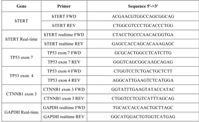
Table 1. Primer sequences for PCR, and Real-time PCR.
Gene Primer Sequence 5'->3'
hTERT hTERT FWD ACGAACGTGGCCAGCGGCAG
hTERT REV CTGGCGTCCCTGCACCCTGG
hTERT Real-time hTERT realtime FWD CTACCTGCCCAACACGGTGA hTERT realtime REV GAGCCACCAGCACAAAGAGC
TP53 exon 7 TP53 exon 7 FWD GCGCACTGGCCTCATCTTG
TP53 exon 7 REV GGGTCAGCGGCAAGCAGAG
TP53 exon 4 TP53 exon 4 FWD CTGGTCCTCTGACTGCTCTT
TP53 exon 4 REV AGGCATTGAAGTCTCATGGA
CTNNB1 exon 3 CTNNB1 exon 3 FWD GGTATTTGAAGTATACCATAC
CTNNB1 exon 3 REV CTGGTCCTCGTCATTTAGCAG
GAPDH Real-time GAPDH realtime FWD TGCACCACCAACTGCTTAGC GAPDH realtime REV GGCATGGACTGTGGTCATGAG
Table 2. Demographic and virological charateristics of 78 HCC patients
Median age, yr (range) 65.8 (25-87)
Men/women 51/27
HBsAg positive/negative 3/75
HCV positive/negative 8/70
Table 3. Demographic and virological charateristics of 41 control patients
Median age, yr (range) 52.25 (23-78)
Men/women 19/22
HBsAg positive/negative 0/41
Table 4. Distribution of CTNNB1 Exon 3 mutations
Tumors Paired Non-tumors Controls
CTNNB1 mutation Exon 3 0 (0%) 0 (0%) 0 (0%)
Table 5. Distribution of TP53 mutations Tumors (n.78) Paired Non-tumors (n.78) Controls (n.41) TP53 mutation R72P 9 (11.5%) 0 (0%)* 0 (0%)# P = 0.04* P = 0.02# TP53 mutation R249S 0 (0%) 0 (0%) 0 (0%)
Table 6. Distribution of hTERT promoter region mutations Tumors (n.78) Paired Non-tumors
(n.78)
Controls
(n.41)
hTERT hot spot mutation -124 bp
(C>T)
29 (37.2%) 0 (0%)* 0 (0%)# P < 0.0001* P < 0.0001#
hTERT hot spot mutation -146 bp (C>T) 0 (0%) 0 (0%) 0 (0%) hTERT mutation -39 bp (C>T) 7 (9%) 0 (0%)* 0 (0%)# P = 0.01* P = 0.04# hTERT mutation -300 bp (C>A) 3 (3,8%) 0 (0%)* 0 (0%)# P = 0.1* P = 0.2# hTERT mutation -245 bp (C>T) 30 (45,5%) 22 (28,2%)* 17 (21,7%)# P = 0.75* P = 0.09#
Figure 1. Representation of Wnt/beta-catenin signaling pathway [40]
Figure 3. hTERT promoter mutations. This figure shows as the hotspot mutations at -146 bp
and -124 bp from the Start Codon lead to formation of new binding sites for Ets/TCF transcription factors [42].
Figure 4. Fold induction of hTERT gene transcription reactivation. Real-time PCR shows that
the mutation at -124 bp in tumors leads to an increase of gene transcription up to 1.5 fold compared to wild-type sequence in tumor and non-tumor tissues.
References
1. Torre, L. A. et al. Global cancer statistics, 2012. CA Cancer J. Clin. 65, 87–108 (2015). 2. El-Serag, H. B. Hepatocellular carcinoma. N. Engl. J. Med. 365, 1118–1127 (2011). 3. Laursen, L. A preventable cancer. Nature 516, S2–S3 (2014).
4. Rogacki, K. et al. Alterations of Wnt/beta-catenin signaling pathway in hepatocellular carcinomas associated with hepatitis C virus. Pol J Pathol 66, 9-21 (2015)
5. Rao, T.P. et al. An updated overview on Wnt signaling pathways: a prelude for more.
Circ. Res. 106, 1798-1806 (2010)
6. MacDonal, B.T. et al. Wnt/beta-catenin signaling: components, mechanisms, and diseases. Dev. Cell. 17, 9-26 (2009)
7. Gregoieff, A. et al. Wnt signaling in the intestinal epithelium: from endoderm to cancer.
Genes Dev 19, 877-890 (2005)
8. Arvalli, R.N. et al. Molecular Mechanisms of Hepatocellular Carcinoma. Hepatology 48, 2047-2063 (2008)
9. Waisberg, J. et al. Wnt/beta-catenin pathway signaling in human hepatocellular carcinoma. World Journal of Hepatology 7, 2631-2635 (2015)
10. Suzuki T., et al. Beta-catenin expression in hepatocellular carcinoma: a possible participation of beta-catenin in dedifferentiation process. J Gastroenterol Hepatol 17, 994-1000 (2002)
11. Lian, Z. et al. Enhanced cell survival of Hep3B cells by the hepatitis B c antigen effector, URG11, is associated with upregulation of beta-catenin, Hepatology 43, 415-424 (2006)
12. Giglia-Mari, G. et al. TP53 mutations in human skin cancers, Hum. Mutat. 21, 217-228 (2003)
13. Pfeifer, G.P., et al. Tobacco smoke carcinogens, DNA damage and p53 mutations in smoking-associated cancers, Oncogene 21, 7435-7451 (2002)
14. Hussain S.P., et al. TP53 mutations and hepatocellular carcinoma: insights into the etiology and pathogenesis of liver cancer. Oncogene 26, 2166-2176 (2007)
15. Donato F., et al. Alcohol and hepatocellular carcinoma: the effect of lifetime intake and hepatitis virus infections in men and women. Am J Epidemiol 155, 323-331 (2002) 16. Petitjean A., et al. Impact of mutant p53 functional properties on TP53 mutation patterns
and tumor phenotype: lessons from recent developments in the IARC TP53 databese.
17. Armanios M., et al. The telomere syndromes. Nat Rev Genet 13, 693-704 (2012)
18. Harley C.B., et al. Telomeres shorten during ageing of human fibroblasts. Nature 345, 458-460 (1990)
19. Horn S., et al. TERT Promoter Mutations in Familial and Sporadic Melanoma. Science
339, 959-961 (2013)
20. Nault J.C., et al. High frequency of telomerase reverse-transcriptase promoter somatic mutations in hepatocellular carcinoma and preneoplastic lesions. Nature
Communications 4, 2218 (2013)
21. Kim N.W., et al. Specific association of human telomerase activity with immortal cells and cancer. Science 266, 2011-2015 (1994)
22. Ramlee M.K., et al. Transcription regulation of the human telomerase reverse transcriptase (hTERT) Gene. Genes 7, 50 (2016)
23. Cong Y.S., et al. The human telomerase catalytic subunit hTERT: Organization of the gene and characterization of the promoter. Hum Mol Genet 8, 137-142 (1999)
24. Saitta C., et al. Evaluation of CTNNB1 and TP53 variability in patients with hepatocellular carcinoma and occult hepatitis B virus infection. Cancer Genetics 208, 513-516 (2015)
25. Oda T., et al. p53 gene mutation spectrum in hepatocellular carcinoma. Cancer Res 52,
6358–64 (1992)
26. Cieply B. et al. Unique phenotype of hepatocellular cancers with exon-3 mutations in
beta-catenin gene. Hepatology 2009;49:821-31.
27. Huang H. et al. Beta-catenin mutations are frequent in human hepatocellular carcinomas associated with hepatitis C virus infection. Am J Pathol 1999;155:1795-801
28. Zucman-Rossi J. et al. Differential effects of inactivated Axin1 and activated beta-catenin mutations in human hepatocellular carcinomas. Oncogene 2007;26:774-80.
29. Guichard C. et al. Integrated analysis of somatic mutations and focal copy-number changes identifies key genes and pathways in hepatocellular carcinoma. Nat Genet 2012;44:694-8 30. Dumont P. et al. The codon 72 polymorphic variants of p53 have markedly different
apoptotic potential. Nat Genet 2003;33:357-65.
31. Irarrázabal C.E. et al. Chilean pilot study on the risk of lung cancer associated with codon 72 polymorphism in the gene of protein p53. Toxicol Lett 2003;144:69-76.
32. Hiyama T. et al. Codon 72 polymorphism in gastric cancer susceptibility in patients with
Helicobacter pylori-associated chronic gastritis. Int J Cancer 2002;100:304-8.
33. Själander A. et al. p53 polymorphisms and haplotypes in breast cancer. Carcinogenesis 1996;17:1313-6
34. Yoon YJ. et al. MDM2 and p53 polymorphisms are associated with the development of hepatocellular carcinoma in patients with chronic hepatitis B virus infection. Carcinogenesis 2008;29:1192-6.
35. Zhu ZZ. et al. A p53 polymorphism modifies the risk of hepatocellular carcinoma among
non-carriers but not carriers of chronic hepatitis B virus infection. Cancer Lett 2005;229:77-83.
36. Leveri M. et al. Codon 72 polymorphism of P53 gene does not affect the risk of cirrhosis
and hepatocarcinoma in HCV-infected patients. Cancer Lett 2004;208:75-9.
37. Totoki Y, Tatsuno K, Covington KR, Ueda H, Creighton CJ, KatoM, et al. Trans-ancestry mutational landscape of hepatocellu-lar carcinoma genomes. Nat Genet 2014;46(12):1267—73 [Epub2014/11/05].
38. Cevik D, Yildiz G, Ozturk M. Common telomerase reverse transcriptase promoter mutations in hepatocellular carcinomas from different geographical locations. World J
Gastroenterol 2015;21(1):311—7.
39. Huang DS, Wang Z, He XJ, Diplas BH, Yang R, Killela PJ,et al. Recurrent TERT promoter mutations identified in a large-scale study of multiple tumour types are associated with increased TERT expression and telomerase activation. Eur JCancer 2015;51(8):969—76.
40. Baron and Kneissel, WNT signaling in bone homeostasis and disease: from human mutations to treatments, Nature Medicine 19, 179-192, 2013
41. Yang et al. Transcriptome analysis of human OXR1 depleted cells reveals its role in regulating the p53 signaling pathway, Scientific Reports 5, 2015
42. Koelsche C. et al, TERT promoter hotspot mutations are recurrent in myxoid liposarcomas but rare in other soft tissue sarcoma entities, Journal of Experimental &
Contents
Introduction . . . 2
Patients and Methods . . . 4
Results . . . 6
Discussion . . . 7
Tables and Figures . . . 9

![Figure 1. Representation of Wnt/beta-catenin signaling pathway [40]](https://thumb-eu.123doks.com/thumbv2/123dokorg/4587878.39019/11.892.207.681.105.470/figure-representation-wnt-beta-catenin-signaling-pathway.webp)

![[3] G K Batchelor: Axial flow in trailing line vortices, Journal of Fluid Mechanics, Vol. 20, 645-658 (1964).](data:image/gif;base64,R0lGODlhAQABAIAAAP///wAAACH5BAEAAAAALAAAAAABAAEAAAICRAEAOw==)