A Large Series of Hyalinizing Trabecular Tumors:
Cytomorphology and Ancillary Techniques on Fine
Needle Aspiration
Marco Dell’Aquila, MD1; Carmen Gravina, MD2; Alessandra Cocomazzi, BD1; Sara Capodimonti, BD1;
Teresa Musarra, MD1; Stefania Sfregola, MD1; Vincenzo Fiorentino, MD1; Luca Revelli, MD2;
Maurizio Martini, MD, PhD1; Guido Fadda, MD1; Liron Pantanowitz, MD 3; Luigi Maria Larocca, MD1;
and Esther Diana Rossi, MD, PhD 1
BACKGROUND: Hyalinizing trabecular tumors (HTTs) are rare, essentially benign, follicular cell–derived thyroid neoplasms characterized by a trabecular growth pattern and nuclear pseudoinclusions. Their cytological findings are misleading, because these tumors are often misinterpreted on fine needle aspirate cytology as malignant lesions, such as papillary thyroid cancer and/or medullary thyroid cancer, leading to unnecessary total thyroidectomy. The aim of this study was to analyze the cytomorphological features and application of ancillary techniques in a series of HTTs. METHODS: Of 26 histological cases of HTT collected from September 2001 to December 2018, 18 cases had concomitant cytopathol-ogy. Cytological cases were processed with liquid-based cytology (LBC). Immunocytochemistry for HBME-1 and galec-tine-3 as well as molecular testing for BRAFV600E mutation were performed on both LBC and histological specimens.
RESULTS: The 18 lesions with fine needle aspirate cytology ranged in size from 5 to 45 mm. Cytological diagnoses included: 1 benign lesion favoring goiter (5.5%), 4 atypia of undetermined significance (22.2%), 6 follicular neoplasms (33.3%), 5 suspicious for malignancy favoring papillary thyroid cancer (28%), and 2 malignant (11%). Hence, 89% HTT had a negative concordant immunopanel, and they were 100% wild-type BRAFV600E. CONCLUSION: The majority of our HTTs
(83.3%) were diagnosed in the indeterminate Bethesda categories, suggesting that their cytomorphological features pose issues for reaching a conclusive cytological diagnosis. The ancillary test results in our series support the fact that HTT is a benign neoplasm. Cancer Cytopathol 2019;127:390-398. © 2019 American Cancer Society.
KEY WORDS: BRAF mutation; hyalinizing trabecular tumor; immunocytochemistry; molecular testing; personalized medicine; thyroid malignancy; thyroid nodule.
INTRODUCTION
Hyalinizing trabecular tumor (HTT) is a rare and distinct follicular-derived neoplasm that affects the thy-roid gland. Despite the fact that HTT was originally described by Zipkin in 1895,1 the concept that HHT is indeed a distinct pathological entity can be attributed to Carney et al in 1987.2,3 The latter authors described a lesion with peculiar histological features composed of a trabecular growth pattern with prominent deposition of hyaline material. Following this description, several authors published case reports or small series demon-strating that HTT is essentially a benign neoplasm requiring conservative management, even though it is worth noting that very rare cases of invasive HTT have been described.4-11
The majority of histological series show that reaching the correct diagnosis of this neoplasm does not pose a diagnostic challenge.4-11 However, a review of the literature indicates that reaching the correct preoperative
Corresponding author: Esther Diana Rossi, MD, PhD, Division of Anatomic Pathology and Histology, Fondazione Policlinico Universitario “Agostino Gemelli”–IRCCS, Università Cattolica del Sacro Cuore, Largo Francesco Vito 1, 00168 Rome, Italy; [email protected]
1 Division of Anatomic Pathology and Histology, Fondazione Policlinico “Agostino Gemelli”, IRCCS, Rome, Italy; 2 Division of Endocrine-Surgery, Fondazione
Policlinico “Agostino Gemelli”, IRCCS, Rome, Italy; 3 Department of Pathology, University of Pittsburgh Medical Center, Pittsburgh, Pennsylvania.
The last two authors contributed equally to this article.
Received: March 12, 2019; Revised: April 9, 2019; Accepted: April 10, 2019 Published online May 28, 2019 in Wiley Online Library (wileyonlinelibrary.com) DOI: 10.1002/cncy.22139, wileyonlinelibrary.com
diagnosis of HTT is more elusive, as the correct inter-pretation was achieved in only 8% of cases.7 More con-cerning, however, is the fact that the remaining 92% of cases resulted in false-positive diagnoses.6-17 The problem in achieving a correct preoperative diagnosis is largely ascribed to the fact that in cytology samples of HTT, the nuclear features and hyaline material mimic thy-roid malignancy. In particular, the typical polygonal or spindle-shaped cells of HTT frequently have nuclear pseudo-inclusions and grooves that may imitate papil-lary thyroid carcinoma (PTC). Furthermore, the recent introduction of noninvasive follicular neoplasm with papillary-like nuclear features characterized by a lack of pathognomonic cytological features poses an additional challenge on the preoperative identification of HTT.7,18-23 On the other hand, the hyaline material secreted by HTT is often misinterpreted either as amyloid of a medullary thyroid carcinoma (MTC) or as colloid from a benign follicular nodule in a goiter. The correct discrimination among HTT, PTC, and MTC is critical to guide the cor-rect management and avoid unnecessary overtreatment with total thyroidectomy.6-9
The cytological application of ancillary techniques, including immunocytochemistry (ICC) and molecular testing, might represent a valid method for those cases with a suspicion of HTT.24-31 From a molecular point of view, HTT harbors RET/PTC rearrangements in up to 45% of cases,24-27 whereas type RAS and wild-type BRAF have been reported in all published series. However, BRAF mutations are only found in approxi-mately 45% of PTCs; therefore, although the presence of a BRAFV600E mutation would rule out a HTT diag-nosis, a wild-type result cannot exclude a diagnosis of HTT.28-31 More recently, Nikiforova et al32 reported that in-frame gene fusion between PAX8 exon 2 and GLIS3 exon 3 is prevalent in HTT but not in PTC, demonstrat-ing for the first time that these tumors have a specific mutation that has no overlap with PTC.
In the current series of HTT from a single medical center, we discuss the cytohistological correlation and role of ancillary techniques in rendering an accurate pre-operative diagnosis.
MATERIALS AND METHODS
A retrospective search was performed of all thyroid his-tological cases diagnosed as HTT and recorded in the archives of the Division of Anatomic Pathology and
Histology of the Fondazione Policlinico Universitario Agostino Gemelli of Rome–IRCCS during the period between September 2001 and December 2018. Of the 26 histological cases retrieved, we selected all cases (n = 18) with preoperative fine needle aspiration (FNA). These patients underwent ultrasound during their thyroid checkup performed in the Centre for Thyroid Diseases at our hospital. Analyzing the ultrasonographic features, none of the patients showed any findings indicative of malignancy. All of the HTTs were solid, with 12 cases showing a hypoechogenic pattern and 6 showing mark-edly hypoechogenic features. The series included 2 male and 16 female patients with a median age of 53 years (range, 29-72 years), and their thyroid neoplasms ranged in size from 1.5 to 4.5 cm (Table 1).
All aspirations (usually 2 passes performed for each lesion) were performed with 25G to 27G needles. No rapid onsite assessment for adequacy of material was performed. All patients consented to their procedure. We received institutional (Catholic University of the Sacred Heart) ethical approval for this study. Cytology
TABLE 1. Clinicopathological Features of Hyaliniz-ing Trabecular Tumor Study Cases
Feature Value
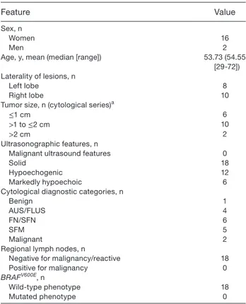
Sex, n
Women 16
Men 2
Age, y, mean (median [range]) 53.73 (54.55 [29-72]) Laterality of lesions, n
Left lobe 8
Right lobe 10
Tumor size, n (cytological series)a
≤1 cm 6
>1 to ≤2 cm 10
>2 cm 2
Ultrasonographic features, n
Malignant ultrasound features 0
Solid 18
Hypoechogenic 12
Markedly hypoechoic 6
Cytological diagnostic categories, n
Benign 1
AUS/FLUS 4
FN/SFN 6
SFM 5
Malignant 2
Regional lymph nodes, n
Negative for malignancy/reactive 18
Positive for malignancy 0
BRAFV600E, n
Wild-type phenotype 18
Mutated phenotype 0
Abbreviations: AUS/FLUS, atypia of undetermined significance/follicular lesion of undetermined significance; FN/SFN, follicular neoplasm/suspi-cious for follicular neoplasm; SFM, suspineoplasm/suspi-cious for malignancy.
samples were processed using the ThinPrep test (Hologic Co., Marlborough, Massachusetts). Prepared slides were fixed in 95% methanol and stained using a Papanicolaou test. Any remaining material was stored in Preservcyt solution (Hologic Co.) for possible prepa-ration of additional slides and investigations (eg, ICC and molecular analysis).
The lower limit for cytological adequacy of each sample was established according to the Bethesda and British Royal College of Pathology classification schemes; specifically, the minimum number of adequate cells in each sample was 6 groups of thyroid follicular epithelial cells per submitted slide, where each of these groups con-tains at least 10 well-visualized epithelial cells.33,34 The cytology cases were classified and diagnosed according to the New Italian Working Group SIAPEC-IAP clas-sification.35,36 These categories are defined as follows: TIR1, inadequate; TIR1C, cystic-hemorrhagic lesions; TIR2, benign nodules; TIR3A, follicular neoplasm (low-risk indeterminate lesions); TIR3B, follicular neoplasm (high-risk indeterminate lesions); TIR4, suspicious for malignancy; and TIR5, positive for malignant neoplasm. All cases were reevaluated and then reclassified according to The Bethesda System for Reporting Thyroid Cytology II (TBSRTC, 2017).19,36 For this study, analyses were con-ducted using TBSRTC terminology. This series included the following distribution of cases: 0% nondiagnostic plus cystic cases, 5.5% benign, 22.2% atypia of unde-termined significance/follicular lesion of undeunde-termined significance (AUS-FLUS), 33.3% follicular neoplasm/ suspicious for follicular neoplasm (FN/SFN), 28% suspi-cious for malignancy (SFM), and 11% malignant.
All cytology and histology cases were reviewed by 2 cytopathologists. Equivocal cases were reviewed by other pathologists to achieve a final consensus agreement.
ICC Analysis
HBME-1 and galectin-3 immunocytochemistry was per-formed using the protocol described by our group.37-40
The minimal percentage of adequate lesional cells for the performance of ICC evaluation was defined at 30% in liquid-based cytology (LBC) samples. Lesional cells were interpreted to be positive when at least 50% of the cells demonstrated strong cytoplasmic staining. To avoid false-negative and/or false-positive yields, this 50% ICC cutoff value was also used for histological tis-sue sections. A case was considered to be positive overall
for malignancy when there was concomitant expression of both immunomarkers. Adequate galectin-3 immuno-reactivity was represented by cyto plasmic staining, and suitable HBME-1 staining was chiefly within the cyto-plasm with accentuation on the cytocyto-plasmic membrane and within the lumen. Positive controls included mes-othelioma for HBME-1 (membranous positivity) and histiocytes for galectin-3 (cytoplasmic). Lymphocytes identified in the majority of the thyroid slides were used as a negative control. Our decision to apply ICC to LBC was based on 2 factors: 1) our cytology team has long-standing success with validated ICC protocols on LBC and 2) our personal experience with ICC on cell blocks resulted in contradictory results when compared with ICC on LBC.
Molecular Analysis for BRAF Mutation
DNA was extracted from both LBC-stored material and paraffin-embedded tissues. The BRAFV600E mutational analysis was performed on DNA extracted from cyto-logical and surgical specimens containing at least 70% tumor. Details about the protocol employed have been published previously by our group.41-44
Histology
All surgical specimens were fixed in 10% buffered for-maldehyde and embedded in paraffin, from which 5-μm-thick sections were prepared and stained with hematoxylin and eosin. In all cases, the entire lesion was completely submitted for histological evaluation. All the perithyroidal adipose tissue was also embedded and examined for lymph nodes. The histological diagnosis of HTT was based on the definition and criteria listed in the 4th edition of the WHO Classification of Tumours of
Endocrine Organs.45 The follow-up period for thyroidec-tomy ranged between 2 and 220 months.
Statistical Analysis
Statistical analysis was performed using GraphPad-Prism 5 software (Graph Pad Software, San Diego, California) and MedCalc version 10.2.0.0 (MedCalc Software, Mariakerke, Belgium). Statistical comparison of continu-ous variables was performed using the Mann-Whitney
U test or paired t test as appropriate. Comparison of
categorical variables was performed using a chi-square statistic using the Fisher’s exact test, and P < .05 was considered statistically significant.
RESULTS
During the 8-year study period, a total of 18/26 (69%) histology-proven HTT cases were included in this study. Table 1 summarizes the clinicopathological features for our series. FNA was performed in 16 females and 2 male patients. Table 2 compares the distribution of cases ac-cording to both the Bethesda and Italian classification systems. These results demonstrate that the majority of HTT are diagnosed in the indeterminate categories for
both reporting systems (22.2% AUS/FLUS; 33.3% FN/ SFN; 28% SFM).
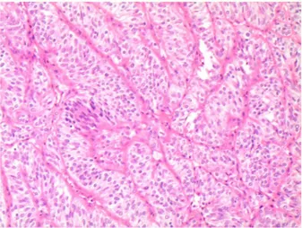
Cytomorphological evaluation on LBC slides (Table 3) confirmed that FNA smears were moderate to highly cellular (Fig. 1) and composed of cohesive clusters and isolated lesional cells. The smears showed very scant hyaline stromal material that was not recognized as a com-ponent of a possible HTT. Tumor cells had low nuclear/ cytoplasmic ratios and were characterized by round-oval and elongated nuclei (Fig. 2) with minimal nuclear atypia, occasional nuclear grooves, and rare nuclear pseudoinclusions. The cells had epithelioid and slightly elongated morphology (Figures 1‒3). The majority of tumor cells had moderate cytoplasm, while a few were characterized by scant cytoplasm. The presence of nuclear pseudoinclusions and grooves were mostly asso-ciated with SFM (Figs. 1‒3) and malignant diagnoses (Table 3). Some cases showed finely granular chromatin with some slightly enlarged and mild atypically nuclei in LBC slides (Fig. 2). The background was characterized by scant colloid and few globular resemblance of colloid- hyaline (Figs. 1 and 3) material and the lack of any inflammatory cells.
On histopathology, we recognized the typical find-ings, including trabecular pattern, polygonal and/or elongated cells with inclusions and/or grooves, and hy-aline material (Fig. 4). The hyhy-aline material was scant
TABLE 2. Comparative Distribution of Hyalinizing Trabecular Tumor Cases According to Cytological Cate-gories per Italian and Bethesda Reporting Systems
SIAPEC-IAP classification TIR 2 TIR 3A TIR3B TIR4 TIR5
TBSRTC II (benign) III (AUS/FLUS) IV (FN/SFN) V (SFM) VI (malignant)
No. of cases (%) 1 (5.5) 4 (22.2) 6 (33.3) 5 (28) 2 (11)
Abbreviations: AUS/FLUS, atypia of undetermined significance/follicular lesion of undetermined significance; FN/SFN, follicular neoplasm/suspicious for follicular neoplasm; SIAPEC/IAP, Societa Italiana di Anatomia Patologica e Citologia; SFM, suspicious for malignancy; TBSRTC, The Bethesda System for Reporting Thyroid Cytopathology; TIR2, benign nodules; TIR3A, follicular neoplasm (low-risk indeterminate lesions); TIR3B, follicular neoplasm (high-risk indeterminate lesions); TIR4, suspicious for malignancy; TIR5, positive for malignant neoplasm.
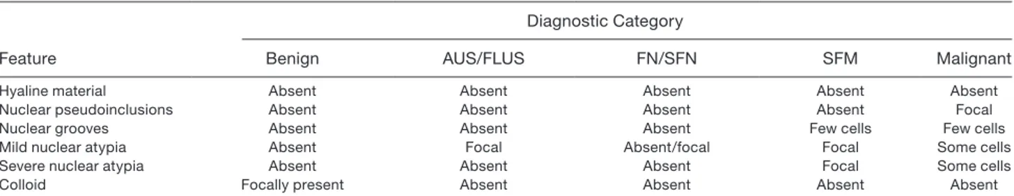
TABLE 3. Cytomorphological Features of Hyalinizing Trabecular Tumor
Feature
Diagnostic Category
Benign AUS/FLUS FN/SFN SFM Malignant
Hyaline material Absent Absent Absent Absent Absent
Nuclear pseudoinclusions Absent Absent Absent Absent Focal
Nuclear grooves Absent Absent Absent Few cells Few cells
Mild nuclear atypia Absent Focal Absent/focal Focal Some cells
Severe nuclear atypia Absent Absent Absent Focal Some cells
Colloid Focally present Absent Absent Absent Absent
AUS/FLUS, atypia of undetermined significance/follicular lesion of undetermined significance; FN/SFN, follicular neoplasm/suspicious for follicular neoplasm; SFM, suspicious for malignancy.
Figure 1. Cytology of hyalinizing trabecular tumor showing
a cellular cluster of elongated, oval nuclei with scant to moderate cytoplasm intermingled with hyaline material (magnification ×200; liquid-based cytology, Papanicolaou stain).
to absent, which might explain why it was missing from the FNAs. We did not find any psammoma bodies and/ or any vascular or capsular invasion. In 6 of the cases, histological specimens revealed a concomitant PTC in the contralateral lobe, while the majority of cases had a histological diagnosis of multinodular goiter.
ICC stain results are reported in Table 4. Tumors diagnosed on FNA as benign lesions and those diag-nosed as FN/SFN had negative staining. For the 4 cases with AUS/FLUS diagnoses, 3 had negative stains, and
1 demonstrated only galectin-3 positivity. For cases grouped in the SFM category, 4 showed negative stain-ing for both markers, while only 1 case revealed positiv-ity for HBME1. The cases interpreted as malignant had negative staining. Those cases with a discordant panel (ie, 1 marker was positive) showed focal positivity for the immune-reactive biomarker. To note, we detected a weak or slightly moderate positivity (membranous for HBME-1 and cytoplasmic for galectin-3) in contrast with the strong positivity reported in our previous series of thyroid carcinoma.37-40 None of the lesions had a posi-tive stain for both markers, confirming the benign nature of these HTT cases.
The results of molecular testing for BRAFV600E demonstrated a wild-type phenotype in all of our cases.
DISCUSSION
Since the first description by Goellner and Carney, the diagnosis of HTT has remained challenging from a morphological perspective due to overlapping histo-pathological features and the long-standing controversy concerning its underlying genetics, potential malignant nature, and relationship with PTC.2,3,17,46,47 In fact, the first description by Carney clearly emphasized that the trabecular pattern and nuclear features (ie, nuclear grooves and pseudoinclusions) overlapped those of PTC, which poses diagnostic difficulties. In the 4th edition of the WHO Classification of Tumours of Endocrine Organs, HTT was accordingly placed in a category of tumors with low malignant potential.45
Figure 2. Cytological details of elongated, oval nuclei showing (A) nuclear grooves and (B) pale nuclei with finely coarse
chromatin. This case, which was diagnosed as follicular neoplasm with spindle-shaped nuclei, resulted in a histological diagnosis of hyalinizing trabecular tumor (magnification ×400; liquid-based cytology, Papanicolaou stain).
Figure 3. Cytological details of nuclear features of a hyalini zing
trabecular tumor case diagnosed as suspicious for malignancy. The cells show nuclear pallor and nuclear pseudoinclusions. The nuclei are oval and elongated; some have moderate, atypical findings. The focal presence of globular material resembles colloid globules (magnification ×200; liquid-based cytology, Papanicolaou stain).
The cytological interpretation of HTT is equally difficult. The difficulty in rendering a cytological diagno-sis of HTT is caused by the cytomorphological features of nuclear pseudoinclusions, nuclear grooves, and hyaline material that can result in a false-positive diagnosis of PTC or MTC.10,48-51 Different published series including
cyto-logical samples of HTT have documented that up to 60% of cases were diagnosed as positive and/or suspicious for malignancy, despite the fact that the samples had a lack of papillary structures and that lesions exhibited elongated nuclei associated with acellular hyaline stroma.7 Up to
75% of HTTs appear to be diagnosed in Bethesda catego-ries spanning from IV to VI, indicating how they can be easily misinterpreted by cytopathologists. As suggested by the 2015 American Thyroid Association guidelines, cyto-logical findings should always be correlated with clinical and ultrasound findings to reduce diagnostic errors.51,52
Indeed, there is often incongruity with HTT where
ultrasound imaging suggests a benign entity, whereas the cytopathology reports favor malignancy.53-56 The imaging features from our cases highlight a solid pattern character-ized by hypoechogenicity, absence of microcalcifications, well-circumscribed smooth margins, and the presence of vascularity. While these findings are not concerning for PTC, they are compatible with neoplasm with papillary- like nuclear features.19-23
Several cytomorphological features have been recog-nized with HTT including cellular aggregates arranged around hyaline material, cells with a low nuclear/ cytoplasmic ratio, fine chromatin (that differs from the optical clear chromatin seen in PTC), abundant nuclear pseudoinclusions and grooves, and samples with a bloody background.1-10 Despite the difficulty in diagnosing HTT with FNA due to these overlapping cytomorpho-logical features, Carney et al demonstrated that HTT could be cytologically diagnosed in 72% of their cases.2,3
In our series, we sought to determine whether the cytomorphological features of HTT were affected by sample preparation using LBC. We found that 85% of our cases had indeterminate diagnoses (categories III-V) and that 11% had a false malignant diagnosis (category VI). Only a minority (5.8%) of our cases were diagnosed as benign lesions.
The fact that most of our diagnoses were indeter-minate confirms the challenge pathologists face with HTT. In our series, only 4 cases were diagnosed as AUS/FLUS, mostly due to sparsely cellular smears. On the other hand, in 6 cases, cytologists were confident to diagnose these lesions as FN/SFN, largely due to the presence of a monomorphic population of follicular cells with microfollicles, nuclear overlapping, and crowding, but without significant nuclear atypia. The subtle and focal nuclear features suggestive of malignancy in these cases, combined with the detection of only a few nuclear pseudoinclusions, led to the diagnosis of either SFM or even malignancy. This may be ascribed to the fact that in our series, all cases were processed with LBC. Compared with conventional smears, in LBC cytology, nuclei are darker, and there is reduced stromal material. In our cases, intranuclear pseudoinclusions were identified, but only infrequently. This may explain why only 11% of the cases were misinterpreted to be malignant in our series.
Immunohistochemistry can assist in diagnosing HTT, even though some of the biomarkers employed may not show significant specificity.7,24-32 In keeping with the
TABLE 4. Immunocytochemistry Results for Hyalini-zing Trabecular Tumor Cases
Benign AUS/FLUS FN/SFN SFM Malignant
H+/G+ 0 0 0 0 0
H−/G− 1 3 6 4 2
H+/G− 0 0 0 1 0
H−/G+ 0 1 0 0 0
Abbreviations: AUS/FLUS, atypia of undetermined significance/follicular lesion of undetermined significance; FN/SFN, follicular neoplasm/suspi-cious for follicular neoplasm; G, galectin-3; H, HBME-1; SFM, suspineoplasm/suspi-cious for malignancy; +, positive; −, negative.
Values represent the numbers of positive cases. All cases had weak to slightly moderate positivity.
Figure 4. Histological picture of hyalinizing trabecular tumor
showing the typical trabecular patterns of the neoplastic clus-ters with the typical elongated nuclei characterized by nuclear clearing and small nuclear pseudoinclusions (magnification ×200; hematoxylin and eosin stain).
follicular cell origin of HTT, different authors, including our group, have used HBME-1 and galectin-3.37-40,57-60 The expression of HBME-1 and galectin-3 in malignant thyroid lesions such as PTC and its variants has been well defined and validated in the literature.
Positive immunostaining with HBME-1 and galec-tin-3 is supportive of a malignant thyroid lesion in up to 77% of cases.61
However, the results seem to be controversial in HTT. In our series, we found that the majority of HTT cases showed an immunoprofile characterized by neg-ative immunoreactivity in 16/18 (89%) lesions. Thus, this supports the notion that HTTs are benign tumors. For example, HTT displays distinctive membrane and cytoplasmic positivity for Ki-67 (using the clone MIB-1) when the reaction is performed at room temperature.24-26 Although not used in our study, mostly due to the dif-ficulties in recognizing the cytological features of HTT, numerous papers have concluded that MIB-1 is also a useful diagnostic tool in cytological samples.7,24-27 HTT shows a distinctive membrane and cytoplasmic positivity for Ki-67 (using the clone MIB-1) when the reaction is performed at room temperature.24-26
Genotyping of HTT has provided further insight into the biological behavior and relationship of this tumor with other malignant entities, including PTC.27-32
Papotti et al62 found that 28.6% of HTT har-bor RET/PTC1 rearrangements, while Cheung et al63 reported this rearrangement in 62% of their HTT cases. No investigators have yet reported RAS or BRAF muta-tions in HTT, which are prevalent in PTC and follicular variant of PTC, suggesting that HTT is perhaps a dis-tinct entity rather than a variant of PTC.27-32,62,63
In our series, BRAFV600E demonstrated a wild-type phenotype in all of our cases. Although a BRAFV600E mutation would exclude the diagnosis of HTT, a wild-type result cannot rule it out. It therefore makes sense that this test could be used in cytology practice as an additional—albeit not conclusive—tool to study HTT.
As noted, Nikiforova et al demonstrated recently that GLIS rearrangements, particularly PAX8-GLIS3, are highly prevalent in HTT but not in PTC.32 In fact, all 14 of their HTT cases were positive for a GLIS rearrangement including PAX8-GLIS3 (93%) and PAX8-GLIS1 (7%) fu-sion. According to their study, it seems that these fusions are responsible for the overexpression of GLIS, upregulation of
extracellular matrix genes, and deposition of the collagen that is so characteristic of HTT. Exactly how GLIS regu-lates these genes remains to be determined. Some studies have begun to address its role in thyroid cells of mice.32 Thus far, it appears that the detection of GLIS fusions preoperatively via thyroid fine needle aspirate cytology may help resolve some indeterminate lesions. In fact, out of 10,165 consecutive FNA samples from thyroid lesions with indeterminate cytology, Nikiforova et al documented PAX8-GLIS3 in 8 cases (0.1%), with a subsequent surgical diagnosis of HTT in 5 of them.32
In conclusion, based on these data, an accurate diagnosis of HTT by FNA is possible if supported by radiological and ancillary testing, despite incongruent cytomorphological findings.10,64,65 When cytomorpho-logical features do not meet clear criteria for malignancy, it is crucial to include HTT in the differential diagnosis. LBC cytological samples may be more challenging given that the nuclear details and background hyaline material content may differ from direct smears. As highlighted in our series, if a clinically indolent tumor is suspected on radiology, but the FNA findings are worrisome, it may be best to place this lesion in an indeterminate category, which will lead to molecular testing and possibly a lobec-tomy instead of an unnecessary thyroideclobec-tomy.
FUNDING SUPPORT
No specific funding was disclosed.
CONFLICT OF INTEREST DISCLOSURES
The authors made no disclosures.
AUTHOR CONTRIBUTIONS
Marco Dell’Aquila: conceptualization, data curation,
investiga-tion, software, visualizainvestiga-tion, writing (original draft). Carmen Gravina: data curation, investigation, software, writing (original draft). Alessandra Cocomazzi: investigation. Sara Capodimonti: investigation. Teresa Musarra: investigation. Stefania Sfregola: investigation. Vincenzo Fiorentino: investigation. Luca Revelli: investigation. Maurizio Martini: formal analysis, investigation.
Guido Fadda: formal analysis, investigation. Liron Pantanowitz:
conceptualization, formal analysis, methodology, supervision, visu-alization, writing (review and editing). Luigi Maria Larocca: con-ceptualization, formal analysis, methodology, resources, supervision, validation, writing (review and editing). Esther Diana Rossi: con-ceptualization, data curation, formal analysis, funding acquisition, investigation, methodology, project administration, resources, supervision, validation, visualization, writing (original draft), writing (review and editing).
REFERENCES
1. Zipkin R. Hyalinanliche collagene kugeln als produkte epitheli-aler zellen in malignen stumen. Virchows Arch Path Anat Physiol. 1895;182:374-406.
2. Carney JA, Ryan J, Goellner JR. Hyalinizing trabecular adenoma of the thyroid gland. Am J Surg Pathol. 1987;11:583-591.
3. Carney JA, Hirokawa M, Lloyd RV, Papotti M, Sebo TJ. Hyalinizing trabecular tumors of the thyroid gland are almost all benign. Am J
Surg Pathol. 2008;32:1877-1889.
4. Galgano MT, Mills SE, Stelow EB. Hyalinizing trabecular adenoma of the thyroid revisited: a histologic and immunohistochemical study of thyroid lesions with prominent trabecular architecture and sclerosis. Am J Surg Pathol. 2006;30:1269-1273.
5. Casey MB, Sebo TJ, Carney JA. Hyalinizing trabecular adenoma of the thyroid gland: identification through MIB-1 staining of fine-nee-dle aspiration biopsy smears. Am J Clin Pathol. 2004;122:506-510. 6. Kim T, Oh YL, Kim KM, Shin JH. Diagnostic dilemmas of
hyalinizing trabecular tumours on fine needle aspiration cytology: a study of seven cases with BRAF mutation analysis. Cytopathology. 2011;22:407-413.
7. Saglietti C, Piana S, La Rosa S, Bongiovanni M. Hyalinizing trabec-ular tumour of the thyroid: fine-needle aspiration cytological diag-nosis and correlation with histology. J Clin Pathol. 2017;70:641-647. 8. Howard BE, Gnagi SH, Ocal IT, Hinni ML. Hyalinizing trabecular tumor masquerading as papillary thyroid carcinoma on fine-needle aspiration. ORL J Otorhinolaryngol Relat Spec. 2014;75:309-313. 9. Jang H, Park CK, Son EJ, et al. Hyalinizing trabecular tumor of the
thyroid: diagnosis of a rare tumor using ultrasonography, cytology, and intraoperative frozen sections. Ultrasonography. 2016;35:131-139. 10. Jones DJ, Kieliszak CR, Patel SS, Selinsky CR. Hyalinizing trabec-ular tumor of the thyroid gland and its significant diagnostic issue.
Thyroid Res. 2017;10:7.
11. Rhee YY, Jung HK, Kim SH, Kim SH. Hyalinizing trabecular tumor of the thyroid gland, a diagnostic challenge in fine-needle aspiration cytology: case report. J Pathol Transl Med. 2018;52:252-256. 12. Arena S, Latina A, et al. Cytological diagnosis difficulties in
hyalinizing trabecular adenoma of the thyroid. J Endocrinol Invest. 2011;34:887-888.
13. Akin MR, Nguyen GK. Fine-needle aspiration biopsy cytology of hyalinizing trabecular adenomas of the thyroid. Diagn Cytopathol. 1999;20:90-94.
14. Baloch ZW, Puttaswamy K, Brose M, LiVolsi VA. Lack of BRAF mutations in hyalinizing trabecular neoplasm. Cytojournal. 2006; 25(3):17.
15. Kuma S, Hirokawa M, Miyauchi A, Kakudo K, Katayama S. Cytologic features of hyalinizing trabecular adenoma of the thyroid.
Acta Cytol. 2003;47:399-404.
16. Bondeson L, Bondeson AG. Clue helping to distinguish hyalinizing trabecular adenoma from carcinoma of the thyroid in fine-needle aspirates. Diagn Cytopathol. 1994;10:25-29.
17. Goellner JR, Carney JA. Cytologic features of fine-needle aspirates of hyalinizing trabecular adenoma of the thyroid. Am J Clin Pathol. 1989;91:115-119.
18. Bakuła-Zalewska E, Cameron R, Gałczyński JP, Domanski HA. Hyaline matrix in hyalinizing trabecular tumor: findings in fine-needle aspiration smears. Diagn Cytopathol. 2015;43:710-713. 19. Ali S, Cibas ES. The Bethesda System for Reporting Thyroid
Cytopathology. 2nd ed. Berlin, Germany: Springer; 2018. 20. Nikiforov YE, Sethala RR, Tallini G, et al. Nomenclature revision
for encapsulated follicular variant fo papillary thyroid carcinoma: a paradigm shift to reduce overtreatment of indolent tumors. JAMA
Oncol. 2016;2:1023-1029.
21. Howitt BE, Chang S, Eslinger M, et al. Fine-needle aspiration diagnoses of noninvasive follicular variant of papillary thyroid car-cinoma. Am J Clin Pathol. 2015;144:850-857.
22. Bizzarro T, Martini M, Capodimonti S, et al. The morphologic analysis of non-invasive follicular thyroid neoplasm with papillary- like nuclear features (NIFTP) on liquid based cytology: some insights of their identification in our institutional experience.
Cancer. 2016;124:699-710.
23. Faquin W, Wong L, Afrogheh A, et al. The impact of reclassifying non invasive FVPC on the risk of malignancy in the Bethesda sys-tem for reporting thyroid cytopathology. Cancer Cytopathol. 2016; 124:181-187.
24. Leonardo E, Volante M, Barbareschi M, et al. Cell membrane reac-tivity of MIB-1 antibody to Ki67 in human tumors: fact or artifact?
Appl Immunohistochem Mol Morphol. 2007;15:220-223.
25. Takada N, Hirokawa M, Ohbayashi C, et al. Re-evaluation of MIB-1 immunostaining for diagnosing hyalinizing trabecular tumour of the thyroid: semi-automated techniques with manual antigen retrieval are more accurate than fully automated techniques.
Endocr J. 2018;65:239-244.
26. Salvatore G, Chiappetta G, Nikiforov YE, et al. Molecular profile of hyalinizing trabecular tumours of the thyroid: high prevalence of RET/PTC rearrangements and absence of B-raf and N-ras point mutations. Eur J Cancer. 2005;41:816-821.
27. Hirokawa M, Carney JA, Ohtsuki Y. Hyalinizing trabecular ade-noma and papillary carciade-noma of the thyroid gland express different cytokeratin patterns. Am J Surg Pathol. 2000;24:877-881. 28. Sheu SY, Vogel E, Worm K, Grabellus F, Schwertheim S, Schmid
KW. Hyalinizing trabecular tumour of the thyroid-differential expression of distinct miRNAs compared with papillary thyroid carcinoma. Histopathology. 2010;56:632-640.
29. Gaffney RL, Carney JA, Sebo TJ, et al. Galectin-3 expression in hy-alinizing trabecular tumors of the thyroid gland. Am J Surg Pathol. 2003;27:494-498.
30. Hino R, Motoi N, Toda K, et al. Stromal tiny black dots, like “sugar- coated”, of von Kossa stain is a diagnostic clue to hyalinizing trabec-ular tumor of the thyroid gland. Pathol Int. 2018;68:176-182. 31. Lenggenhager D, Maggio EM, Moch H, Rössle M. HBME-1
expression in hyalinizing trabecular tumours of the thyroid gland.
Histopathology. 2013;62:1092-1097.
32. Nikiforova MN, Nikitski AV, Panebianco F, et al. GLIS rearrange-ment is a genomic hallmark of hyalinizing trabecular tumor of the thyroid gland. Thyroid. 2019;29:161-173.
33. Gharib H, Goellner RJ, Johnson DA. Fine needle aspiration of the thyroid: a 12 years experience with 11,000 biopsies. Clin Lab Med. 1993;13:699-709.
34. British Thyroid Association, Royal College of Physicians. Report of the Thyroid Cancer Guidelines Update Group. In: Perros P, ed. Guidelines for the Management of Thyroid Cancer. 2nd ed. London, UK: Royal College of Physicians; 2007.
35. Fadda G, Basolo F, Bondi A, et al. Cytological classification of thy-roid nodules. Proposal of the SIAPEC-IAP Italian consensus work-ing group. Pathologica. 2010;102:405-408.
36. Nardi F, Basolo F, Crescenzi A, Fadda G, Frasoldati A, Orlandi F, et al. Italian consensus for the classification and reporting of thyroid cytology. J Endocrinol Invest. 2014;37:593-599.
37. Rossi ED, Martini M, Capodimonti S, et al. Analysis of immuno-cytochemical and molecular BRAF expression in thyroid carcino-mas: a cyto-histological institutional experience. Cancer Cytopathol. 2014;122:527-535.
38. Rossi ED, Mulé A, Russo RM, Pierconti F, Fadda G. Application of liquid based preparation to non-gynecological exfoliative cytology.
Pathologica. 2008;100:461-465.
39. Rossi ED, Raffaelli M, Minimo C, et al. Immunocytochemical evaluation of thyroid neoplasms on thin-layer smears from fine- needle aspiration biopsies. Cancer. 2005;105:87-95.
40. Rossi ED, Bizzarro T, Martini M, et al. Morphological parameters able to predict BRAF(V600E)-mutated malignancies on thyroid
fine-needle aspiration cytology: our institutional experience. Cancer
Cytopathol. 2014;122:883-891.
41. Rossi ED, Martini M, Capodimonti S, et al. Diagnostic and prog-nostic value of immunocytochemistry and BRAF mutation analysis on liquid based biopsies of thyroid neoplasms suspicious for carci-noma. Eur J Endocrinol. 2014;168:853-859.
42. Rossi ED, Martini M, Capodimonti S, et al. BRAF (v600e) muta-tion analysis on LBC-processed aspiramuta-tion biopsies predicts bilater-ality and nodal involvement in papillary thyroid microcarcinoma.
Cancer Cytopathol. 2013;121:291-297.
43. Rossi ED, Martini M, Capodimonti S, et al. Morphology combined with ancillary techniques: An algorithm approach for thyroid nod-ules. Cytopathology. 2018;29:418-427.
44. Rossi ED, Larocca LM, Pantanowitz L. Ancillary molecular testing of indeterminate thyroid nodules. Cancer Cytopathol. 2018;126:654-671. 45. Lloyd RV, Osamura RY, Kloppel G, Rosai J, eds. WHO Classifi-cation of Tumours of Endocrine Organs. 4th ed. Lyon, France: IARC; 2017.
46. McCluggage WG, Sloan JM. Hyalinizing trabecular carcinoma of thyroid gland. Histopathology. 1996;28:357-362.
47. Caraci P, Fulcheri A, Ondolo C, et al. Hyalinizing trabecular tumor of the thyroid: a case report. Head Neck Pathol. 2011;5:423-427. 48. Campanella P, Ianni F, Rota CA, Corsello SM, Pontecorvi A.
Quantification of cancer risk of each clinical and ultrasonographic suspicious feature of thyroid nodules: a systematic review and me-ta-analysis. Eur J Endocrinol. 2014;170:R203-R211.
49. Baloch ZW, LiVolsi VA. Cytologic and architectural mimics of pap-illary thyroid carcinoma. Diagnostic challenges in fine-needle aspi-ration and surgical pathology specimens. Am J Clin Pathol. 2006; 125(suppl):S135-S144.
50. Shikama Y, Osawa T, Yagihashi N. Neuroendocrine differentia-tion in hyalinizing trabecular tumor of the thyroid. Virchows Arch. 2003;443:792-796.
51. Carney JA. Hyalinizing trabecular tumors of the thyroid gland: qua-druply described but not by the discoverer. Am J Surg Pathol. 2008; 32:622-634.
52. Haugen BR, Alexander EK, Bible KC, et al. 2015 American Thyroid Association Management Guidelines for adult patients with thyroid nodules and differentiated thyroid cancer: The American Thyroid Association Guidelines Task Force on thyroid nodules and differenti-ated thyroid cancer. Thyroid. 2016;26:1-133.
53. Lin JS, Aiello Bowles EJ, Williams SB, Morrison CC. Screening for thyroid cancer: updated evidence report and systematic review for the US Preventive Services Task Force. JAMA. 2017;317:1888-1903.
54. Russ G, Bonnema SJ, Erdogan MF, Durante C, Ngu R, Leenhardt L. European Thyroid Association guidelines for ultrasound malig-nancy risk stratification of thyroid nodules in adults: The EU-TIRADS. Eur Thyroid J. 2017;13:225-237.
55. Tajiri K, Hirokawa M, Oshita M, et al. Minute hyalinizing trabecu-lar tumor misinterpreted as a blood vessel on ultrasonography: a case report. Ultrasound Int Open. 2017;3:E42-E44.
56. Kobayashi K, Hirokawa M, Jikuzono T, et al. Hyalinizing trabecu-lar tumor of the thyroid gland: characteristic features on ultrasonog-raphy. J Med Ultrason. 2007;34:43-47.
57. Correia-Rodrigues HG, Nogueira De Pontes AA, Adan LFF. Use of molecular markers in samples obtained from preoperative aspiration of thyroid. Endocr J. 2012;59:417-424.
58. Dabbs D, Abendroth CS, Grenko RT, Wang X, Radcliffe GE. Immunocytochemistry on the Thin-Prep Processor. Diagn
Cytopathol. 1997;17:388-392.
59. Cochand-Priollet B, Dahan H, Laloi-Michelin M, et al. Immunocytochemistry with cytokeratin 19 and HBME-1 in-creases the diagnostic accuracy of thyroid fine-needle aspirations. Preliminary report of 150 liquid-based fine needle aspirations with histological control. Thyroid. 2011;21:1067-1073.
60. Chen H, Izevbaye I, Chen F, Weinstein B. Recent advances in follic-ular variant of papillary thyroid carcinoma. N Am J Med Sci. 2012;5: 212-216.
61. Fadda G, Rossi ED, Raffaelli M, et al. Follicular thyroid neoplasms can be classified as low and high risk according to HBME-1 and galectin 3 expression on liquid based fine needle cytology. Eur J
Endocrinol. 2011;165:447-453.
62. Papotti M, Volante M, Giuliano A, et al. RET/PTC activation in hy-alinizing trabecular tumors of the thyroid. Am J Surg Pathol. 2000; 24:1615-1621.
63. Cheung CC, Boerner SL, MacMilan CM, et al. Hyalinizing trabecular tumor of the thyroid: a variant of papillary carci-noma proved by molecular genetics. Am J Surg Pathol. 2000;24: 1622-1626.
64. Ohtsuki Y, Kimura M, Murao S, Okada Y, Teratani Y, Matsumoto M, Kurabayashi A, Iguchi M, Lee GH, Furihata M. Immunohistochemical and electron microscopy studies of a case of hyalinizing trabecular tumor of the thyroid gland, with special consideration of the hyalinizing mass associated with it. Med Mol
Morphol. 2009;42:189-194.
65. Li J, Yang GZ, Gao LX, Yan WX, Jin H, Li L. Hyalinizing trabecu-lar tumor of the thyroid: case report and review of the literature. Exp